O R I G I N A L A R T I C L E
Galanin pathogenic mutations in temporal lobe epilepsy
Michel Guipponi
1,
*, Amina Chentouf
2
, Kristin E.B. Webling
3
, Krista
Freimann
3,4
, Arielle Crespel
5
, Carlo Nobile
6
, Johannes R. Lemke
7,8
, Jörg
Hansen
9
, Thomas Dorn
9
, Gaetan Lesca
10,11,12,13
, Philippe Ryvlin
14,15,16
, Edouard
Hirsch
17
, Gabrielle Rudolf
17
, Dominique Sarah Rosenberg
18
, Yvonne Weber
19
,
Felicitas Becker
19
, Ingo Helbig
20,21
, Hiltrud Muhle
20
, Annick Salzmann
1
, Malika
Chaouch
22
, Mohand Laid Oubaiche
2
, Serena Ziglio
23
, Corinne Gehrig
1
, Federico
Santoni
1
, Massimo Pizzato
23
, Ülo Langel
3
and Stylianos E. Antonarakis
1,24
1
Department of Genetic Medicine and Development, University of Geneva Medical School and University
Hospitals of Geneva, Geneva, Switzerland,
2Department of Neurology, University Hospital of Oran, Oran 31000,
Algeria,
3Department of Neurochemistry, Arrhenius Laboratories for Natural Science, Stockholm University,
Stockholm, Sweden,
4Institute of Technology, University of Tartu, Tartu, Estonia,
5Epilepsy Unit, Montpellier
University Hospital, Montpellier, France,
6CNR-Institute of Neurosciences, Section of Padua, Padova, Italy,
7
Division of Human Genetics, University Children
’s Hospital Inselspital, Bern, Switzerland,
8Institute of Human
Genetics, University of Leipzig, Leipzig, Germany,
9Swiss Epilepsy Center, Zurich, Switzerland,
10Department of
Medical Genetics, Hospices Civils de Lyon, Lyon, France,
11Claude Bernard Lyon I University, Lyon, France,
12CRNL,
CNRS UMR 5292, Lyon, France,
13INSERM U1028, Lyon, France,
14Department of Clinical Neurosciences, CHUV,
Lausanne, Switzerland,
15Lyon
’s Neuroscience Research Center, INSERM U1028, CNRS 5292, UCBL, Lyon, France,
16Department of Functional Neurology and Epileptology, HCL, Lyon, France,
17IGBMC UMR7104 INSERM U964,
University of Strasbourg, Strasbourg, France,
18Service de Neurologie et Neurophysiologie Clinique, IGCNC - EA
7282, UMR 6284 ISIT, Université d’Auvergne, Clermont Université, CHU Clermont-Ferrand, Hôpital Gabriel
Montpied, Clermont-Ferrand, France,
19Department of Neurology and Epileptology, Hertie Institute for Clinical
Brain Research, University of Tübingen, Tübingen 72076, Germany,
20Department of Neuropediatrics, University
Medical Center Schleswig-Holstein (UKSH), Kiel, Germany,
21Division of Neurology, The Children
’s Hospital of
Philadelphia, Philadelphia, PA, USA,
22Department of Neurology, Benaknoun University Hospital, Algiers 16000,
Algeria,
23Centre for Integrative Biology (CIBIO), University of Trenton, Trenton, Italy and
24Institute of Genetics
and Genomics in Geneva (iGE3), Geneva, Switzerland
*To whom correspondence should be addressed at: Department of Genetic Medicine and Development, University of Geneva Medical School and University Hospitals of Geneva, 1, rue Michel-Servet, 1211 Geneva 4, Switzerland. Tel: +41 223795809; Email: [email protected]
Abstract
Temporal lobe epilepsy (TLE) is a common epilepsy syndrome with a complex etiology. Despite evidence for the participation of genetic factors, the genetic basis of TLE remains largely unknown. A role for the galanin neuropeptide in the regulation of epileptic seizures has been established in animal models more than two decades ago. However, until now there was no report
Received: December 16, 2014. Revised and Accepted: February 11, 2015
© The Author 2015. Published by Oxford University Press. All rights reserved. For Permissions, please email: [email protected] doi: 10.1093/hmg/ddv060
Advance Access Publication Date: 17 February 2015 Original Article
of pathogenic mutations in GAL, the galanin-encoding gene, and therefore its role in human epilepsy was not established. Here, we studied a family with a pair of monozygotic twins affected by TLE and two unaffected siblings born to healthy parents. Exome sequencing revealed that both twins carried a novel de novo mutation ( p.A39E) in the GAL gene. Functional analysis revealed that the p.A39E mutant showed antagonistic activity against galanin receptor 1 (GalR1)-mediated response, and decreased binding affinity and reduced agonist properties for GalR2. These findings suggest that the p.A39E mutant could impair galanin signaling in the hippocampus, leading to increased glutamatergic excitation and ultimately to TLE. In a cohort of 582 cases, we did not observe any pathogenic mutations indicating that mutations in GAL are a rare cause of TLE. The identification of a novel de novo mutation in a biologically-relevant candidate gene, coupled with functional evidence that the mutant protein disrupts galanin signaling, strongly supports GAL as the causal gene for the TLE in this family. Given the availability of galanin agonists which inhibit seizures, ourfindings could potentially have direct implications for the development of anti-epileptic treatment.
Introduction
Temporal lobe epilepsy (TLE) is the most common partial epi-lepsy in adults (1). Originally considered as an acquired condi-tion, twin studies and the description of familial forms have demonstrated the importance of genetic factors in TLE (2,3). Up to date, two genes (LGI1 and DEPDC5) have been found mutated in familial temporal lobe epilepsies; the LGI1 gene (leucine-rich, glioma inactivated 1) has been found mutated in autosomal dominant lateral temporal lobe epilepsy (ADTLE), and these mu-tations lead to failure of glutamate re-uptake resulting in ele-vated glutamate concentration and increased activation of NMDA receptors in the pyramidal neurons, causing epileptic sei-zures (4). Recently, the DEPDC5 gene (dishvelled, Egl-10 and pleckstrin domain-containing protein 5) has been found mutated in familial partial epilepsy with variable foci (FPEVF) (5). DEPDC5 is part of the GATOR complex and negatively regulates the mTOR pathway which controls numerous functions including cellular proliferation, protein synthesis, and transcription (6).
The galanin neuropeptide was discovered more than 30 years ago and described as being able to contract smooth muscle and cause hyperglycemia (7). Galanin is 30-amino-acid peptide produced from the cleavage of a 123-amino-acid protein precur-sor encoded by the galanin/GMAP prepropeptide gene (GAL; NM_015973.3). The GAL gene contains 5 coding exons among which exons 2 and 3 encode for galanin. The galanin neuro-peptide acts as a cellular messenger within the central and per-ipheral nervous systems, modulating diverse physiological functions (8). In 1992, Mazarati and collaboratorsfirst demon-strated that galanin has anticonvulsivant activity in rodents (9). Over the past two decades, significant progress has been made in the understanding of the role of galanin as an endogenous in-hibitor of epileptic activity as well as in deciphering the involve-ment of three G-protein-coupled galanin receptors (GalR1, 2 and 3). Galanin, which is highly expressed in the hippocampus, exerts an inhibitory effect on glutamatergic transmission through the activation of GalR1 and GalR2, ultimately inhibiting epileptic sei-zures (10). The anticonvulsivant effects of galanin have prompted the development of agonists as well as encapsulated galanin-producing cells in the prospect of designing galanin-based anti-epileptic strategy (11,12). Despite all the past knowledge and advanced understanding of the anticonvulsivant properties of galanin, there has not been yet any evidence for pathogenic mu-tations in the human galanin gene related to epilepsy.
Here, we report on the identification of a missense mutation ( p.A39E) in exon 2 of the GAL gene, which encodes for galanin, in patients with TLE. We have shown that this galanin mutant af-fects galanin binding to receptors and downstream signaling and may ultimately lead to TLE. The results of our study further
strengthen the role of galanin in epilepsy and could have direct implications for patient care.
Results
Exome sequencing data
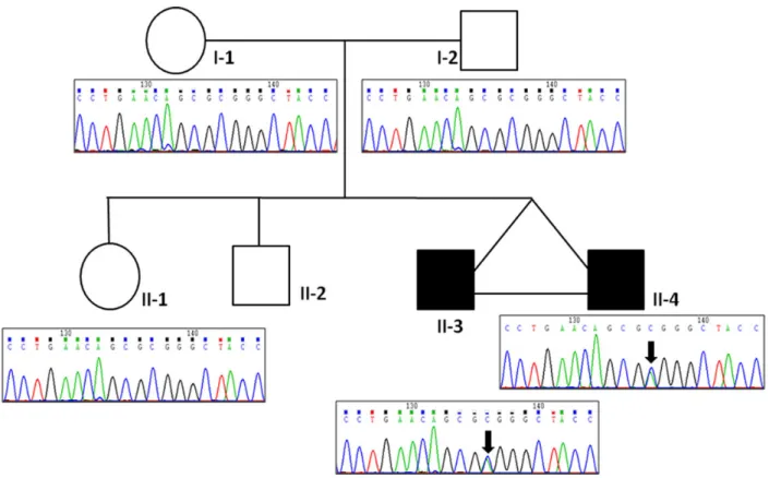
Whole exome sequencing was carried out on the parents–child trio (individuals I-1, I-2 and II-3; Fig.1). On average, we produced 262 (±9.3 SD) million reads per sample, 255 (±8.8 SD) million were properly paired and mapped uniquely to the reference genome (hg19), 233 (±8.2 SD) million reads remained after removal of du-plicate reads, and among these, 165 (±5.3 SD) million were on tar-get. This resulted in an average coverage of at least 8× for 97.43% (±0.04 SD) of the coding part of the RefSeq genes. An average of 27 412 (±210 SD) variants was detected per individual. Supple-mentary Material, Table S1 provides a summary of the exome se-quencing results for each individual.
Identification of de novo and recessive variants
In the absence of positive family history of seizures or other neurological manifestations, we hypothesized that the affected monozygotic twins were sporadic cases and looked for pathogen-ic de novo mutations in one member of the twin pair (individual II-3). We identified 2 de novo single nucleotide variants (SNVs) (Table1). All small insertions/deletions (indels) classified as de novo were rejected during visual inspection mainly due to miscal-ling of indels in homopolymer tracts or trinucleotide repeats (data not shown). Thefirst de novo SNV resulted in an aspartic acid to glycine change at position 137 of the cysteine-rich secre-tory protein LCCL domain-containing 2 precursor (CRISPLD2) gene (c.A410G; p.D137G). The second variant caused an alanine to glutamic acid substitution at position 39 of the galanin/ GMAP prepropeptide (GAL) gene (c.C116A; p.A39E). These two variants were novel, not present in dbSNPv138 nor in the Exome Aggregation Consortium (ExAC) database and were classi-fied as damaging by SIFT, Polyphen2 and Mutation taster (Table1). Up to date, CRISPLD2 has never been associated with seizures or epilepsy but with non-syndromic cleft lip with or without cleft palate in several studies (13–15) except for one (16). The unaffected sister (II-1) did not carry this variant. How-ever, as none of the twins presented with this condition, this gene was not further considered.
We also investigated the possibility of a recessive model and looked for the presence of homozygote or compound heterozy-gote mutations in the twins. We identified three genes (BCR, MYO9A and SH3TC1) with mutations compatible with an
autosomal recessive mode of inheritance (Supplementary Mater-ial, Table S2). However, none of these genes has been linked to epilepsy. The BCR gene is the site of breakpoints used in the gen-eration of the 2 alternative forms of the Philadelphia chromo-some translocation found in chronic myeloid leukemia and acute lymphocytic leukemia (OMIM#151410). The MYO9A and SH3TC1 genes have not been associated with disorders. These genes were not further considered.
The galanin neuropeptide was found to act as a potent antic-onvulsivant and regulate epileptic seizures in animal models (17, for review). However, until now its role in human epilepsy was not established. All family members were subsequently screened for the p.(A39E) mutation using Sanger sequencing. We con-firmed the wild-type allele in both parents and the unaffected sister and the mutated allele in both affected twins (Fig.1).
The 30-amino-acid galanin is located at position 33 to 62 of the 123-amino-acid galanin/GMAP prepropeptide which also comprises a 5′-hydrophobic signal peptide and a 3′-galanin mes-sage-associated peptide (GMAP). These peptides are released upon cleavage at two Lysine-Arginine (KR) dibasic sites located
on either side of galanin (18). The p.(A39E) mutation affects the alanine at position 39 of the GAL prepropeptide (galanin precur-sor), which corresponds to the 7th residue of the 30-amino-acid mature galanin peptide. Thefirst 15 residues of galanin are highly conserved throughout evolution and crucial for its biological ac-tivity (Fig.2). The last 15 residues, which are less conserved, lack receptor affinity and are believed to protect the C-terminal half from proteolysis (19–21).
Screening of the GAL gene for mutations in patients with focal epilepsy (FE)
To further appreciate the contribution of galanin mutations in FE in humans, we performed mutation analysis of coding sequences and splice site junctions of exons 2 and 3 of the GAL gene, which encode for the galanin neuropeptide, in 530 individuals with TLE and 52 patients with various types of focal epilepsies. We also analyzed exon 1 to seek for potential nonsense and frameshift mutations. We did notfind any potential pathogenic mutations suggesting that GAL mutations are a rare cause of TLE.
Figure 1. Pedigree structure of family EPI-ORA-AFF. Sanger sequencing traces showing the c.C116A ( p.A39E) mutation in exon 2 of the GAL gene. The black arrows indicate the variant present in both affected twins (II-3 and II-4).
Table 1. List of validated de novo variants
Chr Position Gene (Acc. number) Nucl. change AA change Cov.a SIFT PP2 MT GERP
11 68’453’096 GAL (NM_015973.3) c.C116A p.A39E 370 0 0.93 0.98 2.45
16 84’883’042 CRISPLD2 (NM_031476.3) c.A410G p.D137G 140 0.02 0.99 0.99 5.03
SIFT, Sorting Intolerant from Tolerant algorithm; PP2, Polyphen2; MT, MutationTaster; GERP, Genomic Evolutionary Rate Profiling score. Cov.a: Number of reads after the removal of duplicates that covers this position.
DP4#: Number of reads after the removal of duplicates that supports from left to right: forward reference allele—reverse reference allele—forward mutant allele—reverse mutant allele. Sum can be smaller than coverage because low-quality bases are not counted.
Functional analysis of the p.(A39E) mutation
Galanin-receptor affinity analysis
Displacement studies of 125I-galanin with hGal(WT) or hGal
(A39E) peptides, were performed on cell membranes from human Bowes melanoma cells endogenously expressing human GalR1 (hGalR1), CHO cells stably transfected with human GalR2 (hGalR2) andfinally Flp-In T-REx 293 cells with inducible expres-sion of human GalR3 (hGalR3). When compared to that of hGal (WT), hGal(A39E) peptide showed similar binding affinities to hGalR1 (t-test tailed P-value = 0.25) and hGalR3 (t-test two-tailed; P-value = 0.10) but significantly decreased affinities to
hGalR2 (t-test two-tailed P-value = 0.03; Fig.3and Table2). hGal (A39E) peptide also showed preferred binding to hGalR1 with a 8-fold selectivity when compared to hGalR2 and 259-fold select-ivity when compared to hGalR3 (Table2).
Galanin signaling analysis
Bowes cells expressing hGalR1 were pre-incubated with increas-ing concentrations of hGal(A39E) peptide (from 0.1 n to 10 μ) for 1 h before being stimulated with 100 n hGal(WT). Cell index was monitored before and after the addition of the pep-tides. hGal(WT) peptide showed a half maximal effective Figure 2. Multiple species alignment of the predicted amino-acid sequences of the galanin peptide andflanking cleavage sites produced using the 100-way multiZ new (hg19) option in Galaxy (https://usegalaxy.org/). Theflanking dibasic Arg-lys (KR) cleavage sites are depicted in green. Red arrows point to the residue found mutated in TLE patients ( p.A39E).
concentration (EC50) value of 22.4 ± 2.56 n (Fig.4). hGal(A39E)
peptide exhibited a dose-dependent inhibition of the hGal(WT) response, with an half maximal inhibitory concentration (IC50)
value of 486.5 ± 12.46 n, suggesting that hGal(A39E) acts as an antagonist of galanin GalR1-mediated signaling (Fig.4).
Signal transduction was examined by assessing the ability to stimulate inositol phosphate (IP) production in CHO cells expres-sing hGalR2. Both hGal(WT) and hGal(A39E) peptides stimulated IP production (Fig.5). hGal(WT) stimulated IP production with a EC50value of 171 ± 9.37 n, whereas hGal(A39E) showed a
statis-tically significant higher EC50value of 1243 ± 103 n (t-test
two-tailed, P-value = 0.0005) indicating that this mutant is a 7-fold less potent activator of hGalR2 (Fig.5). The additive effect of dif-ferent concentrations of hGal(A39E) on IP production obtained with 100 n hGal(WT) was also assessed. hGal(A39E) showed a statistically significant additive effect starting from concentra-tion of 1μ (one-way ANOVA, P-value < 0.001; Fig.6). Indeed, an increase in IP production was observed when CHO cells were co-stimulated with hGal(WT) and hGal(A39E) compared to stimu-lation with hGal(WT) alone suggesting that hGal(A39E) acts as a
hGalR2 agonist (Fig.6). hGal(A39E) at concentration below than 1μ did not show significant synergistic effect on the IP produc-tion induced by 100 n hGAL(WT) (Fig.6).
Discussion
Here, we report on the identification of a de novo p.(A39E) muta-tion in the galanin/GMAP prepropeptide (GAL) gene in a family with TLE. The GAL gene encodes for the galanin neuropeptide which possesses anticonvulsant activities in animal models. Functional analysis of the mutant galanin peptide showed that it impairs galanin function in a dominant negative manner.
A role of galanin in epilepsy wasfirst demonstrated 22 years ago by Mazarati and collaborators who showed that galanin de-creased the severity of picrotoxin-kindled convulsions in rats and that seizures-induced galanin depletion may contribute to the maintenance of seizure activity, whereas the increase of ga-lanin concentration may favor the cessation of seizures (9,22). In addition, transgenic mice that overexpress galanin showed in-creased resistance to seizure induction, whereas mice deficient for galanin showed higher seizure susceptibility (23). Galanin eli-cits a range of biological effects by interactions with three G-pro-tein-coupled receptors (GalR). Both GalR1 and GalR2, which are expressed in the hippocampus, mediate anticonvulsivant effect of galanin (24). GalR3, which is mainly expressed in the preop-tic/hypothalamic area, has not been reported to play a role in epi-lepsy (25). The hippocampal formation, which is involved in seizures underlying TLE, receives galaninergic and excitatory glutamatergic innervations. Galaninfibers suppress this excita-tory action through the opening of potassium channel which triggers membrane hyperpolarization and eventually inhibits glutamate release from presynaptic terminals (26). Our data on the function of the p.A39E mutant are consistent with this model; indeed, the p.A39E mutant, which has been shown to re-duce galanin signaling, is predicted to increase neuronal excit-ability in the hippocampus, ultimately leading to TLE. The discovery of galanin mutations suggests that genetic variation in galanin may be important in TLE.
Analysis of GAL for mutations in 530 individuals with TLE and 52 patients with various types of focal epilepsies did not reveal additional pathogenic mutations indicating that galanin muta-tions are not important contributors to the genetics of TLE. How-ever, mutation screening of other genes involved in galanin signaling, for instance the galanin receptors, would be necessary to comprehensively assess the contribution of galaninergic sys-tem to the genetics of TLE. Indeed, GalR1 has been shown to ex-acerbate hippocampal neuronal loss after status epilepticus (27) and CYM2503, a GalR2-positive allosteric modulator, has been shown to exhibit anticonvulsant effects in animal models (11).
Seventy two missense and loss-of-function mutations in the galanin/GMAP prepropeptide (GAL) gene have been reported by ExAC (http://exac.broadinstitute.org/). Among these mutations, only ten were predicted to affect galanin synthesis and/or Figure 3. Galanin-receptors binding studies: Displacement of
porcine-[125I]-galanin from membranes by peptide hGal(WT) (A) or hGal(A39E) (B). Membranes were from human Bowes melanoma cells expressing GalR1 (closed circle), CHO cells expressing GalR2 (closed square) and Flp-InT-REx 293 cells expressing GalR3 (closed triangle). The data are from three representative experiments performed in duplicates, presented as mean ± SEM.
Table 2. Ki values of hGal(WT) and hGal(A39E) towards galanin receptors 1, 2 and 3 (GalR1, 2 and 3) Ki (nM; mean ± SEM)
Ligand GalR1 GalR2 GalR3 Ki GalR2/
Ki GalR1 Ki GalR3/ Ki GalR1 hGal(WT) 6.071 ± 2.134 1.789 ± 0.684 80.90 ± 27.08 0.29 13.32 hGal(A39E) 3.001 ± 0.848 24.90 ± 7.01 780 ± 337 8.29 259.91 P-value (t-test) 0.252 0.03 0.1
function including six loss-of-function mutations and four mis-sense mutations (Supplementary Material, Table S3). For in-stance, the p.Trp25leufsTer47 frameshift mutation is predicted to disrupt the synthesis of the galanin peptide which is located at position 33 to 62 of the 123-amino-acid galanin/GMAP prepro-peptide. The other variants in the galanin/GMAP prepropeptide were missense mutations located in exons which do not encode for galanin and were therefore unlikely to affect galanin function. The pathogenic significance of these 10 variants is currently un-known as (i) there is no experimental data on their potential ef-fect on galanin function and (ii) the phenotypic status of the carriers is not available. Even if considering that they are all pathogenic, their cumulative allele frequency was calculated at 0.0246% which is well-below the prevalence of TLE estimated at 0.17% (28). Our functional analysis of the hGal(A39E) mutant fa-vored dominant negative effect over haploinsufficiency as a po-tential mechanism for causation. Among the ten variants predicted to affect galanin, the missense mutations are most likely to act in a dominant negative manner. Their cumulative frequency was calculated at 0.0075% which is compatible with their involvement as a very rare cause of TLE.
The anticonvulsivant effects of galanin have prompted the development of various approaches targeting the galanin system for the treatment of epilepsy. For example, Lu et al. (11) have de-veloped a GalR2-positive allosteric modulator, which potentiates the galanin action. Rodents with induced seizures that had re-ceived this compound showed increased seizures threshold and reduced seizures duration. The p.A39E mutant, which showed antagonist effects on GalR1 and reduced agonist effects on GalR2, is likely to have direct implications for the development of galanin-based anti-epileptic molecules.
Materials and Methods
Monozygotic twins with TLE
Monozygotic twins affected by TLE, born to healthy and non-con-sanguineous parents, were evaluated by one of the authors (AC) at the Neurology Department of Oran University Hospital (Al-geria) (Fig.1). Clinical data for the other members of the family (with the exception of II-2) were obtained in afield trip during which history and examination were performed by the same neurologist. All members of this family underwent an electroen-cephalography (EEG). All family members gave their informed consent for this study.
Based on detailed clinical seizure description in the twins, the diagnosis of TLE was established. Patient II-3, now 30 years old, denied both febrile seizures (FS) and central nervous system (CNS) infections. He has had a normal development. He started having isolated auras characterized by abdominal discomfort, in-coherent speech, palpitations and blurred vision for one or two minutes at the age of 13 years. He did not come to medical atten-tion until he had hisfirst secondarily generalized seizure one year later. His EEG activity showed burst of theta oscillations in the temporal lobe. He had complex partial seizures at 19 years of age. Brain MRI did not show hippocampal sclerosis or any other anomalies. He is treated with carbamazepine (600 mg/ day) and is seizure-free.
Patient II-4 is treated for TLE since the age of 13 years. His seizure description was suggestive of mesial temporal origin (auditory hallucinations, slow ideation, forced thoughts and Figure 6. Inositol phosphate production in CHO cells expressing human GalR2. CHO cells were pre-incubated with 1μCi [3H]-myo-inositol for 24 h before being stimulated with increasing concentrations of hGal(A39E) in the presence of 100 n of hGal(WT). Data are presented as percent of control (no peptide) and mean ± SEM of three separate experiments. ***P < 0.001; one-way ANOVA, Tukey HSD post hoc comparison test.
Figure 4. Inhibition of hGal(WT) stimulation by hGal(A39E) peptide. Serum-starved Bowes cells expressing GalR1 were pre-incubated for 1 h with hGal (A39E) peptide (0.1n to 10 μ). Subsequently, cells were stimulated with 100 n hGal(WT). Data are presented as mean ± SEM. Cell index values were normalized with respect to the cell index at the time of ligand addition and baseline-corrected by subtracting the cell index obtained with PBS treatment.
Figure 5. Inositol phosphate production in CHO cells expressing human GalR2. CHO cells were pre-incubated with 1μCi [3H]-myo-inositol for 24 h before being stimulated with peptides hGal(WT) or hGal(A39E) at increasing concentrations. Data are presented as percent of control (no peptide) and mean ± SEM of three separate experiments. Significant level compared to controls: ***P < 0.001; one-way ANOVA, Tukey HSD post hoc comparison test.
déjà-vu). The complex partial seizures included loss of aware-ness and staring. On occasion, they evolved into generalized tonic-clonic seizures. EEG showed a temporal focus and photo-sensitivity. Brain MRI analysis did not show hippocampal sclerosis or other anomalies. He became seizure-free with Leve-tiracetam (1 g/day).
Samples for screening of galanin mutations
Patients with partial epilepsy
Hundred and eighty-one unrelated individuals of European an-cestry were admitted to the Epilepsy Unit at Montpellier Univer-sity Hospital (France) with a diagnosis of FE according to ILAE Classification of 1989 (29). These patients suffered from a severe form of epilepsy with poor seizure control, although they were treated by anti-epileptic drugs (AEDs). Diagnosis was based on patient history, clinical examination, interictal and ictal EEG ana-lysis carried out with monitoring video-EEG, and magnetic reson-ance imaging. The study group consisted of patients who suffered from non-lesional and lesional FE (including vascular malformation, cortical dysplasia, and nervous system tumor). These FE patients showed the following distribution of epilepsy syndromes: 129 patients with TLE (71.4%), 25 patients with front-al epilepsy (13.7%), 12 patients with frontotemporfront-al epilepsy (6.6%), 4 patients with temporo-occipital epilepsy (2.2%), 3 pa-tients with mesial-parietal epilepsy (1.6%), 2 papa-tients with par-ietal epilepsy (1.1%), 2 patients with parieto-occipital epilepsy (1.1%), 1 patient with multifocal epilepsy (0.5%), 1 patient with temporo-parietal epilepsy (0.5%) and 2 patients with undeter-mined FE (1.1%). This study was approved by the ethics commit-tee of the University Hospitals of Montpellier.
Thirty-seven unrelated patients were recruited in Lyon (n = 14) (GL, PR), Strasbourg (n = 18) (GR, EH) and Clermont Ferrand (n = 5) (DSR) through a multicenter research project (Projet Hospitalier de Recherche Clinique 2007-A00481-52 from the French ministry of health, coordinator Dr Philippe Ryvlin) that was approved by the local Ethics Committee (CPP SUD-EST IV). All these patients had a diagnosis of drug-resistant TLE based on patient history, clinical examination, interictal and ictal EEG analysis carried out with monitoring video-EEG, and magnetic resonance imaging.
Hundred and thirty-one unrelated adult individuals of Euro-pean ancestry were recruited at the Swiss Epilepsy Center in Zur-ich (Switzerland). All Individuals were diagnosed with TLE, two of which had a bilateral temporal focus. Within this cohort, severity of phenotypes, subgroups of TLE, familiarity as well as response to AEDs was diverse. The study was approved by the ethics com-mittee of the Kanton Zurich.
Hundred andfifty-nine patients with sporadic lateral tem-poral epilepsy (LTE) were collected as part of a collaborative study supported by the Genetics Commission of the Italian Lea-gue Against Epilepsy (LICE). Diagnosis of LTE was made according to a clinical history of at least two lifetime seizures with auditory symptoms such as ringing, humming, sounds, voices, music, or sudden hearing loss. Sporadic LTE patients were selected accord-ing to the followaccord-ing inclusion criteria: (i) absence offirst and se-cond-degree relatives (siblings, children, parents, grandparents, aunts and uncles) with epileptic seizures; (ii) absence of neuror-adiological abnormalities including mesial temporal sclerosis; (iii) absence of mutations in the LGI1 gene. Exclusion criteria were an insufficient or doubtful family history and lack of neuroi-maging data. All patients underwent repeated EEG recordings ac-cording to the 10–20 International System during wakefulness and, when possible, during diurnal sleep induced by sleep
deprivation. Each patient underwent high resolution MRI scan of the brain. This study was approved by LICE and local ethics committees.
Sixty-four unrelated patients were recruited at the Depart-ment of Neurology and Epileptology (Tübingen) and DepartDepart-ment of Neuropediatrics (Kiel). Thirty-two children were diagnosed with focal epilepsies (FE), 18 children with TLE, 10 of which had abnormal MRI scan (mesial temporal sclerosis (n = 1), cortical dysplasia (n = 5), 1 with heterotopia (n = 1), arachnoïdal cysts (n = 2), proencephalic cyst temporal (n = 1)). Among these 18 TLE pa-tients, 11 had normal intelligence, 6 had intellectual disability and 1 was not assessed. Six patients were diagnosed with frontal lobe epilepsy (FLE), two of which had abnormal MRI scan (cortical dysplasia frontal lobe (n = 1), unspecific gliosis (n = 1). Among these six FLE patients, one had behavior problems, one had mild intellectual disability and four had normal intelligence. Eight children with undetermined focal seizures (unclassified focal seizures/secondary generalized seizures), two of which had abnormal MRI scan [subcortical heterotopias suspected with hippocampus malrotation (n = 1), unspecific anomalies (n = 1)]. Among these eight patients, one had behavior problems, one had mild intellectual disability and six had normal intelligence. Fourty-nine unrelated Caucasian individuals (mainly of Ger-man origin) with mesial TLE were recruited as a part of the gen-etic and pharmacogengen-etic studies which were approved by the local Ethics Committee (Department of Neurology and Epileptol-ogy University of Tübingen). All reported patients gave written informed consent previous to the study inclusion. The diagnosis of mesial TLE occurred according to patient history, clinical examination, EEG analysis and magnetic resonance imaging. A hippocampal sclerosis was detected in almost all cases (n = 48; 97%). Most of the patients (n = 40, 81%) suffered from refractory epilepsy, only 14% became seizure-free (n = 5 without surgery and n = 2 after surgery). In two cases, the outcome was unknown. Before study inclusion, the patients had an average of 5.4 (n = 49) trials of anti-epileptic medication and 6.6 (n = 42) seizures per month on average.
Exome capture and sequencing
Exome capture from the parents and one of the twins of family EPI-ORA-AFF (Fig.1, individuals I-1, I-2 and II-3) was carried out on 2μg of genomic DNA extracted from blood using the SureSelect Human ALL Exon v5 kit (Agilent Technologies). High-throughput sequencing was performed on a HiSeq2000. Demultiplexed fastq files were obtained for each sample using the Illumina CASAVA v1.8.2 software and processed by our‘in-house’ pipeline running on the Vital-IT Center for high-performance computing of the Swiss Institute of Bioinformatics (SIB; (http://www.vital-it.ch). Specifically, Burrows–Wheeler Aligner (BWA) was used to align the sequencing reads to the human reference genome NCBI build (GRCh37/hg19). SAMtools was used to remove duplicate reads. SNVs and small insertions and deletions (INDELs) were called using bcftools and Pindel 0.2.4, respectively. The minimum number of reads required for allele calling was set at eight. SNVs and INDELS variants were then functionally annotated using the ANNOVAR package.
Identification of de novo and recessive variants
Potential pathogenic variants under de novo or recessive models were identified using VariantMaster (30). Variants detected in the proband with Samtools and PINDEL quality scores≥150 and ≥600, respectively were retained for subsequent analysis. These
variants were furtherfiltered so as to exclude variants found out-side of exons or splice sites (±2) and variants with a minimum al-lele frequency greater than 0.02 in dbSNPv138 (Database of Single Nucleotide Polymorphism) and/or the Exome Variant Server (EVS) database. VariantMaster used the raw data (BAMfiles) to ro-bustly estimate the probability of the remaining variants to be present in the parents and siblings. Variants were classified as de novo only if both parents were found to be homozygous for the reference allele. Homozygote or compound heterozygote var-iants in the proband that were predicted as damaging by at least two of the three prediction algorithms used (SIFT, Polyphen2 and Mutation Taster) were classified as recessive only if both parents were carriers and the genotype of the unaffected sister was dif-ferent from that of the proband. All variants classified as de novo or recessive by VariantMaster, were subsequently visually inspected using the SAMtools text alignment viewer. The re-maining candidate variants were then validated using Sanger se-quencing on an ABI 3730xl DNA Analyzer (Applied Biosystems).
Peptide synthesis
Peptides (Human galanin1–30 wild type [hGal(WT)]:GWTLNSA GYLLGPHAVGNHRSFSDKNGLTS and Human galanin A39E mu-tant [hGal(A39E)]: GWTLNSEGYLLGPHAVGNHRSFSDKNGLTS) were synthesized at 0.1 mmol scale on an automated microwave peptide synthesizer (Biotage® Initiator + Alstra™) using Fmoc (fluorenylmethyloxycarbonyl) solid-phase peptide synthesis strategy with HMPB-ChemMatrix 0.4 mmol/g resin as solid phase to obtain peptide acid. Thefirst amino acid was attached to resin using symmetrical anhydride. All other coupling reac-tions were carried out using OxymaPure/DIC in DMF with DIEA as an activator base. Thefinal cleavage was performed using standard protocol (95% TFA/2.5% TIS/2.5% H2O). Peptides were purified by reversed-phase HPLC using BioBasic C-8 column (Thermo Scientific, Sweden) and a gradient of 20–80% aceto-nitrile/water containing 0.1% TFA. The identity of peptides was analyzed by MALDI-TOF mass spectrometry Voyager-DE STR (Applied Biosystems) in positive linear mode using α-cyano-4-hydroxycinnamic acid as matrix.
Galanin-receptor binding assays
Cells for125I-galanin-receptor displacement studies were seeded in 150-mm dishes and cultured 3–4 days until confluent. Cell dishes were washed and scraped into phosphate-buffered saline (PBS) and centrifuged twice at 4°C, 3000 g for 5 min. The pellet was re-suspended in assay buffer (20 m HEPES, 5 m MgCl2,
pH 7.4) supplemented with EDTA (5 m EDTA) and incubated on ice for 45 min before centrifugation at 4°C, 8500 g for 15 min. After washing, the pellet was re-suspended in assay buffer sup-plemented with 1% protease inhibitor cocktail (Sigma–Aldrich, St. Louis, MO, USA) to a protein concentration of 1 mg/ml. Protein concentration was determined according to Lowry (BioRad, Stockholm, Sweden). Displacement studies on cell membranes were performed in afinal volume of 200 μl, containing 0.15 n porcine-[125I]-galanin (2200 Ci/mmol; Perkin–Elmer Life Science,
Boston, MA, USA), 30μg cell membrane and various concentra-tions of peptide (10−5–10−9M). Peptide solutions were made in
assay buffer supplemented with 0.3% bovine serum albumin (BSA). Samples were shaken at 37°C for 30 min and filtered through a MultiScreen-FBfilter plate (Millipore, Billerica, MA, USA) pre-soaked in 0.3% polyethylenimine solution (Sigma–Al-drich). Thefilters were washed thrice with assay buffer and the
retained radioactivity was determined in aβ-counter (Tri-Carb Li-quid Scintillation Analyzer, model 2500 TR; Packard Instrument Company, Meriden, CT, USA) using OptiPhase Supermix Cocktail (Perkin–Elmer Life Science, Boston, MA, USA) as scintillation fluid. IC50values for the peptides were calculated using Prism
5.0 (GraphPad Software Inc., San Diego, CA, USA) and converted into Kivalues using the equation of Cheng–Prusoff (31).
xCELLigence cellular impedance assay
The impedance-based readout used by the xCELLigence System (Roche Applied Science) is based on the principle that the adhe-sion of cultured cells directly onto microelectrodes induces changes in the local ionic environment at the electrode/solution interface, conferring an increase in electrode impedance. As a re-sult, any changes in cell physiological properties that modulate the physical contact between cell and electrode will be reflected by changes in the measured impedance, defined by the cell index variable. xCELLigence System can be used for functional screening of G-protein-coupled receptors (GPCRs) activity, based on its ability to modulate the actin cytoskeleton and cell adhesion (32–34). Bowes cells expressing GalR1 were seeded at 20 000 cells per well on 96-well E-Plates (ACEA Biosciences Inc.) and placed on the Real-time xCELLigence Cell Analyzer (Roche Applied Science) platform at 37°C and 5% CO2. After 24 h incuba-tion, growth media was changed for assay buffer (HBSS contain-ing 0.1% BSA). Three hours later, hGal(A39E) peptide was added (0.1 n to 10 µ), after 1 h incubation, hGal(WT) was added at 100 n. The xCELLigence System was employed to measure changes in cellular impedance following ligand stimulation. Re-sulting dose–response relationships were plotted using mean va-lues from four replicates. Cell index vava-lues were normalized by dividing the cell index at the time of ligand addition and base-line-corrected by subtracting the cell index obtained with PBS treatment. Data analysis was done with built-in xCELLigence Sys-tem software and presented with GraphPad Prism.
Inositol phosphate accumulation assay
CHO cells stably expressing human GalR2 were seeded in 12-well plates and cultured until confluent. Then cells were incubated 24 h with 1μCi [3H]-myo-inositol in M-199 medium containing 100 U ml−1penicillin and 100μg ml−1streptomycin. CHO GalR2 cells were washed twice with HEPES Krebs Ringer (HKR) buffer (5 m HEPES, 137 m NaCl, 2.68 m KCl, 2.05 m MgCl2, 1.8 m CaCl2, 1 g/l glucose, pH7.4) followed by 10 min pre-incu-bation in 800μl HKR buffer with 10 m LiCl at 37°C. hGal(A39E) peptide (10−5–10−8M) with or without hGal(WT) (10−7M) was
added to afinal volume of 1000 μl and incubated for 60 min at 37°C. The reaction was terminated by addition of 200μl ice cold 20% perchloric acid followed for 10 min followed by the addition of 1.5 M KOH/75 m HEPES (pH7). Anion exchange chromatog-raphy was performed over 1 cm 50:50 Dowex (AG 1-X8 Resin, 200– 400 mesh formate; BioRad, Hercules, CA, USA). The columns were washed with 5 ml distilled water before the IPs were eluted with 5 ml of 0.1 formic acid/1.2 ammonium formate. Radio-activity of the eluate was determined using scintillation count-ing in a β-counter (Tri-Carb Liquid Scintillation Analyzer, model 2500 TR, Packard Instrument Company, Meriden, CT, USA) using Utima Flo AF (Perkin–Elmer Life Science, Boston, MA, USA) as scintillationfluid. Each sample was normalized against the total count obtained before anion exchange chromatography.
Supplementary Material
Supplementary Material is available at HMG online.
Acknowledgements
We thank Dr Michael Lang and Prof. Dr Herbert Schreiber (Prac-tice for Neurology and Psychiatry, Ulm, Germany) for their contri-bution to the recruitment of the patients from Tübingen. Conflict of Interest statement. None declared.
Funding
We thank the Swiss National Science Foundation (SNF-144082) and NCCR Synapsy grant for supporting the S.E.A. laboratory. C. N. was supported by Telethon-Italy (Grant no. GGP12078) and by the LICE Genetics Commission. U.L. is supported by the Swed-ish research Council.
References
1. Manford, M., Hart, Y.M., Sander, J.W. and Shorvon, S.D. (1992) National General Practice Study of Epilepsy (NGPSE): partial seizure patterns in a general population. Neurology, 42, 1911–1917.
2. Berkovic, S.F., McIntosh, A., Howell, R.A., Mitchell, A., Shef-field, L.J. and Hopper, J.L. (1996) Familial temporal lobe epi-lepsy: a common disorder identified in twins. Ann. Neurol., 40, 227–235.
3. Vadlamudi, L., Scheffer, I.E. and Berkovic, S.F. (2003) Genetics of temporal lobe epilepsy. J. Neurol. Neurosurg. Psychiatry, 74, 1359–1361.
4. Schulte, U., Thumfart, J.O., Klocker, N., Sailer, C.A., Bildl, W., Biniossek, M., Dehn, D., Deller, T., Eble, S., Abbass, K. et al. (2006) The epilepsy-linked Lgi1 protein assembles into pre-synaptic Kv1 channels and inhibits inactivation by Kvbeta1. Neuron, 49, 697–706.
5. Dibbens, L.M., de Vries, B., Donatello, S., Heron, S.E., Hodgson, B.L., Chintawar, S., Crompton, D.E., Hughes, J.N., Bellows, S.T., Klein, K.M. et al. (2013) Mutations in DEPDC5 cause familial focal epilepsy with variable foci. Nat. Genet., 45, 546–551. 6. Bar-Peled, L., Chantranupong, L., Cherniack, A.D., Chen, W.
W., Ottina, K.A., Grabiner, B.C., Spear, E.D., Carter, S.L., Meyer-son, M. and Sabatini, D.M. (2013) A Tumor suppressor com-plex with GAP activity for the Rag GTPases that signal amino acid sufficiency to mTORC1. Science, 340, 1100–1106. 7. Tatemoto, K., Rokaeus, A., Jornvall, H., McDonald, T.J. and
Mutt, V. (1983) Galanin—a novel biologically active peptide from porcine intestine. FEBS Lett., 164, 124–128.
8. Mechenthaler, I. (2008) Galanin and the neuroendocrine axes. Cell. Mol. Life Sci., 65, 1826–1835.
9. Mazarati, A.M., Halaszi, E. and Telegdy, G. (1992) Anticonvul-sive effects of galanin administered into the central nervous system upon the picrotoxin-kindled seizure syndrome in rats. Brain Res., 589, 164–166.
10. Lerner, J.T., Sankar, R. and Mazarati, A.M. (2008) Galanin and epilepsy. Cell Mol. Life Sci., 65, 1864–1871.
11. Lu, X., Roberts, E., Xia, F., Sanchez-Alavez, M., Liu, T., Baldwin, R., Wu, S., Chang, J., Wasterlain, C.G. and Bartfai, T. (2010) GalR2-positive allosteric modulator exhibits anticonvulsant effects in animal models. Proc. Natl. Acad. Sci U S A, 107, 15229–15234.
12. Nikitidou, L., Torp, M., Fjord-Larsen, L., Kusk, P., Wahlberg, L. U. and Kokaia, M. (2014) Encapsulated galanin-producing cells attenuate focal epileptic seizures in the hippocampus. Epilepsia, 55, 167–174.
13. Chiquet, B.T., Lidral, A.C., Stal, S., Mulliken, J.B., Moreno, L.M., Arcos-Burgos, M., Valencia-Ramirez, C., Blanton, S.H. and Hecht, J.T. (2007) CRISPLD2: a novel NSCLP candidate gene. Hum. Mol. Genet., 16, 2241–2248.
14. Shi, J., Jiao, X., Song, T., Zhang, B., Qin, C. and Cao, F. (2010) CRISPLD2 polymorphisms are associated with non-syndrom-ic cleft lip with or without cleft palate in a northern Chinese population. Eur. J. Oral Sci., 118, 430–433.
15. Letra, A., Menezes, R., Cooper, M.E., Fonseca, R.F., Tropp, S., Govil, M., Granjeiro, J.M., Imoehl, S.R., Mansilla, M.A., Murray, J.C. et al. (2011) CRISPLD2 variants including a C471 T silent mutation may contribute to nonsyndromic cleft lip with or without cleft palate. Cleft. Palate Craniofac. J., 48, 363–370.
16. Girardi, A., Martinelli, M., Carinci, F., Morselli, P.G., Caramelli, E. and Scapoli, L. (2011) No evidence for a role of CRISPLD2 in non-syndromic cleft lip with or without cleft palate in an Ital-ian population. Eur. J. Oral. Sci., 119, 102–105.
17. Webling, K.E., Runesson, J., Bartfai, T. and Langel, U. (2012) Galanin receptors and ligands. Front. Endocrinol. (Lausanne), 3, 146.
18. Evans, H., Baumgartner, M., Shine, J. and Herzog, H. (1993) Genomic organization and localization of the gene encoding human preprogalanin. Genomics, 18, 473–477.
19. Land, T., Langel, U. and Bartfai, T. (1991) Hypothalamic deg-radation of galanin(1–29) and galanin(1–16): identification and characterization of the peptidolytic products. Brain Res., 558, 245–250.
20. Land, T., Langel, U., Low, M., Berthold, M., Unden, A. and Bart-fai, T. (1991) Linear and cyclic N-terminal galanin fragments and analogs as ligands at the hypothalamic galanin receptor. Int. J. Pept. Protein Res., 38, 267–272.
21. Bedecs, K., Langel, U. and Bartfai, T. (1995) Metabolism of galanin and galanin (1–16) in isolated cerebrospinal fluid and spinal cord membranes from rat. Neuropeptides, 29, 137–143.
22. Mazarati, A.M., Liu, H., Soomets, U., Sankar, R., Shin, D., Kat-sumori, H., Langel, U. and Wasterlain, C.G. (1998) Galanin modulation of seizures and seizure modulation of hippocam-pal galanin in animal models of status epilepticus. J. Neurosci., 18, 10070–10077.
23. Mazarati, A.M., Hohmann, J.G., Bacon, A., Liu, H., Sankar, R., Steiner, R.A., Wynick, D. and Wasterlain, C.G. (2000) Modula-tion of hippocampal excitability and seizures by galanin. J. Neurosci., 20, 6276–6281.
24. Lang, R., Gundlach, A.L. and Kofler, B. (2007) The galanin pep-tide family: receptor pharmacology, pleiotropic biological ac-tions, and implications in health and disease. Pharmacol. Ther., 115, 177–207.
25. Mennicken, F., Hoffert, C., Pelletier, M., Ahmad, S. and O’Don-nell, D. (2002) Restricted distribution of galanin receptor 3 (GalR3) mRNA in the adult rat central nervous system. J. Chem. Neuroanat., 24, 257–268.
26. Zini, S., Roisin, M.P., Langel, U., Bartfai, T. and Ben-Ari, Y. (1993) Galanin reduces release of endogenous excitatory amino acids in the rat hippocampus. Eur. J. Pharmacol., 245, 1–7.
27. Schauwecker, P.E. (2010) Galanin receptor 1 deletion exacer-bates hippocampal neuronal loss after systemic kainate ad-ministration in mice. PLoS One, 5, e15657.
28. Tellez-Zenteno, J.F. and Hernandez-Ronquillo, L. (2012) A re-view of the epidemiology of temporal lobe epilepsy. Epilepsy Res. Treat, 2012, 630853.
29. Proposal for revised classification of epilepsies and epileptic syndromes. (1989) Commission on Classification and Terminology of the International League Against Epilepsy. Epilepsia, 30, 389–399.
30. Santoni, F.A., Makrythanasis, P., Nikolaev, S., Guipponi, M., Robyr, D., Bottani, A. and Antonarakis, S.E. (2014) Simultan-eous identification and prioritization of variants in familial, de novo, and somatic genetic disorders with VariantMaster. Genome Res., 24, 349–355.
31. Cheng, Y. and Prusoff, W.H. (1973) Relationship between the inhibition constant (K1) and the concentration of inhibitor
which causes 50 per cent inhibition (I50) of an enzymatic re-action. Biochem. Pharmacol., 22, 3099–3108.
32. Solly, K., Wang, X., Xu, X., Strulovici, B. and Zheng, W. (2004) Application of real-time cell electronic sensing (RT-CES) tech-nology to cell-based assays. Assay Drug Dev. Technol., 2, 363– 372.
33. Scott, C.W. and Peters, M.F. (2010) Label-free whole-cell as-says: expanding the scope of GPCR screening. Drug Discov. Today, 15, 704–716.
34. Stallaert, W., Dorn, J.F., van der Westhuizen, E., Audet, M. and Bouvier, M. (2012) Impedance responses reveal beta(2)-adren-ergic receptor signaling pluridimensionality and allow classi-fication of ligands with distinct signaling profiles. PLoS One, 7, e29420.