Fibrin gel as a three dimensional matrix in cardiovascular
tissue engineering
q
Qing Ye
a, Gregor ZuÈnd
a,*, Peter Benedikt
a, Stefan Jockenhoevel
a, Simon P. Hoerstrup
a,
Shelly Sakyama
b, Jeffrey A. Hubbell
b, Marko Turina
aaClinic for Cardiovascular Surgery, University Hospital Zurich, RaÈmistrasse 100, 8091 Zurich, Switzerland bDepartment of Materials and Institute for Biomedical Engineering, ETH-Zurich, Switzerland
Received 7 September 1999; received in revised form 19 January 2000; accepted 26 January 2000
Abstract
Objective: In tissue engineering, three-dimensional biodegradable scaffolds are generally used as a basic structure for cell anchorage, cell proliferation and cell differentiation. The currently used biodegradable scaffolds in cardiovascular tissue engineering are potentially immu-nogenic, they show toxic degradation and in¯ammatory reactions. The aim of this study is to establish a new three-dimensional cell culture system within cells achieve uniform distribution and quick tissue development and with no toxic degradation or in¯ammatory reactions. Methods: Human aortic tissue is harvested from the ascending aorta in the operation room and worked up to pure human myo®broblasts cultures. These human myo®broblasts cultures are suspended in ®brinogen solution and seeded into 6-well culture plates for cell develop-ment for 4 weeks and suppledevelop-mented with different concentrations of aprotinin. Hydroxyproline assay and histological studies were performed to evaluate the tissue development in these ®brin gel structures. Results: The light microscopy and the transmission electron microscopy studies for tissue development based on the three-dimensional ®brin gel structures showed homogenous cell growth and con¯uent collagen production. No toxic degradation or in¯ammatory reactions could be detected. Furthermore, ®brin gel myo®broblasts structures dissolved within 2 days in medium without aprotinin, but medium supplemented with higher concentration of aprotinin retained the three-dimensional structure and had a higher collagen content (P , 0:005) and a better tissue development. Conclusions: A three-dimensional ®brin gel structure can serve as a useful scaffold for tissue engineering with controlled degradation, excellent seeding effects and good tissue development. q 2000 Elsevier Science B.V. All rights reserved.
Keywords: Tissue engineering; Scaffold; Fibrin gel; Cardiovascular
1. Introduction
Heart valve replacements are routinely performed cardiac operations for treating severely diseased valves. Commonly used valve substitutes include mechanical valves, biopros-thetic valves and homografts. These prosbiopros-thetic valves func-tion well, but each of them has its own inherent limitafunc-tions. This leads to a series of studies to determine, if tissue engi-neering principles can be used to develop valve tissue substitutes out of isolated autologous cells to avoid the known limitations of the common heart valve prosthesis [1±4].
The most common mode of engineering heart valve tissue is based on using a three-dimensional biodegradable
scaf-fold for cell anchorage. The scafscaf-fold serves as a synthetic template for cell growth and tissue development during in vitro culture. First, cells have to be seeded on the scaffold and then the cells are going to attach the ®bers for further proliferation and differentiation. The scaffold degrades while tissue development [5].
Ideally, scaffolds for tissue engineering should provide a high surface area for cell-polymer interactions, suf®cient space for extracellular matrix regeneration, and minimal diffusional constraints during in vitro culture. The scaffolds should resorb once it has served its purpose of providing a template for regeneration tissue, and the scaffold degrada-tion rate should be adjustable to match the rate of tissue regeneration [6]. The currently used materials for maintain-ing three-dimensional structures in tissue engineermaintain-ing are either polymers composed of chemical substances like poly-glycolic acid or polyhydroxybutyrate or gels out of extra-cellular matrix proteins such as collagen. However, these materials are still far from ideal, they are expensive and
1010-7940/00/$ - see front matter q 2000 Elsevier Science B.V. All rights reserved. PII: S1010-7940(00)00373-0
www.elsevier.com/locate/ejcts
qPresented at the 13th Annual Meeting of the European Association for
Cardio-thoracic Surgery, Glasgow, Scotland, UK, September, 5±8, 1999. * Corresponding author. Tel.: 141-1-255-38-01; fax: 141-1-255-43-69. E-mail address: [email protected] (G. ZuÈnd)
potentially immunogenic and in addition they show toxic degradation and in¯ammatory reactions [6±8].
Fibrin gel can be produced of the patients blood and be used as an autologous scaffold for the seeded ®broblasts to create a three-dimensional structure and furthermore no toxic degradation or in¯ammatory reactions will be expected.
2. Material and methods
2.1. Human aortic myo®broblast cell expansion
Human aortic tissue was harvested from the ascending aorta in the operating room. After harvesting, the explants were rinsed with phosphate-buffered saline (PBS) and stripped off adventitia with scissors under a laminar ¯ow hood. The tissue was then cut into small pieces of 2 £ 2 mm for primary culture in 75 cm2vented polystyrene cell culture
¯asks (Falcon 3111, Becton Dickinson, Lincoln Park, NJ) with Dulbecco's modi®ed Eagle's medium supplemented with 10% fetal bovine serum and 1% streptomycin (Gibco BRL±Life Technologies, Grand Island, NY). After 3±4 weeks, human aortic myo®broblasts grew into con¯uent monolayers and were serially passaged by trypsinization (trypsin/EDTA solution, 0.05/0.02%, Gibco BRL-Life Technologies, 3±5 min) and subcultured to obtain suf®cient cell numbers for cell seeding (passage 3±4).
2.2. Preparation of ®brin gel
Fifty milligrams plasminogen-free ®brinogen from pooled human plasma (Fluka) was dissolved in 3 ml distilled water and dialyzed vs. Tris-buffered saline (TBS) overnight at room temperature. The solution was ®ltered through a 5 mm and 0.22 mm ®lter for sterilization. The ®brinogen concentration was determined by measuring the absorbance at 280 nm. The volume required to obtain 3.5 mg of ®brinogen in the seeding was calculated based on the determined ®brinogen concentrations.
2.3. Seeding
Seedings were performed in polystyrene 6-well ¯at-bottom culture plates (Costar 3516, Cambridge, MA). To construct a 1-mm thick cell-®brin gel structure a 6-well culture plate, 750 000 human myo®broblasts were suspended in 1 ml ®brinogen solution composed of 50 mM CaCl250 ml, 100 ml thrombin (20 U/ml in TBS)(Sigma
Inc.), 350 ml TBS, 500 ml ®brinogen. The ®nal ®brin gels contains 3.5 mg ®brinogen, 2.5 mM Ca11, and 2 NIH units
thrombin. The culture plates were then put in cell culture incubator for polymerization for 1 h. Afterwards 5 ml medium was added to the seeded plates.
2.4. Culture
The cell-®brin gel structure was cultured for 1 month with
Dulbecco's modi®ed Eagle's medium with 10% fetal bovine serum and 1% streptomycin (Gibco BRL±Life Tech-nologies). The medium was further supplemented with 1.0 mM l-ascorbic acid 2-phosphate, 0.4 M proline (Sigma Chemical Co, St. Louis, MO) and different concentrations of aprotinin (5, 10 and 20 mg/ml) and was changed on a daily base.
2.5. Structure assessment 2.5.1. Collagen content
Collagen content was estimated by hydroxyproline assay according to Reddy's method [9]. Brie¯y, samples were lyophilized and hydrolyzed with 2 M NaOH. Hydrolyzed free hydroxyproline was oxidized with chloramine-T and the addition of Ehrlich's reagent resulted in the formation of a chromophore that was measured at 550 nm.
2.5.2. Histological evaluation
Specimens for light microscope examination were ®xed in 4% formalin, embedded in paraf®n and sectioned. The sections were stained by haematoxylin and eosin (HE) or Masson's trichrome. Transmission electron microscopy specimens were ®xed with Na-Cacodylat-buffered glutaral-dehade (2.5%) and paraformaldehyde (0.8%) solution, and were post-®xed with 1% osmium tetroxide, dehydrated in a series of alcohol, and embedded. Ultra-thin sections were stained with uranyl acetate and lead citrate. The specimens were observed under a transmission electron microscope. 2.5.3. Statistics
Results data were expressed as mean ^ standard devia-tion (SD). Comparisons between groups were performed by ANOVA test. Statistical signi®cance was set at P , 0:05. Linear regression analysis was utilized to evaluate the corre-lation. Data and graphs were proceeded with StatView 4.5 (Abacus Concepts, Inc, Berkeley, CA).
3. Results
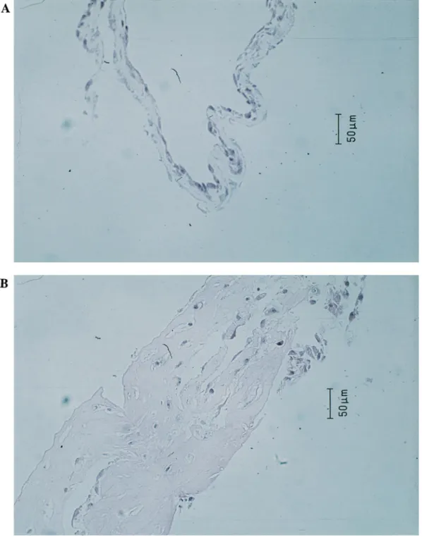
The light microscopy and the transmission electron microscopy studies for tissue development based on the three-dimensional ®brin gel structures showed homogenous cell growth and collagen production. The thickness of the tissue was reduced from 1 to 0.8 mm during the culturing period. The tissue showed a uniform cell distribution with a mean cell density of 34 ^ 5.7 cells/®eld. No toxic degrada-tion reacdegrada-tions could be detected (Figs. 1 and 2). In the group with no aprotinin the ®brin gel was dissolved within 2 days. In the group with low aprotinin concentration (5 mg/ml) the gel was phagozyted by the cells after 1 week and the struc-ture appeared to be a thin cellular layer (Fig. 1A). A certain ®brinolysis appeared in the group with 15 mg/ml after 3 weeks but the ®brin gel did not degrade completely. In the group with 20 mg/ml aprotinin no ®brinolysis was visible during the whole culture period. In addition, a
three-dimen-sional structure with multilayer myo®broblasts surrounded by extracellular matrix was detected in this group (Fig. 1B). Furthermore, the transmission electron microscope docu-ments con¯uent collagen production and viable cells in the group with high aprotinin concentration (Fig. 2).
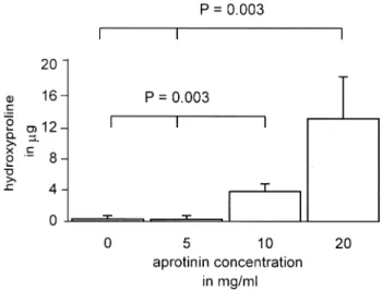
After 31 days the hydroxyproline content was measured, which is directly related to the collagen content. A signi®-cantly (P 0:003) higher amount of hydroxyproline in the groups with 15 and 20 mg/ml aprotinin compared with the
groups with no or low aprotinin concentration was observed (Fig. 3). The biochemical results con®rmed the light micro-scopy and electron micromicro-scopy ®ndings.
4. Discussion
Cell distribution and attachment in three dimensions are mainly determined by the gravity [10]. When cells are
Fig. 1. Light microscopy pictures of cell ®brin gel cultured in different aprotinin concentration. (A) Cultured in lower concentration of aprotinin (5 mg), ®brin gel degraded and the structure appeared to be a thin cellular layer. (B) Cultured in high concentration of aprotinin (20 mg/ml), the cell-®brin gel structure demonstrated a multilayer-structure with myo®broblasts surrounded by extracellular matrix (hematoxylin and eosin staining).
seeded onto the scaffolds not all of them will attach. Some will be unattached and are lost for further tissue develop-ment. A fast cell attachment on the scaffold provides a spatially uniform distribution of seeded cells. Furthermore, an initial high homogenous cell distribution is associated with high rates of extracellular matrix production [11±13].
Polyglycolic acid polymers (PGA) as scaffolds have been widely studied. They are biodegradable and content a signif-icant surface area for cell attachment. In addition their high porosity guaranties an excellent nutrition supply. Unfortu-nately the cell attachments on PGA meshes are low and, therefore, they need to be precoated with cell adhesion factors to enhance the cell attachments [14]. Collagen gels as three-dimensional scaffold provides uniform distribution with good cell attachments, but they are not biodegradable and potentially immunogenic. Furthermore there is a big variation between produced collagen batches [15].
In the presented study a three-dimensional ®brin gel structure is used as a scaffold for tissue engineering. Fibrin plays an important role in natural wound healing and is used as sealant and adhesive in surgery. It is formed by the enzy-matic polymerization of ®brinogen. A special feature of ®brin, both formed naturally or therapeutically, is that it is degraded and remodelled by cell-associated enzymatic
activity during cell migration and wound healing. Aprotinin, a proteinase inhibitor, can slow down or stop ®brinolysis. Aprotinin acts as an inhibitor of human trypsin, plasmin, and plasma and tissue kallikrein by forming reversible-enzyme-inhibitor complexes. It stops ®brinolysis via inhi-biting plasmin. In the presented study, different concentra-tions of aprotinin were used to control the degradation of three dimensional ®brin gel matrix. The results demonstrate, that the degradation of three-dimensional ®brin gel struc-tures is directly adjusted to the supplemented aprotinin concentrations.
In vitro studies recently reported that ®brin gels might have other properties such as promoting cell migration, proliferation, and matrix synthesis through the release of platelet-derived growth factors and the transforming growth factor beta [16]. In addition there is the possibility to incor-porate cell mediators like growth factors and other bioactive peptides and proteins into the ®brin gel [17±19]. This would render a more tissue speci®c environment for the isolated cells and further amelioration of cell function might be expected The gel structure also serves as a semi-permeable membrane, that separates the cells from direct contact with the medium. Collagen and other newly synthesized extra-cellular matrix components can accumulate in the intercel-lular space rather than diffusing into surrounding medium. In case of engineering autologous re-implant tissue, the whole procedure might be carried out in an autologous system, in which autologous plasma from the patient itself can be used for producing ®brin gel. The potential possibi-lity of antigenity would completely be eliminated.
In conclusion, a three-dimensional ®brin gel structure can serve as a useful scaffold for tissue engineering with controlled degradation, excellent seeding effects and good tissue development. At that moment the developed tissue with a thickness of 1 mm is not stable enough to create cardiovascular grafts on arterial side. Further investigations
Fig. 2. Transmission electron microscopy documents con¯uent collagen production and viable cells in the 20 mg/ml aprotinin group and ®brin gel remain undergraded. Scale bar 3 mm.
Fig. 3. Comparison of Hyp content in different aprotinin concentration group. More Hyp content was detected in high aprotinin concentration (10 and 20 mg/ml) groups. Hyp hydroxyproline.
to improve the mechanical properties of the new developed tissue with growth factors, de®ned mechanical forces are in process. Possible methods of the production of cardiac valves from this tissue are the formation like the production of valves from pericardium. Beyond the formation of cardi-ovascular structures with the elastic ®brin gel by casting is imaginable in the future.
Acknowledgements
Many thanks goes to Professor Grosscurth and Mrs Erni for performing electron microscopy studies. This work was supported by a grant from the Bonizzi-Theler Foundation. References
[1] Shinoka T, Breuer CK, Tanel RE, ZuÈnd G, Miura T, MA PX, Langer R, Vacanti JP, Mayer JE. Tissue engineering heart valves: valve lea¯et replacement study in a lamb model. Ann Thorac Surg 1995;60:S513±S516.
[2] Shinoka T, Ma PX, Shum Tim D, Breuer CK, Cusick RA, ZuÈnd G, Langer R. Tissue engineering heart valves: Autologous valve lea¯et replacement in a lamb model. Circulation 1996;94(Suppl II):II164± II168.
[3] Zund G, Hoerstrup SP, Schoeberlein A, Lachat M, Uhlschmid G, Vogt PR, Turina M. Tissue engineering: a new approach in cardio-vascular surgery; Seeding of human ®broblasts followed by human endothelial cells on resorbable mesh. Eur J Cardio-thorac Surg 1998;13:160±164.
[4] Zund G, Breuer CK, Tanel RE, MA PX, Langer R, Mayer JE, Vacanti JP. The in vitro construction of a tissue engineered bioprosthetic heart valve. Eur J Cardio-thorac Surg 1997;11:493±497.
[5] Mikos AG, Sarakinos G, Leite SM, Vacanti JP, Langer R. Laminated three-dimensional biodegradable foams for use in tissue engineering. Biomaterials 1993;14:323±330.
[6] Freed LE, Vunjak Novakovic G, Biron RJ, Eagles DB, Lesnoy DC,
Barlow SK, Langer R. Biodegradable polymer scaffolds for tissue engineering. Biotechnol N Y 1994;12:689±693.
[7] Grande DA, Halberstadt C, Naughton G, Schwartz R, Manji R. Evaluation of matrix scaffolds for tissue engineering of articular carti-lage grafts. J Biomed Mater Res 1997;34:211±220.
[8] Sims CD, Butler PE, Cao YL, Casanova R, Randolph MA, Black A, Vacanti CA, Yaremchuk MJ. Tissue engineered neocartilage using plasma derived polymer substrates and chondrocytes. Plast Reconstr Surg 1998;101:1580±1585.
[9] Reddy GK, Enwemeka CS. A simpli®ed method for the analysis of hydroxyproline in biological tissues. Clin Biochem 1996;29:225±229. [10] Sittinger M, Bujia J, Rotter N, Minuth WW, Burmester GR. Tissue engineering and autologous transplant formation: practical approaches with resorbable biomaterials and new culture techniques. Biomaterials 1996;17:237±242.
[11] Vunjak Novakovic G, Obradovic B, Martin I, Bursac PM, Langer R, Freed LE. Dynamic cell seeding of polymer scaffolds for cartilage tissue engineering. Biotechnol Prog 1998;14:193±202.
[12] Kim BS, Putnam AJ, Kulik TJ, Mooney DJ. Optimizing seeding and culture methods to engineer smooth muscle tissue on biodegradable polymer matrices. Biotechnol Bioeng 1998;57:46±54.
[13] Vunjak Novakovic G, Freed LE, Biron RJ, Langer R. Effects of mixing on the composition and morphology of tissue-engineered cartilage. Am Inst Chem Eng J 1996;42:850±860.
[14] Zund G, Ye Q, Hoerstrup SP, Schoeberlein A, Schmid AC, Grunen-felder J, Vogt PR, Turina M. Tissue engineering in cardiovascular surgery: MTT, a rapid and reliable quantitative method to assess the optimal human cell seeding on polymeric meshes. Eur J Cardio-thorac Surg 1999;15:519±524.
[15] Kim BS, Mooney DJ. Development of biocompatible synthetic extra-cellular matrices for tissue engineering. Trends Biotechnol 1998;16:224±230.
[16] Sierra D, Saltz R. Surgical adhesives and sealants: current technology and application, Lancaster, PA: Technomic, 1996.
[17] Lewis KB, Teller DC, Fry J, Lasser GW, Bishop PD. Crosslinking kinetics of the human transglutaminase, factor XIII[A2], acting on
®brin gels and chain peptides. Biochemistry 1997;36:995±1001. [18] Hubbell JA. Bioactive biomaterials. Curr Opin Biotechnol
1999;10:123±129.
[19] Hubbell JA. Biomaterials in tissue engineering. Biotechnol N Y 1995;13:565±576.