Modulation of drug sensitivity in yeast cells by the
ATP-binding domain of human DNA topoisomerase
IIa
Nathalie Vilain, Monika Tsai-P¯ugfelder
1,2, Audrey Benoit
2,3, Susan M. Gasser
1,2and
Didier Leroy
2,3,*
Swiss Institute for Experimental Cancer Research, Ch. des Boveresses 155, CH-1066 Epalinges s/Lausanne, Switzerland,1Department of Molecular Biology and 2NCCR Frontiers in Genetics, University of Geneva,
Quai Ernest-Ansermet 30, CH-1211 Geneva, Switzerland and3Proteomics Platform, Geneva, Switzerland
Received May 15, 2003; Revised and Accepted July 28, 2003
ABSTRACT
Epipodophyllotoxins are effective antitumour drugs that trap eukaryotic DNA topoisomerase II in a co-valent complex with DNA. Based on DNA cleavage assays, the mode of interaction of these drugs was proposed to involve amino acid residues of the cata-lytic site. An in vitro binding study, however, revealed two potential binding sites for etoposide within human DNA topoisomerase IIa (htopoIIa), one in the catalytic core of the enzyme and one in the ATP-binding N-terminal domain. Here we have tested how N-terminal mutations that reduce the af®nity of the site for etoposide or ATP affect the sensitivity of yeast cells to etoposide. Surprisingly, when introduced into full-length enzymes, muta-tions that lower the drug binding capacity of the N-terminal domain in vitro render yeast more sensi-tive to epipodophyllotoxins. Consistently, when the htopoIIa N-terminal domain alone is overexpressed in the presence of yeast topoII, cells become more resistant to etoposide. Point mutations that weaken etoposide binding eliminate this resistance pheno-type. We argue that the N-terminal ATP-binding pocket competes with the active site of the holo-enzyme for binding etoposide both in cis and in trans with different outcomes, suggesting that each topoisomerase II monomer has two non-equivalent drug-binding sites.
INTRODUCTION
DNA topoisomerase II (topoII) plays an essential role in the disjunction of sister chromatids and in chromosome condensa-tion during mitosis (for a review see 1). This nuclear protein is strongly expressed in proliferating cells and is therefore a useful target for antitumour agents. Both non-intercalating topoII inhibitors, such as etoposide and teniposide, and intercalators, such as ellipticine and amsacrine, act by trapping
the enzyme in a covalent complex with DNA (2±4) and are widely used for the treatment of cancer. Unfortunately, high dose chemotherapy frequently leads to drug resistance among tumour cells. In many cases this resistance correlates with changes in the expression (reviewed in 5±7) or primary structure of topoII itself (4,8±10).
The identi®cation of point mutations in topoII that modify drug sensitivity has not led to a coherent characterisation of the drug-binding site(s), since such mutations were found throughout the protein (11±18; for reviews see 19,20). Several studies have mapped a ternary complex of topoII and intercalating drugs stabilised in a covalent complex with DNA, suggesting that the drug-binding site is near the catalytic site (21±23). Epipodophyllotoxins and bisdioxo-piperazine derivates do not bind DNA, however, and thus may inhibit topoII in other ways. Recently, it was shown that the bisdioxopiperazine ICRF-193 inhibits the ATPase activity of a truncated form of human topoII that contains the N-terminal domain only, supporting a direct interaction between this drug and the ATP-binding domain (24). Using an in vitro drug-binding assay with recombinant enzyme in the absence of DNA, we have identi®ed at least two potential binding sites for etoposide in human and yeast topoII (25). One, as expected, encompasses the active site of the core enzyme (amino acids 430±1214 of human topoIIa) and another one is found within the N-terminal ATPase domain (amino acids 1± 440 of human topoIIa). We could show that, at low levels of ATP, the N-terminal ATP-binding pocket binds etoposide, a situation reminiscent of the inhibition of the bacterial topoisomerase II gyrase B (GyrB) by the antibiotic novobiocin (26). Structural similarities between etoposide and novobiocin could explain this result. In the presence of DNA we suspect that drugs may bind preferentially to the catalytic core, because an N-terminal deleted form of the Drosophila topoII enzyme can be trapped in the typical `cleavable' enzyme± DNA complex induced by teniposide or ICRF-159 (27). To reconcile these results, we propose two hypotheses: either the two sites cooperate to create a single drug-binding pocket or they are distinct and interact with the drug independently one of the other, possibly under different binding conditions.
*To whom correspondence should be addressed at NCCR Frontiers in Genetics, University of Geneva, Quai Ernest-Ansermet 30, CH-1211 Geneva, Switzerland. Tel: +41 22 379 65 90; Fax: +41 22 379 68 68; Email: didier.leroy@frontiers-in-genetics.org
Here we use yeast to examine in vivo the effects of mutations that have been shown to alter the interaction of drugs with the htopoIIa N-terminus in vitro. To characterise the physiological relevance of the proposed N-terminus±drug interaction, the mutations were reconstituted into the full-length htopoIIa and expressed in a yeast strain lacking its endogenous enzyme. All recombinant forms are stable and the two mutants that can complement a top2 deletion in yeast render the cells more sensitive to etoposide. Importantly, when the N-terminal domain alone is overexpressed, the wild-type form enhances the drug resistance of the transformed cells, while the mutated forms do not, suggesting that the ATPase domain of topoII can bind etoposide and modulate the effects of antitumour drugs in vivo.
MATERIALS AND METHODS Expression of htopoIIa and domains
The indicated point mutations in htopoIIa are described in Leroy et al. (25). Mutations were reconstituted within full-length htopoIIa by ligation of the MluI±EcoRI fragment of pMalC2 and the EcoRI±CelII fragment of pHT212 (ARS-CEN, LEU2, carrying the cDNA encoding full-length htopoIIa fused to c-Myc and 6-His; a gift of Dr A. Andersen) into the pHT212 backbone digested with MluI and CelII. To express the wild-type or the mutated htopoIIa as the unique topoII in yeast, the ClaI±SacI fragment from the corresponding pHT212 was subcloned in pRS414 (ARS-CEN, TRP1). This latter plasmid was used to transform the strain GA559 (MATa, ade2-1, ura3-1, trp1-1, his3-15, leu2-3, top2::LEU2), carrying the plasmid pBB6 (2m, URA3, TOP2). The top2::LEU2 disruption is a null allele, removing amino acids 161±1429. After selection of transformants on medium lacking tryptophan, colonies were streaked on medium containing 0.1% 5-¯uoroorotic acid (5-FOA) to force loss of pBB6, in a process called plasmid `shuf¯ing'. Whole cell extracts were prepared by trichloroacetic acid (TCA) pre-cipitation or by spheroplasting cells. The pRS414 vector carrying wild-type and mutated htopoIIa cDNAs was digested with SmaI and SacI. The fragments were subcloned at the ®lled BamHI site and at the SacI site downstream of the GAL1 UAS in the p316 plasmid (2m, URA3). Then the htopoIIa cDNAs were digested at EcoRI sites and blunt end-ligated after ®lling in. This resulted in fusion of the ATPase domain and the htopoIIa nuclear localisation signal (NLS), which was expressed in the yeast strain GA1072 (MATa, ade2-1, ade3-130, ura3-1, trp1-1, cyh2, pdr1::LEU2, pdr3::hisG).
The relative expression levels of the N-terminal domains and full-length topoII forms were determined as follows. Yeast cells (originally GA559) kept alive by the plasmid pRS414-hTOP2 (ARS-CEN, TRP1, expressing htopoIIa) were transformed by pRS426-Gal1-Cyc1, which carries either wild-type or mutated forms of the topoII N-terminal domains under the control of the GAL1 promoter. Both the wild-type and the N-terminal forms of htopoIIa are tagged with a single Myc epitope, allowing us to quantify their relative expression levels with the same antibody. Cultured yeast cells were harvested and total protein extracts were prepared by TCA precipitation and dissolution in NaOH. Analysis of the relative overexpression of the N-terminal domains over the wild-type
form of htopoIIa was performed by SDS±PAGE and western blotting using 9E10 anti-Myc monoclonal, anti-pentaHis (Qiagen), anti-yeast topoII (28) and rat anti-p42RNase H(a gift
of U. Wintersberger, University of Vienna) antibodies, with peroxidase-derivatised secondary antibodies.
In vitro drug binding assays
Drug binding assays were performed with puri®ed N-terminal htopoII domains and [3H]etoposide as described (25). Binding
ef®ciency is calculated as the ratio between [3H]etoposide and
[g-32P]ATP bound to the protein and normalised to 1 for the
wild-type domain. Assays were performed in the absence of DNA, resulting in a low yield of ATP hydrolysis.
Drug sensitivity assays
Etoposide (VP16), teniposide (VM26), m-AMSA and o-AMSA were from Sandoz Pharma AG (Basel, Switzerland) or kind gifts of Dr Y. Pommier (NIH, Bethesda, MD). For growth curves, yeast strains were cultured in YPAD to saturation and diluted 2000-fold. After growth to 2±5 3 106/ml, cells were adjusted to 5 3 105/ml (65%,
veri®ed by counting) and distributed in triplicate into a 96-well plate (135 ml/well) containing 15 ml of 10% DMSO or 10 mM± 2 mM drug in 10% DMSO. The microtitre plate was incubated at 30°C for 3 h, then cell density was monitored at 660 nm every 15 min over 16 h (Spectra MAX plus spectrophotometer; Molecular Devices). The blank values were obtained by mixing culture medium with the corresponding drug concentration. All kinetics were determined multiple times. Immuno¯uorescence
The indicated transformed yeast cells were grown overnight in 2% galactose and were harvested at a density of 2 3 107cells/
ml. Immuno¯uorescence was performed by standard protocols after pre-®xation (29) using monospeci®c primary antibodies (af®nity puri®ed rabbit anti-ytopoII; see 28) and anti-nuclear pore mAb414 (Berkeley Ab Co., Berkeley, CA). Detection was on a Zeiss LSM 510 confocal microscope [100 3 PlanApo (NA = 1.4) objective], as previously described (29). RESULTS
Mutations in the N-terminal domain of topoII enhance drug sensitivity
We have previously shown that both the N-terminal and the catalytic core domains of htopoIIa bind epipodophyllotoxins in vitro (25). Guided by molecular modelling based on the bacterial GyrB subunit, mutations K123A, V137W and E155F
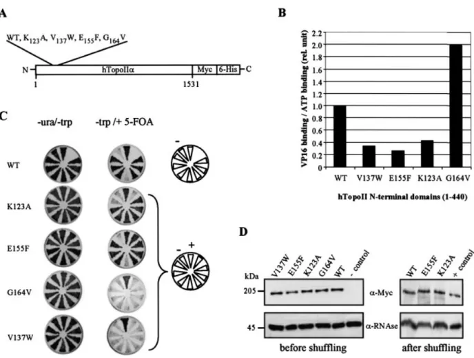
were created at the predicted sites of contact between etoposide and the ATP-binding cleft of the enzyme (Fig. 1A). All three mutations reduce the drug binding ef®ciency of a recombinant N-terminal domain (amino acids 1±440) in an in vitro binding assay, although they do not affect the binding of ATP (Fig. 1B), con®rming that etoposide can interact with the N-terminus of topoII. However, from our modelling, we propose that etoposide ®ts in the upper part of the cleft, impeding the free access of ATP instead of substituting for ATP, as novobiocin does in GyrB. The mutation we created in the Walker A motif (G164V) reduces
to radiolabelled etoposide (see 25) (Fig. 1B). The relative af®nities of the mutated domains for VP16 and ATP are summarised in Figure 1B. To examine the physiological relevance of these observations, we next introduced these mutations into the cDNA encoding the full-length htopoIIa fused at its C-terminus to the epitope tags Myc and 6-His for expression in budding yeast (Fig. 1A).
To study the in¯uence of these mutations on drug sensitivity, the wild-type and mutated forms of htopoIIa were expressed in a yeast strain lacking its endogenous enzyme. It is known that the wild-type human enzyme can complement the absence of yeast topoII (30) and that yeast show sensitivity to etoposide (31). Increased drug resistance in strains expressing the mutant forms might indicate that the N-terminal binding site is indeed critical for trapping topoII in a covalent complex with DNA. On the other hand, if drug
binding to the core domain is suf®cient for drug-induced cytotoxicity, then the N-terminal mutations might have no effect. A third possibility is that the two drug-binding sites compete for a limited pool of etoposide. In this case, reducing af®nity for the N-terminal binding site might render cells more sensitive to a limited intracellular pool of etoposide, since drug interaction with the catalytic core would be favoured. These possibilities were tested by introducing plasmids encoding either the wild-type or mutated form of htopoIIa into the top2::LEU2 yeast strain GA559 (Materials and Methods and Fig. 1C). During transformation, the essential TOP2 functions are provided by pBB6 (CEN-URA3-S.c. TOP2), which is later lost by plating cells on 5-FOA. The surviving populations then express htopoIIa as the only source of topoII. In these populations we could score the mutant forms for complementation (30), as well as for altered drug resistance.
Figure 1. Wild-type and two mutated htopoIIa complement the absence of yeast topoII. (A) The wild-type and point mutated forms of full-length htopoIIa cDNA were fused to c-Myc and 6-His epitope tags in a yeast expression vector (see Material and Methods). All constructs were sequenced to ensure that no additional mutations were introduced. (B) Radiolabelled [3H]VP16 and [a-32P]ATP were used in a ligand binding assay performed in the presence of the
indi-cated recombinant wild-type and mutated N-terminal domains (amino acids 1±440) of htopoII, as previously described (25). The ratio of binding ef®ciencies of both ligands was determined for each mutated form after each ef®ciency was normalised to the values obtained for the wild-type domain. (C) Individual transformants of strain GA559 expressing the indicated mutated form of htopoIIa were streaked on selective media in the presence (±ura ±trp) or absence (±trp +FOA) of pBB6, a URA3-based vector expressing wild-type yeast TOP2. Controls include this same top2::LEU2 strain transformed with the empty vector pRS414 (±) or with vector pRS414 expressing wild-type htopoIIa (+). Growth is visible as a dark triangular patch. We see no dominant negative effects from the expression of any of the htopoIIa proteins in yeast. Only the wild-type and the point mutants E155F and K123A are able to substitute for yeast
topoII to support mitotic growth in at least six of the eight colonies tested. (D) Full-length wild-type and the indicated mutant forms of htopoIIa are expressed at equal levels, both in the presence (left panel) and in the absence (right panel) of the endogenous yeast topoII. Western blots were performed on whole cell extracts of the indicated transformants using mouse anti-Myc (9E10) and rat anti-p42RNase H, an abundant cytosolic protein that serves as an internal standard.
Western blot analysis of cells expressing both yeast and human enzymes showed that the wild-type and mutated forms of htopoIIa (a-Myc) are expressed equally and migrate with the expected mobility (p42RNase Hwas used for normalisation;
Fig. 1D, left panel). None of the recombinant forms, wild-type or mutant, had a dominant negative effect on cell growth (data not shown). After shuf¯ing, we ®nd that the wild-type, the K123A and the E155F forms of htopoIIa support mitotic growth
in yeast, while the ATP-binding site mutant, G164V, and the
V137W substitution do not (Fig. 1C). The non-viability of
these two mutants probably does not re¯ect insuf®cient expression levels or misfolding of the human proteins in vivo, as all are detected on western blots as full-length proteins and their relative abundance suggests that they are as stable as the wild-type enzyme (Fig. 1D). The G164V mutation in the
ATP-binding site has been shown previously to inactivate yeast topoII (32) and would therefore account for the lack of complementation. We speculate that the bulky tryptophan substitution (V137W) may also interfere with ATP
turnover, although the mutated protein domain was able to bind ATP (25).
Drug sensitivity of the viable top2::LEU2 yeast strains expressing the wild-type, K123A or E155F forms of htopoIIa
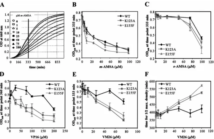
was monitored by following growth rates in a 96-well plate in the absence or presence of increasing drug concentrations (see Materials and Methods). This automated procedure has been shown to be an accurate and reliable means to compare the effects of drugs on the growth of several strains in a single experiment. Cell density was monitored at 660 nm for 16 h (an example of the raw data is shown in Fig. 2A) and, to facilitate comparisons, the optical density was plotted for each strain at the optimal point in the growth curve (between 333 and 360 min; Fig. 2B±E) as a function of the drug concentration. To ensure drug solubility, all assays were performed in 1% DMSO, which alone has no effect in the growth rate assay (data not shown). In the presence of increasing concentrations of etoposide (VP16), we observed a decrease in cell growth that was more severe for strains expressing mutant htopoIIa than for those expressing the wild-type form (Fig. 2D). This effect was similar for VM26, an etoposide-related inhibitor, which is active at even lower concentrations (5±100 mM; Fig. 2E). Plotting t1
2max, the time at which cells reached half of
their maximal density, as a function of VM26 concentration (Fig. 2F), con®rms that the K123A and E155F expressing strains
are signi®cantly more sensitive to epipodophyllotoxins than the strain expressing wild-type htopoIIa. In contrast,
Figure 2. Drug sensitivity of top2::LEU2 yeast expressing the wild-type or mutated htopoIIa. Yeast growth is monitored in microtitre plates for 16 h in increasing concentrations of the indicated drug, as described in Materials and Methods. (A) Typical examples of growth curve data obtained for strain GA559 expressing htopoIIa E155F in increasing concentrations of m-AMSA. (B±E) The OD660for a given strain which was recorded at a ®xed time point (333 or
360 min as indicated) and is plotted as a function of increasing drug concentration [(B) m-AMSA; (C) o-AMSA; (D) VP16; (E) VM26] for the strains express-ing the wild-type (diamond), K123A (square) or E155F (triangle) htopoIIa. The time point was chosen as being closest to half-maximal growth for the strain in
the absence of drug, but resulting graphs were similar between 300 and 500 min growth [see panel (A)]. (F) Time to half-maximal yeast density is plotted as a function of VM26 concentration for strains expressing the wild-type, K123A or E155F form of htopoIIa. Error bars result from growth curves analysed in
increasing amounts of m-AMSA slowed growth of the wild-type and the K123A mutants similarly and slightly more
ef®ciently for the E155F mutant (Fig. 2B). As expected, the
control compound, o-AMSA, had toxic effects only at very high concentrations, and these were identical for all strains tested (Fig. 2C). In conclusion, we ®nd that both mutants E155F and K123A show enhanced sensitivity to the
epipodo-phyllotoxins VP16 and VM26, while only E155F is slightly
more sensitive to m-AMSA.
Overexpression of the N-terminal domain confers drug resistance in yeast
The increased sensitivity to epipodophyllotoxins described above could re¯ect either altered turnover rate or enhanced expression levels of the mutated forms (19,33), yet we exclude changes in protein levels by western blot analysis (see Fig. 1D). On the other hand, since both the N-terminal domain and the catalytic core can bind etoposide, this result might alternatively re¯ect a competition for the drug between the two domains in vivo. If true, mutated N-termini with lower af®nity for etoposide should trap the drug less ef®ciently, thus increasing the pool of etoposide available for binding near the enzyme active site. To test this hypothesis, we expressed the wild-type N-terminal domain of htopoIIa at high levels to see if it is able to titrate the available drug and confer drug resistance.
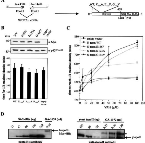
The wild-type and mutant N-terminal ATPase domains were fused in-frame with the C-terminal 83 amino acids of htopoIIa, which provide a NLS (Fig. 3A). These proteins are tagged with c-Myc and 6-His epitopes and can be expressed in the yeast strain GA1072 under control of the pGAL promoter. As for the full-length enzymes, all forms are expressed to equivalent levels and none has a dominant negative effect on yeast growth in the absence of drug (Fig. 3B). To determine the drug sensitivity of yeast cells expressing these N-terminal domains, growth was analysed as described above and t1
2max
was plotted as a function of etoposide concentration. Figure 3C shows that cells overexpressing the wild-type N-terminal domain are more resistant to etoposide than cells transformed with the empty vector (Fig. 3C). This resistance was similar for the G164V mutated domain, which binds VP16 but not
ATP, yet it is abolished by the E155F mutation and reduced by
the mutation K123A. The results indicate that the resistance
correlates well with the drug binding capacity of the overexpressed fragment (Fig. 3C).
To interpret the variations in drug sensitivity, it was important to quantify the levels of DNA topoII in yeast. To this end, whole cell extracts were prepared after carefully monitoring cell number and samples were western blotted with an antibody raised against yeast topoII. Increasing amounts of puri®ed yeast topoII of known concentrations were loaded on the same gel for quanti®cation (Fig. 3D, right panel). Considering the average radius of the G1phase yeast
nucleus as 0.9 mm, we calculated a concentration of 15 mM for the endogenous topoII. The same calculation was made to quantify the intranuclear concentration of the human enzyme expressed in the absence of yeast topoII. The reference protein used in this case was the yeast silencing protein Sir2-6His detected with an antibody raised against the penta-His antigen (Fig. 3D, left panel). Here, the concentration of htopoIIa was between 5 and 10 mM.
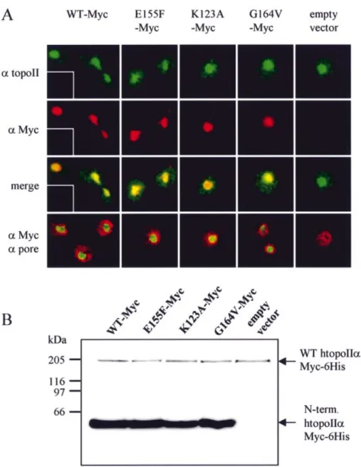
We also checked for mislocalisation of the N-terminal domains and the endogenous ytopoII. Endogenous topoII (detected by immuno¯uorescence) remains localised in the nucleus, whether or not the small domain is expressed (Fig. 4A). Overexpressed wild-type and mutated htopoIIa domains are also exclusively nuclear and co-localise with the yeast topoII, although quantitative western blots indicate that the individual domains are expressed at >20-fold the level of the full-length enzyme (Fig. 4B). Finally, fractionation assays show that both the full-length htopoIIa and all the N-terminal domains are associated with a chromatin fraction (data not shown), suggesting that the ability of a domain to compete for etoposide does not necessarily re¯ect its ability to associate with chromatin.
DISCUSSION
We have previously characterised two distinct etoposide binding sites in topoII in vitro. One was found within the catalytic core, as suggested by other studies (23,27), while the second was mapped to the N-terminal ATPase domain (25). Several point mutations have been identi®ed within the ATP-binding pocket of htopoIIa that lower the af®nity of the domain for etoposide in vitro (25). However, because an N-terminally truncated form of topoII is still capable of forming the covalent cleavable complex with DNA in the presence of drug (27) and because the Kdof etoposide binding
to the catalytic core domain is slightly lower than to the N-terminal domain (25), it is likely that the binding site in the core domain mediates the cytotoxic effects of the inhibitors. As recently shown, each monomer of topoII binds the drug and forms a cleavable complex with DNA independently of the other (34).
Nonetheless, we ®nd that overexpression of the N-terminal ATPase domain confers resistance to high concentrations of etoposide in yeast. The phenotype does not result from the ATPase activity of the N-terminal domain, because expression of the G164V mutant, which binds etoposide but cannot use
ATP (25,32), confers the same resistance phenotype as the wild-type domain. On the other hand, point mutations that weaken etoposide binding in the N-terminal domain eliminate the drug resistance. We propose that an excess of the topoII N-terminal domain enhances drug resistance by competing with the full-length enzyme for a limiting pool of inhibitor (see Fig. 5A).
This model is consistent with other data showing that enhanced drug resistance correlates with the expression of topoII subfragments (reviewed in 6,19,20). In most of these cases the subfragments remain in the cytoplasm, where they do not directly interfere with topoII function in the nucleus, but still appear to compete with nuclear topoII for the inhibitor. This was true for a C-terminally truncated form of yeast topoII, which was analysed in yeast (31). Similar topoII mutations have been detected in drug-resistant human small cell lung cancer cells (35±37).
When expressed as the unique source of topoII in yeast cells, htopoIIa forms carrying point mutations in the ATPase pocket (K123A and E155F) support mitotic growth as ef®ciently
as the wild-type enzyme, demonstrating that these mutant forms are catalytically active. As shown above, they render yeast more sensitive to epipodophyllotoxins, even though
these mutations reduce the af®nity of the N-terminus for etoposide in vitro. Hypersensitivity is not due to altered levels of protein expression nor is it likely to re¯ect changes in the catalytic activity of topoII. If it were, the cells would be hypersensitive to all inhibitors that stabilise the cleavable complex, yet we show here that the K123A mutation does not
render cells more sensitive to m-AMSA. Finally, both mutant enzymes are equally active as the wild-type enzyme in yeast cell extracts in vitro (data not shown).
To explain this, we propose that the ATP-binding cleft competes for a limiting pool of drug even in the context of the holoenzyme (Fig. 5). Under normal conditions, the catalytic
core domain of the DNA-bound topoII is probably the primary target for topoII poisons that trigger cytotoxicity through creation of DNA breaks. In this conformation, the ATPase domain may not be able to bind the drug and would, instead, ef®ciently bind and hydrolyse ATP (38). However, a cytoplasmic or a nuclear, DNA-free population of topoII should also be able to bind and sequester the drug, thereby reducing the pool of free etoposide available to the active site of the enzyme without inducing DNA damage (Fig. 5B). When the N-terminal domain is mutated such that it binds etoposide with a lower af®nity, then more inhibitor may be available for the core domain, rendering cells more sensitive
Figure 3. Overexpression of the htopoIIa N-terminal domain confers drug resistance in yeast. (A) Scheme of the fusion between htopoIIa wild-type or mutant N-terminal domains and the NLS. (B) Time required to reach half-maximal culture density (t1
2max) was determined for the transformed yeast strain
GA1072, expressing the indicated htopoIIa N-terminal domains. The contents of eight microtitre wells of each transformant were analysed by western blotting with antibodies against Myc or p42RNase H. (C) The increase in t1
2maxis shown as a function of VP16 concentration for GA1072 transformants expressing the
indicated htopoIIa N-terminal domain: no protein (diamond), N-term.NLS fusions wild-type (square), E155F (triangle), K123A (cross) and G164V (disc). Note
that GA1072 carries transporter mutations and is more sensitive to VP16 than normal yeast strains. The t1
2maxvalues in the absence of drug were normalised
to the strain carrying an empty vector. Error bars result from growth curves analysed in triplicate. Each experiment was performed three times. (D) The levels of expression of both the human and the yeast topoII were analysed in yeast. The strains GA1659 (expressing only wild-type htopoIIa, left panel) and GA1072 (expressing endogenous ytopoII, right panel) were grown and counted when harvested. Whole cell extracts were prepared and analysed by quantita-tive western blotting with anti-pentaHis or anti-ytopoII antibodies, respecquantita-tively. Increasing amounts of the yeast transcription regulator Sir2-6His and yeast topoII, both puri®ed to homogeneity, were used as standards for the quanti®cation. * indicates a non-speci®c signal obtained with the antibody raised against ytopoII.
to a given concentration of drug (Fig. 5C). The modulation of drug sensitivity through this mechanism is plausible only if intracellular drug concentrations are quite low. This appears to be the case in yeast, as it was previously found that low level expression of intact topoII or of cytoplasmically localised subfragments of the enzyme correlates with enhanced drug resistance (31). Moreover, to be valid, the model we propose requires an effective drug concentration that is not in vast excess as compared to the yeast endogenous topoII concen-tration. The concentrations of 15 and 5±10 mM found, respectively, for the endogenous ytopoII and for the htopoII expressed in yeast after shuf¯ing is slightly lower than 40 mM, the concentration of drug shown to produce a signi®cant difference in drug sensitivity in vivo (Figs 2 and 3). However,
these values are within the same range. Moreover, the concentration of the drug added extracellularly is certainly higher than the effective intracellular concentration. Indeed, yeast drug ef¯ux pumps and non-speci®c interactions with intracellular structures are mechanisms that decrease the concentration of free drug available to topoII in the nucleus. Importantly, the effective drug concentration is very close to the dissociation constants found for the drug binding sites (20 mM for the N-terminal domain and the full-length enzyme and 9 mM for the catalytic core domain) (25).
Our data are consistent with studies reporting mutations in the ATP-binding domain of topoII that affect drug sensitivity and provide evidence that the ATPase domain of topoII can bind at least some topoII inhibitors in vivo. However, in
Figure 4. Highly overexpressed htopoIIa N-terminal domains localise to the yeast nucleus. (A) After 16 h induction, cells expressing the indicated htopoIIa N-terminal domain or carrying an empty vector were formaldehyde ®xed and immunostained for ytopoII (af®nity puri®ed atopoII antibody is visualised in green) and for the htopoIIa domains (a-Myc, visualised in red). Co-localisation is shown in yellow. Insets show reactions in the absence of primary anti-bodies. Where indicated, the nuclear envelope is detected with mAb414 (a-pore, in red). (B) The level of overexpression of the htopoIIa N-terminal domains was determined in a strain expressing full-length wild-type htopoIIa from a single copy plasmid. Both constructs carry a single c-Myc epitope, which allows direct comparison of the relative levels of overexpression. Quantitation indicates at least 20-fold excess of the small domains over full-length protein.
contrast to novobiocin for GyrB (26) and bisdioxopiperazine for topoII (24), we consider that the site of interaction of etoposide within the ATP-binding domain of the enzyme does not necessarily represent the key mechanism of action of this drug, but rather a secondary interaction site that modi®es the effectiveness of antitumour agents. Useful paradigms for future drug development may well rest upon the observation that both intra- and intermolecular competition can modulate epipodophyllotoxin-mediated cytotoxicity.
ACKNOWLEDGEMENTS
We thank T. Laroche for help with microscopy, G.T.C. Alghisi and P. Szankasi for strains, Y. Pommier for drugs, A. Andersen and O. Westergaard for plasmids, F. Martino for the puri®ed Sir2-6His, M. Parkan for computer assistance and Drs J. Paulson, L. Bjergbaek and F. Cubizolles for critical help on the manuscript. D.L. thanks the ARC, RhoÃne-Alpes Exchange Program and EMBO for fellowships. These studies were funded by grants from the Swiss Cancer League, the Swiss National Science Foundation and the EU BioMED
Program to S.M.G. S.M.G. and D.L. thank the NCCR Frontiers in Genetics program for funding.
REFERENCES
1. Nitiss,J.L. (1998) Investigating the biological functions of DNA topoisomerases in eukaryotic cells. Biochim. Biophys. Acta, 1400, 63±81. 2. D'Arpa,P. and Liu,L.F. (1989) Topoisomerase-targeting antitumor drugs.
Biochim. Biophys. Acta, 989, 163±177.
3. Corbett,A.H. and Osheroff,N. (1993) When good enzymes go bad: conversion of topoisomerase II to a cellular toxin by antineoplastic drugs. Chem. Res. Toxicol., 6, 585±597.
4. Pommier,Y. (1997) Cancer Therapeutics: Experimental and Clinical Agents. Humana Press, Totowa, NJ, pp. 153±173.
5. Beck,W.T., Danks,M.K., Wolverton,J.S., Kim,R. and Chen,M. (1993) Drug resistance associated with altered DNA topoisomerase II. Adv. Enzyme Regul., 33, 113±127.
6. Watt,P.M. and Hickson,I.D. (1994) Structure and function of type II DNA topoisomerases. Biochem. J., 303, 681±695.
7. Larsen,A.K. and Skladanowski,A. (1998) Cellular resistance to topoisomerase-targeted drugs: from drug uptake to cell death. Biochim. Biophys. Acta, 1400, 257±274.
8. Lee,M.S., Wang,J.C. and Beran,M. (1992) Two independent amsacrine-resistant human myeloid leukemia cell lines share an identical point mutation in the 170 kDa form of human topoisomerase II. J. Mol. Biol., 223, 837±843.
9. Campain,J.A., Gottesman,M.M. and Pastan,I. (1994) A novel mutant topoisomerase II alpha present in VP-16-resistant human melanoma cell lines has a deletion of alanine 429. Biochemistry, 33, 11327±11332. 10. Freudenreich,C.H., Chang,C. and Kreuzer,K.N. (1998) Mutations of the
bacteriophage T4 type II DNA topoisomerase that alter sensitivity to antitumor agent 4¢-(9-acridinylamino)methanesulfon-m-anisidide and an antibacterial quinolone. Cancer Res., 58, 1260±1267.
11. Hinds,M., Deisseroth,K., Mayes,J., Altschuler,E., Jansen,R., Ledley,F.D. and Zwelling,L.A. (1991) Identi®cation of a point mutation in the topoisomerase II gene from a human leukemia cell line containing an amsacrine-resistant form of topoisomerase II. Cancer Res., 51, 4729±4731.
12. Sabourin,M., Byl,J.A., Hannah,S.E., Nitiss,J.L. and Osheroff,N. (1998) A mutant yeast topoisomerase II (top2G437S) with differential sensitivity to anticancer drugs in the presence and absence of ATP. J. Biol. Chem., 273, 29086±29092.
13. Sehested,M., Wessel,I., Jensen,L.H., Holm,B., Oliveri,R.S., Kenwrick,S., Creighton,A.M., Nitiss,J.L. and Jensen,P.B. (1998) Chinese hamster ovary cells resistant to the topoisomerase II catalytic inhibitor ICRF-159: a Tyr49Phe mutation confers high-level resistance to
bisdioxopiperazines. Cancer Res., 58, 1460±1468.
14. Strumberg,D., Nitiss,J.L., Rose,A., Nicklaus,M.C. and Pommier,Y. (1999) Mutation of a conserved serine residue in a quinolone-resistant type II topoisomerase alters the enzyme-DNA and drug interactions. J. Biol. Chem., 274, 7292±7301.
15. Wessel,I., Jensen,L.H., Jensen,P.B., Falck,J., Rose,A., Roerth,M., Nitiss,J.L. and Sehested,M. (1999) Human small cell lung cancer NYH cells selected for resistance to the bisdioxopiperazine topoisomerase II catalytic inhibitor ICRF-187 demonstrate a functional R162Q mutation in the Walker A consensus ATP binding domain of the alpha isoform. Cancer Res., 59, 3442±3450.
16. Dong,J., Walker,J. and Nitiss,J.L. (2000) A mutation in yeast topoisomerase II that confers hypersensitivity to multiple classes of topoisomerase II poisons. J. Biol. Chem., 275, 7980±7987. 17. Hammonds,T.R., Foster,S.R. and Maxwell,A. (2000) Increased
sensitivity to quinolone antibacterials can be engineered in human topoisomerase IIalpha by selective mutagenesis. J. Mol. Biol., 300, 481±491.
18. Patel,S., Jazrawi,E., Creighton,A.M., Austin,C.A. and Fisher,L.M. (2000) Probing the interaction of the cytotoxic bisdioxopiperazine ICRF-193 with the closed enzyme clamp of human topoisomerase IIalpha. Mol. Pharmacol., 58, 560±568.
19. Vassetzky,Y.S., Alghisi,G.-C. and Gasser,S.M. (1995) DNA
topoisomerase II mutations and resistance to anti-tumor drugs. Bioessays, 17, 767±774.
20. Nitiss,J.L. and Beck,W.T. (1996) Antitopoisomerase drug action and resistance. Eur. J. Cancer, 32A, 958±966.
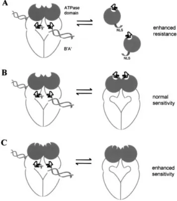
Figure 5. A model for a regulatory role of the N-terminal domain in etopo-side action. (A) To explain the drop in VP16 sensitivity conferred by over-expression of the wild-type htopoIIa N-terminal domain (dark circle) in yeast, we propose that it sequesters the drug (black squiggle) and thereby enhances resistance of the transformed strain. This is compromised by muta-tions that interfere with VP16 binding. (B) Under drug-limiting condimuta-tions that may occur in yeast, the N-terminal and the core domain appear to com-pete intra- or intermolecularly for etoposide (see Fig. 2). We propose that the N-terminal site functions mainly when topoII is not DNA (double helix) bound. Drug association with this domain appears not to induce a cytotoxic cleavage. (C) When mutations that reduce etoposide binding are introduced into the N-terminal domain, more inhibitor may become available to the site in the core domain, increasing cytotoxicity.
21. Freudenreich,C.H. and Kreuzer,K.N. (1994) Localization of an aminoacridine antitumor agent in a type II topoisomerase-DNA complex. Proc. Natl Acad. Sci. USA, 91, 11007±11011.
22. Froelich-Ammon,S.J., Patchan,M.W., Osheroff,N. and Thompson,R.B. (1995) Topoisomerase II binds to ellipticine in the absence or presence of DNA. Characterization of enzyme-drug interactions by ¯uorescence spectroscopy. J. Biol. Chem., 270, 14998±15004.
23. Burden,D.A., Kingma,P.S., Froelich-Ammon,S.J., Bjornsti,M.A., Patchan,M.W., Thompson,R.B. and Osheroff,N. (1996) Topoisomerase II.etoposide interactions direct the formation of drug-induced enzyme-DNA cleavage complexes. J. Biol. Chem., 271, 29238±29244. 24. Hu,T., Sage,H. and Hsieh,T.-S. (2002) ATPase domain of eukaryotic
DNA topoisomerase II. Inhibition of ATPase activity by the anti-cancer drug bisdioxopiperazine and ATP/ADP-induced dimerization. J. Biol. Chem., 277, 5944±5951.
25. Leroy,D., Kajava,A.V., Frei,C. and Gasser,S.M. (2001) Analysis of etoposide binding to subdomains of human DNA topoisomerase II alpha in the absence of DNA. Biochemistry, 40, 1624±1634.
26. Lewis,R.J., Singh,O.M., Smith,C.V., Skarzynski,T., Maxwell,A., Wonacott,A.J. and Wigley,D.B. (1996) The nature of inhibition of DNA gyrase by the coumarins and the cyclothialidines revealed by X-ray crystallography. EMBO J., 15, 1412±1420.
27. Chang,S., Hu,T. and Hsieh,T.-S. (1998) Analysis of a core domain in Drosophila DNA topoisomerase II. Targeting of an antitumor agent ICRF-159. J. Biol. Chem., 273, 19822±19828.
28. Klein,F., Laroche,T., Cardenas,M.E., Hofmann,J.F.X., Schweizer,D. and Gasser,S.M. (1992) Localization of RAP1 and topoisomerase II in nuclei and meiotic chromosomes of yeast. J. Cell Biol., 117, 935±948. 29. Gotta,M., Laroche,T. and Gasser,S.M. (1999) Analysis of nuclear
organization in Saccharomyces cerevisiae. Methods Enzymol., 304, 663±672.
30. Jensen,S., Andersen,A.H., Kjeldsen,E., Biersack,H., Olsen,E.H., Andersen,T.B., Westergaard,O. and Jakobsen,B.K. (1996) Analysis of functional domain organization in DNA topoisomerase II from humans and Saccharomyces cerevisiae. Mol. Cell. Biol., 16, 3866±3877. 31. Vassetzky,Y.S., Alghisi,G.C., Roberts,E. and Gasser,S.M. (1996) Ectopic
expression of inactive forms of yeast DNA topoisomerase II confers resistance to the anti-tumour drug, etoposide. Br. J. Cancer, 73, 1201±1209.
32. Lindsley,J.E. and Wang,J.C. (1993) On the coupling between ATP usage and DNA transport by yeast DNA topoisomerase II. J. Biol. Chem., 268, 8096±8104.
33. Lage,H., Helmbach,H., Dietel,M. and Schadendorf,D. (2000) Modulation of DNA topoisomerase II activity and expression in melanoma cells with acquired drug resistance. Br. J. Cancer, 82, 488±491.
34. Bromberg,K.D., Burgin,A.B. and Osheroff,N. (2003) A two-drug model for etoposide action against human topoisomerase IIalpha. J. Biol. Chem., 278, 7406±7412.
35. Feldhoff,P.W., Mirski,S.E., Cole,S.P. and Sullivan,D.M. (1994) Altered subcellular distribution of topoisomerase II alpha in a drug-resistant human small cell lung cancer cell line. Cancer Res., 54, 756±762. 36. Wessel,I., Jensen,P.B., Falck,J., Mirski,S.E., Cole,S.P. and Sehested,M.
(1997) Loss of amino acids 1490Lys-Ser-Lys1492 in the COOH-terminal region of topoisomerase IIalpha in human small cell lung cancer cells selected for resistance to etoposide results in an extranuclear enzyme localization. Cancer Res., 57, 4451±4454.
37. Mirski,S.E., Sparks,K.E., Yu,Q., Lang,A.J., Jain,N., Campling,B.G. and Cole,S.P. (2000) A truncated cytoplasmic topoisomerase IIalpha in a drug-resistant lung cancer cell line is encoded by a TOP2A allele with a partial deletion of exon 34. Int. J. Cancer, 85, 534±539.
38. Wang,H., Mao,Y., Zhou,N., Hu,T., Hsieh,T.-S.D. and Liu,L.F. (2001) ATP-bound topoisomerase II as a target for antitumor drugs. J. Biol. Chem., 276, 15990±15995.