HAL Id: hal-01470553
https://hal.sorbonne-universite.fr/hal-01470553
Submitted on 17 Feb 2017HAL is a multi-disciplinary open access archive for the deposit and dissemination of sci-entific research documents, whether they are pub-lished or not. The documents may come from teaching and research institutions in France or abroad, or from public or private research centers.
L’archive ouverte pluridisciplinaire HAL, est destinée au dépôt et à la diffusion de documents scientifiques de niveau recherche, publiés ou non, émanant des établissements d’enseignement et de recherche français ou étrangers, des laboratoires publics ou privés.
Hydrozoan insights in animal development and evolution
Lucas Leclère, Richard Copley, Tsuyoshi Momose, Evelyn Houliston
To cite this version:
Lucas Leclère, Richard Copley, Tsuyoshi Momose, Evelyn Houliston. Hydrozoan insights in animal development and evolution. Current Opinion in Genetics and Development, Elsevier, 2016, Devel-opmental mechanisms, patterning and evolution, 39, pp.157-167. �10.1016/j.gde.2016.07.006�. �hal-01470553�
Current Opinion in Genetics and Development 2016, 39:157–167
http://dx.doi.org/10.1016/j.gde.2016.07.006
Hydrozoan insights in animal development and evolution
Lucas Leclère, Richard R. Copley, Tsuyoshi Momose and Evelyn Houliston
Sorbonne Universités, UPMC Univ Paris 06, CNRS, Laboratoire de Biologie du Développement de Villefranche‐sur‐mer (LBDV), 181 chemin du Lazaret, 06230 Villefranche‐sur‐mer, France. Corresponding author: Leclère, Lucas (leclere@obs‐vlfr.fr).
Abstract
The fresh water polyp Hydra provides textbook experimental demonstration of positional information gradients and regeneration processes. Developmental biologists are thus familiar with Hydra, but may not appreciate that it is a relatively simple member of the Hydrozoa, a group of mostly marine cnidarians with complex and diverse life cycles, exhibiting extensive phenotypic plasticity and regenerative capabilities. Hydrozoan species offer extensive opportunities to address many developmental mechanisms relevant across the animal kingdom. Here we review recent work from non‐Hydra hydrozoans – hydromedusae, hydroids and siphonophores – shedding light on mechanisms of oogenesis, embryonic patterning, allorecognition, stem cell regulation and regeneration. We also highlight potential research directions in which hydrozoan diversity can illuminate the evolution of developmental processes at micro‐ and macro‐evolutionary time scales.Introduction
Although the relationships between non‐bilaterian animals are currently somewhat controversial, the position of cnidarians as the closest relatives of bilaterians seems relatively undisputed. This has helped to establish the anthozoan cnidarian Nematostella vectensis as a key model for molecular studies in evolutionary aspects of developmental biology. Anthozoa is one side of the deepest split in the cnidarian lineage, the other being Medusozoa, a group united by the presence of a medusa (i.e. a jellyfish) stage in their life‐cycle. Medusae show high morphological complexity, including features such as striated muscle and elaborate sensory structures, and in some instances even camera eyes. Medusozoa is composed of four clades: Hydrozoa, Staurozoa, Cubozoa and Scyphozoa (Figure 1a) [1•]. Hydrozoa is the biggest and most diverse of these, comprising more than 3500 species. Detailed observation and experimentation on marine hydrozoan species fueled many discoveries and conceptual advances in the second half of the 19th century, relating notably to germ plasm theory, evolution of germ layers, and regeneration. Weismann, Haeckel, Morgan, Huxley, Driesch, the Hertwig brothers and Metchnikov all worked extensively on marine hydroids and hydromedusae. During the 20th century, studies in the freshwater polyp Hydra dominated hydrozoan research in cell and developmental biology, resulting in important findings related to organizer activity, morphogen gradients, pluripotent stem cells, and more recently, evolution of aging (e.g. [2]) and tumorigenesis (e.g. [3]), and interactions between animals, symbionts and microbiota (see [4]). Hydra is still, at the time of writing, the only hydrozoan for which a genome sequence has been published [5]. Several excellent recent reviews have covered Hydra research (see [4,6–12•]) and so here, we will focus on current work on other hydrozoan species to highlight their contribution and potential for understanding fundamental developmental processes and evolutionary questions. Unlike Hydra, some of these species allow easy experimental access to sexual reproduction and embryonic development. In recent years, marine hydrozoan species have been used increasingly for molecular studies, notably Clytia hemisphaerica [13], Hydractinia echinata and its close American relative Hydractinia symbiolongicarpus [14], Cladonema radiatum [15] and Podocoryna carnea [16]. Improved molecular technologies and decreasing sequencing costs are aiding emergence of additional models such as Turritopsis dohrnii [17,18], Eleutheria dichotoma [19,20] and the siphonophore Nanomia bijuga [21,22•,23] (summarized in Table 1).Phenotypic plasticity and life cycle complexity
The typical hydrozoan life cycle comprises three basic forms: the motile ‘planula’ larva, sessile polyps (which typically propagate to form extensive colonies by asexual reproduction), and the pelagic medusa, a sexual form usually generated by budding from a polyp (in contrast to the scyphozoan medusa, which forms by a process known as strobilation [24••]). The evolutionary plasticity of this presumably ancestral program is remarkable, as illustrated by the loss of the medusa in the lineages leading to Hydra and Hydractinia. Molecular based phylogenies imply more than 50 independent losses of the medusa and at least 2 losses of the polyp within Hydrozoa, and show that major changes in life cycles and in colonial organization have occurred over short evolutionary time‐scales [25–35•] (Figure 1b‐c). Hydrozoan life cycles can include sexual and asexual reproduction at almost any stage, and the transitions can occur in atypical sequences, such as the famous medusa to polyp transformation (‘reverse development’) of Turritopsis dohrnii [17]. Determining the molecular mechanisms (including epigenetic modifications and response to environmental factors), and evolutionary processes triggering morphological and life cycle plasticity are important challenges for future research [36]. The history of genes expressed preferentially in the medusa (relating for instance to striated muscle or sense organs) has started to be addressed [37,38], and the first comparisons of transcriptomes from Podocoryna and Hydractinia, related species with and without medusae respectively, have been made [39•]. A successful approach that allowed identification of cnidarian‐specific genes regulating tentacle patterning in Hydra involved subtractive hybridization between cDNAs from morphologically distinct Hydra strains, followed by functional validation via transgenesis [40]. Additional life cycle complexity in hydrozoans arises via the formation in many species of complex benthic or pelagic colonies comprising connected polyps specialized for different functions (e.g. budding, feeding, defense). The siphonophores are pelagic colonies which have attained extreme complexity including many different polyp types, but also specialized attached medusa forms serving distinct functions (e.g. reproduction, swimming) [21,22•,41]. Very little is known about how the polyp and medusa components of complex colonies acquire different forms and functions during development and evolution, although the advent of molecular analysis in siphonophores is promising [22•,23]. New transcriptomic data for several polyp types in Hydractinia [42–44] provide many candidates for future investigations of the developmental mechanisms leading to a division of labor between polyps within a colony (see Table 1).
In some hydrozoans groups, polyp colonies of the same species in close contact can fuse or reject depending on how closely genetically related they are. This phenomenon has been successfully
dissected at the molecular level in Hydractinia, starting with the seminal work of von Hauenschild in the 1950s (see [45]). A long and outstanding genetic investigation, involving crosses of many related and unrelated strains [46], led to the discovery of an allorecognition complex composed of two genetic loci, alr1 [47] and alr2 [48]. The Alr1 and Alr2 proteins show huge diversity in natural populations [49••,50]. They have recently been shown to be homophilic ligands binding only to themselves [51••]. In natural populations additional genetic factors may also be involved in the allorecognition process [52]. Allorecognition regulation has important consequences for individual and colonial biology and genotype survival. Fusion between sexually generated polyps has recently being proposed as the mode of colony formation in the genus Ectopleura [35•]. Rejection between colonies can induce necrosis in Hydractinia [53]. Contact between genetically distinct colonies results in mixing of stem cells, which can lead to invasion, takeover and eventually changes in morphology. A fascinating corollary is that evolutionary selection in this context might act at the level of the stem cells, as also proposed in the colonial ascidian Botryllus [54].
Medusa evolution and development
Cnidarians are often represented as having a simple morphology and relatively few cell types, reflecting the situation in the common bilaterian‐cnidarian ancestor. The medusa form, however, has a sophisticated organization, including specialized sensory and reproductive organs and the fast‐ contracting striated muscle of the bell for swimming [15,13,55] (Figure 2a), providing an opportunity to compare the developmental and evolutionary origins of these structures in cnidarians and bilaterians. Molecular studies of hydrozoan medusae sensory structures have so far focused mainly on light detection. Eyes of Cladonema medusae express Opsin photoproteins and Six/Pax/Eya transcription factors [15,56,57], suggesting a common origin of the molecular mechanisms of eye development and convergence of the eyes as complex structures between Hydrozoa and Bilateria. The medusa stage itself has been the subject of historical dispute, with opposing camps regarding it as a primitive cnidarian trait lost in Anthozoa, or a derived feature of Medusozoa. From a phylogenetic perspective, recent debate has revolved around potential paraphyly of Anthozoa and the position of Staurozoa (so‐called ‘stalked jellyfish’). It is now widely accepted that the medusa is a derived character of Medusozoa, although the most recent phylogeny recovers monophyletic Anthozoa, as sister to Medusozoa, with Hydrozoa (rather than Staurozoa) as sister group to the other medusozoans [1•].Gene expression data relating to medusa development is accumulating gradually (e.g. [58–61•]), but remains scarce compared to planula and polyp data, while gene function studies in the medusa are still largely lacking [20]. Detailed comparative anatomical, molecular and functional studies between medusae of the different medusozoan groups will be insightful for deciphering the origin(s) of the medusa form. The evolutionary origin of hydrozoan striated muscle, traditionally considered homologous between cnidarians and bilaterians [62], was addressed by Steinmetz and coworkers [63••] (Figure 2a). Genomic analyses and in situ hybridization approaches showed that key bilaterian striated muscle proteins, notably components of the Z‐disk, are not present in Clytia, while most of the bilaterian striated muscle genes are either expressed broadly or in other cell types. This study thus provides compelling evidence for convergent evolution of striated muscle in hydrozoans and bilaterians.
Gametogenesis and spawning
The transparency and accessibility of many hydrozoan sexual forms makes them particularly good models for studying the widely conserved regulatory mechanisms of animal gametogenesis, and dissecting the mechanisms of oocyte maturation, spawning and fertilization (discussed in detail in [64], Figure 2b). Studies in several medusae including Cladonema, Cytaeis and Clytia are allowing analysis of successive regulatory steps. For technical reasons (notably access to microinjection of Morpholinos/mRNAs/indicators), the main research focus so far has been on the fully grown oocyte and spawned egg. Spawning in both males and females is triggered by light, which induces somatic cells of the gonad to secrete a small diffusible hormone that awakens the dormant gametes. Recent studies suggest that neuropeptide‐related molecules participate in this process [65]. The essential downstream response of oocytes to the hormone is an immediate rise in cytoplasmic cAMP levels and PKA activation [66]. This stimulates both release of meiotic arrest and entry into the meiotic divisions and parallel activation of the Mos‐MAP kinase pathway responsible for regulation of oocyte‐specific processes such as polar body formation and post‐meiotic cell cycle arrest [67]. MAP kinase pathway inactivation following fertilization, along with cytoplasmic Ca++ release, is also involved in regulating polyspermy [68•]. Concerning the regulation of early steps of gametogenesis such as entry into meiosis and oocyte growth, which represent major bottlenecks in research in reproductive biology, the advent of gene editing techniques is opening the powerful possibility of combining gene function analysis and imaging using transparent hydrozoan gonads.Body axis evolution and development
Studying axis formation in a range of cnidarians, including hydrozoans, offers an informative perspective for the understanding of body plan evolution in metazoans. It is still debated whether the oral‐aboral axis of cnidarians is homologous to any of the body axes of bilaterians, but it seems very likely that localized Wnt signaling was a feature of the gastrulation pole of the last common ancestor of these major animal groups. Each hydrozoan form, planula, polyp and medusa, is organized with respect to a principal axis of polarity termed oral‐aboral (Figure 3a). For instance the planula larva has an aboral specialization of the nervous system [69], and the medusa has integrating nerve rings around the bell margin [55]. Key to establishment and maintenance of this axis is the oral pole organizer. The key molecular pathway of oral organizer activity in Hydra is Wnt/‐catenin signaling [11], activated by the diffusible ligand Wnt3 and maintained at the oral tip by a complex auto‐feedback loop [70]. Oral organizers have now been described at the oral pole of the planula larvae and polyp in several marine hydrozoan species [71,72], with the Wnt/‐catenin pathway established as a key player in the regulation of the planula and polyp organizer. In Clytia larvae, functional studies involving morpholino and mRNA injection into the egg have shown that axis formation is initiated by maternal mRNAs coding for the ligand Wnt3 (Figure 3a, b), and for two Frizzled family receptors [73,74] which become localized in the egg during oocyte growth and maturation [75]. During early development, Wnt3 and Fz1 together activate the Wnt/‐catenin pathway at the oral pole, while Fz3 acts as a negative regulator at the aboral pole. In parallel, the transmembrane protein Strabismus interacts with Fz1 across neighboring cell boundaries to direct morphogenesis along the oral‐aboral axis via a typical PCP mechanism [76••]. PCP coordinates the alignment of the ectodermal cilia to ensure unidirectional swimming, and orients cell movements participating in elongation of the developing larva (Figure 3b). Common involvement of Frizzled proteins in establishing a Wnt/‐catenin activity gradient and PCP is likely to be an ancestral mechanisms to couple morphological and molecular polarity during development [76••]. A transcriptomic study identified potential Wnt/‐catenin downstream targets and genes regulated by the PCP pathways [77••], providing several promising candidates for roles in organizer activity. Analyses of Wnt pathway regulation in Hydractinia have focused mainly on the polyp. Wnt3 signaling via a TCF transcription factor induces development of oral structures [78]. Other studies in Hydractinia have addressed the dramatic metamorphosis that transforms the planula into the polyp,and notably the extensive caspase dependent apoptosis at the oral pole during metamorphosis [79– 82]. This apoptosis is essential for the formation of the primary polyp [83], playing a constructive role [81,84] as it does during regeneration in Hydra [85]. Interestingly, Wnt5a expressing cells at the oral pole of Hydractinia larvae appear to be protected from apoptosis during metamorphosis [71].
Cell type plasticity and stem cell systems in Hydrozoa
Hydrozoans characteristically exhibit a very strong ability to regenerate one or all their phenotypic forms, a capacity underpinned by extreme cell type plasticity. This includes bone fide transdifferentiation, as demonstrated using Podocoryna carnea by Schmid and collaborators, who convincingly showed transdifferentiation of striated muscle cells isolated from the medusa into several cell types including smooth muscle, sensory cells, gland cells and nematocytes (stinging cells characteristic of Cnidaria) [16]. Hydrozoan regeneration involves active stem cell systems, first characterized in Hydra, where three types of stem cell coexist: ectodermal epithelial stem cells, endodermal epithelial stem cells and ‘insterstitial’ cells (i‐cells) (see [7]) (Figure 3c). The first two basic epithelial cell types support the constant renewal of the ectodermal and endodermal epithelia. I‐cells are a hydrozoan‐specific type of undifferentiated multipotent stem cell that can differentiate into nerve cells, nematocytes, gland cells and also germ cells. I‐cell developmental potential varies between species. Thus, in Hydractinia echinata, i‐cells can differentiate not only into nerve cells and nematocytes, but also into epithelial cells, as demonstrated using mutants, BrdU labeling and grafting experiments [86], and confirmed using transgenic tools [87] (Figure 3c). Progress in understanding the molecular regulation and cellular dynamics of hydrozoan stem cells was greatly accelerated by the development of transgenic lines both in Hydractinia [88••] and Hydra [89], which allowed tracking of stem cells in vivo, notably during regeneration [88••] (Figure 3d). Pioneering studies in Hydra have compared molecular signatures of different cell types by transcriptome analysis of fluorescence‐tagged sorted cells from transgenic lines [90] and established proteomic methods [91••]. In Hydractinia a POU‐domain containing transcription factor of the class 3/5 involved in regulating pluripotency has been identified [92•], of particular interest given the role of the (class 5) POU‐domain factor Oct 4 in mammals. Similarly, in Clytia, several Sox family transcription factors (groups B, C, E and F) were found expressed either in stem‐cells or differentiated cells, suggesting a regulatory role in multipotency maintenance operating widely across the animal kingdom [93]. A recent study has shown a correlation between nuclear localizationof the Hippo pathway component Yorkie and cell division in Clytia medusae [94], suggesting that regulation of cell proliferation by this transcriptional cofactor is conserved between Cnidaria and Bilateria. I‐cells in Clytia [95,96•] and Hydractinia [88••,92•,97,98], as in Hydra (e.g. [90,99]), also express orthologues of genes typically associated with germ line or stem cell function such as Piwi, Nanos1, and Vasa. I‐cell formation during embryogenesis was addressed in Clytia, and appears to involve both determinant inheritance and inductive mechanisms [96•]. I‐cells originate at the onset of gastrulation in a region inheriting localized Piwi, Nanos1, Vasa and Pl10 mRNAs from the animal pole of the egg, evoking the ‘preformation’ mode of germ‐line specification by maternal ‘germ plasm’ well known in insects and frog embryos. I‐cells can also form in Clytia planulae in the absence of the key egg animal pole region, implying the involvement of inductive mechanisms [96•].
Conclusion
Hydrozoans have much to contribute to our understanding of basic developmental and cell biology processes and their evolution, as well as to relatively neglected areas of biological complexity such as colony level organization and asexual reproduction. Rapid technological advances mean that it is now feasible with quite modest budgets to undertake genome sequencing, large scale gene expression/transcriptomics studies, functional analyses at all life cycle stages via gene editing, as well as cell/molecule tracking using transgenic lines carrying diverse fluorescent reporters. With these accumulating resources, we expect in the next few years that a wide range of marine hydrozoan species will emerge as valuable experimental models.Acknowledgements
Our recent research on hydrozoans has been funded by grants from the ANR (MEDUSEVO – ANR‐13‐ PDOC‐0016 and OOCAMP – ANR‐13‐BSV2‐0008), Sorbonne Universités («Convergence» program and the André Picard Network), the EMBRC‐Fr Infrastructure project and CNRS core funding to the LBDV. We thank Chiara Sinigaglia and Michaël Manuel for helpful comments on the manuscript.References and recommended reading
Papers of particular interest, published within the period of review, have been highlighted as: • of special interest •• of outstanding interest 1. Zapata F, Goetz FE, Smith SA, Howison M, Siebert S, Church SH, Sanders SM, Ames CL, McFadden CS, France SC, et al.: Phylogenomic analyses support traditional relationships within Cnidaria. PLoS One 2015, 10:e0139068. • This paper gathers transcriptomes from the major cnidarian groups and recovers monophyletic anthozoans and medusozoans, showing that hydrozoans are likely to be the sister group to the other medusozoans. More work remains to be done to confirm these results, in particular, richer taxon sampling of cerianthids, staurozoans, cubozoans and hydrozoans. 2. Boehm A‐M, Khalturin K, Anton‐Erxleben F, Hemmrich G, Klostermeier UC, Lopez‐Quintero JA, Oberg H‐H, Puchert M, Rosenstiel P, Wittlieb J, et al.: FoxO is a critical regulator of stem cell maintenance in immortal Hydra. Proc Natl Acad Sci USA 2012, 109:19697–19702. 3. Domazet‐Lošo T, Klimovich A, Anokhin B, Anton‐Erxleben F, Hamm MJ, Lange C, Bosch TCG: Naturally occurring tumours in the basal metazoan Hydra. Nat Commun 2014, 5:4222. 4. Bosch TCG: Rethinking the role of immunity: lessons from Hydra. Trends Immunol 2014, 35:495–502. 5. Chapman JA, Kirkness EF, Simakov O, Hampson SE, Mitros T, Weinmaier T, Rattei T, Balasubramanian PG, Borman J, Busam D, et al.: The dynamic genome of Hydra. Nature 2010, 464:592–596. 6. Buzgariu W, Al Haddad S, Tomczyk S, Wenger Y, Galliot B: Multi‐functionality and plasticity characterize epithelial cells in Hydra. Tissue Barriers 2015, 3:e1068908. 7. Hobmayer B, Jenewein M, Eder D, Eder M‐K, Glasauer S, Gufler S, Hartl M, Salvenmoser W: Stemness in Hydra ‐ a current perspective. Int J Dev Biol 2012, 56:509–517. 8. Steele RE: The Hydra genome: insights, puzzles and opportunities for developmental biologists. Int J Dev Biol 2012, 56:535–542. 9. David CN: Interstitial stem cells in Hydra: multipotency and decision‐making. Int J Dev Biol 2012, 56:489–497. 10. Nishimiya‐Fujisawa C, Kobayashi S: Germline stem cells and sex determination in Hydra. Int J Dev Biol 2012, 56:499–508. 11. Bode HR: The head organizer in Hydra. Int J Dev Biol 2012, 56:473–478. 12. Galliot B: Hydra, a fruitful model system for 270 years. Int J Dev Biol 2012, 56:411–423. • Together with [7‐11] and more than 10 other review articles: comprehensive special issue on current Hydra research.13. Houliston E, Momose T, Manuel M: Clytia hemisphaerica: a jellyfish cousin joins the laboratory. Trends Genet 2010, 26:159–167. 14. Plickert G, Frank U, Müller WA: Hydractinia, a pioneering model for stem cell biology and reprogramming somatic cells to pluripotency. Int J Dev Biol 2012, 56:519–534. 15. Suga H, Schmid V, Gehring WJ: Evolution and functional diversity of jellyfish opsins. Curr Biol 2008, 18:51–55. 16. Schmid V, Alder H: Isolated, mononucleated, striated muscle can undergo pluripotent transdifferentiation and form a complex regenerate. Cell 1984, 38:801–809. 17. Schmich J, Kraus Y, De Vito D, Graziussi D, Boero F, Piraino S: Induction of reverse development in two marine hydrozoans. Int J Dev Biol 2007, 51:45–56. 18. Piraino S, Boero F, Aeschbach B, Schmid V: Reversing the Life Cycle: Medusae Transforming into Polyps and Cell Transdifferentiation in Turritopsis nutricula (Cnidaria, Hydrozoa). Biol Bull 1996, 190:302–312. 19. Kamm K, Schierwater B, Jakob W, Dellaporta SL, Miller DJ: Axial patterning and diversification in the cnidaria predate the Hox system. Curr Biol 2006, 16:920–926. 20. Jakob W, Schierwater B: Changing hydrozoan bauplans by silencing Hox‐like genes. PLoS One 2007, 2:e694. 21. Costello JH, Colin SP, Gemmell BJ, Dabiri JO, Sutherland KR: Multi‐jet propulsion organized by clonal development in a colonial siphonophore. Nat Commun 2015, 6:8158. 22. Siebert S, Goetz FE, Church SH, Bhattacharyya P, Zapata F, Haddock SHD, Dunn CW: Stem cells in Nanomia bijuga (Siphonophora), a colonial animal with localized growth zones. EvoDevo 2015, 6:22. • This pioneering molecular study in the siphonophore Nanomia bijuga identified ‘i‐cell’ stem cells and examined their distribution using a combination of high quality in situ hybridization and cell proliferation mapping. In the complex growing Nanomia ‘colonies’, i‐cells are concentrated within the growth zones and gametogenic regions, but not at all the identified cell proliferation sites, implying the existence of other stem cell/progenitor cell populations. 23. Siebert S, Robinson MD, Tintori SC, Goetz F, Helm RR, Smith SA, Shaner N, Haddock SHD, Dunn CW: Differential gene expression in the siphonophore Nanomia bijuga (Cnidaria) assessed with multiple next‐generation sequencing workflows. PLoS One 2011, 6:e22953. 24. Fuchs B, Wang W, Graspeuntner S, Li Y, Insua S, Herbst E‐M, Dirksen P, Böhm A‐M, Hemmrich G, Sommer F, et al.: Regulation of polyp‐to‐jellyfish transition in Aurelia aurita. Curr Biol 2014, 24:263–273. •• This outstanding work identified a secreted protein encoding arginine rich repeats that induces strobilation (i.e. medusa formation) in the scyphomedusa Aurelia. The nuclear hormone receptor RxR is also implicated, raising the possibility of similarity with other animal metamorphosis events. 25. Dunn CW, Pugh PR, Haddock SHD: Molecular phylogenetics of the siphonophora (Cnidaria), with implications for the evolution of functional specialization. Syst Biol 2005, 54:916–935.
26. Collins AG, Schuchert P, Marques AC, Jankowski T, Medina M, Schierwater B: Medusozoan phylogeny and character evolution clarified by new large and small subunit rDNA data and an assessment of the utility of phylogenetic mixture models. Syst. Biol 2006, 55:97–115. 27. Dunn CW, Wagner GP: The evolution of colony‐level development in the Siphonophora (Cnidaria:Hydrozoa). Dev Genes Evol 2006, 216:743–754. 28. Govindarajan AF, Boero F, Halanych KM: Phylogenetic analysis with multiple markers indicates repeated loss of the adult medusa stage in Campanulariidae (Hydrozoa, Cnidaria). Mol Phylogenet Evol 2006, 38:820–834. 29. Leclère L, Schuchert P, Manuel M: Phylogeny of the Plumularioidea (Hydrozoa, Leptothecata): evolution of colonial organisation and life cycle. Zool Scr 2007, 36:371–394. 30. Collins AG, Bentlage B, Lindner A, Lindsay D, Haddock SHD, Jarms G, Norenburg JL, Jankowski T, Cartwright P: Phylogenetics of Trachylina (Cnidaria: Hydrozoa) with new insights on the evolution of some problematical taxa. J Mar Biol Assoc UK 2008, 88:1673–1685. 31. Leclère L, Schuchert P, Cruaud C, Couloux A, Manuel M: Molecular phylogenetics of Thecata (Hydrozoa, Cnidaria) reveals long‐term maintenance of life history traits despite high frequency of recent character changes. Syst Biol 2009, 58:509–526. 32. Cartwright P, Nawrocki AM: Character evolution in Hydrozoa (phylum Cnidaria). Integr Comp Biol 2010, 50:456–472. 33. Nawrocki AM, Schuchert P, Cartwright P: Phylogenetics and evolution of Capitata (Cnidaria: Hydrozoa), and the systematics of Corynidae. Zool Scr 2010, 39:290–304. 34. Miglietta MP, Cunningham CW: Evolution of life cycle, colony morphology, and host specificity in the family Hydractiniidae (Hydrozoa, Cnidaria). Evolution 2012, 66:3876–3901. 35. Nawrocki AM, Cartwright P: A novel mode of colony formation in a hydrozoan through fusion of sexually generated individuals. Curr Biol 2012, 22:825–829. • This phylogenetic study puts Hydra into evolutionary perspective within Hydrozoa. It also demonstrates that in the genus Ectopleura, closely related to Hydra, colonies are not formed through an asexual budding process, but instead by fusion of sexually generated polyps. 36. Bosch TCG, Adamska M, Augustin R, Domazet‐Loso T, Foret S, Fraune S, Funayama N, Grasis J, Hamada M, Hatta M, et al.: How do environmental factors influence life cycles and development? An experimental framework for early‐diverging metazoans. BioEssays 2014, 36:1185–1194. 37. Nawrocki AM, Cartwright P: Expression of Wnt pathway genes in polyps and medusa‐like structures of Ectopleura larynx (Cnidaria: Hydrozoa). Evol Dev 2013, 15:373–384. 38. Sanders SM, Cartwright P: Patterns of Wnt signaling in the life cycle of Podocoryna carnea and its implications for medusae evolution in Hydrozoa (Cnidaria). Evol Dev 2015, 17:325–336. 39. Sanders SM, Cartwright P: Interspecific differential expression analysis of RNA‐Seq data yields insight into life cycle variation in hydractiniid hydrozoans. Genome Biol Evol 2015, 7:2417– 2431.
• By comparing RNA‐Seq data from related hydrozoans, one including a medusa stage in its life‐cycle, and the other not, this work attempts to identify key genes involved in medusa formation. Hydrozoans present great opportunities for generating rich gene expression datasets from key life cycle stages and structures, and we expect many more such studies in the future. 40. Khalturin K, Anton‐Erxleben F, Sassmann S, Wittlieb J, Hemmrich G, Bosch TCG: A novel gene family controls species‐specific morphological traits in Hydra. PLoS Biol 2008, 6:e278. 41. Mapstone GM: Global diversity and review of Siphonophorae (Cnidaria: Hydrozoa). PLoS One 2014, 9:e87737. 42. Soza‐Ried J, Hotz‐Wagenblatt A, Glatting K‐H, del Val C, Fellenberg K, Bode HR, Frank U, Hoheisel JD, Frohme M: The transcriptome of the colonial marine hydroid Hydractinia echinata. FEBS J 2010, 277:197–209. 43. Sanders SM, Shcheglovitova M, Cartwright P: Differential gene expression between functionally specialized polyps of the colonial hydrozoan Hydractinia symbiolongicarpus (Phylum Cnidaria). BMC Genomics 2014, 15:406. 44. Plachetzki DC, Sabrina Pankey M, Johnson BR, Ronne EJ, Kopp A, Grosberg RK: Gene co‐ expression modules underlying polymorphic and monomorphic zooids in the colonial hydrozoan, Hydractinia symbiolongicarpus. Integr Comp Biol 2014, 54:276–283. 45. Rosengarten RD, Nicotra ML: Model systems of invertebrate allorecognition. Curr Biol 2011, 21:R82‐92. 46. Cadavid LF, Powell AE, Nicotra ML, Moreno M, Buss LW: An invertebrate histocompatibility complex. Genetics 2004, 167:357–365. 47. Nicotra ML, Powell AE, Rosengarten RD, Moreno M, Grimwood J, Lakkis FG, Dellaporta SL, Buss LW: A hypervariable invertebrate allodeterminant. Curr Biol 2009, 19:583–589. 48. Rosa SFP, Powell AE, Rosengarten RD, Nicotra ML, Moreno MA, Grimwood J, Lakkis FG, Dellaporta SL, Buss LW: Hydractinia allodeterminant alr1 resides in an immunoglobulin superfamily‐like gene complex. Curr Biol 2010, 20:1122–1127. 49. Gloria‐Soria A, Moreno MA, Yund PO, Lakkis FG, Dellaporta SL, Buss LW: Evolutionary genetics of the hydroid allodeterminant alr2. Mol Biol Evol 2012, 29:3921–3932. •• One the latest contributions to the outstanding body of work from L. Buss and collaborators to identifying and characterizing the allorecognition complex in Hydractinia. 50. Rosengarten RD, Moreno MA, Lakkis FG, Buss LW, Dellaporta SL: Genetic diversity of the allodeterminant alr2 in Hydractinia symbiolongicarpus. Mol Biol Evol 2011, 28:933–947. 51. Karadge UB, Gosto M, Nicotra ML: Allorecognition proteins in an invertebrate exhibit homophilic interactions. Curr Biol 2015, 25:2845–2850. •• This elegant study demonstrates that the allorecognition proteins Alr1 and Alr2 are self‐ligands that bind to each other across adjacent cell membranes providing a molecular explanation for the self/non‐self recognition process in Hydractinia.
52. Powell AE, Moreno M, Gloria‐Soria A, Lakkis FG, Dellaporta SL, Buss LW: Genetic background and allorecognition phenotype in Hydractinia symbiolongicarpus. G3 2011, 1:499–504. 53. Buss LW, Anderson C, Westerman E, Kritzberger C, Poudyal M, Moreno MA, Lakkis FG: Allorecognition triggers autophagy and subsequent necrosis in the cnidarian Hydractinia symbiolongicarpus. PLoS One 2012, 7:e48914. 54. Laird DJ, De Tomaso AW, Weissman IL: Stem cells are units of natural selection in a colonial ascidian. Cell 2005, 123:1351–1360. 55. Koizumi O, Hamada S, Minobe S, Hamaguchi‐Hamada K, Kurumata‐Shigeto M, Nakamura M, Namikawa H: The nerve ring in cnidarians: its presence and structure in hydrozoan medusae. Zoology 2015, 118:79–88. 56. Suga H, Tschopp P, Graziussi DF, Stierwald M, Schmid V, Gehring WJ: Flexibly deployed Pax genes in eye development at the early evolution of animals demonstrated by studies on a hydrozoan jellyfish. Proc Natl Acad Sci USA 2010, 107:14263–14268. 57. Graziussi DF, Suga H, Schmid V, Gehring WJ: The “eyes absent” (eya) gene in the eye‐bearing hydrozoan jellyfish Cladonema radiatum: conservation of the retinal determination network. J Exp Zoolog B Mol Dev Evol 2012, 318:257–267. 58. Chiori R, Jager M, Denker E, Wincker P, Da Silva C, Le Guyader H, Manuel M, Quéinnec E: Are Hox genes ancestrally involved in axial patterning? Evidence from the hydrozoan Clytia hemisphaerica (Cnidaria). PLoS One 2009, 4:e4231. 59. Hroudova M, Vojta P, Strnad H, Krejcik Z, Ridl J, Paces J, Vlcek C, Paces V: Diversity, phylogeny and expression patterns of Pou and Six homeodomain transcription factors in hydrozoan jellyfish Craspedacusta sowerbyi. PLoS ONE 2012, 7:e36420. 60. Seipel K, Yanze N, Schmid V: The germ line and somatic stem cell gene Cniwi in the jellyfish Podocoryne carnea. Int J Dev Biol 2004, 48:1–7. 61. Kraus JEM, Fredman D, Wang W, Khalturin K, Technau U: Adoption of conserved developmental genes in development and origin of the medusa body plan. EvoDevo 2015, 6:23. • There are several long standing theories of how the medusa body plan relates to the polyp form in cnidarians. Using a hydrozoan (Clytia) and a scyphozoan (Aurelia), Kraus and co‐workers examine the expression patterns of key developmental genes in polyps and medusa, suggesting that the medusa bell corresponds to fused tentacle anlage and that scyphozoan and hydrozoan medusae are likely to be homologous. One can argue with the choice of genes, but given how little was known in this area, the work is a step forward. 62. Seipel K, Schmid V: Evolution of striated muscle: jellyfish and the origin of triploblasty. Dev Biol 2005, 282:14–26. 63. Steinmetz PRH, Kraus JEM, Larroux C, Hammel JU, Amon‐Hassenzahl A, Houliston E, Wörheide G, Nickel M, Degnan BM, Technau U: Independent evolution of striated muscles in cnidarians and bilaterians. Nature 2012, 487:231–234. •• Jellyfish striated muscles show strong structural similarity with those of bilaterians, but it turns out they have separate evolutionary histories. Molecular phylogeny studies and gene expression
analyses reported here show that the many of proteins that comprise these structures have derived from related but distinct cellular proteins already present in pre‐metazoan cells. 64. Amiel A, Chang P, Momose T, Houliston E: Clytia hemisphaerica: a cnidarian model for studying oogenesis. in Oogenesis: the universal process. Edited by Verlhac MH, Villeneuve A, John Wiley & Sons 2010, 81‐106. 65. Takeda N, Nakajima Y, Koizumi O, Fujisawa T, Takahashi T, Matsumoto M, Deguchi R: Neuropeptides trigger oocyte maturation and subsequent spawning in the hydrozoan jellyfish Cytaeis uchidae. Mol Reprod Dev 2013, 80:223–232. 66. Takeda N, Kyozuka K, Deguchi R: Increase in intracellular cAMP is a prerequisite signal for initiation of physiological oocyte meiotic maturation in the hydrozoan Cytaeis uchidae. Dev Biol 2006, 298:248–258. 67. Amiel A, Leclère L, Robert L, Chevalier S, Houliston E: Conserved functions for Mos in eumetazoan oocyte maturation revealed by studies in a cnidarian. Curr Biol 2009, 19:305–311. 68. Arakawa M, Takeda N, Tachibana K, Deguchi R: Polyspermy block in jellyfish eggs: collaborative controls by Ca(2+) and MAPK. Dev Biol 2014, 392:80–92. • Elegant work using the transparent eggs of Cytaeis uchidae dissected the dependence of different aspects of egg physiology on MAP kinase and Ca2+ signaling. Both sperm attraction and sperm‐egg fusion are dependent on MAP kinase activity, with the regulation of sperm attraction showing striking reversibility. 69. Piraino S, Zega G, Di Benedetto C, Leone A, Dell’Anna A, Pennati R, Carnevali DC, Schmid V, Reichert H: Complex neural architecture in the diploblastic larva of Clava multicornis (Hydrozoa, Cnidaria). J Comp Neurol 2011, 519:1931–1951. 70. Nakamura Y, Tsiairis CD, Özbek S, Holstein TW: Autoregulatory and repressive inputs localize Hydra Wnt3 to the head organizer. Proc Natl Acad Sci USA 2011, 108:9137–9142. 71. Stumpf M, Will B, Wittig K, Kasper J, Fischer B, Schmich J, Seipp S, Leitz T: An organizing region in metamorphosing hydrozoan planula larvae ‐ stimulation of axis formation in both larval and in adult tissue. Int J Dev Biol 2010, 54:795–802. 72. Mayorova T, Kosevich I, Dulin N, Savina E, Kraus Y: Organizer regions in marine colonial hydrozoans. Zoology 2015, 118:89–101. 73. Momose T, Houliston E: Two oppositely localised frizzled RNAs as axis determinants in a cnidarian embryo. PLoS Biol 2007, 5:e70. 74. Momose T, Derelle R, Houliston E: A maternally localised Wnt ligand required for axial patterning in the cnidarian Clytia hemisphaerica. Development 2008, 135:2105–2113. 75. Amiel A, Houliston E: Three distinct RNA localization mechanisms contribute to oocyte polarity establishment in the cnidarian Clytia hemisphaerica. Dev Biol 2009, 327:191–203. 76. Momose T, Kraus Y, Houliston E: A conserved function for Strabismus in establishing planar cell polarity in the ciliated ectoderm during cnidarian larval development. Development 2012, 139:4374–4382.
•• This study demonstrates a highly conserved role for the transmembrane protein Strabismus in setting up the Planar Cell Polarity (PCP) required for oriented ciliogenesis and embryo elongation. Common involvement of Frizzled family proteins in PCP and Wnt/β‐catenin signaling ensures intimate coupling of morphological and molecular polarity along the developing axis, a key feature which may date back to the origins of metazoans. 77. Lapébie P, Ruggiero A, Barreau C, Chevalier S, Chang P, Dru P, Houliston E, Momose T: Differential responses to Wnt and PCP disruption predict expression and developmental function of conserved and novel genes in a cnidarian. PLoS Genet 2014, 10:e1004590. •• A screen based on transcriptome comparisons between Wnt and PCP manipulated embryos allowed systematic prediction of gene sets expressed in oral versus aboral territories, and a set expressed in ingressing cell populations at the gastrulation pole. Many members of the expected families of developmental regulators known from bilaterians were identified, but also many taxon‐ specific genes, reminding us that evolution of development in cnidarians has probably not solely relied on the 'shared metazoan toolkit'. 78. Duffy DJ, Plickert G, Kuenzel T, Tilmann W, Frank U: Wnt signaling promotes oral but suppresses aboral structures in Hydractinia metamorphosis and regeneration. Development 2010, 137:3057–3066. 79. Seipp S, Schmich J, Leitz T: Apoptosis ‐ a death‐inducing mechanism tightly linked with morphogenesis in Hydractina echinata (Cnidaria, Hydrozoa). Development 2001, 128:4891– 4898. 80. Seipp S, Schmich J, Will B, Schetter E, Plickert G, Leitz T: Neuronal cell death during metamorphosis of Hydractina echinata (Cnidaria, Hydrozoa). Invertebr Neurosc 2010, 10:77–91. 81. Wittig K, Kasper J, Seipp S, Leitz T: Evidence for an instructive role of apoptosis during the metamorphosis of Hydractinia echinata (Hydrozoa). Zoology 2011, 114:11–22. 82. Duffy DJ, Millane RC, Frank U: A heat shock protein and Wnt signaling crosstalk during axial patterning and stem cell proliferation. Dev Bio. 2012, 362:271–281. 83. Seipp S, Wittig K, Stiening B, Böttger A, Leitz T: Metamorphosis of Hydractinia echinata (Cnidaria) is caspase‐dependent. Int J Dev Biol 2006, 50:63–70. 84. Duffy DJ: Instructive reconstruction: a new role for apoptosis in pattern formation. BioEssays 2012, 34:561–564. 85. Chera S, Ghila L, Dobretz K, Wenger Y, Bauer C, Buzgariu W, Martinou J‐C, Galliot B: Apoptotic cells provide an unexpected source of Wnt3 signaling to drive Hydra head regeneration. Dev Cell 2009, 17:279–289. 86. Müller WA, Teo R, Frank U: Totipotent migratory stem cells in a hydroid. Dev Biol 2004, 275:215–224. 87. Künzel T, Heiermann R, Frank U, Müller W, Tilmann W, Bause M, Nonn A, Helling M, Schwarz RS, Plickert G: Migration and differentiation potential of stem cells in the cnidarian Hydractinia analysed in eGFP‐transgenic animals and chimeras. Dev Biol 2010, 348:120–129.
88. Bradshaw B, Thompson K, Frank U: Distinct mechanisms underlie oral vs aboral regeneration in the cnidarian Hydractinia echinata. eLife 2015, 4:e05506. •• Polyp head regeneration in Hydractinia involves recruitment of stem cells from distant sites and their local proliferation, while aboral polyp fragments show transitory conversion into stolon‐like structures before a polyp reforms. This study is one of a series from the U. Frank and G. Plickert groups that illustrates the experimental possibilities opening up in Hydractinia, notably stem cell tracking using transgenic lines and RNAi‐based gene knockdown. 89. Wittlieb J, Khalturin K, Lohmann JU, Anton‐Erxleben F, Bosch TCG: Transgenic Hydra allow in vivo tracking of individual stem cells during morphogenesis. Proc Natl Acad Sci USA 2006, 103:6208–6211. 90. Hemmrich G, Khalturin K, Boehm A‐M, Puchert M, Anton‐Erxleben F, Wittlieb J, Klostermeier UC, Rosenstiel P, Oberg H‐H, Domazet‐Loso T, et al.: Molecular signatures of the three stem cell lineages in Hydra and the emergence of stem cell function at the base of multicellularity. Mol Biol Evol 2012, 29:3267–3280. 91. Petersen HO, Höger SK, Looso M, Lengfeld T, Kuhn A, Warnken U, Nishimiya‐Fujisawa C, Schnölzer M, Krüger M, Özbek S, et al.: A comprehensive transcriptomic and proteomic analysis of Hydra head regeneration. Mol Biol Evol 2015, 32:1928–1947. •• First comparative transcriptomic and proteomic analyses in a hydrozoan species suggesting a decoupled regulation between the transcriptional and translational levels during head regeneration in Hydra. 92. Millane RC, Kanska J, Duffy DJ, Seoighe C, Cunningham S, Plickert G, Frank U: Induced stem cell neoplasia in a cnidarian by ectopic expression of a POU domain transcription factor. Development 2011, 138:2429–2439. • The first functional study of an i‐cell expressed gene in Hydractinia, the POU‐domain protein Pln. Strikingly, forced Pln expression in epithelial cells induces stem cell neoplasms, while RNAi‐mediated Pln downregulation promotes differentiation. It is tempting to draw parallels with the stem cell regulatory role of mammalian Oct4, although existing molecular phylogenies do not seem to position these two transcription factors as closely related within the large POU‐domain family. 93. Jager M, Quéinnec E, Le Guyader H, Manuel M: Multiple Sox genes are expressed in stem cells or in differentiating neuro‐sensory cells in the hydrozoan Clytia hemisphaerica. EvoDevo 2011, 2:12. 94. Coste A, Jager M, Chambon J‐P, Manuel M: Comparative study of Hippo pathway genes in cellular conveyor belts of a ctenophore and a cnidarian. EvoDevo 2016, 7:4. 95. Denker E, Manuel M, Leclère L, Le Guyader H, Rabet N: Ordered progression of nematogenesis from stem cells through differentiation stages in the tentacle bulb of Clytia hemisphaerica (Hydrozoa, Cnidaria). Dev Biol 2008, 315:99–113. 96. Leclère L, Jager M, Barreau C, Chang P, Le Guyader H, Manuel M, Houliston E: Maternally localized germ plasm mRNAs and germ cell/stem cell formation in the cnidarian Clytia. Dev Biol 2012, 364:236–248.
• The multiple fate possibilities of i‐cells include germ cells, so these hydrozoan stem cells can in some ways be equated with the fate‐restricted 'germ lines' of many bilaterians. This parallel was thrown into the spotlight by the discovery in Clytia of 'germ plasm' containing a shared set of maternal mRNAs, which appears to be inherited by i‐cell precursors. How i‐cell fate is specified remains to be resolved, since Clytia embryo fragments with or without germ plasm can develop i‐ cells. 97. Rebscher N, Volk C, Teo R, Plickert G: The germ plasm component Vasa allows tracing of the interstitial stem cells in the cnidarian Hydractinia echinata. Dev Dyn 2008, 237:1736–1745. 98. Kanska J, Frank U: New roles for Nanos in neural cell fate determination revealed by studies in a cnidarian. J Cell Sci 2013, 126:3192–3203. 99. Juliano CE, Reich A, Liu N, Götzfried J, Zhong M, Uman S, Reenan RA, Wessel GM, Steele RE, Lin H: PIWI proteins and PIWI‐interacting RNAs function in Hydra somatic stem cells. Proc Natl Acad Sci USA 2014, 111:337–342. 100. Fourrage C, Swann K, Gonzalez Garcia JR, Campbell AK, Houliston E: An endogenous green fluorescent protein‐photoprotein pair in Clytia hemisphaerica eggs shows co‐targeting to mitochondria and efficient bioluminescence energy transfer. Open Biol 2014, 4:130206. 101. Church SH, Siebert S, Bhattacharyya P, Dunn CW: The histology of Nanomia bijuga (Hydrozoa: Siphonophora). J Exp Zoolog B Mol Dev Evol 2015, 324:435–449.
Figure legends
Figure 1. Hydrozoan phylogeny and life cycle diversity and evolution. (a) Phylogeny of Cnidaria following [1•], with Bilateria as the outgroup. Hydrozoa is sister to the other medusozoans (Staurozoa, Scyphozoa and Cubozoa). Red, orange and blue dots indicate presence of planula, polyp or medusa stages respectively in the last common ancestor of these groups. Acquisitions of the medusa and polyp stages are indicated by orange and blue diamonds respectively. Staurozoa adult stages show a mix of polyp and medusa characters. (b) Phylogeny and evolution of Hydrozoa following [32]. The groups ‘Filifera’, ‘Trachymedusae’ and ‘Limnomedusae’ are probably not monophyletic [30, 32]. Losses of the polyp stage in the clade Trachylina are indicated by white diamonds. The planula stage was likely lost in the common ancestor of Aplanulata [35•]. It is still equivocal whether the medusa stage was lost concomitantly, or later, within this clade [35•]. Mature siphonophores are composed of specialized polyps and medusae that remain attached within the colony. (c) Examples of the four main life cycle types found in Hydrozoa. In all cases sexual reproduction generates planula larvae. (i) Clytia hemisphaerica; polyps develop from planulae by metamorphosis; medusae by budding from polyps. Two polyp types co‐exist within the colony, specialized for medusa budding (gonozoid; left) and feeding (gastrozoid; right). Image from [13]. (ii) Aglantha digitale; the planula larva metamorphoses directly into a medusa. (iii) Hydractinia echinata; The colony contains four types of polyp (from left to right): gonozooid, gastrozooid, dactylozooid, tentaculozooid. The common part of the colony also includes protective spines. (iv) Nanomia bijuga; the larva develops into a complex polygastric colony comprising different types of medusa‐ (e.g. nectophores, propelling the colony – in blue) and polyp‐ (e.g. gastrozooid – in orange) derived structures, distributed in ordered arrays. Image in (c‐i) reproduced from [13]. Images courtesy of Alexander Semenov (c‐ii), Uri Frank (c‐iii), Brad Gemmell (c‐iv). Figure 2. Clytia medusa: muscle and egg formation. (a) Medusa organization and development. (i) Diagram of a mature Clytia jellyfish; cc: circular canal, ex‐u: ex‐umbrella, go: Gonad, ma: manubrium tb: tentacle bulb, te: tentacle, rc: radial canal, sub‐u: sub‐umbrella surface, ve: velum. (ii) Convergent evolution of striated muscles in Bilateria and Cnidaria [63••]. (iii) Subumbrellar striated muscle of a young medusa stained with phalloidin (red in iii and iv; grey in v and vi) and nuclei stained with Hoechst (blue). (iv) Newly budded medusa – at this stage the striated muscle layer (sm) covers most of the sub‐umbrella surface and is sandwiched between an endodermal (end) and ectodermal smooth muscle (ect) layers. gp: gonad primordium, other labels as in (i). Image courtesy of Johanna Kraus [61•]. (v) Confocal section of a medusa bud within the gonozoid. Prominent features are thedeveloping manubrium (ma), striated muscles (sm) and tentacle bulbs (tb). (vi) Confocal section of a an early medusa bud composed of ectoderm (ect), endoderm (end), and a third layer, the entocodon (ent), which generates the striated muscle of the medusa. The entocodon is a unique feature of hydrozoan medusae; homology to mesoderm has been proposed but is not widely accepted (see [62]). (b) Clytia oocyte maturation [64, 67, 75]. (i) Female gonad containing fully grown oocyte ready to mature (FGO). (ii) Fully‐grown oocyte with germinal vesicle (GV) close to the cell surface on the ectoderm side (ect), opposite the contacts with endodermal cells (end). (iii) The same oocyte about 15 min after a light cue, undergoing germinal vesicle breakdown (GVBD). A light cue initiates the release of maturating inducing factor (MIH) by the gonad ectoderm, which unblocks the oocyte’s meiotic arrest at prophase I via an cytoplasmic cAMP release / PKA activation, leading to activation both of MPF and of the Mos‐MAPK pathway. (iv) Spawning of the same oocyte, about 100 min after light; involves rupture of the ectodermal epithelium just after second polar body (pb) emission. (v) Clytia egg with the heads of attracted sperm (sp) visible in the surrounding jelly. Fertilization induces MAPK inactivation and increase of intracellular calcium, shown in Cytaeis to prevent polyspermy [68•]. Scales bars: 100 µm, except for (a‐iii): 10 µm. Figure 3. Embryonic axis establishment and stem cell plasticity. (a) Schematic representation of Wnt3 expression at five embryonic stages in Clytia. From left to right: fertilized egg, blastula, early gastrula, late planula, primary polyp. Wnt3 mRNA is localized at the egg animal pole, and oral (top in all figure parts) expression is maintained at all stages. Drawings modified from [13]. (b) Wnt3 Morpholino injected embryos show no oral‐aboral polarity. Images reproduced from [74]. Strabismus Morpholino larvae show disrupted PCP and ciliogenesis. Images reproduced from [76••]. Arrowheads indicate microvilli marking sites where ciliogenesis has failed. (c) Schematic representation of cell lineages in Hydra (top) and Hydractinia (bottom). Epithelial cells (EC) can be generated from interstitial stem cells (i‐cells) in Hydractinia, but not in Hydra. Intermediate differentiation stages are not represented [86]. (d) Summary of Hydractinia polyp head regeneration. Proliferating i‐cells (blue dots) migrate towards the oral pole after bisection and participate in the formation of the new head structures. Tools routinely used in Hydractinia regeneration studies include stable transgenic fluorescent reporter lines and proliferation assays (BrDU). Images from [88••]. Scales bars: 100 µm, except the four right panels in (b): 5 µm.
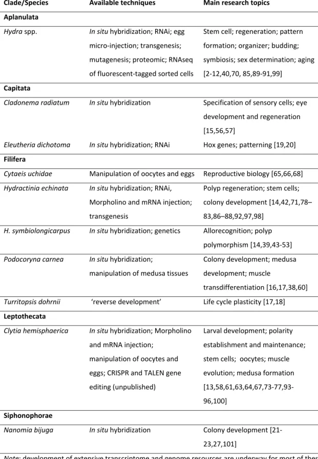
Table 1 – Hydrozoans used for genetic/molecular studies
Clade/Species Available techniques Main research topics Aplanulata Hydra spp. In situ hybridization; RNAi; egg micro‐injection; transgenesis; mutagenesis; proteomic; RNAseq of fluorescent‐tagged sorted cells Stem cell; regeneration; pattern formation; organizer; budding; symbiosis; sex determination; aging [2‐12,40,70, 85,89‐91,99] Capitata
Cladonema radiatum In situ hybridization Specification of sensory cells; eye
development and regeneration [15,56,57]
Eleutheria dichotoma In situ hybridization; RNAi Hox genes; patterning [19,20]
Filifera
Cytaeis uchidae Manipulation of oocytes and eggs Reproductive biology [65,66,68]
Hydractinia echinata In situ hybridization; RNAi, Morpholino and mRNA injection; transgenesis Polyp regeneration; stem cells; colony development [14,42,71,78– 83,86–88,92,97,98]
H. symbiolongicarpus In situ hybridization; genetics Allorecognition; polyp polymorphism [14,39,43‐53] Podocoryna carnea In situ hybridization; manipulation of medusa tissues Colony development; medusa development; muscle transdifferentiation [16,17,38,60]
Turritopsis dohrnii ‘reverse development’ Life cycle plasticity [17,18]
Leptothecata Clytia hemisphaerica In situ hybridization; Morpholino and mRNA injection; manipulation of oocytes and eggs; CRISPR and TALEN gene editing (unpublished) Larval development; polarity establishment and maintenance; stem cells; oocytes; muscle evolution; medusa formation [13,58,61,63,64,67,73‐77,93‐ 96,100] Siphonophorae
Nanomia bijuga In situ hybridization Colony development [21‐
23,27,101]
Note: development of extensive transcriptome and genome resources are underway for most of these species. In situ hybridization technique and/or transcriptome resources have been developed for a few others species
(e.g. [1,37,59]).
Figure 3