aBSTRacT
Background: Central pain sensitization is often refractory to drug treat-ment. Dextromethorphan, an N-methyl-d-aspartate receptor antagonist, is antihyperalgesic in preclinical pain models. The hypothesis is that dextro-methorphan is also antihyperalgesic in humans.
Methods: This randomized, double-blind, placebo-controlled, crossover study explores the antihyperalgesic effect of single and repeated 30-mg dose of oral dextromethorphan in 20 volunteers, using the freeze-injury pain model. This model leads to development of primary and secondary hyperalgesia, which develops away from the site of injury and is associated with central sensitization and activation of N-methyl-d-aspartate receptor in the spinal cord. The primary outcome was antihyperalgesia calculated with the area under the curve of the percentage change in mechanical pain threshold (electronic von Frey) on the area of secondary hyperalgesia. The secondary outcomes were mechanical pain threshold on the area of primary hyperalgesia and cognitive (reaction time) effect. Results: Single 30-mg results are reported. Antihyperalgesia (% ∙ min) is significantly higher on the area of secondary hyperalgesia with dextrometho-rphan than placebo (median [interquartile range]: 3,029 [746; 6,195] vs. 710 [–3,248; 4,439], P = 0.009, Hedge’s g = 0.8, 95% CI [0.1; 1.4]). On primary hyperalgesia area, mechanical pain threshold 2 h after drug intake is signifi-cantly higher with dextromethorphan (P = 0.011, Hedge’s g = 0.63, 95% CI [0.01; 1.25]). No difference in antinociception is observed after thermal painful stimuli on healthy skin between groups. Reaction time (ms) is shorter with placebo than with dextromethorphan (median [interquartile range]: 21.6 [–37.4; 0.1] vs. –1.2 [–24.3; 15.4], P = 0.015, Hedge’s g = 0.75, 95% CI [0.12; 1.39]). Nonserious adverse events occurrence (15%, 3 of 20 volun-teers) was similar in both groups.
conclusions: This study shows that low-dose (30-mg) dextromethorphan is antihyperalgesic in humans on the areas of primary and secondary hyperalge-sia and reverses peripheral and central neuronal sensitization. Because dex-tromethorphan had no intrinsic antinociceptive effect in acute pain on healthy skin, N-methyl-d-aspartate receptor may need to be sensitized by pain for dextromethorphan to be effective. (ANESTHESIOLOGY 2019; 131:356–68)
Dextromethorphan
Analgesia in a Human
Experimental Model of
Hyperalgesia
E. Martin, Ph.D., C. Narjoz, Ph.D., Pharm.D.,
X. Decleves, Ph.D., L. Labat, Ph.D., C. Lambert, M.Sc., M.-A. Loriot, Ph.D., Pharm.D., G. Ducheix, M.D., C. Dualé, Ph.D., M.D., B. Pereira, Ph.D., G. Pickering, Ph.D., M.D., Pharm.D.
Anesthesiology 2019; 131:356–68
This article is featured in “This Month in Anesthesiology,” page 1A. This article is accompanied by an editorial on p. 233. Supplemental Digital Content is available for this article. Direct URL citations appear in the printed text and are available in both the HTML and PDF versions of this article. Links to the digital files are provided in the HTML text of this article on the Journal’s Web site (www.anesthesiology.org). This article has a video abstract. This article has a visual abstract available in the online version.
Submitted for publication March 8, 2018. Accepted for publication March 13, 2019. From University Clermont Auvergne, Department of Fundamental and Clinical Pharmacology of Pain, NeuroDol, F-63000 Clermont-Ferrand, France (E.M., C.D., G.P.); Inserm UMR-S1147, Saints-Pères University Centre, Paris, France (C.N., M.-A.L.); University Paris Descartes, Sorbonne Paris Cité, Paris, France (C.N., M.-A.L.); Assistance Publique—Paris Hospital (AP-HP), Georges Pompidou European Hospital, Biochemistry Department, Paris, France (C.N., M.-A.L.); Cochin Hospital, HUPC, Hôpitaux de Paris (AP-HP) Paris, France (X.D., L.L.); Pharmacy Faculty, University Paris Descartes Inserm UMR-S1144, Paris, France (X.D.); Clermont-Ferrand, Research and Innovation Department, Clermont-Ferrand, France (C.L., B.P.); and University Hospital Clermont-Ferrand, Clinical Pharmacology Department/ Clinical Research Centre, Inserm 1405, F-63003 Clermont-Ferrand, France (G.D., C.D., G.P).
Copyright © 2019, the American Society of Anesthesiologists, Inc. All Rights Reserved. Anesthesiology 2019; 131:356–68. DOI: 10.1097/ALN.0000000000002736
N
europathic pain, defined as pain caused by a lesion or a disease of the somatosensory nervous system,1affects 7 to 10% of the general population2 and is associated
with central sensitization involving N-methyl-d-aspartate (NMDA) receptors.3 Neuropathic pain presents
abnor-mal pain manifestations including allodynia and hyper-algesia1 and is accompanied by impaired quality of life.
Management of neuropathic pain is still not satisfactory4
and in recent years, special attention has been focused on NMDA receptor antagonists, ketamine, memantine, and
ediTOR’S PeRSPecTiVe
What We Already Know about this topic
• Neuropathic pain, which presents abnormal pain manifestations including allodynia and hyperalgesia, is associated with central sensitization involving N-methyl-d-aspartate receptors
• In the freeze-injury hyperalgesia model, a cold burn leads to development of both primary hyperalgesia and secondary hyperalgesia, which develops away from the site of injury without apparent tissue modification, and is associated with central sensitization and activation of N-methyl-d-aspartate receptors in the spinal cord
• Dextromethorphan, which is an N-methyl-d-aspartate receptor antagonist, is antihyperalgesic in preclinical pain models
What this Article tells us that Is New
• Using the freeze-injury pain model in a randomized, double-blind, placebo-controlled crossover trial of 30-mg doses of oral dextrometh-orphan in 20 male volunteers, dextromethdextrometh-orphan was antihyperalgesic and reversed peripheral and central neuronal sensitization
• Because dextromethorphan had no intrinsic antinociceptive effect in acute pain on healthy skin, N-methyl-d-aspartate receptors may need to be sensitized by pain for dextromethorphan to be effective
dextromethorphan. The analgesic mechanism of action of dextromethorphan, mainly used as a cough suppressant with safety hazards,5 remains incompletely known, however.
A preclinical study in a spinal nerve ligation animal model showed that postsurgical dextromethorphan administration induced a significant decrease of allodynia and hyperalgesia while preserving mobility and cognition in the Y-maze test.6
These positive findings in animals and in some clinical stud-ies showing neuropathic pain alleviation after trauma7,8 and
diabetes,9 although not universal,10,11 triggered an ongoing
clinical trial (NCT02271893)12 in patients with refractory
postsurgery neuropathic pain and central pain sensitization. To complement this translational approach, human experimen-tal pain models may mimic some neuropathic pain charac-teristics and central pain sensitization, and help to understand the pharmacology of dextromethorphan. In the freeze-injury hyperalgesia model, a cold burn leads to the development of primary hyperalgesia,13 and of secondary hyperalgesia that
develops away from the site of injury without apparent tis-sue modification, and is associated with central sensitization, rather than neuropathic pain per se, and activation of NMDA receptor in the spinal cord.14 Such an experimental model
has not been used so far with dextromethorphan and might allow a better understanding of the antinociceptive effect of dextromethorphan in pain central sensitization.
Dextromethorphan is known to bind noncompetitively with low-moderate affinity to the phencyclidine site in the NMDA receptor channel.15 It also has a moderate-affinity
ago-nist activity with σ1 receptor sites and is an antagoago-nist of nico-tinic acetylcholine receptors. It is metabolized by cytochrome P450 (CYP), CYP2D6, to dextrorphan, its main active metab-olite, and by CYP3A4 to inactive metabolites.16 Genetic
poly-morphism may be a variability factor of dextromethorphan metabolism (CYP2D6, CYP3A4), efflux (P-glycoprotein encoded by ABCB1 gene),17–19 and analgesic effect.7,8 This
analgesic effect has been attributed to dextromethorphan itself8
or to dextrorphan.7 Dextromethorphan has also an impact on
cognition20 and has been considered as an opioid, although
some publications rather suggest a nonopioid effect21,22
(inhib-itory constant for dextromethorphan: 1,280 nM; inhib(inhib-itory constant for dextrorphan: 420 nM).23 Its role on the autonomic
nervous system is still unclear.24 These characteristics may be
assessed respectively by cognitive tests and by pupillometry, as miosis—a known effect of opioids—pupil size and reactivity to light, reflect the iris autonomic nervous system.25
Using the freeze-injury–induced hyperalgesia model, this placebo-controlled study aims to explore the anti-hyperalgesic effect of single and repeated doses of 30-mg dextromethorphan with mechanical painful stimuli, central activity (reaction time and pupillary diameter), and metab-olism (drug measurements) in healthy volunteers.
Materials and Methods
This randomized, double-blind, placebo-controlled and crossover study was conducted in the Clinical Pharmacology
Department and Clinical Research Center, University Hospital of Clermont-Ferrand, Clermont-Ferrand, France, from November 2015 to February 2016. It was approved by the referent ethics committee CPP (Committee for the Protection of Persons) Sud-Est VI, Clermont-Ferrand, France, (AU1213) and the French competent author-ity (151147A-32). It was registered on EudraCT (2015-003171-30) and ClinialTrials.gov (NCT02596360).
subjects
Subjects were preselected from the Healthy Volunteers File of the Clinical Pharmacology Department and Clinical Research Center. All subjects were compensated for their participation in the study.
Caucasian healthy male volunteers were eligible if they were between 18 and 45 yr old, with a body mass index of at least 19 and no greater than 30 kg/m2, were extensive or
intermediate CYP2D6 metabolizers, as determined during the prescreening visit, and were required to be free of any medication for at least 7 days before inclusion. Volunteers were excluded with a known hypersensitivity to dextro-methorphan, aspartate transaminase, alanine transaminase and total bilirubin twice the normal range, consumption of alco-hol, tobacco, or any drug addiction. In order to avoid interfer-ing with the psychometric tests results, volunteers were asked not to consume magnesium, citrus juice, drinks with theine, or caffeine during the assessment days. Volunteers lacking concentration and not able to evaluate pain thresholds were excluded. Eligible volunteers were informed about the proto-col and provided a signed informed consent before inclusion.
study Design
After inclusion, volunteers were familiarized (one session) with the psychophysics experiments. Blood samples were collected from all participants to assay serum aspartate trans-aminase, alanine transtrans-aminase, and bilirubin levels, CYP2D6, CYP3A4, and ABCB1 genotyping. The selected volunteers were randomly assigned to receive oral dextromethorphan bromhydrate tablets (Pulmodexane 30 mg [23 mg dextro-methorphan base]; Bailly-Creat Laboratory, France, maxi-mal dose 120 mg daily) or placebo similar in appearance to dextromethorphan tablets (lactose, Cooper Laboratory, France) in two randomized periods 11 days apart according to a randomization list. The randomization sequence was generated using random blocks and was established before-hand by a research assistant who was not involved in the trial. Volunteers and all personnel involved in the trial con-duct were blinded to the treatment assignment.
On day 0, after freeze injury induction, volunteers had baseline tests (t0) and received 30 mg of dextromethorphan or placebo. Treatment was given by a nurse independent of the study. Tests were repeated at 1 h, 2 h, and 3 h to assess the effects of a single dose. Thereafter, to measure the effects of drugs after repeated dosages, while respecting the maximal
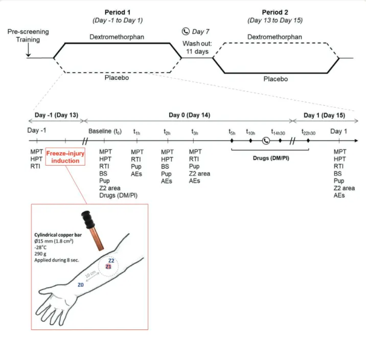
dosage recommendations, volunteers took 30 mg of dextro-methorphan or placebo four times at home: three times on day 0 (5 h, 10 h, and 14.5 h after baseline) and once on day 1 (22.5 h after baseline). Tests were repeated on day 1. After a washout period of 11 days, the same procedure was repeated in a crossover fashion during the second session starting at day 13 and ending at day 15. Drug adverse events were collected during the sessions by a nurse independent of the study on day 0 and day 14 between the third and the fourth dextro-methorphan doses and during the washout period. A detailed overview of the experimental design is given on figure 1.
Experimental Pain Model Induction
In the majority of human pain models,26 the induced
hyperalgesia is dose-dependent, is not reproducible, is not stable with time, and does not last long enough (1–3 h with capsaicin and mustard oil) to cover the duration of action of repeated drug administration. In this present study, the freeze-injury–induced hyperalgesia model described by Kilo et al.13 was chosen to induce reproducible primary and
secondary hyperalgesia that may stay stable for 72 h13,27 with
the absence of carryover effect as described previously.27
This experimental cutaneous hyperalgesia model consists
Fig. 1. Design and chronology of the study. AEs, adverse events; Bs, blood sample; DM/Pl, dextromethorphan or placebo administra-tion; HPt, heat pain thresholds; MPt, mechanical pain thresholds; Pup, pupillometry; RtI, reaction time Cambridge Neuropsychological test Automated Battery test; t1h, 1 h after baseline; t2h, 2 h after baseline; t3h, 3 h after baseline; t5h, 5 h after baseline; t10h, 10 h after baseline; t14h30, 14.5 h after baseline; t22h30, 22.5 h after baseline; Z0, healthy skin; Z1, primary hyperalgesia; Z2, secondary hyperalgesia; Z2 area, secondary hyperalgesia surface measurement.
of applying for 8 s on the anterior glabrous part of the fore-arm (dominant fore-arm in the first study period and nondom-inant arm in the second) the tip of a 15-mm-diameter and 290-g-weight cylindrical copper bar frozen at –28°C. The induced first-degree burn leads to two types of hyperalge-sia: a primary hyperalgesia area associated with a sharply defined erythema, corresponding to the surface in contact with the copper bar, and a localized secondary hyperalgesia area surrounding primary hyperalgesia in undamaged skin (fig. 1).13 A control skin area was determined on the injured
arm at a distance of 20 cm from the lesion.
The primary endpoint was the comparison of the areas under the curve (AUC) of the percentage change in mechanical pain threshold (% MPT, providing force to 0.1 g) between baseline and 3 h after drug intake (AUC %MPTt0-t3h; % ∙ min) in the secondary hyperalgesia area between dex-tromethorphan versus placebo according to the formula
AUC%MPTt0-t3h (% ∙ min) = 60 × ((((t1h – t0) × 100) / t0) + (((t2h – t0) × 100) / t0)) / 2 + 60 × ((((t2h – t0) × 100) / t0) + (((t3h – t0) × 100) / t0)) / 2,
with t0, t1h, t2h, and t3h corresponding to mechanical pain threshold at baseline, 1 h, 2 h, and 3 h after drug, respectively.
Secondary outcome measures were the assessment of mechanical pain threshold in primary hyperalgesia and healthy skin areas, the evolution of the secondary hyperal-gesia area, thermal pain thresholds, pupillary reactivity, cog-nitive status, plasma dextromethorphan and dextrorphan concentrations, and drug adverse events.
Procedures of Pain Assessment
Mechanical Test. The electronic von Frey (Somedic, France) test consists in applying pressure with a 0.2-mm-diameter probe tip on the middle of primary hyperalgesia, secondary hyperalgesia, and control skin areas with a constant slope of increasing punctate pressure up to the detection of mechan-ical pain thresholds (grams), corresponding to the first pain sensation, signaled by the volunteer by a response push button. The mean of three measurements was taken as the mechanical pain threshold. An increase of mechanical pain threshold signifies that the individuals cope better with pain.
Secondary Hyperalgesia Area Measurement. The borders of the secondary hyperalgesia area were determined with a handheld 588-mN von Frey hair by concentric stimu-lations along six linear paths arranged radially around the lesioned site. When the volunteer reported a clear change in sensation, this was defined as the border of the secondary hyperalgesia area. Secondary hyperalgesia areas (cm2) were
calculated with the software ImageJ28 (W.S. Rasband, M.Sc.,
U.S. National Institutes of Health, Bethesda, Maryland; ImageJ, https://imagej.nih.gov/ij/). Primary hyperalgesia area was defined as the area of erythema corresponding to the skin area in contact with the frozen copper bar (1.8 cm2).
Thermal Tests. Heat pain threshold was assessed using the Advanced Thermal Stimulator thermode (30 × 30 mm)
connected to the Medoc Pathway system (Medoc Ltd., Israel) applied on normal skin of the control arm. From the baseline value of 32°C, the Medoc Pathway system deliv-ers an adjustable temperature peak in heat with a slope increase of 1°C increments and controlled by rapid feed-back. Heat pain threshold was determined as the mean of three measures.
Evaluation of Central and Cognitive Effects of Dextromethorphan
Pupillary Reactivity Measurement. Pupillometry recordings were performed with a noninvasive monocular porta-ble infrared pupillometer (NeuroLight Algiscan; IDMED, France) to measure a baseline scotopic pupil size (mm), then quantitative pupillary light reflex, which represents the reduction in pupil size after light stimulation. Scotopic con-ditions were obtained with the device’s light-tight occlusive silicone collar between the camera and the edges of the orbit and with the subject shielding his contralateral eye tightly with a hand. After application, the subject was asked to keep the eye open, and pupil diameter was noted after stable values were obtained (30 s in general). All measure-ments were undertaken on the right pupil, or on the left in case of abnormality of the right eye.
Reactivity Measurement. Reaction time, measured to 0.1-ms precision, was assessed using the Cambridge Neuropsychological Test Automated Battery (Cambridge Cognition, United Kingdom). Reaction time (ms) is the time taken to release the press pad in response to the visual stimulus (yellow dot). Movement time (ms) is the time taken to touch the yellow dot on the touchscreen after the press pad has been released. Differences from baseline (delta) were compared between dextromethorphan and placebo.
Pharmacogenetics and Plasma Concentrations
Considering genetic polymorphism in dextromethorphan effect, CYP2D6 extensive and intermediate metabolizer volunteers have been selected in this study to homoge-nize the population on this highly polymorphic enzyme. Interindividual variations in the disposition of dextro-methorphan/dextrorphan have been investigated.
Genomic DNA samples, collected during the prescreen-ing visit, were extracted from blood mononuclear cells by use of a commercial kit (Maxwell 16 LEV Blood DNA Kit, Promega, France) according to the manufacturer’s protocols.
CYP2D6 Genotyping. The CYP2D6*6 allele was detected by use of the long polymerase chain reaction method for the whole-gene amplification, followed by a subsequent nested polymerase chain reaction and restriction enzyme analy-sis.29 Gene deletion (CYP2D6*5 allele) and gene
duplica-tion (responsible for ultrarapid phenotype) were analyzed by the long polymerase chain reaction method as previously described.30 CYP2D6*3, CYP2D6*4, and CYP2D6*6
were detected by use of Taq Man Drug Metabolism Genotyping Assays (C__32407232_50, C__27102431_Day 0, C__32407243_20; Applied Biosystems-Thermo Fisher Scientific, France).
Dextromethorphan and Dextrorphan Plasma Concentrations. Plasma concentrations were quantified after acetonitrile precipitation using a validated high-per-formance liquid chromatography tandem mass spec-trometry method (TSQ Quantum Ultra, Fisher, USA). Separation was carried out on an Accucore Phenyl Hexyl column (100 mm × 2.6 mm, 2.6 µm, Fisher). The mobile phase consisted of solvent A (0.1% formic acid in water) and solvent B (0.1% formic acid in acetonitrile) with gra-dient program. Briefly, aliquots of plasma (200 µl) were added to 100 µl of internal standard (dextrorphan-d3, 200 ng/mL, LGC, United Kingdom) and 600 µL of ace-tonitrile. After centrifugation and evaporation of organic phase under nitrogen flow at 30°C, the residue was dis-solved in 100 µL of water/methanol (95/5%) with 0.1% formic acid and 10 µl used for injection.
Detection in triple quadrupole mass spectrometry used an electrospray ionization probe and operated in the posi-tive ion mode. The multiple reaction monitoring transitions used for quantification were 272.1/215.0 for dextrometho-rphan, 258.1/201.1 for dextrodextrometho-rphan, and 261.1/157.1 for dextrorphan-d3. The linear calibration ranges in plasma (correlation coefficients greater than 0.999) were 2.5 to 250 ng/ml for dextromethorphan and 1 to 500 ng/ml for dextrorphan. The metabolic ratio was calculated by divid-ing dextrorphan concentration by dextromethorphan concentration.
statistical Analysis
For type I and type II errors of 5% (two-sided) and 20%, respectively, and with intraindividual correlation coefficient equals to 0.5 (owing to the crossover design and no carry-over effect assumed), 19 patients are needed to highlight a difference of at least 3,400 on the primary outcome for an effect size around 0.70. This primary outcome is the area under the curve of the percentage change in mechanical pain threshold between baseline and 3 h after drugs intake (AUC %MPTt0-t3h) in secondary hyperalgesia after treatment administration (with a SD of 4,900), according to the study results previously detailed in the literature.26 According to
these estimations, 20 patients were included per group. Statistical analysis was performed using the Stata soft-ware (Version 13, StataCorp, USA) where hypothesis test-ing was two-sided with significance interpreted as P < 0.05. For continuous parameters, mean ± SD or median and interquartile range were calculated, according to statistical distribution. The Shapiro–Wilk test was used to study nor-mality assumption of continuous data. Then, the primary endpoint and secondary endpoints were compared between groups using a random-effects model for crossover designs
while taking into account the following effects: treatment group, sequence, subject (as random-effect), and carryover. A Sidak’s type I error correction was applied to take into account the multiple comparisons. The normality of resid-uals was studied using the Shapiro–Wilk test when appro-priate, and a logarithmic transformation has been proposed to achieve the normality. The results were expressed using Hedges’s g effect size and 95% CI. For categorical param-eters, a Stuart–Maxwell test for paired data or generalized linear mixed model (binomial distribution with log link for dichotomous endpoint) was applied. Random effect mod-els were also carried out to analyze repeated measures to study fixed effects (group, timepoints and interaction group × time). A cumulative proportion of responder’s analysis (AUC %MPTt0-t3h, 0, 1,000, 2,000, 6,000, 10,000, 12,000, 14,000, 16,000) comparing dextromethorphan to placebo was performed to provide a visual representation of the likelihood of response over a full range of response levels for the two groups. The analysis was conducted using random effects model to measure group and response level effects and their interaction, taking into account between- and within-subject variability.
Concerning noncrossover comparisons, usual statistical tests were performed: independent Student’s t test or Mann– Whitney test (when assumptions of t test were not met: nor-mality and homoscedasticity) for quantitative parameters and chi-square test or Fisher exact test for categorical variables. The Kruskal–Wallis test and Spearman correlation coeffi-cient (rather than Pearson correlation due to non-Gaussian statistical distribution) was used to identify relations between dextromethorphan and dextrorphan plasma concentrations, pain, central, and cognitive parameters.
Results
study subjects
For this study, 34 healthy males were prescreened, 23 were randomized, 3 were excluded from the analysis (consent withdrawal, unstable pain model, and flu-like syndrome), and 20 volunteers were analyzed. The flowchart is presented in figure 2.
Characteristics of Induced Hyperalgesia
Stability of the Pain Model with Time and Induced Hyperalgesia. At baseline, a significant decrease of the mechanical pain threshold in primary and secondary hyper-algesia (P < 0.001, effect size = –0.81 [–1.26; –0.36]) was observed when compared to control skin area (measured before the pain model induction) in both treatment groups. With the placebo, the freeze-injury–induced hyperalge-sia was maintained (comparison vs. control skin) in pri-mary hyperalgesia at each timepoint in day 0 and day 1 (P < 0.001, effect size = –1.02 [–1.67; –0.37]), and in sec-ondary hyperalgesia at each time at day 0, 1 h after dosing
(P < 0.001, effect size = –0.82 [–1.46; –0.18]), 2 h after dos-ing (P < 0.001, effect size = –0.83 [–1.47; –0.19]), 3 h after dosing (P < 0.001, effect size = –0.70 [–1.66; –0.04]) but for only 10 out of 20 individuals (50%) at day 1 (P = 0.069, effect size = –0.37 [–0.99; 0.25]).
Primary Study Endpoint. Evaluation of secondary hyperalge-sia shows on day 0, between 1 and 3 h after dosing, a signif-icant increase of the area under the curve of the percentage change in mechanical pain threshold (AUC%MPTt0-t3h) between dextromethorphan compared to placebo (P = 0.009, effect size = 0.8 [0.1; 1.4]; table 1). The cumu-lative proportion of responder’s analysis, comparing dextro-methorphan to placebo, provides a statistically significant proportion of responders (P < 0.001). More precisely, the significance of AUC %MPTt0-t3h is P = 0.003 (proportion of responders = 85% vs. 45%) at level 0 and P = 0.011 (proportion of responders = 75% vs. 40%; fig. 3) at level 1,000. From t0 to day 1, the surface of secondary hyperalge-sia shrank concentrically toward primary hyperalgehyperalge-sia, and
this was not significantly different between dextrometho-rphan and placebo at t0 (P = 0.625, effect size = 0.2 [–0.5; 0.8]), at 3 h after dosing (P = 0.991, effect size = 0.0 [–0.6; 0.6]) and at day 1 (P = 0.743, effect size = 0.1 [–0.5; 0.7]; table 1). Raw values of mechanical pain thresholds in sec-ondary hyperalgesia, primary hyperalgesia and healthy skin are shown in Supplemental Digital Content 2 (http://links. lww.com/ALN/B949).
Primary Hyperalgesia. Mechanical pain thresholds (delta) on primary hyperalgesia increased significantly for dex-tromethorphan compared to placebo between t0 and t1h (deltat0-t1h, dextromethorphan: 4.6 ± 11.6 g, placebo: –3.6 ± 17.6 g, P = 0.038, effect size 0.53 [0.09; 1.15]), between baseline and 2 h after dosing (deltat0-t2h, dextromethorphan: 7.9 ± 12.8 g, placebo: –2.8 ± 19.7 g, P = 0.011, effect size 0.63 [0.01; 1.25]) and between baseline and day 1 (deltat0-day1, dextromethorphan: 8.4 ± 15.6 g, placebo: –2.9 ± 21.1 g, P = 0.015, effect size 0.60 [0.03; 1.27]). The primary hyper-algesia surface did not change during the entire study period.
Fig. 2. Flowchart of participants during the trial.
Thermal Thresholds. No significant difference was observed between treatments in heat pain threshold on the control skin area on the opposite arm of the freeze-injured arm (table 1).
Cognitive Parameters
With the placebo, repeated reaction time decreased compared to baseline, suggesting a learning process. This diminution was
Table 1. Effect of Dextromethorphan on Primary and secondary Outcome Measures
dextromethorphan (n = 20) Placebo (n = 20) P Value
AuC%MPtt0-t3h (% ∙ min)
3,029 [746; 6,195] –710 [–3,248; 4,439] 0.009
secondary hyperalgesia surface (cm2)
t0 42.65 ± 18.79 39.86 ± 18.26 0.625
t3h 29.45 ± 19.49 29.38 ± 17.84 0.991
Day 1 23.12 ± 19.30 21.55 ± 15.92 0.743
Heat pain thresholds (°C)
t0 42.1 ± 2.3 42.5 ± 1.7 0.313
t2h 42.4 ± 2.3 42.6 ± 2.0 0.631
Day 1 43.4 ± 1.9 43.3 ± 1.6 0.819
Movement time, delta from baseline (ms)
t1h – t0 28.6 [–37.6; 108.4] –26.5 [–71.5; 53.1] 0.229
t3h – t0 19.8 [–53.9; 105.9] 10.3 [–69.5; 78.2] 0.650
Day 1 – t0 –29.6 [–66.2; 53.3] –47.2 [–86.0; 47.6] 0.518
Pupillary light reflex (%)
t0 40 ± 5 40 ± 4 0.921
t1h 40 ± 5 40 ± 4 0.959
t2h 39 ± 5 39 ± 5 0.847
t3h 39 ± 5 39 ± 5 0.952
Day 1 39 ± 5 39 ± 5 0.639
Effect of dextromethorphan on mechanical pain threshold in secondary hyperalgesia (area under the curve of the percentage change in mechanical pain threshold in secondary hyperalgesia [AuC%MPtt0-t3h]), secondary hyperalgesia surface, heat pain thresholds, movement time, and pupillary light reflex (mean ± sD or median [interquartile range]).
Fig. 3. Cumulative proportion of responder’s analysis with area under the curve of the percentage change in mechanical pain threshold in secondary hyperalgesia (AuC%MPtt0-t3h >0; %.min) in dextromethorphan and placebo groups. A statistically significant proportion of responders (P < 0.001) was observed, more precisely at AuC %MPtt0-t3h level 0 P = 0.003 and level 1,000 P = 0.011. P values were estimated applying a sidak’s correction type I error.
not observed with dextromethorphan, and the repeated test showed that delta reaction time between baseline and day 1 was larger with dextromethorphan than with placebo (P = 0.015, effect size = 0.75 [0.12; 1.39]; fig. 4). No significant difference between treatments was observed in movement time at any time, but variability was very high (table 1). Raw values of reaction time and movement time are shown in Supplemental Digital Content 2 (http://links.lww.com/ALN/B949).
Pupillary Reactivity
Two hours after drugs intake (t2h), corresponding to the plasmatic peak of dextromethorphan,31 a significant
differ-ence of pupil diameter was observed between treatments with a larger diameter for dextromethorphan compared to placebo (P = 0.017, effect size = 0.49 [0.06; 0.94]; fig. 5A). No significant difference was observed between treatments concerning pupillary light reflex (table 1).
Pharmacogenetics and Plasma Concentrations
The study was designed with a homogenous population by selecting extensive or intermediate CYP2D6 metabolizers. Correlations between concentrations of dextromethorphan and dextrorphan, hyperalgesia, central, and cognitive parame-ters have been studied. At day 1, there was a positive significant correlation between dextrorphan concentration and reaction time (ρ = 0.715, P = 0.001). A negative significant correlation was observed between metabolic ratio and pupillary diameter change 2 h after dosing (ρ = –0.559, P = 0.012; fig. 5B). A neg-ative correlation was observed for dextrorphan concentration (ρ = –0.267) and a positive correlation with dextromethorphan concentration (ρ = 0.225; fig. 5B). Concentrations of dextro-methorphan and dextrorphan at day 0 (dextrodextro-methorphan: 1.7 ± 1.6 ng/ml, 95% CI [1.0; 2.4]; dextrorphan: 3.4 ± 1.5 ng/ml, 95% CI [2.7; 4.0]) and at day 1 (dextromethorphan: 3.8 ±
5.5 ng/ml, 95% CI [1.4; 6.2]; dextrorphan: 6.5 ± 2.8 ng/ml, 95% CI [5.3; 7.8]) are shown in Supplemental Digital Content 1 (http://links.lww.com/ALN/B948).
Adverse Events
The proportion of subjects experiencing possible drug-re-lated nonserious adverse events was 15% (3 of 20 volun-teers) with both dextromethorphan and placebo treatments (P > 0.999). With dextromethorphan, adverse events were of mild severity and were those commonly reported for this drug (dry mouth, fatigue, n = 3). With the placebo, subjects experienced fatigue and stomach ache (n = 3). No serious adverse event was reported.
discussion
This study in healthy volunteers shows in humans that low-dose (30 mg) oral dextromethorphan significantly dimin-ishes hyperalgesia in a freeze-injury pain model, whereas previous studies mainly assessed this effect in animals or with higher dosages in humans (e.g., 270 mg7; 960 mg/
day32). The secondary hyperalgesia induced by the model
was significantly decreased (P = 0.009), confirming the antihyperalgesic effect of dextromethorphan. This is con-sistent with the literature reporting that NMDA receptor noncompetitive antagonists, by blocking the NMDA recep-tor channel, could limit or even reverse central sensitization symptoms.6,33 The main mechanism of secondary
hyper-algesia results from central neuronal sensitization medi-ated by low-threshold myelinmedi-ated mechanoreceptors and nociceptors (stimulated by punctate hyperalgesia),34,35 and
descending facilitation of spinal nociception contributes to the maintenance of secondary hyperalgesia.36
The study also showed that dextromethorphan significantly reduced primary hyperalgesia to punctate stimuli (P < 0.03),
Fig. 4. Effect of dextromethorphan on reaction time. With placebo, reaction time decreased compared to baseline, suggesting a learning process that was not observed with dextromethorphan. Delta reaction time between baseline and Day 1 was larger with dextromethorphan than with placebo (P = 0.015). the plus sign and the straight line represent, respectively, the mean and the median of values. P values were estimated applying a sidak’s correction type I error. t1h, 1 h after baseline; t3h, 3 h after baseline.
Fig. 5. Effect of dextromethorphan on basal pupillary diameter. (A) two hours after dosing, a significant larger pupil diameter was observed with dextromethorphan compared to placebo (P = 0.017). (B) A negative significant correlation (ρ = –0.559, P = 0.012) was observed between pupillary diameter change (between 2 h after dosing and baseline) and metabolic ratio 2 h after dosing (metabolic ratio = dextro-rphan/dextromethorphan). A positive correlation (ρ = 0.225, P = 0.338) and a negative correlation (ρ = –0.267, P = 0.254) were observed between pupillary diameter change (between 2 h after dosing and baseline) with dextromethorphan and dextrorphan, respectively. t0, base-line; t1h, i h after baseline; t2h, 2 h after baseline; t3h, 3 h after baseline.
possibly via blockade of voltage-gated sodium channels37 or
peripheral antiinflammatory action.38,39 These findings
under-line the effectiveness of dextromethorphan on both central and peripheral sensitization with mechanical stimuli. Although dextromethorphan had a significant antihyperalgesic effect, the secondary and primary hyperalgesia surfaces diminished simi-larly with time for dextromethorphan and placebo, suggesting that dextromethorphan does not interfere with the sponta-neous wound healing processes. Such an observation has not been reported in the literature, and skin biopsies could shed some light on this hypothesis. Concerning other pain modali-ties, while dextromethorphan had an effect on mechanical pain stimuli, it did not have any on acute thermal painful stimuli in healthy skin, confirming previous studies of a poor effect of dextromethorphan on thermal challenges.40,41 This may
sug-gest that dextromethorphan does not behave like an agonist of μ opioid receptors that are expressed in peptidergic pain fibers and regulate the heat pain responsiveness.42
Concerning the effect of dextromethorphan on pain, our sample included only CYP2D6 extensive or interme-diate metabolizers. A pharmacogenetic approach,8
includ-ing CYP3A4 and ABCB1 polymorphisms, needs to be explored further as dextromethorphan-induced analgesia is considered to be mediated by dextromethorphan8 or by its
metabolite dextrorphan.7
The study also explored the central effect of dextro-methorphan by pupillometry, and a lesser constriction of the pupillary diameter was reported, (fig. 5A) as described in previous publications43,44 that stressed the occurrence of
mydriasis after dextromethorphan intake. Even though nor-adrenaline concentration was not measured, the mydriasis could be partly explained by the inhibition of noradrenaline reuptake24 as suggested by the mydriatic effect of
noradrena-line reuptake inhibitors like venlafaxine and reboxetin anti-depressants.45,46 Dextromethorphan and dextrorphan have
been reported to inhibit in vitro noradrenaline uptake into rat brain with inhibitory constant values of 240 nM and 340 nM, respectively.23 Another possible explanation is that
dextromethorphan could behave as an α agonist (inhibitory constant = 3000 nM) 24and directly dilate the radial muscles
of the iris. It is well accepted that a decreased arousal of the central nervous system (often induced by sedation) is accompanied by miosis and that sedation is a well-known adverse event of dextromethorphan.32 It is interesting to
note that the pupillary light reflex remained stable in our study, underlining that the dosage of dextromethorphan (30 mg/day up to 120 mg/day) did not induce central ner-vous system depression. However, we observed a significant negative correlation between the change of pupil diameter and metabolic ratio, showing that larger dextrorphan con-centrations might be correlated with a miosis (fig. 5B). We also observed a negative correlation for dextrorphan con-centration (ρ = –0.267) and a positive correlation for dex-tromethorphan concentration (ρ = 0.225) with pupillary diameter change (fig. 5B). This discrepancy may be caused
by contradictory actions of dextromethorphan and dextro-rphan. While mydriasis could be exerted by the noradren-ergic or α agonist activities of dextromethorphan, miosis is believed to rather be mediated by dextrorphan. Therefore, the pupillary diameter change cannot be attributed solely to plasma concentrations of either of these compounds, but appears to be a result of their combined effects. According to Slanar et al.,47 we may hypothesize that there is a
poten-tial cutpoint of metabolic ratio discriminating whether or not a significant mydriatic reaction occurs after dextro-methorphan intake. This needs to be confirmed by mea-suring the effect of each drug—dextromethorphan and dextrorphan—alone and in combination, on pupil diameter and sedation.
Concerning cognitive parameters, reaction time (fig. 4) but not movement time was impaired by dextrometho-rphan, suggesting that dextromethorphan impaired the timing of decision and response programming processes but not motor preparation and motor response. The learning effect during the reaction time test, illustrated by a mod-est decrease of reaction time after each repeated tmod-est, was amplified with placebo compared to dextromethorphan (P < 0.02). Such a defect in learning with dextrometho-rphan could be explained by the impairment of learning and memorization mediated by antagonists of the NMDA receptor,12 rather than by a sedative effect that was
sim-ilarly little reported in both groups. This specific impact of dextromethorphan on new learning would bene-fit from exploration in future studies with other specific cognitive tests like Paired Associates Learning (Cambridge Neuropsychological Test Automated Battery). More spe-cifically, it appears that it is dextrorphan that is associated with the increased reaction time (P = 0.001), a test that is related to cognitive function and may reflect cognitive impairment.48 These findings concur with the current view
that adverse effects of dextromethorphan would be related to its metabolite dextrorphan.49
This study has several limitations. First, the study was linked to the hyperalgesia model described by Kilo et al.13
that was chosen because it was described to induce a 72-h stable hyperalgesia.13,27 Assessment of this pain model
sta-bility in the placebo group showed, however, that sec-ondary hyperalgesia was maintained after day 1 for only 10 out of 20 individuals (50%), limiting the assessment of the antihyperalgesic clinical effect with repeated dextro-methorphan dosages because of the variability of the model itself. A second limitation is that this study may not be gen-eralized to the general population due to the absence of females, non-Caucasians, and volunteers outside the 18- to 45-yr-old age range, and with a diversity of CYP450 pro-files. In order to generalize our results, it would be inter-esting in the future to include patients suffering from pain with hyperalgesic characteristics of both sexes, all ages, and a stratification on the pharmacogenetics profile. A third
limitation is that this model is a surrogate of central pain sensitization, but not of neuropathic pain.
Collective data show that dextromethorphan is antihy-peralgesic and that it reverses peripheral and central neu-ronal sensitization in the freeze-injury pain model. Results suggest that NMDA receptor must be sensitized by pain for dextromethorphan to be effective, as it showed no intrinsic antinociceptive effect in acute pain on healthy skin. The major item of information this study provided is the anti-hyperalgesic efficacy in humans of dextromethorphan at a low single dose of 30 mg, whereas previous studies mainly assessed this effect on animals or with higher dosage in humans (e.g., 270 mg7; 960 mg/day32). The effects on
pupil-lary diameter showed in humans that the antihyperalgesic effect of dextromethorphan is not accompanied by a seda-tive effect. It also underlined that dextromethorphan does not have opioid-like effect, a consideration often encoun-tered in the literature. Finally, our results suggest that the main dextromethorphan metabolite, dextrorphan, may be responsible for deleterious cognitive impairment. Future trials with dextromethorphan combined with CYP450 modulators, inhibitors,8,9 and inducers in patients
suffer-ing from hyperalgesia are required to confirm these new findings and provide a therapeutic option for vulnerable patients with refractory pain.
Acknowledgments
The authors thank the volunteers who participated in this study and the staff of the Clinical Pharmacology Department/ Clinical Research Center, University Hospital of Clermont-Ferrand, Clermont-Clermont-Ferrand, France. The authors also thank Bernard Calvino, Ph.D. (École supérieure de physique et de chimie industrielles de la ville de Paris, Paris, France) for pathophysiological explanations on the pain model.
Research support
Support was provided solely from institutional and/or departmental sources.
Competing Interests
The authors declare no competing interests.
Reproducible science
Full protocol available at: [email protected]. Raw data available at: [email protected].
Correspondence
Address correspondence to Dr. Pickering, Centre de Pharmacologie Clinique, Bâtiment 3C, CIC Inserm 1405, CHU Ferrand, BP 69, F-63003 Clermont-Ferrand Cedex 1, France. [email protected]. This
article may be accessed for personal use at no charge through the Journal Web site, www.anesthesiology.org.
References
1. IASP: Taskforce on Taxonomy. 2011. Available at http://www.iasp-pain.org/Taxonomy. Accessed June 15, 2018
2. Hecke O van, Austin SK, Khan RA, Smith BH, Torrance N: Neuropathic pain in the general popu-lation: A systematic review of epidemiological studies. Pain 2014; 155:654–62
3. Latremoliere A, Woolf CJ: Central sensitization: A gen-erator of pain hypersensitivity by central neural plas-ticity. J Pain 2009; 10:895–926
4. Dworkin RH, Backonja M, Rowbotham MC, Allen RR, Argoff CR, Bennett GJ, Bushnell MC, Farrar JT, Galer BS, Haythornthwaite JA, Hewitt DJ, Loeser JD, Max MB, Saltarelli M, Schmader KE, Stein C, Thompson D, Turk DC, Wallace MS, Watkins LR, Weinstein SM: Advances in neuropathic pain: Diagnosis, mechanisms, and treatment recommendations. Arch Neurol 2003; 60:1524–34
5. Wilson MD, Ferguson RW, Mazer ME, Litovitz TL: Monitoring trends in dextromethorphan abuse using the National Poison Data System: 2000-2010. Clin Toxicol (Phila) 2011; 49:409–15
6. Morel V, Pickering G, Etienne M, Dupuis A, Privat AM, Chalus M, Eschalier A, Daulhac L: Low doses of dex-tromethorphan have a beneficial effect in the treatment of neuropathic pain. Fundam Clin Pharmacol 2014; 28:671–80
7. Carlsson KC, Hoem NO, Moberg ER, Mathisen LC: Analgesic effect of dextromethorphan in neuropathic pain. Acta Anaesthesiol Scand 2004; 48:328–36
8. Ehret GB, Daali Y, Chabert J, Rebsamen M, Wolff A, Forster A, Moursli F, Fritschy D, Rossier MF, Piguet V, Dayer P, Gex-Fabry M, Desmeules JA: Influence of CYP2D6 activity on pre-emptive analgesia by the N-methyl-D-aspartate antagonist dextromethorphan in a randomized controlled trial of acute pain. Pain Physician 2013; 16:45–56
9. Shaibani AI, Pope LE, Thisted R, Hepner A: Efficacy and safety of dextromethorphan/quinidine at two dosage levels for diabetic neuropathic pain: A dou-ble-blind, placebo-controlled, multicenter study. Pain Med 2012; 13:243–54
10. Ben Abraham R, Marouani N, Weinbroum AA: Dextromethorphan mitigates phantom pain in cancer amputees. Ann Surg Oncol 2003; 10:268–74
11. Gilron I, Booher SL, Rowan MS, Smoller MS, Max MB: A randomized, controlled trial of high-dose dex-tromethorphan in facial neuralgias. Neurology 2000; 55:964–71
12. Martin E, Morel V, Joly D, Villatte C, Delage N, Dubray C, Pereira B, Pickering G: Rationale and design of a randomized double-blind clinical trial in breast cancer: Dextromethorphan in chemotherapy-induced periph-eral neuropathy. Contemp Clin Trials 2015; 41:146–51 13. Kilo S, Schmelz M, Koltzenburg M, Handwerker HO:
Different patterns of hyperalgesia induced by experi-mental inflammation in human skin. Brain 1994; 117 (Pt 2):385–96
14. Meyer RA, Ringkamp M, Campbell JN, Raja SN: Neural mechanisms of hyperalgesia after tissue injury. Johns Hopkins Apl Tech Dig 2005; 26:56–66
15. Murray TF, Leid ME: Interaction of dextrorotatory opioids with phencyclidine recognition sites in rat brain membranes. Life Sci 1984; 34:1899–911
16. Capon DA, Bochner F, Kerry N, Mikus G, Danz C, Somogyi AA: The influence of CYP2D6 polymor-phism and quinidine on the disposition and antitus-sive effect of dextromethorphan in humans. Clin Pharmacol Ther 1996; 60:295–307
17. Marier JF, Deschênes JL, Hage A, Seliniotakis E, Gritsas A, Flarakos T, Beaudry F, Vachon P: Enhancing the uptake of dextromethorphan in the CNS of rats by concomitant administration of the P-gp inhibitor ver-apamil. Life Sci 2005; 77:2911–26
18. Bisaga A, Kos T, Wójcikowski J, Daniel WA, Popik P: Brain levels of dextromethorphan and the intensity of opioid withdrawal in mice. Drug Alcohol Depend 2008; 95:147–51
19. Kim RB, Leake BF, Choo EF, Dresser GK, Kubba SV, Schwarz UI, Taylor A, Xie HG, McKinsey J, Zhou S, Lan LB, Schuetz JD, Schuetz EG, Wilkinson GR: Identification of functionally variant MDR1 alleles among European Americans and African Americans. Clin Pharmacol Ther 2001; 70:189–99
20. Carter LP, Reissig CJ, Johnson MW, Klinedinst MA, Griffiths RR, Mintzer MZ: Acute cognitive effects of high doses of dextromethorphan relative to tri-azolam in humans. Drug Alcohol Depend 2013; 128:206–13
21. Mutschler J, Koopmann A, Grosshans M, Hermann D, Mann K, Kiefer F: Dextromethorphan withdrawal and dependence syndrome. Dtsch Arztebl Int 2010; 107:537–40
22. Chen ZR, Irvine RJ, Somogyi AA, Bochner F: Mu receptor binding of some commonly used opioids and their metabolites. Life Sci 1991; 48:2165–71
23. Codd EE, Shank RP, Schupsky JJ, Raffa RB: Serotonin and norepinephrine uptake inhibiting activity of cen-trally acting analgesics: Structural determinants and role in antinociception. J Pharmacol Exp Ther 1995; 274:1263–70
24. Taylor CP, Traynelis SF, Siffert J, Pope LE, Matsumoto RR: Pharmacology of dextromethorphan: Relevance
to dextromethorphan/quinidine (Nuedexta®) clinical use. Pharmacol Ther 2016; 164:170–82
25. Barvais L, Engelman E, Eba JM, Coussaert E, Cantraine F, Kenny GN: Effect site concentrations of remifentanil and pupil response to noxious stimulation. Br J Anaesth 2003; 91:347–52
26. Amerongen G van, Boer MW de, Groeneveld GJ, Hay JL: A literature review on the pharmacological sensi-tivity of human evoked hyperalgesia pain models. Br J Clin Pharmacol 2016; 82:903–22
27. Chassaing C, Schmidt J, Eschalier A, Cardot JM, Dubray C: Hyperalgesia induced by cutaneous freeze injury for testing analgesics in healthy volunteers. Br J Clin Pharmacol 2006; 61:389–97
28. Schneider CA, Rasband WS, Eliceiri KW: NIH Image to ImageJ: 25 years of image analysis. Nat Methods 2012; 9:671–5
29. Sachse C, Brockmöller J, Bauer S, Roots I: Cytochrome P450 2D6 variants in a Caucasian population: Allele frequencies and phenotypic consequences. Am J Hum Genet 1997; 60:284–95
30. Johansson I, Lundqvist E, Dahl ML, Ingelman-Sundberg M: PCR-based genotyping for duplicated and deleted CYP2D6 genes. Pharmacogenetics 1996; 6:351–5
31. Hughes AM, Rhodes J, Fisher G, Sellers M, Growcott JW: Assessment of the effect of dextromethorphan and ketamine on the acute nociceptive threshold and wind-up of the second pain response in healthy male volunteers. Br J Clin Pharmacol 2002; 53:604–12 32. Sang CN, Booher S, Gilron I, Parada S, Max MB:
Dextromethorphan and memantine in painful dia-betic neuropathy and postherpetic neuralgia: Efficacy and dose-response trials. Anesthesiology 2002; 96:1053–61
33. Carpenter KJ, Dickenson AH: NMDA receptors and pain–Hopes for novel analgesics. Reg Anesth Pain Med 1999; 24:506–8
34. Ringkamp M, Meyer RA: Physiology of Nociceptors, The Senses: A Comprehensive Reference, Vol. 5. Edited by Elsevier Inc., 2010, pp 97–114
35. Treede RD, Meyer RA, Raja SN, Campbell JN: Peripheral and central mechanisms of cutaneous hyperalgesia. Prog Neurobiol 1992; 38:397–421
36. Heinricher MM, Tavares I, Leith JL, Lumb BM: Descending control of nociception: Specificity, recruit-ment and plasticity. Brain Res Rev 2009; 60:214–25 37. Trube G, Netzer R: Dextromethorphan: Cellular
effects reducing neuronal hyperactivity. Epilepsia 1994; 35(suppl 5):S62–7
38. Madeira JM, Schindler SM, Klegeris A: A new look at auranofin, dextromethorphan and rosiglitazone for reduction of glia-mediated inflammation in neu-rodegenerative diseases. Neural Regen Res 2015; 10:391–3
39. Li MH, Luo YH, Lin CF, Chang YT, Lu SL, Kuo CF, Hong JS, Lin YS: Dextromethorphan efficiently increases bactericidal activity, attenuates inflammatory responses, and prevents group a streptococcal sepsis. Antimicrob Agents Chemother 2011; 55:967–73 40. Duedahl TH, Dirks J, Petersen KB, Romsing J, Larsen
NE, Dahl JB: Intravenous dextromethorphan to human volunteers: Relationship between pharmacokinetics and anti-hyperalgesic effect. Pain 2005; 113:360–8 41. Kauppila T, Grönroos M, Pertovaara A: An attempt
to attenuate experimental pain in humans by dex-tromethorphan, an NMDA receptor antagonist. Pharmacol Biochem Behav 1995; 52:641–4
42. Scherrer G, Imamachi N, Cao YQ, Contet C, Mennicken F, O’Donnell D, Kieffer BL, Basbaum AI: Dissociation of the opioid receptor mechanisms that control mechani-cal and heat pain. Cell 2009; 137:1148–59
43. Jasinski DR: Abuse potential of morphine/dextro-methorphan combinations. J Pain Symptom Manage 2000; 19(1 suppl):S26–30
44. Logan BK, Goldfogel G, Hamilton R, Kuhlman J: Five deaths resulting from abuse of dextromethorphan
sold over the internet. J Anal Toxicol 2009; 33:99–103
45. Saletu B, Grünberger J, Anderer P, Linzmayer L, Semlitsch HV, Magni G: Pharmacodynamics of ven-lafaxine evaluated by EEG brain mapping, psychome-try and psychophysiology. Br J Clin Pharmacol 1992; 33:589–601
46. Phillips MA, Bitsios P, Szabadi E, Bradshaw CM: Comparison of the antidepressants reboxetine, flu-voxamine and amitriptyline upon spontaneous pupillary fluctuations in healthy human volunteers. Psychopharmacology (Berl) 2000; 149:72–6
47. Slanar O, Nobilis M, Kvetina J, Mikoviny R, Zima T, Idle JR, Perlík F: Miotic action of tramadol is deter-mined by CYP2D6 genotype. Physiol Res 2007; 56:129–36
48. Jakobsen LH, Sorensen JM, Rask IK, Jensen BS, Kondrup J: Validation of reaction time as a measure of cognitive function and quality of life in healthy subjects and patients. Nutrition 2011; 27:561–70
49. Burns JM, Boyer EW: Antitussives and substance abuse. Subst Abuse Rehabil 2013; 4:75–82