Toxin-positive Clostridium difficile latently infect mouse colonies
and protect against highly-pathogenic C. difficile
Lucie Etienne-Mesmin1, Benoit Chassaing1, Oluwaseyi Adekunle2, Lisa M. Mattei3, Frederic D. Bushman4, and Andrew T. Gewirtz1,*
1Center for Inflammation Immunity and Infection, Institute for Biomedical Sciences, Georgia State University, Atlanta, Georgia, USA.
2Immunology and Molecular Pathogenesis Graduate Program, Emory University, Atlanta, Georgia, USA.
3PennCHOP Microbiome, Children’s Hospital of Philadelphia, Philadelphia, Pennsylvania, USA. 4Department of Microbiology, University of Pennsylvania School of Medicine, Philadelphia, Pennsylvania, USA.
Abstract
Objective—Clostridium difficile is a toxin-producing bacterium and a leading cause of
antibiotic-associated disease. The ability of C. difficile to form spores and infect antibiotic-treated persons at low MOI underlies its large disease burden. However, C. difficile-induced disease might also result from long-harbored C. difficile that blooms in individuals administered antibiotics.
Design—Mice purchased from multiple vendors and repeatedly testing negative for this pathogen by qPCR bloomed C. difficile following antibiotic treatment. This endogenous C. difficile strain, herein termed LEM1, which formed spores and produced toxin, was compared to highly pathogenic C. difficile strain VPI10463.
Results—Whole-genome sequencing revealed that LEM1 and VPI10463 shared 95% of their genes, including all known virulence genes. In contrast to VPI10463, LEM1 did not induce overt disease when administered to antibiotic-treated or germ-free mice, even at high doses. Rather, blooms of LEM1 correlated with survival following VPI10463 inoculation, and exogenous
*Corresponding author: Andrew T. Gewirtz, PhD, Center for Inflammation, Immunity, and Infection, Georgia State University,
Atlanta GA 30303, [email protected], Ph: 404-413-3586. SUPPLEMENTARY MATERIALS
Supplementary files include methods, tables and figures.
COMPETING INTERESTS
None
ETHICS STATEMENT
All animals were housed at Georgia State University (Atlanta, Georgia, USA). All procedures were performed under institutionally approved animal use (Georgia State University Institutional Animal Care and Use Committee, IACUC protocol number A14033) whose responsibilities are mandated by the Animal Welfare Act (Public Law 98–198), Guide for Care and Use of Laboratory Animals and Public Health Service Policy on Humane Care and Use of Laboratory Animals.
CONTRIBUTORS
L.E.M., B.C., and A.T.G. designed the experiments. L.E.M., B.C., O.A. performed the experiments, L.E.M., B.C., and A.T.G.
HHS Public Access
Author manuscript
Gut
. Author manuscript; available in PMC 2019 May 01.Published in final edited form as:
Gut. 2018 May ; 67(5): 860–871. doi:10.1136/gutjnl-2016-313510.
A
uthor Man
uscr
ipt
A
uthor Man
uscr
ipt
A
uthor Man
uscr
ipt
A
uthor Man
uscr
ipt
administration of LEM1 before or shortly following VPI10463 inoculation prevented C. difficile-induced death. Accordingly, despite similar growth properties in vitro LEM1 strongly
outcompeted VPI10463 in mice even at 100-fold lower inocula.
Conclusions—These results highlight the difficulty of determining whether individual cases of C. difficile infection resulted from a bloom of endogenous C. difficile or a new exposure to this pathogen. In addition to impacting the design of studies utilizing mouse models of C. difficile-induced disease, this study identified, isolated and characterized an endogenous murine spore forming C. difficile strain able to decrease colonization, associated disease and death induced by a pathogenic C. difficile strain.
Keywords
C. difficile; antibiotics; mouse; germ-free; whole-genome sequencing
INTRODUCTION
The spore-forming obligate anaerobic Gram-positive toxin-producing bacterium Clostridium difficile (C. difficile) is a leading cause of nosocomial disease [1, 2, 3]. C. difficile-induced disease includes diarrhea, severe colitis, and, in about 30,000 cases per year in the U.S., death [2, 4]. That room locations of patients manifesting C. difficile infections (CDI) have been observed to cluster within hospitals [5], and that its spores are highly stable and shed in abundance by infected hosts, indicates that many cases of C. difficile result from susceptible patients encountering spores that had been shed by nearby C. difficile-infected patients [6]. However, about 50% of CDI are from strains that not directly related to any previous case of C. difficile indicating the origin of many cases remains unclear [7].
Susceptibility to CDI is primarily conferred by use of antibiotics that ablate the microbiota, which normally serves as a strong barrier to C. difficile colonization [8, 9, 10, 11, 12]. Concomitantly, rodents are normally impervious to C. difficile but become prone CDI upon gut microbiota depletion, achieved by antibiotics or germ-free approaches [1, 8, 13, 14, 15]. However, when setting up a CDI model, we observed that mice from multiple sources that had repeatedly tested negative for C. difficile by qPCR would frequently display readily detectable C. difficile following treatment with antibiotics but prior to being exposed to C. difficile. Herein, we isolated endogenous C. difficile that "bloomed" in this manner, termed LEM1, and characterized its genome and phenotypic impact in mice. We demonstrate that C. difficile LEM1 is a common albeit very low-abundance member of the microbiota of mice from major suppliers, is not highly virulent, and rather, can protect against highly pathogenic isolate C. difficile VPI10463.
MATERIALS and METHODS (see supplement for details)
Bacterial strains: Table S1. CDI model
Eight-week-old male C57BL/6 mice, bred at GSU or purchased from indicated suppliers, received antibiotics and C. difficile spores as previously described [14].
A
uthor Man
uscr
ipt
A
uthor Man
uscr
ipt
A
uthor Man
uscr
ipt
A
uthor Man
uscr
ipt
DNA isolation and quantification of C. difficile tcdA gene in feces
Fecal DNA was phenol-chloroform extracted and subjected to qPCR using primers specific to C. difficile toxin A (tcdA) (table S2) to measure C. difficile loads (quantification limit: 1708 genomic copy number of C. difficile tcdA per g of feces (CT=33.41)).
DNA extraction and quantification of C. difficile VPI10463 and LEM1 loads using specific primers
Whole-genome sequencing identified unique sequences that enabled primers specific to LEM1 and VPI10463 strains (table S2) (limit of quantification 1220 genomic copy number of LEM1 per g of feces (CT=31.2) and 1708 genomic copy number of VPI10463 per g of
feces (CT=30.99)).
Statistical analysis
Significance was determined using Student’s or Mann-Whitney t-test (GraphPad Prism software, La Jolla, CA). Differences were noted as significant *P<0.05.
RESULTS
C. difficile positivity in antibiotic-treated non-inoculated mice
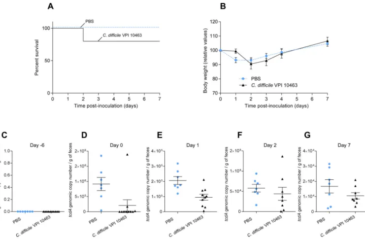
Our initial goal was to set up a model of C. difficile-induced disease wherein mice were inoculated with VPI10463 spores and C. difficile levels in feces sensitively quantitated by nucleic acid-based measurements, allowing efficient parallel quantitation of microbial genomes and/or gene expression. First, we verified that a Taqman qPCR analysis of C. difficile toxin A gene, tcdA, could be reliably quantitate purified C. difficile VPI10463 spores. While VPI10463 spores were not reliably extracted by commercially DNA isolation kits, bead-beating followed by phenol/chloroform extraction afforded quantitation of purified spores that could reliably detect as few as two copies of tcdA per mg of feces (figure S1). While mice are relatively resistant to C. difficile colonization, a model of C. difficile-induced disease has been established by Kelly wherein mice are orally administered multiple antibiotics via drinking water for 3 days, followed 2 days later by intraperitoneal injection of clindamycin [14]. This treatment renders mice susceptible to C. difficile 24h post-clindamycin, as assayed by levels in feces and clinical indicators of disease, such as colitis, weight-loss and death [14, 16]. In accord, we observed that treatment of C57BL/6 mice with the oral antibiotic cocktail only, followed by inoculation with C. difficile VPI10463 spores, did not result in observable disease (figure S2) or detectable levels of tcdA in feces at any time point assayed (figure S2). In further accord, adding a single dose of clindamycin to this regimen one day prior to inoculation with VPI10463 resulted in some mortality (figure 1A) and weight loss (figure 1B) and, moreover shedding of C. difficile at multiple time points, as indicated by levels of fecal tcdA (figure 1C–G). Yet, disease penetrance was less than previously reported. More surprisingly, many mice had detectable levels of tcdA 1 day following clindamycin treatment regardless of whether or not they had been inoculated with VPI10463. Levels of fecal tcdA were at least as high in the sham-inoculated (i.e. PBS) group as that observed in the VPI-sham-inoculated group.
A
uthor Man
uscr
ipt
A
uthor Man
uscr
ipt
A
uthor Man
uscr
ipt
A
uthor Man
uscr
ipt
Our initial presumption was that our non-inoculated control mice exhibiting tcdA positivity reflected an unintended exposure of these mice to VPI10463 or another C. difficile strain present in our vivarium or in the food or bedding it utilizes. To address this possibility, we maintained multiple groups of non-inoculated control mice in distinct rooms, performed similar experimentation on mice from distinct breeding colonies, and utilized mice newly obtained from Jackson Laboratories that were placed in our ABSL2 facility upon arrival and, thereafter, maintained in autoclaved cage with sterile food and water. While the extent of disease penetrance induced by VPI10463 varied in these experiments, we consistently observed that most antibiotic-treated mice displayed tcdA positivity 1–3 days following clindamycin treatment, even if never inoculated with VPI10463. In contrast, we did not observe tcdA positivity in germ-free animals removed from their sterile isolators and then transferred and maintained in our ASBL2 facility (figure 2A). Moreover, challenge of such formerly germ-free with even a modest number of VPI10463 spores resulted in tcdA positivity (figure 2B) and rapid mortality (figure 2C), in accord with previous work [8]. Together, these results suggested that tcdA positivity observed in our non-inoculated mice did not result from antibiotic-treated mice being exposed to C. difficile, but rather was a consequence of endogenous C. difficile that had been harbored at a low (i.e. undetectable) level that "bloomed" upon antibiotic treatment.
We next investigated why C. difficile colonization that resulted from such blooms lacked any evidence of associated disease. We performed fecal transplant from conventional mice that had been inoculated with VPI10463 or mice that had bloomed C. difficile to germ-free mice (figure 2D). Germ-free mice receiving fecal transplants from VPI-inoculated mice exhibited tcdA positivity and mortality, whereas those receiving feces from mice that had bloomed C. difficile displayed relatively high levels of fecal tcdA but lacked evidence of disease (figure 2E–F). Levels of tcdA correlated with fecal toxin levels and persisted for at least 4 weeks post-transplant (figure 2G–J). These results indicated that mice obtained from multiple sources had, upon antibiotherapy, bloomed endogenous C. difficile that, although toxin positive, lacked virulence in mice.
Isolation of murine Clostridium difficile strain LEM1
We next sought to isolate and characterize the endogenous C. difficile that seemed to be latent and widespread, albeit at very low levels, in the murine intestine. Fecal samples that had bloomed C. difficile upon antibiotic treatment were grown on TCCFA medium under anaerobic conditions. This yielded an isolate that was positive for tcdA and tcdB genes and that sporulated on BHIS and 70:30 media (figure S3A). PCR ribotyping, in order to amplify the variable-length intergenic spacer region between the 16S and 23S rRNA genes, did not reveal a match between any of the reference C. difficile strains tested (figure S3B),
suggesting it was a strain that had not been previously described. To gain insight into why it lacked VPI10463's virulence and enable design of PCR primers to distinguish it from VPI10463, we performed whole-genome-sequencing of this endogenous strain, termed C. difficile LEM1, in parallel with C. difficile VPI10463 (figure 3). GenBank accession numbers for LEM1 and VPI are pending.
A
uthor Man
uscr
ipt
A
uthor Man
uscr
ipt
A
uthor Man
uscr
ipt
A
uthor Man
uscr
ipt
Complete genomes of LEM1 and VPI10463 were aligned using Circos software [17]. The alignment reveals numerous genomic translocations and rearrangements but, nonetheless, a high overall degree of similarity between the two C. difficile strains was observed, with some strain-specific regions (figure 3C). Genome Subtractor, a high-throughput in silico subtractive hybridization analytical tool [18], was used to compare the two C. difficile genomes and demonstrated 90–95% identity between the two strains at the DNA level. In accordance, the LEM1 and VPI10463 genomes had very similar metabolic profiles as analyzed by MG-RAST, which assigns sequences to metabolic categories based on their best Blast hits against the SEED database (figure S4) [19, 20]. Nor did LEM1 and VPI10463 differ appreciably in any known virulence genes. De novo genome analysis of LEM1 pathogenicity locus (PaLoc) based on established standards within the C. difficile field [21, 22], revealed that LEM1 is toxinotype 0, like VPI10463, with no differences in TcdA and TcdB sequences themselves neither in their respective promoters. However, we observed that LEM1's strain produces lower amounts of toxin A than VPI10463 strain both in vitro and in vivo.
Genome Subtractor revealed a specific pool of 206 coding DNA sequences (CDS) that were present in LEM1 but not in VPI10463, among which 38 CDS are unique to LEM1 with no identity with any other CDS contained in NCBI. We also identified 371 CDS that were present in VPI10463 but not in LEM1 (92 of these CDS were unique to VPI10463). Such unique CDS in LEM1 and VPI10463 provided potential PCR targets that could differentiate these strains. Testing of over 20 sets of primers resulted in two primer sets that reliably quantitated levels of LEM1 and VPI10463 individually and when mixed together (table S2, figure S5).
Presence of LEM1 correlates with differential susceptibility to virulent strain VPI10463
We next examined the extent to which the heterogeneity in susceptibility to VPI10463-induced disease could be explained by endogenous blooms of LEM1. We retrospectively assayed fecal levels of VPI10463 and LEM1 in mice (n=10) purchased from Jackson, administered antibiotics, and inoculated with VPI10463 spores. One cage of such mice (n=5) had appeared ill, lost weight and succumbed to death and was termed succumbers while the other cage (n=5) lacked evidence of disease and was termed resistors (figure 4A– B). qPCR for LEM1 and VPI10463 indicated that all of the resistant mice had bloomed LEM1 prior to VPI10463 inoculation, whereas none of the mice that succumbed to VPI10463 exhibited detectable levels of LEM1 at any time point assayed (figure 4C). Blooming of LEM1 in the resistant mice correlated with lower levels of fecal VPI10463 (figure 4D). Assay of C. difficile tcdA, detecting both LEM1 and VPI10463, corroborated these results (figure 4E). To investigate our hypothesis that LEM1 detected in this cage of mice reflected an expansion of LEM1 that had been harbored at a very low level, we assayed fecal samples for LEM1 prior to antibiotic administration by a highly sensitive, albeit only semi-quantitative, nested-PCR assay. This assay revealed that in the “resisting” cage, 2 of the 5 mice had detectable levels of LEM1 prior to antibiotic administration, while all mice in the “succumbing” cage were negative for this bacterium (figure 4F). These results suggest that while some mice probably became colonized by LEM1 as a result of being exposed to it
A
uthor Man
uscr
ipt
A
uthor Man
uscr
ipt
A
uthor Man
uscr
ipt
A
uthor Man
uscr
ipt
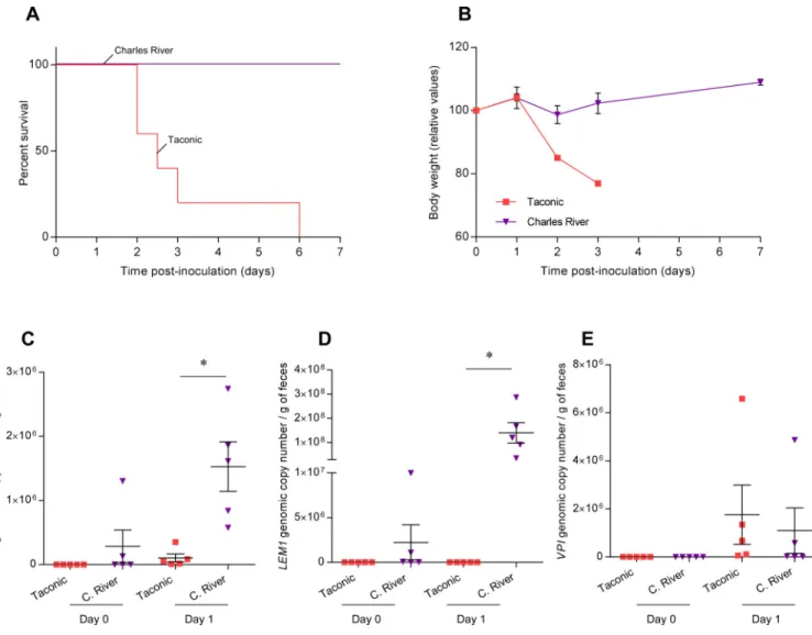
from cage mates following antibiotherapy, others, i.e. those positive for LEM1 by nested PCR at day -6, had truly bloomed LEM1 that they had been harboring at very low levels. Next, we examined the extent to which LEM1 might bloom and correlate with susceptibility to VPI10463 in mice obtained from other vendors. Like mice purchased from Jackson Labs, mice from Charles River Research bloomed LEM1 following antibiotic cocktail treatment, as revealed by specific LEM1 primers (figure 5, S3C). Here also, such blooms correlated with lack of disease in response to inoculation with VPI10463 (figure 5A–B). In contrast, mice from Taconic did not bloom LEM1 and exhibited high mortality following inoculation with VPI10463 (figure 5C–E). Thus, although there is heterogeneity in mice from different sources and also within mice from a single source, colonization with LEM1 appears to be a correlate of susceptibility to VPI10463-induced disease.
LEM1 lacks high virulence in mice
We next examined the consequences of exogenous inoculation of mice with purified spores of LEM1. First, we utilized the antibiotic-induced susceptibility model, in which
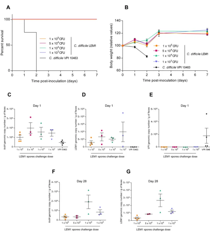
administration of 1 × 10⁵ spores of VPI10463 resulted in 60% lethality (figure S6). Infection with the same dose of LEM1 spores resulted in a similar level of C. difficile colonization (figure S6), but did not cause the weight loss or death induced by VPI10463. When the LEM1 inoculum was increased 100-fold (the highest dose achieve), we observed a modest increase in C. difficile fecal loads that was associated with transient weight loss but not death (figure S6). Next, we tested germ-free mice, which are highly sensitive to C. difficile infection and lack the confounding variable of potential endogenous bacterial competitors. While inoculation with as little as 1000 spores of VPI10463 resulted in an acute disease and rapid death by 48h post-infection (figure 2A, 6A–B), inoculation with up to 100-fold higher levels of LEM1 spores (highest dose tested) did not result in clinical evidence of disease (figure 6A–B). In both antibiotic and germ-free models, LEM1 persisted at detectable levels (without additional treatment) for at least 4 weeks post LEM1 inoculation (figure 6C–G). Moreover, euthanizing mice at 48h post-inoculation indicated the presence of colitis, based on levels of inflammatory markers (myeloperoxidase and lipocalin-2), cytokine levels, and histopathology in VPI10463-treated mice that was less evident in mice administered LEM1 (figure S7). These results suggest that LEM1 colonized the murine intestine as efficiently as VPI10463 but lacked high virulence even in mice prone to C. difficile-induced disease.
LEM1 protects against VPI10463-induced disease via outcompeting it
We next directly considered the possibility that LEM1 might protect against the pathogenic strain VPI10463. The frequent but unpredictable blooms of endogenous LEM1 complicate its use in the antibiotic model. Nonetheless, we observed that administration of LEM1 3 days prior to VPI10463 inoculation eliminated VPI-induced mortality (figure 7A–B). In the absence of LEM1 treatment, some mice exhibited delayed blooms of endogenous LEM1 and survived VPI10463 challenge, while some mice did not bloom LEM1 and succumbed to the pathogenic strain (figure 7A, D). Moreover, prophylactic administration of LEM1 resulted in reduced levels of VPI10463 at days 5–8 post clindamycin treatment (figure 7E). These results suggest that LEM1, when blooming endogenously or when administered
A
uthor Man
uscr
ipt
A
uthor Man
uscr
ipt
A
uthor Man
uscr
ipt
A
uthor Man
uscr
ipt
exogenously, protects against VPI-induced mortality at least in part by preventing colonization of virulent C. difficile strain.
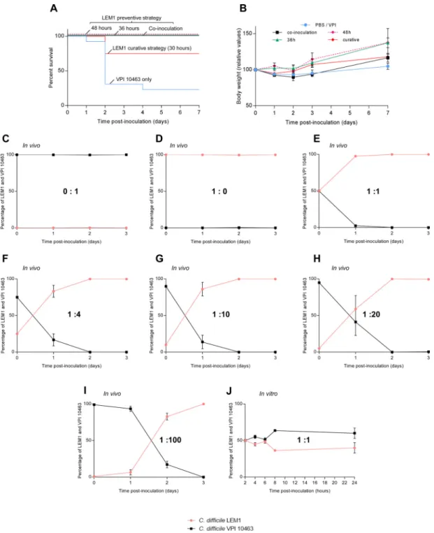
Next, we examined how LEM1 and VPI10463 would impact each other in the absence of microbiota via the use of germ-free mice. Strikingly, the high rate of mortality induced by VPI10463 in germ-free animals was completely prevented by administration of LEM1 prior to, or concurrent with, VPI10463 inoculation (figure 8A). Moreover, curative administration of LEM1 as late as 30 hours after VPI10463 inoculation was able to reduce mortality induced by VPI10463 (figure 8A). Preventive administration of LEM1 fully protected against mortality-induced by VPI10463 even in response to a 10-fold higher dose of VPI10463 spores, that otherwise resulted in 100 percent lethality (figure S8). Lastly, to better understand how LEM1 protects against C. difficile-induced disease, we directly measured the ability of these strains to compete against each other in vivo and in vitro. While germ-free mice on both C57Bl/6 and Swiss-Webster backgrounds are highly prone to VPI-induced disease, the latter are somewhat more resistant thus enabling formal
demonstration that either VPI10463 or LEM1 can persist a few days post-inoculation (figure 8C–D). However, when administered together, at equal ratios or 100-fold higher levels of VPI10463 (highest tested), LEM1 dramatically out-competed VPI10463 thus eliminating it by 3 days post-challenge (figure 8E–I). Such ability of LEM1 to outcompete VPI10463, thus nearly eliminating it even the absence of commensal microbes, correlated with complete protection against VPI-induced disease. LEM1 and VPI10463 did not impact each other's growth in vitro. Rather, these strains grew in broth medium at similar rates when cultured individually or together (figure 8JS9A–B), thus suggesting that LEM1's ability to
outcompete VPI10463 does not reflect a direct interaction between these C. difficile strains but, rather, is a consequence of the murine colonic environment.
DISCUSSION
The recent development of mouse models of CDI, which are genetically and
immunologically tractable, promised to allow mechanistic study of disease determinants [13, 14]. However, a paucity of expertise in anaerobic culture in many research groups has remained a barrier. Hence, our initial goal was to setup a model of C. difficile-induced disease in mice using spores that would permit high-throughput and culture-independent quantification of pathogen loads using frozen fecal samples [23, 24]. This would enable a broad range of researchers using mouse-based approaches to test C. difficile strains and query host determinants. Real-time qPCR-based measure of fecal levels of C. difficile toxin A, following infection of mice with stable C. difficile spores, proved to be a relatively simple and sensitive means to quantitate C. difficile levels. However, we made an unanticipated observation, namely that many colonies of mice, including those from some commercial suppliers, harbored endogenous C. difficile, a strain of which we herein named LEM1. While, in the absence of antibiotics, LEM1 levels were below the detection limit of our qPCR assay, use of semi-quantitative nested-PCR indicated it was nonetheless present, at least in some mice that later bloomed it following antibiotherapy. The notion that C. difficile can be present but yet undetectable by culture was demonstrated by Lawley and colleagues, whom observed that, even in stringently hygienic conditions, mice that had seemingly fully resolved a CDI would bloom the bacteria upon antibiotherapy [25]. While such blooms
A
uthor Man
uscr
ipt
A
uthor Man
uscr
ipt
A
uthor Man
uscr
ipt
A
uthor Man
uscr
ipt
observed by Lawley were associated with clinical-type indicators of disease, LEM1 lacked virulence and rather protected mice against pathogenic C. difficile VPI10463. These observations have important implications for mouse modeling of CDI and understanding the pathophysiology of C. difficile-induced disease in humans.
The frequent, albeit sporadic, blooms of LEM1 observed in our antibiotic-treated mice illustrate the need for animal models of C. difficile infection to be carefully controlled. Specifically, control animals subjected to all treatments except C. difficile inoculation are needed and should be sampled throughout the experiment. Further, assays should utilize a specific means to be sure that the C. difficile strain of interest is the one being quantitated. Regarding this point, hamsters from commercial suppliers also harbored endogenous C. difficile, different from LEM1 based on its ribotype [26]. In many cases, it may prove difficult to eliminate blooms of endogenous C. difficile, but the presence of such organisms can be taken into account during data interpretation.
LEM1 is clearly distinct from human isolates in that it contains about 100 CDS not found in any previously sequenced human isolate. Based on ribotyping and the PCR protocol developed herein using one of LEM1’s novel CDS, we find “LEM1-like” C. difficile is a frequent albeit low-abundance resident of the murine intestine but note that sequencing of additional isolates is needed to discern how closely related to such isolates are to LEM1. The extent to which LEM1's low virulence in mice even when administered at high doses might be explained by its modest toxin production relative to VPI10463 (figure S9C). In support of this possibility, we have observed that LEM1 is virulent in hamsters (data not shown), which are far more sensitive to C. difficile toxins and highly susceptible to C. difficile-induced mortality [27]. Concomitantly, LEM1's ability to protect against a virulent strain is analogous to observations that non-toxigenic C. difficile strains can prevent C. difficile-induced mortality in hamsters [28, 29, 30] and reduce the rate of recurrence in humans [31]. While such non-toxigenic C. difficile strains may soon be utilized to treat C. difficile-associated disease, their failure to eliminate recurrence and difficile-associated adverse events highlights the importance of mechanistically understanding how such strains confer
protection, thus allowing more effective and specific strategies to prevent recurrence [30, 32, 33]. We are hopeful that mouse modeling of protection against virulent C. difficile by an avirulent strain, as shown herein, will prove a useful platform for achieving this goal. The mechanism by which LEM1 protects against VPI10463 remains unclear. We initially favored the hypothesis that LEM1 might activate an immune response that affects VPI10463 more than LEM1. However, deletion of several host genes that mediate innate immunity (MyD88, Tlr5, and Nlrc4) and adaptive immunity (Rag1) had minimal effects on ability of LEM1 to protect against VPI10463-induced disease (data not shown) arguing against this possibility. Another possible mechanism for LEM1's ability to outcompete VPI10463 is that bacteriophages and/or anti-microbial compounds may be produced by LEM1 in vivo that may directly impede VPI10463; analogous to hypotheses from others [34, 35], although that LEM1 did not impede growth of VPI10463 fails to support this scenario. Hence, we view the ability of LEM1 to outcompete VPI10463 in germ-free mice to reflect that LEM1 outcompetes VPI10463 for a limiting niche in the mouse gastro-intestinal tract [8, 25]. Such ability of LEM1 to protect against VPI10463 was not shared by known probiotic strains or
A
uthor Man
uscr
ipt
A
uthor Man
uscr
ipt
A
uthor Man
uscr
ipt
A
uthor Man
uscr
ipt
non-pathogenic E. coli strains, both of which, in our hands, had only modest ability to protect against VPI10463-induced disease even when administered prophylactically (data not shown). Nor did administration of fecal transplants to antibiotic-treated mice offer strong protection against VPI10463 (data not shown), thus highlighting the specificity, strength, and perhaps ultimate potential usefulness of the protection conferred by LEM1.
Extrapolating the results from this study in mice to C. difficile-induced disease in humans questions the reliability of the widely held assumption that newly diagnosed CDI result from exposure of the antibiotic-treated host to spores of C. difficile. Indeed, while Gerding et al. demonstrated that this is one major route of transmission at least in the 1980s, Eyre and colleagues sequence-based study of hospital-manifesting CDI in Oxfordshire, U.K from 2007–11 2013 found that the majority of such cases were not transmitted from another symptomatic patient [5, 7]. This study revealed that almost half (45%) of all C. difficile cases in the Oxfordshire area were genetically distinct from all other previous cases of C. difficile [7]. In this context, we view our results to suggest that some CDI that manifest in hospitals are, in fact, the result of C. difficile that was harbored at very low, perhaps undetectable levels. While studies analyzing the microbiome of healthy humans have not frequently reported the presence of C. difficile [36], such studies typically 1) lack the depth to detect low-abundance bacteria, 2) use DNA extraction methods that work poorly on C. difficile spores, and 3) target regions of the 16S RNA genes that do not resolve C. difficile from other Clostridia (Tor Savidge, Baylor College of Medicine, personal communication). Indeed, our use of 16S sequencing, using relatively standard methodology and a depth of over 20,000 sequences per sample failed to detect any sequences classified as C. difficile even in mice that had clearly bloomed it by qPCR. Hence, we submit that the true rate of carriage of C. difficile in the general population is not easily discerned from existing studies. While culture-based studies indicate it is quite high (up to 70%) in infancy and relatively low in healthy persons over age 2 [37], we speculate that use of highly sensitive
methodologies such as nested-PCR might ultimately reveal that low-level carriage of C. difficile is relatively high.
Our observation that colonization of mice by a C. difficile that did not induce overt disease protected against a highly virulent strain is analogous to observations that hospital patients who were positive for C. difficile, upon initial hospital culture, had less clinical
manifestations of disease [5]. However, this does not exclude the possibility that some strains of C. difficile that are harbored at low levels may bloom and become highly virulent. Indeed, as demonstrated by Lawley, the ready-ability of other administered C. difficile strains to establish latency in mice argues that the lack of virulence is not prerequisite for long-term maintenance of latency [38, 39]. We view our results to suggest that many arising instances of C. difficile positivity, including strains that induce symptomatic disease and/or protect against more virulent strains, may have resulted from blooms of C. difficile long harbored by affected individuals.
Supplementary Material
Refer to Web version on PubMed Central for supplementary material.
A
uthor Man
uscr
ipt
A
uthor Man
uscr
ipt
A
uthor Man
uscr
ipt
A
uthor Man
uscr
ipt
Acknowledgments
We thank Dr. Shonna McBride and Dr. Adrianne N. Edwards (Emory University) for outstanding guidance and technical expertise with C. difficile. We thank the Gewirtz lab members for technical assistance, especially Dr. Emilie Viennois and Zhan Zhang for help with histology, and helpful discussions. We thank Dr. Richard Plemper (Georgia State University) for providing Vero cells.
FUNDINGS
This work was supported by NIH grants DK099071 and DK083890 to ATG. BC is a recipient of the Career Development Award from the Crohn’s and Colitis Foundation of America (CCFA).
Abbreviations
CDS coding DNA sequences
C. difficile Clostridium difficile
References
1. Kociolek LK, Gerding DN. Breakthroughs in the treatment and prevention of Clostridium difficile infection. Nature reviews Gastroenterology & hepatology. 2016; 13:150–60. [PubMed: 26860266] 2. Lessa FC, Mu Y, Bamberg WM, Beldavs ZG, Dumyati GK, Dunn JR, et al. Burden of Clostridium difficile infection in the United States. The New England journal of medicine. 2015; 372:825–34. [PubMed: 25714160]
3. Rupnik M, Wilcox MH, Gerding DN. Clostridium difficile infection: new developments in epidemiology and pathogenesis. Nature reviews Microbiology. 2009; 7:526–36. [PubMed: 19528959]
4. Dubberke ER, Olsen MA. Burden of Clostridium difficile on the healthcare system. Clinical infectious diseases : an official publication of the Infectious Diseases Society of America. 2012; 55(Suppl 2):S88–92. [PubMed: 22752870]
5. Johnson S, Clabots CR, Linn FV, Olson MM, Peterson LR, Gerding DN. Nosocomial Clostridium difficile colonisation and disease. Lancet. 1990; 336:97–100. [PubMed: 1975332]
6. Martin JS, Monaghan TM, Wilcox MH. Clostridium difficile infection: epidemiology, diagnosis and understanding transmission. Nature reviews Gastroenterology & hepatology. 2016; 13:206–16. [PubMed: 26956066]
7. Eyre DW, Cule ML, Wilson DJ, Griffiths D, Vaughan A, O'Connor L, et al. Diverse sources of C. difficile infection identified on whole-genome sequencing. The New England journal of medicine. 2013; 369:1195–205. [PubMed: 24066741]
8. Reeves AE, Koenigsknecht MJ, Bergin IL, Young VB. Suppression of Clostridium difficile in the gastrointestinal tracts of germfree mice inoculated with a murine isolate from the family Lachnospiraceae. Infection and immunity. 2012; 80:3786–94. [PubMed: 22890996] 9. Seekatz AM, Young VB. Clostridium difficile and the microbiota. The Journal of clinical
investigation. 2014; 124:4182–9. [PubMed: 25036699]
10. Theriot CM, Koenigsknecht MJ, Carlson PE Jr, Hatton GE, Nelson AM, Li B, et al. Antibiotic-induced shifts in the mouse gut microbiome and metabolome increase susceptibility to Clostridium difficile infection. Nature communications. 2014; 5:3114.
11. Theriot CM, Young VB. Interactions Between the Gastrointestinal Microbiome and Clostridium difficile. Annual review of microbiology. 2015; 69:445–61.
12. Rolhion N, Chassaing B. When pathogenic bacteria meet the intestinal microbiota. Philosophical transactions of the Royal Society of London Series B, Biological sciences. 2016; 371
13. Best EL, Freeman J, Wilcox MH. Models for the study of Clostridium difficile infection. Gut microbes. 2012; 3:145–67. [PubMed: 22555466]
A
uthor Man
uscr
ipt
A
uthor Man
uscr
ipt
A
uthor Man
uscr
ipt
A
uthor Man
uscr
ipt
14. Chen X, Katchar K, Goldsmith JD, Nanthakumar N, Cheknis A, Gerding DN, et al. A mouse model of Clostridium difficile-associated disease. Gastroenterology. 2008; 135:1984–92. [PubMed: 18848941]
15. Wilson KH, Silva J, Fekety FR. Suppression of Clostridium difficile by normal hamster cecal flora and prevention of antibiotic-associated cecitis. Infection and immunity. 1981; 34:626–8. [PubMed: 7309245]
16. Reeves AE, Theriot CM, Bergin IL, Huffnagle GB, Schloss PD, Young VB. The interplay between microbiome dynamics and pathogen dynamics in a murine model of Clostridium difficile Infection. Gut microbes. 2011; 2:145–58. [PubMed: 21804357]
17. Krzywinski M, Schein J, Birol I, Connors J, Gascoyne R, Horsman D, et al. Circos: an information aesthetic for comparative genomics. Genome research. 2009; 19:1639–45. [PubMed: 19541911] 18. Shao Y, He X, Harrison EM, Tai C, Ou HY, Rajakumar K, et al. mGenomeSubtractor: a web-based
tool for parallel in silico subtractive hybridization analysis of multiple bacterial genomes. Nucleic acids research. 2010; 38:W194–200. [PubMed: 20435682]
19. Aziz RK, Bartels D, Best AA, DeJongh M, Disz T, Edwards RA, et al. The RAST Server: rapid annotations using subsystems technology. BMC genomics. 2008; 9:75. [PubMed: 18261238] 20. Overbeek R, Olson R, Pusch GD, Olsen GJ, Davis JJ, Disz T, et al. The SEED and the Rapid
Annotation of microbial genomes using Subsystems Technology (RAST). Nucleic acids research. 2014; 42:D206–14. [PubMed: 24293654]
21. Rupnik M. Clostridium difficile toxinotyping. Methods in molecular biology. 2010; 646:67–76. [PubMed: 20597003]
22. Rupnik M, Avesani V, Janc M, von Eichel-Streiber C, Delmee M. A novel toxinotyping scheme and correlation of toxinotypes with serogroups of Clostridium difficile isolates. Journal of clinical microbiology. 1998; 36:2240–7. [PubMed: 9665999]
23. Belanger SD, Boissinot M, Clairoux N, Picard FJ, Bergeron MG. Rapid detection of Clostridium difficile in feces by real-time PCR. Journal of clinical microbiology. 2003; 41:730–4. [PubMed: 12574274]
24. Mutters R, Nonnenmacher C, Susin C, Albrecht U, Kropatsch R, Schumacher S. Quantitative detection of Clostridium difficile in hospital environmental samples by real-time polymerase chain reaction. The Journal of hospital infection. 2009; 71:43–8. [PubMed: 19041162]
25. Lawley TD, Clare S, Walker AW, Stares MD, Connor TR, Raisen C, et al. Targeted restoration of the intestinal microbiota with a simple, defined bacteriotherapy resolves relapsing Clostridium difficile disease in mice. PLoS pathogens. 2012; 8:e1002995. [PubMed: 23133377]
26. Peterfreund GL, Vandivier LE, Sinha R, Marozsan AJ, Olson WC, Zhu J, et al. Succession in the gut microbiome following antibiotic and antibody therapies for Clostridium difficile. PloS one. 2012; 7:e46966. [PubMed: 23071679]
27. Buckley AM, Spencer J, Maclellan LM, Candlish D, Irvine JJ, Douce GR. Susceptibility of hamsters to Clostridium difficile isolates of differing toxinotype. PloS one. 2013; 8:e64121. [PubMed: 23704976]
28. Borriello SP, Barclay FE. Protection of hamsters against Clostridium difficile ileocaecitis by prior colonisation with non-pathogenic strains. Journal of medical microbiology. 1985; 19:339–50. [PubMed: 4009689]
29. Nagaro KJ, Phillips ST, Cheknis AK, Sambol SP, Zukowski WE, Johnson S, et al. Nontoxigenic Clostridium difficile protects hamsters against challenge with historic and epidemic strains of toxigenic BI/NAP1/027 C. difficile. Antimicrobial agents and chemotherapy. 2013; 57:5266–70. [PubMed: 23939887]
30. Sambol SP, Merrigan MM, Tang JK, Johnson S, Gerding DN. Colonization for the prevention of Clostridium difficile disease in hamsters. The Journal of infectious diseases. 2002; 186:1781–9. [PubMed: 12447764]
31. Gerding DN, Meyer T, Lee C, Cohen SH, Murthy UK, Poirier A, et al. Administration of spores of nontoxigenic Clostridium difficile strain M3 for prevention of recurrent C. difficile infection: a randomized clinical trial. Jama. 2015; 313:1719–27. [PubMed: 25942722]
A
uthor Man
uscr
ipt
A
uthor Man
uscr
ipt
A
uthor Man
uscr
ipt
A
uthor Man
uscr
ipt
32. Brouwer MS, Roberts AP, Hussain H, Williams RJ, Allan E, Mullany P. Horizontal gene transfer converts non-toxigenic Clostridium difficile strains into toxin producers. Nature communications. 2013; 4:2601.
33. Gerding DN, Johnson S. Management of Clostridium difficile infection: thinking inside and outside the box. Clinical infectious diseases : an official publication of the Infectious Diseases Society of America. 2010; 51:1306–13. [PubMed: 20979491]
34. Gebhart D, Williams SR, Bishop-Lilly KA, Govoni GR, Willner KM, Butani A, et al. Novel high-molecular-weight, R-type bacteriocins of Clostridium difficile. Journal of bacteriology. 2012; 194:6240–7. [PubMed: 22984261]
35. Govind R, Vediyappan G, Rolfe RD, Dupuy B, Fralick JA. Bacteriophage-mediated toxin gene regulation in Clostridium difficile. Journal of virology. 2009; 83:12037–45. [PubMed: 19776116] 36. Schubert AM, Rogers MA, Ring C, Mogle J, Petrosino JP, Young VB, et al. Microbiome data
distinguish patients with Clostridium difficile infection and non-C. difficile-associated diarrhea from healthy controls. mBio. 2014; 5:e01021–14. [PubMed: 24803517]
37. Jangi S, Lamont JT. Asymptomatic colonization by Clostridium difficile in infants: implications for disease in later life. Journal of pediatric gastroenterology and nutrition. 2010; 51:2–7. [PubMed: 20512057]
38. Deakin LJ, Clare S, Fagan RP, Dawson LF, Pickard DJ, West MR, et al. The Clostridium difficile spo0A gene is a persistence and transmission factor. Infection and immunity. 2012; 80:2704–11. [PubMed: 22615253]
39. Lawley TD, Clare S, Walker AW, Goulding D, Stabler RA, Croucher N, et al. Antibiotic treatment of clostridium difficile carrier mice triggers a supershedder state, spore-mediated transmission, and severe disease in immunocompromised hosts. Infection and immunity. 2009; 77:3661–9.
[PubMed: 19564382]
A
uthor Man
uscr
ipt
A
uthor Man
uscr
ipt
A
uthor Man
uscr
ipt
A
uthor Man
uscr
ipt
SUMMARY BOX What is already known about this subject?
Clostridium difficile is a leading cause of antibiotic-associated disease. Its disease burden presumably results from its ability to form spores and infect antibiotic-treated persons at low doses. However, C. difficile infections might also result from long-harbored C. difficile blooming upon antibiotic therapy.
What are the new findings?
Mice from various sources bloomed an endogenous C. difficile strain, herein termed LEM1, which shared 95% of its genome with VPI10463. LEM1 was avirulent in mice. Rather, blooms of LEM1 correlated with lack of disease following VPI10463 challenge. Moreover, exogenous administration of LEM1 strongly outcompeted and protected against VPI10463-induced disease in antibiotic-treated and germ-free mice.
How might it impact on clinical practice in the foreseeable future?
These results demonstrate the difficulty of determining whether individual cases of C. difficile resulted from a bloom or new exposure to the pathogen. Furthermore, we have identified, isolated and characterized an endogenous murine spore forming C. difficile strain that prevents colonization and disease induced by pathogenic C. difficile.
A
uthor Man
uscr
ipt
A
uthor Man
uscr
ipt
A
uthor Man
uscr
ipt
A
uthor Man
uscr
ipt
Figure 1. C57BL6 WT mice bred at GSU exhibited unanticipated tcdA positivity and moderate disease upon VPI10463 challenge
(A). Kaplan-Meier survival curve of mice uninfected (PBS, n=7) or infected with 1 × 10⁵
spores of C. difficile VPI10463 (n=10) at day 0. (B). Average body weight measured daily starting from the day of infection (day 0). (C–G). C. difficile load in feces was quantified by q-PCR using Taqman probe and primers specific to the gene encoding C. difficile toxin A at day -6 (C), day 0 (D), day 1 (E), day 2 (F) and day 7 (G).
Each point represents an individual animal from 2 colonies of wild-type mice bred at Georgia State University. Results are the means ± SEM (n=7–10).
A
uthor Man
uscr
ipt
A
uthor Man
uscr
ipt
A
uthor Man
uscr
ipt
A
uthor Man
uscr
ipt
Figure 2. Germ-free mice exhibit disease in response to VPI10463 spores and VPI10463-containig feces but not feces exhibiting C. difficile blooms
(A–B) C. difficile load in feces was quantified by q-PCR using Taqman probe and primers specific to the gene encoding C. difficile toxin A at day 0 (A) and day 1 (B). (C). Kaplan-Meier survival curve of C57Bl6 germ-free mice uninfected (PBS, n=3) or infected with 1 × 10³ spores of C. difficile VPI10463 (n=4) at day 0. (D). Schematic representation of the experimental design. (E). Kaplan-Meier survival curve of Swiss Webster germ-free mice receiving fecal microbiota transplant from conventional antibiotic-treated mice without a
A
uthor Man
uscr
ipt
A
uthor Man
uscr
ipt
A
uthor Man
uscr
ipt
A
uthor Man
uscr
ipt
bloom (fecal sample previously tcdA negative by qPCR, n=2), with a bloom (fecal sample previously tcdA positive by qPCR, n=4) or C. difficile VPI-infected mice (fecal sample previously tcdA positive by qPCR, n=4). (F). Average body weight measured daily starting from the day of infection (Day 0). (G and I). C. difficile load in feces was quantified by q-PCR using Taqman probe and primers specific to the gene encoding C. difficile toxin A at day 1 (G) and day 28 post-infection (I). (H and J). Vero cell cytotoxic assay was used to determine the log10 reciprocal cytotoxin dilution per gram of fecal sample at day 1 (H) and day 28 post-infection (J).
Each point represents an individual animal. Results represent the means ± SEM (n=3–4 for panels B and C); results represent the means (n=2–4 for panels G to J).
A
uthor Man
uscr
ipt
A
uthor Man
uscr
ipt
A
uthor Man
uscr
ipt
A
uthor Man
uscr
ipt
Figure 3. Genomic comparison of C. difficile strains LEM1 and VPI10463
(A). Comparison of C. difficile LEM1 and C. difficile VPI10463 genomes. (B). Venn diagram representing the number of sequences unique or shared between the two strains, where 206 sequences specific to C. difficile LEM1 genome and 371 sequences specific to C. difficile VPI10463 genome were identified. (C). Comparative analysis of the genome sequences of C. difficile VPI10463 and C. difficile LEM1 using Circos software tool where blue lines represent high homology between the two genomes, orange lines visualize the
A
uthor Man
uscr
ipt
A
uthor Man
uscr
ipt
A
uthor Man
uscr
ipt
A
uthor Man
uscr
ipt
differences (modest homology) and the darker blue lines represent overlapping areas of homology.
A
uthor Man
uscr
ipt
A
uthor Man
uscr
ipt
A
uthor Man
uscr
ipt
A
uthor Man
uscr
ipt
Figure 4. Variation in susceptibility to C. difficile VPI10463-induced disease amongst Jackson laboratories mice correlates with LEM1 bloom
(A). Kaplan-Meier survival curve of infected with 1 × 10⁵ spores of C. difficile VPI10463 (n=5 mice for resisting cage, n=5 for succumbing cage). (B). Average body weight measured daily starting from the day of infection (Day 0). (C–E). C. difficile loads in feces were quantified by q-PCR using SYBR Green technology and primers specific to C. difficile LEM1 (C) or primers specific to C. difficile VPI10463 (D) or using Taqman technology with probe and primers specific to the gene encoding C. difficile toxin A (E) at day-6, day 0
A
uthor Man
uscr
ipt
A
uthor Man
uscr
ipt
A
uthor Man
uscr
ipt
A
uthor Man
uscr
ipt
and day 1 post-infection. (F). Agarose gel electrophoresis of nested PCR products in fecal DNA samples of succumbing and resisting animals on fecal samples collected upon receipt from Jackson laboratories (day-6). M: molecular size DNA marker (1kb Plus DNA ladder), lanes 1–5: succumbing animals, lanes 6–10: resisting animals, lanes 11–12: C. difficile LEM1 (positive control), lanes 13–14: negative control. Lanes 1, 2 and 3 have non-specific bands.
In C–E, each point represents an individual animal. Results are the means ± SEM (n=5). Significance was determined by Mann-Whitney test, * indicates p<0.05.
A
uthor Man
uscr
ipt
A
uthor Man
uscr
ipt
A
uthor Man
uscr
ipt
A
uthor Man
uscr
ipt
Figure 5. Variation of C. difficile susceptibility in mice from Charles River Laboratories and Taconic farms
(A). Kaplan-Meier survival curve of infected Charles River (n=5) or Taconic (n=5) mice with 1 × 10⁵ spores of C. difficile VPI10463. (B). Average body weight measured daily starting from the day of infection (Day 0). (C–E). C. difficile loads in feces were quantified by q-PCR using Taqman probe and primers specific to the gene encoding C. difficile toxin A (C) or using SYBR Green technology and primers specific to C. difficile LEM1 (D) or primers specific to C. difficile VPI10463 (E) at day 0 and day 1 post-infection. Each point represents an individual animal. Results are the means ± SEM (n=5). Significance was determined by Mann-Whitney test, * indicates p<0.05.
A
uthor Man
uscr
ipt
A
uthor Man
uscr
ipt
A
uthor Man
uscr
ipt
A
uthor Man
uscr
ipt
Figure 6. C. difficile LEM1 strain lacks virulence in germ-free mice
(A). Kaplan-Meier survival curve of Swiss Webster germ-free mice infected with C. difficile LEM1 (1 × 10⁵, n=4; 5 × 10⁴, n=3; 1 × 10⁴, n=3; 1 × 10³ spores n=3) or C. difficile
VPI10463 (1 × 10³ spores, n=4). (B). Average body weight measured daily starting from the day of infection (Day 0). (C–G). C. difficile loads in feces were quantified by q-PCR using Taqman technology with probe and primers specific to the gene encoding C. difficile toxin A at day 1 (C) and day 28 post-infection (F) or using SYBR Green technology and primers
A
uthor Man
uscr
ipt
A
uthor Man
uscr
ipt
A
uthor Man
uscr
ipt
A
uthor Man
uscr
ipt
specific to C. difficile LEM1 at day 1 (D) and day 28 post-infection (G) or primers specific to C. difficile VPI10463 at day 1 post-infection (E).
Each point represents an individual animal. Results are the means ± SEM (n=3–4).
A
uthor Man
uscr
ipt
A
uthor Man
uscr
ipt
A
uthor Man
uscr
ipt
A
uthor Man
uscr
ipt
Figure 7. C. difficile LEM1 protects mice against C. difficile VPI10463-induced disease and death in conventional antibiotic-treated mice
(A). Kaplan-Meier survival curve of Jackson mice uninfected (n=5) or infected with C. difficile LEM1 (1 × 10⁵ spores, n=5, the dotted line represents the mice that did not show an
endogenous bloom for C. difficile LEM1) and challenged 72 hours later with C. difficile VPI10463 (1 × 10⁵ spores). (B). Average body weight measured daily starting from the day of intra-peritoneal injection of clindamycin. (C–E). C. difficile loads in feces were
quantified by q-PCR using Taqman technology with probe and primers specific to the gene
A
uthor Man
uscr
ipt
A
uthor Man
uscr
ipt
A
uthor Man
uscr
ipt
A
uthor Man
uscr
ipt
encoding C. difficile toxin A (C) or using SYBR Green technology and primers specific to C. difficile LEM1 (D) or primers specific to C. difficile VPI10463 (E) at day 2, 3, 4 5, 6 and 8 post-clindamycin respectively.
Each point represents an individual animal. Results are the means ± SEM (n=5). Significance was determined by Student's t-test, * indicates p<0.05.
A
uthor Man
uscr
ipt
A
uthor Man
uscr
ipt
A
uthor Man
uscr
ipt
A
uthor Man
uscr
ipt
Figure 8. C. difficile LEM1 outcompetes C. difficile VPI10463 and protects against VPI-induced disease in germ-free mice
(A). Kaplan-Meier survival curve of germ-free C57Bl6 mice infected with 1 × 10³ spores of C. difficile VPI10463 (n=15) or given 1 × 10⁵ spores of C. difficile LEM1 48 hours (n=2),
36 hours (n=6) prior to or concomitant to (n=2) VPI10463 challenge. In another experiment, germ-free mice were challenged with C. difficile VPI10463 (1 × 10³ spores) and treated 30
hours later with C. difficile LEM1 (1 × 10⁵ spores). (B). Average body weight measured
daily starting from the day of infection (Day 0). (C–J) Competition experiments between C.
A
uthor Man
uscr
ipt
A
uthor Man
uscr
ipt
A
uthor Man
uscr
ipt
A
uthor Man
uscr
ipt
difficile VPI10463 (black line) and LEM1 (pink line) were performed in vivo in germ-free Swiss Webster mice (C–I) and in vitro (J); the number of bacteria recovered in feces or TY medium was quantified by quantitative PCR of C. difficile VPI10463 and C. difficile LEM1 (with C. difficile LEM1 being defined as 1) and then expressed as a competitive index (CI). The CI was defined as: (LEM1 / VPI10463) output / (LEM1 / VPI10463) input and then expressed as a percentage of each strain. (C–I). Mixed spores suspensions were
administered to germ-free mice at different ratio (J) C. difficile VPI10463 and LEM1 were grown in equal amount of each bacteria in TY medium.
Results are the means ± SD (n=2–6).