ANTI-INFLAMMATORY DRUGS FOR MODULATION OF HOST RESPONSE TO
BIOMATERIALS AND APPLICATION IN DIABETES THERAPY
ARCHNES
By
AASA40 7.,ITITUTETHUY TRAM DANG
JUL
19
2"12
B.Sc. Chemical Engineering, 2006 University of Illinois, Urbana-Champaign
AIiES
Submitted to the Department of Chemical Engineering in Partial Fulfillment of the Requirements for the Degree of
Doctor of Philosophy
At the
Massachusetts Institute of Technology September 2012
© 2012 Massachusetts Institute of Technology. All rights reserved.
Signature of Author:
...
Thuy Tram Dang Department of Chemical Engineering
July 1 0 th
2012
Certified by: ... ... ... *r... Robert S. Langer, Sc.D. Institute Professor Thesis Supervisor Certified by: Daniel G. Anderson, Ph.D. Professor of Chemical Engineering& Division of Health Sciences and Technology
Thesis Supervisor Accepted by:
... .... ...
Patrick Doyle, Ph.D. Professor of Chemical Engineering Chairman, Committee for Graduate Students
Anti-inflammatory drugs for modulation of host response to
biomaterials and application in diabetes therapy
By THUY TRAM DANG
Submitted to the Department of Chemical Engineering on July 11 , 2012 in partial fulfillment of the requirements for the degree of Doctor of Philosophy in Chemical Engineering
ABSTRACT
Host response to implanted biomaterials and medical devices poses tremendous challenges to their clinical applications. Today, the quest to mitigate this immunological attack for improved longevity of these devices remains daunting. This thesis aims to explore the use of anti-inflammatory drugs in minimizing the host response
and improve the efficacy of implantable and transplantable therapeutics.
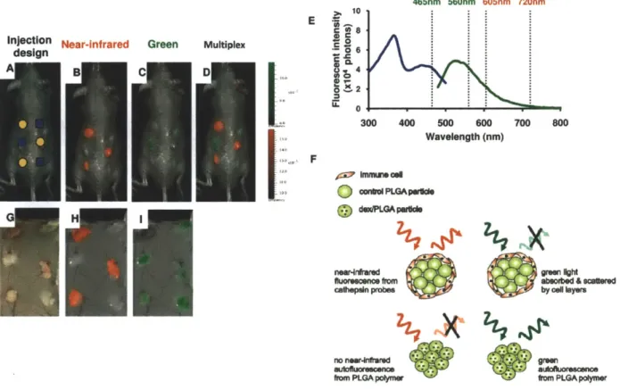
Firstly, we developed a new non-invasive in vivo imaging technique to study the activity of early immune cells in the host response to implanted biomaterials. A fluorescent imaging probe (Prosense680*, Perkin Elmer) activatable by cathepsins, a class of inflammatory proteases secreted from immune cells, was used for simultaneous biocompatibility screening of up to 8 different materials per animal in immunocompetent hairless SKH1E mice. In this assay, the different biocompatibility properties of polystyrene beads, alginate and saline were correlated with varying levels of cathepsin activities as acquired by imaging. Comparison of the imaging results
with traditional histological analysis validated that this new fluorescent imaging technique can be used to assess
material biocompatibility efficiently and rapidly.
We applied this new fluorescent imaging technique to investigate the in vivo spatial and temporal host response to a subcutaneously-injected, controlled-release anti-inflammatory drug formulation. Poly-lactic-co-glycolic (PLGA) microparticles with low loading (1.3wt%) of dexamethasone locally inhibited the activity of cathepsin enzymes from immune cells, while high drug loading formulation (26wt%) resulted in systemic immunosuppression. We also showed that incorporation of dexamethasone at a low loading (1.3wt%) attenuated the coverage of polymeric microparticles by immune cell layers. Temporal monitoring of the drug effect confirmed that incorporation of dexamethasone decreased early enzymatic activity and long-term cellular infiltration to
implanted materials.
Next, we performed in vivo subcutaneous screening of 16 small molecule anti-inflammatory drugs (NSAIDs, polyphenols, glucocorticoids and other non-steroidal immunosuppressants) encapsulated in PLGA microparticles in immunocompetent hairless SKH-1E mice. Using non-invasive fluorescent imaging coupled with parallel bioluminescent imaging, we identified dexamethasone and curcumin as the most effective drugs in inhibiting the
activities of inflammatory proteases and reactive oxygen species respectively. Histological analysis also showed
that dexamethasone and curcumin encapsulated in PLGA microparticles decreased subsequent cellular infiltration and fibrosis formation surrounding the subcutaneously injected PLGA microparticles for up to 4 weeks and 2 weeks respectively.
Lastly, we designed hybrid alginate hydrogel microcapsules co-encapsulating pancreatic rat islets and
dexamethasone or curcumin. Uniform spherical microcapsules containing homogeneously distributed
dexamethasone (2mg/ml) or curcumin (1mg/ml) were transplanted into streptozotocin-induced C57B6/J diabetic mice. Using a marginal islet mass of 250 islet equivalents, curcumin-loaded capsules effectively improved glycemic control by increasing the graft survival time to 30 days compared to 15 and 21 days by control and
dexamethasone-containing capsules respectively. Curcumin also significantly reduced fibrotic overgrowth on the
encapsulated islets explanted on day 60 as evidenced by DNA fluorescent staining of the fibrotic cell layers on the surface of the retrieved capsules.
This doctoral thesis was successfully defended in public on Tuesday, July 1 0 th 2012 at
1:00PM at the Koch Institute in partial fulfillment of the degree of Doctor of Philosophy in Chemical Engineering at the Massachusetts Institute of Technology. This thesis has been
examined by the following Thesis Committee:
Thesis Advisors
Robert S. Langer, Sc.D.
Institute Professor
Massachusetts Institute of Technology
Daniel G. Anderson, Ph.D.
Associate Professor of Chemical Engineering Massachusetts Institute of Technology
Thesis Committee
Gordon Weir, M.D.
Professor of Medicine Harvard Medical School
Michael Strano, Ph.D.
Associate Professor of Chemical Engineering Massachusetts Institute of Technology
ACKNOWFEDGEMENTS
I would like to take this opportunity to express my profound gratitude to my advisors, Professor
Robert Langer and Professor Daniel Anderson. It is through their constant support and guidance that this work can finally be accomplished. Their research vision, high standards and great enthusiasm have been the source of motivation during my study.
I am grateful to Professor Gordon Weir and Professor Michael Strano who kindly served in my
thesis committee and provided insightful suggestions and feedbacks.
I would also like to thank all my colleagues in Professor Langer and Professor Anderson's Groups.
Though I cannot mention all of their names, I would like to thank all of them for their valuable help and intellectually stimulating scientific discussions. I thank Dr. Qiaobing Xu, Dr. Kaitlin Bratlie, Dr. Minglin Ma, Dr. Wendy Liu, Dr. Arturo Vegas, Dr. Todd Hoare, Dr. Zhen Gu, Dr. Hao Cheng, Dr Yair Levy, Dr. Christopher Levins, Dr. Christian Kastrup, Dr. Daniel Siegwart, Dr. Weiwei Gao, Dr. Juliana Chan, Dr.
Paulina Hill, Dr. Omar Fisher, Dr. Manos Karagiannis, Dr. Daniel Heller, Said Boratyrev and Yadira Soto.
I am indebted to my team of excellent students including my research technicians, Anh Thai and
Jeremy Slosberg and my undergraduate research assistants (UROPS), Ivy Xiao Chen, Catherine Fan, Diviya Chhabra , and Evgeny Kiner. They have been one of the most wonderful highlights of my graduate study at MIT and fuelled my passion in mentoring students.
I am extremely grateful to my collaborators in Professor Gordon Weir's group at the Joslin Diabetes
Centers, especially Dr. Esther O'Sullivan, Dr. Jennifer Hollister-Lock, Karolina Siniakowicz, Josh Cohen, Dr. Francisco Caballero-Gonzalez, Dr. Amedeo Vetere and Professor Dale Greiner's team at University of Massachusetts Medical School, especially Dr. Rita Bortell and Elaine Norowski, as well as Professor Stephan Lyle at University of Massachusetts.
The staff at various MIT's core facilities has also been instrumental in my successful completion of this doctoral thesis. My sincere appreciation goes to the dedicated staff at MIT Division of Comparative Medicine, the Koch Institute's Swanson Biotechnology Center, the Institute of Soldier Nanotechnology and the Keck Microscopy Facility at the Whitehead Institute.
I also gratefully acknowledge the Singapore A*STAR National Science Graduate Fellowship
(NSS-PhD), the MIT Edward Clark Walsh Presidential Fellowship and the Juvenile Diabetes Research Foundation for research funding.
Most importantly, I would like to show my deepest gratitude to my parents, Dang Ngoc Dong and Phan Thi Hong Anh, to my sister, Dang Thao Huong for their love and support throughout my life and especially to my husband, Nguyen Hong Tam, to whom I owe the most for his endless understanding, support and encouragement. I am also extremely grateful to receive the most beautiful gift of life, my newborn daughter - Nguyen Marie Minh Chau, during my last year of graduate study. Finally, I would
TABLE OF CONTENTS
ACKNOWLEDGEMENTS
... 4TABLE
OF CONTENTS ...
5
LIST OF TABLES ...
...-. 9
LIST OF
FI(IJRES
...
.. 10
INTO1DUCTION ...
12
CHAPTER
I
-
BACK(GROUND ...
14
1.1. HOST RESPONSE TO BIOMATERIALS AND MEDICAL DEVICES ... 14
1.1.1. Biological mechanisms underlying the host response ... 14
1.1.2. Existing techniques to characterize host response ... 14
1.2. FAILURE OF IMPLANTABLE MEDICAL DEVICES DUE TO HOST RESPONSE ... 15
1.2.1. Encapsulated islets in diabetes therapy ... 15
1.2.3. Other implantable biomedical devices ... 16
1.3. ANTI-INFLAMMATORY DRUGS TO IMPROVE DURABILITY OF IMPLANTABLE DEVICES ... ... ...- 17
CHAPTER 2 - NON-INVASIVE IMAGING OF
EARLY
HOSTRESPONSE...
192.1. ABSTRACT ... ... 19
2.2. INTRODUCTION ... 19
2.3. MATERIALS AND METHODS...20
2.3.1. M olar Absorptivity... 20
2.3.2. Ethics Statem ent... 20
2.3.3. A nim als. ... 21
2.3.4. Injections... 21
2.3.5. Im aging. ... 21
2.4.3. Histology ... 24
2.5. DISCUSSION ... 25
2.6. CONCLUSION...26
CHAPTER 3
-
IMAGING SPATIO-TEMPORAL EFFECTS OF A CONTROLLED-RELEASE
ANTI-INFIAMMATORY DRUG ...
33
3.1. ABSTRACT ... 33
3.2. INTRODUCTION...33
3.3. MATERIALS AND METHODS...35
3.3.1. Fabrication and characterization of PLGA microparticles ... 35
3.3.2. In vitro drug release kinetics... 35
3.3.3. Animal care ... 36
3.3.4. Subcutaneous injection of polymeric microparticles ... 36
3.3.5. In vivo fluorescent imaging of whole animals ... 36
3.3.6. Tissue harvest and histology processing ... 37
3.3.7. Histology analysis by laser scanning cytometry ... 37
3.3.8. Statistical analysis... 37
3.4. RESULTS AND DISCUSSION ... 38
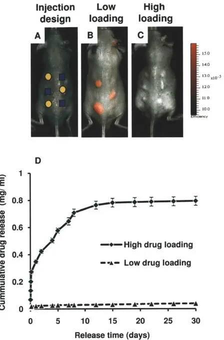
3.4.1. Spatial effect of a controlled-release anti-inflammatory drug... 38
3.4.1.1. Effect of drug loading on controlled-release properties ... 38
3.4.1.2. Anti-inflammatory drug attenuated coverage of implanted polymer by immune cell layers...39
3.4.2 Temporal effect of a controlled-release anti-inflammatory drug... 40
3.4.2.1. Tim e-evolution of cathepsin activity ... 40
3.4.2.2. Tim e-evolution of cellular infiltration ... 41
3.5. CONCLUSION...42
3.6. SUPPLEMENTAL INFORMATION...47
CHAPTER 4
-
ANTI-INFIAMMATORY DRUGS FOR IMPROVE) EFFI(ACY OF
ENCAPSULATED
ISLETS... 49
4.3.2. Fabrication and characterization of PLGA microparticles ... 52
4.3.3. Subcutaneous injection of PLGA microparticles... 52
4.3.4. Non-invasive fluorescent and bioluminescent imaging of SKH-1E mice ... 53
4.3.5. Tissue retrieval and histology processing of subcutaneously injected PLGA microparticles ... 53
4.3.6. Isolation of rat pancreatic islets... 53
4.3.7. Fabrication of hybrid alginate microcapsules co-encapsulating drug and islets... 54
4.3.8. Transplantation of encapsulated islets into STZ-induced diabetic C57B6/J mice ... 55
4.3.9. Blood glucose monitoring and Intraperitoneal Glucose Tolerance Test ... 55
4.3.9.1. Daily blood glucose m onitoring ... 55
4.3.9.2. Intraperitoneal glucose tolerance test (IPGTT):...55
4.3.10. Retrieval of transplanted capsules from the intraperitoneal cavity... 55
4.3.11. Quantification of fibrosis by DNA fluorescent staining... 56
4.3.12. Statistical analysis... 56
4.4. RESULTS ... 56
4.4.1. In vivo subcutaneous screening of small molecule anti-inflammatory drugs ... 56
4.4.2. Effect of selected drugs on the subcutaneous cellular dynamics and fibrosis formation ... 57
4.4.3. Improved glycemic control by alginate microcapsules co-encapsulating drug and islets in diabetic m ice ... 59
4.4.4. Reduced fibrotic overgrowth on explanted hybrid drug-islet capsules... 61
4.5. DISCUSSION ... 62
4.6. CONCLUSION...65
4.7. SUPPLEMENTARY INFORMATION ... 72
4.7.1. Supplementary results... 72
4.7.1.1. Subcutaneous screening of different formulations of anti-inflammatory drugs ... 72
4.7.1.2. Temporal evolution of inflammation markers in the host response...73
4.7.1.3. Analysis of excised PLGA microparticles to determine the presence of residual drugs ... 74
4.7.1.4. Determination of marginal islet mass for transplantation in diabetic mice ... 75
4.7.1.5. Establishing DNA fluorescent staining as a quantitative method for fibrosis assessment ... 76
4.7.1.6. Residual drugs from hybrid islet-drug capsules explanted after two months ... 77
4.7.2. Supplementary materials and methods... 78
5.2. INTRODUCTION ... 79
5.3. MATERIALS AND METHODS...80
5.3.1. Fabrication of alginate hydrogel capsules ... 80
5.3.2. Asymmetric surface modification of hydrogel capsules... 81
5.3.3. Cell culture ... 81
5.3.4. Viability analysis of encapsulated cells ... 81
5.3.5. Static glucose-stimulated insulin secretion ... 82
5.4. RESULTS AND DISCUSSION ... 82
5.4.1. Properties of template meshes for successful capsule fabrication ... 82
5.4.2. Fabrication of hydrogel capsules with different shapes and asymmetrically modified surfaces 83 5.4.3. Fabrication of hydrogel capsules containing insulin-secreting cells... 84
5.4.4. Assessment of viability, proliferation and function of encapsulated cells ... 84
5.4.3.1. Fabrication of hydrogel capsules containing insulin-secreting cells ... 84
5.4.3.2. Static glucose-stim ulated insulin secretion ... 85
5.5. CONCLUSION...85
CIAPTER 6
-CONCLUSION AND RECOMMENDATION FOR FUTURE WORK
... 93
6.1. CONCLUSION...93
6.2. RECOMMENDATION FOR FUTURE WORK... 93
REFERENCES...95
APPENDIX A -
ABBREVIATIONS...105
LIST OF TABLS
Table 4.1: Small molecule anti-inflammatory drugs investigated in the in vivo subcutaneous
screening...66
Table 5.1: Relationship between the feasibility of microcapsule formation and the properties
of a variety of thermoplastic meshes...87
LIST OF FIGURES
Figure 1.1: Temporal variation of tissue response to implanted biodegradable microspheres.
...
...--
14
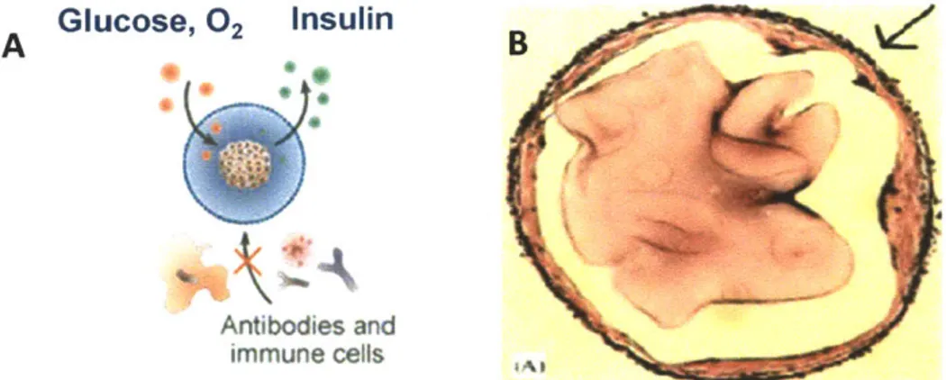
Figure 1.2: Immuno-isolation of islets and fibrosis response after transplantation...16
Figure 1.3: Schematic illustration of an amperometric glucose sensor and potential sources of declining sensor signals ... 17
Figure 2.1: Subcutaneous Injection arrays...28
Figure 2.2: Time evolution of cathepsin activity in response to injected materials fluorescently im aged... ... ... ... 29... 29
Figure 2.3: Time evolution of macrophage response to injected materials fluorescently im aged... ... ... ... ... ... .... ... ... ... 30
Figure 2.4: Histological scores of materials subcutaneously injected...31
Figure 2.5: H&E staining of representative sections subcutaneously injected materials...32
Figure 3.1: Effect of drug loading on the localization of anti-inflammatory properties...43
Figure 3.2: Anti-inflammatory drug attenuated coverage of implanted polymer by immune cell layers... ... ... ... ... . ... ... 44
Figure 3.3: Quantitative temporal monitoring of cathepsin activity...45
Figure 3.4: Quantitative monitoring of cellular infiltration to the inter-particle spaces...46
Figure 3.5: In vitro and in vivo fluorescent images of PLGA microparticles with different glycolide monomer contents... .47
Figure 3.6: Incorporation of dexamethasone did not alter the physical properties of the microparticles... ... ... 48
Figure 4.1: In vivo subcutaneous screening of anti-inflammatory drugs encapsulated in PLGA m icroparticles... ... .... ... .... .... .... ... ... 67
Figure 4.4: Effects of hybrid drug-islet capsules on glycemic control of STZ-induced diabetic
mice transplanted with a marginal islet mass of 250 IE.
...
70
Figure 4.5: Characterization of fibrotic pericapsular overgrowth on microcapsules retrieved 60
days after transplantation into STZ-induced C57B6J diabetic mice.
...
71
Figure 4.6: In vivo subcutaneous screening of anti-inflammatory drugs encapsulated in PLGA
m icroparticles...72
Figure 4.7: Temporal evolution of cathepsin enzymes and ROS in the host response to PLGA
microparticles with and without drugs.
...
73
Figure 4.8: Ex-vivo analysis of PLGA microparticles excised from SKH1E mice at day 28...74
Figure 4.9: Blood glucose concentrations in STZ-induced diabetic C57B6/J mice depended on
the transplanted mass of encapsulated Sprague-Dawley rat islets...75
Figure 4.10: DNA fluorescent staining correlated with fibrosis scoring by observation...76
Figure 4.11: Comparison of alginate capsules containing islets before transplantation and
after retrieval from C57B6/J diabetic mice on day 60...77
Figure 5.1: Schematic illustration of the procedure to fabricate alginate microcapsules
...
88
Figure 5.2: Images of polypropylene meshes used for microcapsule fabrication...89
Figure 5.3: Microcapsules with different geometries and asymmetric modification...90
Figure 5.4: Light and fluorescent microscopy images of alginate microcapsules containing
INS-1
cells...91
Figure 5.5: Viability and homogeneous distribution of cell clusters in a single microcapsule..92
INTRODUCTION
The fields of material biocompatibility and drug-device combination have progressed rapidly in the past decades. However, with the exception of the steroid-eluting pacemaker leads and the drug-eluting stents, no other clinical success has been achieved for medical devices utilizing controlled drug release technology to improve device biocompatibility. This thesis research aims to address several challenges that remain the bottlenecks for achieving further understanding and improved performance of medical devices.
The commentary in Chapter 1 discusses the background on the host response to implanted biomaterials and medical devices. This chapter outlines the biological mechanisms underlying the host response to implanted foreign objects, existing techniques to characterize this phenomenon and examples of medical device failures due to this host reaction. Existing studies on the incorporation of controlled-release anti-inflammatory drugs as a strategy to improve device performance and durability were also reviewed.
Chapter 2 reports the development of a new non-invasive imaging technique to study the activity of early immune cells in the early host response to implanted biomaterials. Fluorescent imaging probes activatable by inflammatory proteases secreted from immune cells were used for simultaneous biocompatibility screening of multiple materials in an immune-competent mouse model. Comparison of the imaging results with traditional histological analysis validated that the new fluorescent imaging technique can be used to assess material biocompatibility efficiently and rapidly.
The new fluorescent imaging technique in Chapter 2 was applied to study the spatio-temporal effects of a controlled release anti-inflammatory drug as outlined in Chapter 3. Subcutaneously injected poly (lactic-co-glycolic) (PLGA) microparticles with and without dexamethasone were investigated in hairless immunocompetent SKH-1E mice. The influence of drug loading and release kinetics on the local and systemic inhibition of inflammatory cellular activities was investigated by fluorescent imaging and parallel semi-quantitative histology analysis. Temporal monitoring of host response showed that the inhibition of inflammatory proteases in the early phase correlated with decreased cellular infiltration in the later phase of this reaction.
effectively inhibited the activities of inflammatory proteases while curcumin, a polyphenol drug, significantly decreased the presence of reactive oxygen species secreted by early immune cells. These drugs also decreased subsequent cellular infiltration and fibrosis formation surrounding subcutaneously injected polymeric microparticles.
The second part of chapter 4 focuses on applying the favorable finding from the subcutaneous drug screening in a medically relevant context with a focus on improving the treatment of type I diabetes by immuno-isolated islets. Hybrid alginate hydrogel microcapsules co-encapsulating pancreatic rat islets and selected drugs were developed and optimized to achieve good capsule morphology and drug loading. Curcumin effectively reduced the fibrotic response against encapsulated islets and improved their efficacy with better glycemic control in a mouse model of chemically-induced type I diabetes.
In a different effort to improve the design and production of islet-encapsulating microcapsules,
Chapter 5 introduces a new fabrication method that allows for rapid, homogenous microencapsulation
of insulin-secreting cells with varying microscale geometries and asymmetrically modified surfaces. Micromolding systems were developed using polypropylene mesh, and the material/surface properties associated with efficient encapsulation were identified. Cells encapsulated using these methods maintain desirable viability and preserve their ability to proliferate and secrete insulin in a glucose-responsive manner. This new cell encapsulation approach enables a practical route to an inexpensive and convenient process for the generation of cell-laden microcapsules without requiring any specialized equipment or microfabrication process.
Lastly, the commentary in Chapter 6 summarizes the collective finding from this thesis research and recommends directions for future research.
CHAPTER
I
-
BACKGROUND
1.1. HOST RESPONSE TO BIOMATERIALS AND MEDICAL DEVICES
1.1.1. Biological mechanisms underlying the host response
During the implantation of a biomaterial or biomedical device, tissue injury activates the inflammation cascade which leads to migration of inflammatory cells to the wound site, release of cytokines and growth factors that promote cell proliferation and protein synthesis as well was activation of complements, blood clotting and fibrinolytic cascades [1]. Typical events following the implantation of a material or device are depicted in Figure 1.1 below [2, 3]. The acute and chronic inflammatory responses are of short duration occurring over the first several weeks post-implantation[2]. Eosinophils and polymorphonuclear cells are typically present during the acute phase while macrophages and fibroblasts are observed during the chronic phase [4]. If not controlled, this sequence of inflammatory events can trigger the proliferation of fibroblasts which synthesize and deposit extracellular matrix to form granulation tissues and subsequently fibrous scars surrounding the implanted subject[5]. The duration of granulation tissue development, foreign body reaction, and fibrosis formation varies depending upon the characteristics of the implanted materials [2]
*-ACUTE - CHRONIC- GRANULATION TISSUE
-Neutrophils --- - - - -----
Macrophages Neovascularization
--
-- -- -- -- -- -- -- ---- Foreign Body Giant Cells
Fibroblasts
- d- Fibrosis
... ... Mononuclear
Leucocytes
Time
Traditionally, host response to biomaterial is characterized via histology analysis of excised samples ex vivo. This approach has primarily relied on visual evaluation by trained pathologists[6]. Computer-aided image analysis systems and immunohistochemical staining techniques have also been introduced to gain semi-quantitative information and improve data consistency and accuracy [7-9]. These histological approaches provide an informative end-point assessment with useful static information on cell types, quantity and distribution. However, the quality of data acquisition and interpretation remained variable [10].
Host response is a dynamic process involving the constant migration and recruitment of different population of immune cells whose secretion of active biomolecules such as cytokines and enzymes play an important role in determining the immunological response to an implanted object [2, 3]. For example, when evaluating a polymer system encapsulating a therapeutic drug, in vivo cellular secretory products such as inflammatory enzymes or cell signaling molecules might affect the degradation rate of the polymeric matrix [11-13] used to encapsulate drugs, and are partly responsible for the discrepancy between in vitro and in vivo release kinetics [14].
There remains a substantial need for new methods to provide more information in the characterization of biocompatibility phenomena, especially quantitative approaches that acquire kinetic information on the dynamic activities of the immune cell populations participating in the host response.
1.2. FAILURE OF IMPLANTABLE MEDICAL DEVICES DUE TO HOST
RESPONSE
1.2.1. Encapsulated islets in diabetes therapy
Implantable biomedical devices often suffer from loss of function in vivo due to changes caused in the tissue surrounding the devices caused by surgical injuries during implantation [15]. For example, immuno-isolated islets suffer transplanted capsules. Encapsulated islets as shown schematically in Figure 1.2A have been investigated as a technology which allows transplantation of non-autologous insulin-secreting cells into diabetic patients in the absence of long-term systemic administration of immunosuppressants [16-19]. The semi-permeable alginate hydrogel membrane surrounding the islets
A
Glucose,
02
Insulin
Antibodies and immune cells
Figure 1.2: Immuno-isolation of islets and fibrosis response after transplantation.
(A) Schematic illustration of microencapsulated islets in alginate hydrogel membrane[20]. (B) Deposition
of fibroblastic overgrowth on an alginate microcapsules retrieved from rat[21].
However, current development of islet encapsulation is still facing the problem of graft rejection and the lack of long-term survival of the islet grafts[22]. The capsules suffer from attachment by the components of the immune system such as antibodies and inflammatory cells which may induce capsular overgrowth as shown in Figure 1.2B , especially in the case of xenografts [23, 24]. This cellular layer can block the transport of nutrients and oxygen resulting in islet starvation. In addition, it was also suggested that non-specific inflammation caused by surgical trauma can lead to further recruitment of immune cells which secrete soluble cytokines[25]. These cytokines might be able to penetrate the alginate layers causing early mass loss and impaired function of the transplanted beta cells[26].
1.2.3. Other implantable biomedical devices
Glucose biosensors are also typical examples of biomedical devices whose functions are adversely affected by such tissue responses. Figure 1.3 illustrates a subcutaneously implanted amperometric glucose sensor with several potential sources of declining sensor signals [15]. Glucose diffuses through the sensor's outer membrane and is enzymatically converted to species that are detected by the electrode. Sensors fail due to host tissue response includes membrane biofouling, fibrous encapsulation and membrane biodegradation as well as electrode passivation due to protein adsorption and cell attachment [15].
fibrous encapsulation membrane electrical biodegradaon failure delamination enzyme of membranes degradation
Figure 1.3: Schematic illustration of an amperometric glucose sensor and potential sources of declining sensor signals [15].
Causes of device failure includes membrane biofouling, fibrous encapsulation and membrane
biodegradation as well as electrode passivation due to protein adsorption and cell attachment [15].
1.3. ANTI-INFLAMMATORY
DRUGS TO IMPROVE DURABILITY
OF
IMPLANTABLE DEVICES
Administration of inhibitory therapeutics such as anti-inflammatory drugs can help to improve
device performance by mitigating early tissue response and subsequent fibrosis [27]. Drug-incorporated
medical devices represent an emerging trend to localized delivery of therapeutics specifically to the site
of implantation [27]. This strategy promises to avoid the side effects of systemic administration,
minimize the effective dosage and ensure continuous drug release over a prolonged period of time [27].
However, development of devices incorporating anti-inflammatory therapeutics has mostly focused on
one or two model drugs with success in some devices but failure in others [28, 29]. The lack of an
efficient and systematic approach to characterize and compare drug efficacy in vivo hinders progress in
selecting optimal drug dosages and formulations for applications to specific devices.
Host response as discussed above significantly impairs the functions of devices that require specific
electrochemical or biochemical communication with the host microenvironment. Potent steroidal
anti-inflammatory drugs have been incorporated in medical devices to inhibit acute inflammation and
at the electrode-tissue interface[28]. This response can be attenuated by local delivery of glucocorticosteroids from the steroid-eluting tip of the electrode. Clinical studies comparing identical electrode configurations with and without local release of dexamethasone showed the superior performance of steroid-eluting pacemaker leads [32]. These steroid-eluting pacemaker leads are now widely used for patients with slow heart rates, abnormal rhythms or heart failure.
Recently, several groups have attempted to adapt this approach to improve the performance of glucose sensors. A composite of dexamethasone-loaded PLGA microparticles with poly-vinyl alcohol hydrogel has been reported to demonstrate some ability to modulate acute and chronic inflammation in vivo and was proposed to be used as a coating for implantable biosensors[33]. However, most of the recent research on combining anti-inflammatory drugs and medical devices has focused on a limited number of model drugs. Dexamethasone has been most widely used in attempts to improve biosensor performance, possibly due to its high potency and historical success with pacemaker leads. However, no significant improvement in sensor performance has been demonstrated. Ward et al reported that the lifetime of amperometric glucose sensor subcutaneously implanted in dogs for several weeks was not improved by localized delivery of dexamethasone[29]. Given the difference in the modes of communication (electric vs electrochemical) and the sites of implantation (heart muscle vs subcutaneous or intradermal) of the pacemaker lead and the glucose sensor, it is possible that an even stronger inhibitory agent or a combination of several drugs is needed to minimize tissue response and ensure acceptable communication between the sensor and the host environment. On the other hand, dexamethasone and other potent synthetic steroids are known to have diabetogenic effect and inhibit insulin secretion in islets[34] .Even though these potent glucocorticoids might be effective in mitigating the host response, they might not be the most efficacious for incorporation into cell-based therapeutics as they might adversely affects cell viability and function [35, 36]. Other classes of anti-inflammatory drugs such as Non-steroidal Anti-inflammatory Drugs (NSAIDs), polyphenols or non-steroidal immunosuppressants might be useful in inhibiting fibrosis while supporting cellular functionality and survival.
CHAPTER
2 -
NON-INVASIVE IMAGING OF EARLY HOST RESPONSE
The work reported in this chapter was conducted in collaboration primarily with Dr Kaitlin M. Bratlie. The content of this chapter has been published in whole or in part in the following peer-reviewed journal article:
Bratlie KM, Dang TT Lyle S, Nahrendorf M, Weissleder R, Langer R, Anderson DG. "Rapid biocompatibility analysis of materials by in vivo fluorescent imaging of inflammatory response". PLoS
ONE 2010; 5(4): e10032. doi:10.1371/ journal.pone.0010032
2.1. ABSTRACT
Many materials are unsuitable for medical use because of poor biocompatibility. Recently, advances in the high throughput synthesis of biomaterials has significantly increased the number of potential biomaterials, however current biocompatibility analysis methods are slow and require histological analysis. Here we develop rapid, non-invasive methods for in vivo quantification of the inflammatory response to implanted biomaterials. Materials were placed subcutaneously in an array format and monitored for host responses. Host cell activity in response to these materials was imaged kinetically, in vivo using fluorescent whole animal imaging. Data captured using whole animal imaging displayed similar temporal trends in cellular recruitment of phagocytes to the biomaterials compared to histological analysis. Histological analysis similarity validates this technique as a novel, rapid approach for screening biocompatibility of implanted materials. Through this technique there exists the possibility to rapidly screen large libraries of polymers in vivo.
2.2. INTRODUCTION
To our knowledge, there are no methods for in vivo visualization of biocompatibility or inflammatory responses to implanted biomaterials. Traditionally, biocompatibility is determined via histology. Histology allows for the determination of cell type and number near the implant, including those belonging to the immune system. However, histology is an endpoint measurement, allowing examination of only one time point per animal. Fluorescence imaging represents a set of powerful techniques that have traditionally been employed as a method for examining tumor models[37-42],
cells can lead to such complications as: bio-instability of glucose sensors[49]; overgrowth of encapsulated pancreatic islets for diabetes therapy causing ischemia and, eventually, necrosis of the islets[50]; and constrictive fibrosis following silicone implants in mammary augmentation[51]. Granulation tissue will then be formed and may appear as early as 3 to 5 days following implantation[48]. In general, granulation tissue will ultimately form a fibrous capsule surrounding the implant[48].
Immunological responses are dynamic processes and, as such, cell type and population at the implant site change during the healing process[2]. The sequence of local events following implantation is generally regarded as the tissue response continuum in which each individual event leads to the subsequent: injury progresses to acute inflammation, which proceeds to chronic inflammation, followed
by granulation tissue formation, foreign body reaction and fibrous encapsulation[2, 52]. The presence
of eosinophils and polymorphonuclear (PMN) cells typify acute inflammatory responses while macrophages and fibroblasts signify the chronic form[531. Neutrophils, together with monocytes and macrophages, release cathepsins during the process of degranulation [54, 55]. Cathepsins are proteolytic enzymes responsible for digesting foreign material [48].
Here, we describe the first methods for examining biomaterial biocompatibility in vivo, using fluorescence reflectance screening. The novelty of this technique lies in its ability to repeatedly analyze foreign body responses in the same animal. The macrophage recruitment and protease enzyme activity, both of which serve as markers of biocompatibility, were monitored in vivo, in real-time. We believe the methods developed here provide the first rapid techniques for parallel determination of biomaterial biocompatibility in vivo in a non-invasive manner.
2.3. MATERIALS AND METHODS
2.3.1. Molar Absorptivity.
The absorbance of the two fluorophores, ProSense-680 and F4/80 pan macrophage monoclonal antibody conjugated to Fluorescein isothiocyanate (FITC), were monitored using UV/Vis absorbance spectroscopy over the 200 to 800 nm range. Solutions were diluted in 0.9% w/v NaCl and housed in 1 cm path-length quartz cuvettes. Absorbances were measured on a Cary 100 Bio UV/Vis
The research protocol was approved by the local animal ethics committees at Massachusetts Institute of Technology (Committee on Animal Care) and Children's Hospital Boston (Institutional Animal Care and Use Committee) prior to initiation of the study.
2.3.3. Animals.
8-12 week old male SKH1 mice were obtained from Charles River Laboratories (Wilmington, MA).
The mice were maintained at the animal facilities of Massachusetts Institute of Technology, accredited
by the American Association of Laboratory Animal care, and were housed under standard conditions
with a 12-hour light/dark cycle. Both water and food were provided ad libitum.
2.3.4. Injections.
Injections were performed in accordance with ISO 10993-6: 2001. Prior to injection all materials were sterilized. Saline was sterilized via 0.22 pm filtration; alginate was autoclaved for 20 min. at 121*C; and polystyrene particles were washed in 70% ethanol and re-suspended in sterile saline. The mice were anesthetized via isoflurane inhalation at a concentration of 1-4% isoflurane/balance 02 to minimize movement. Their backs were scrubbed with 70% isopropyl alcohol and the animals were injected with saline, a solution of 2%-w/v alginate (Protanal LF 10/60, FMC BioPolymer, Newark, DE, having high guluronic acid composition (65-75%), mean molecular weight of 180kDa), or 10%-w/v polystyrene beads (3.0 pm, Sigma Aldrich, St. Louis, MO) in an array format on the mouse's back. Eight injections were made in each mouse in a random fashion to establish position-dependent inflammatory responses. Injection volumes ranged from 30 - 100 p. All experiments were conducted in
quadruplicate for each imaging time-point. In addition, a set four mice were imaged at every time-point and sacrificed at the 28 day time-point.
2.3.5. Imaging.
The following two imaging agents were co-injected into the tail vein 24 hours before in vivo fluorescence imaging: ProSense-680 (VisEn Medical, Woburn, MA, excitation wavelength 680 ± 10 nm, emission 700 ± 10 nm)[40] for imaging cathepsin activity, 2 nmol in 150 pl sterile Phosphate Buffered
imaging. Exposure time and f/stop - the relative size of the opening of the aperture - were optimized for each acquired image. Data were acquired and analyzed using the manufacturer's proprietary Living Image 3.1 software. All images are presented in fluorescence efficiency which is defined as the ratio of the collected fluorescent intensity to an internal standard of incident intensity at the selected imaging configuration. Regions of interest (ROls) were determined around the site of injection. ROI signal intensities were calculated in fluorescent efficiency. Images were obtained 1, 3, 7, 14, 21, and 28 days post-injection with four replicates imaged at each time point. A separate set of four replicates were imaged at all six time points.
2.3.6. Histology.
Histology evaluated the severity of inflammation resulting from the injected biomaterials. Mice were euthanized via CO2 asphyxiation and the injected biomaterial and surrounding tissue were excised.
The tissues were then fixed in 10% formalin, embedded in paraffin, cut into 5 pm sections, and stained using hematoxylin and eosin (H&E) for histological analysis by a board certified pathologist. Fibrosis was rated on a scale where a zero involved no fibrosis, a one indicated partial coverage with one to two layers of fibrosis, a two is designated a thicker fibrotic layer that nearly covered the implant, and a three denoted concentric fibrotic coverage of the polymer. Both polymorphonuclear (PMN) cells and macrophages were rated on a scale where no observed cells were indicated with a zero, scattered cells scored a one, numerous cells clustering on the sides of the polymer scored a two, and numerous cells surrounding the material resulted in a three.
2.3.7.
Statistical Analysis.
The values of the histologic scores and the ROls were averaged and expressed as the mean standard error of the mean. Comparisons of values were performed by the Student's unpaired two-tailed t-test. P values less than 0.05 were considered significant.
2.4. RESULTS
F
=K'(O -I)
(1)
Where I is the intensity of the incident excitation beam and I is the detected fluorescence intensity after traversing a length b of the medium - in this case - the tissue of the animal. The constant K' depends upon the quantum efficiency of the fluorescence process. In order to relate F to the concentration c of the fluorescing species, Beer's law can be written in the form:
I
=10~*b
10 (2)
Where
e
is the molar absorptivity of the fluorescing molecules and ebc is the absorbance. Inserting Beer's law into equation 1, we obtain:F = K'I 0
(1-_10-e*c).
3Which can be approximated for absorbances less than 0.05 to:
F = 2.3K'EscI
0(4)
Assuming that I is constant, the fluorescence intensity is linearly proportional to concentration at low absorbances. The molar absorptivities were determined by UV-visible absorbance to be 2.90 ± 0.04
x 106 M1 cm' and 2.30 ± 0.06 x 10' M1 cm1 for ProSense-680 and FITC mAb-F4/80, respectively. With an in vivo penetration depth for visible light of ~5 mm in reflectance mode,[37] the onset of nonlinear relations between fluorescence and concentration would present at doses 5 and 2.5 times larger than those injected for FITC mAb-F4/80 and ProSense-680, respectively, indicating the ability for relative quantitative analysis.
2.4.2. In Vivo Imaging of Cathepsin Activity and Macrophages.
Mice were injected with alginate, polystyrene, or saline in an array format (Figure 2.1) in volumes of 30, 50, 70, and 100 ptl. Alginate is a bio-inert material used in a variety of biomedical applications including encapsulation of insulin producing islets for diabetes therapy [56-59], wound healing[60, 61], implants for cardiac remodeling following infarction[62, 63]. In contrast, Polystyrene exhibits high cellular adhesive properties, induces a strong inflammatory response and was chosen as a positive control. Polystyrene particles below 10 ptm activate macrophages and are easily phagocytosed[64],
Fluorescent regions of interest (ROls) were quantified for each image and are presented in Figures
2.2D,E and 2.3D,E for cathepsin activity and macrophages recruitment.
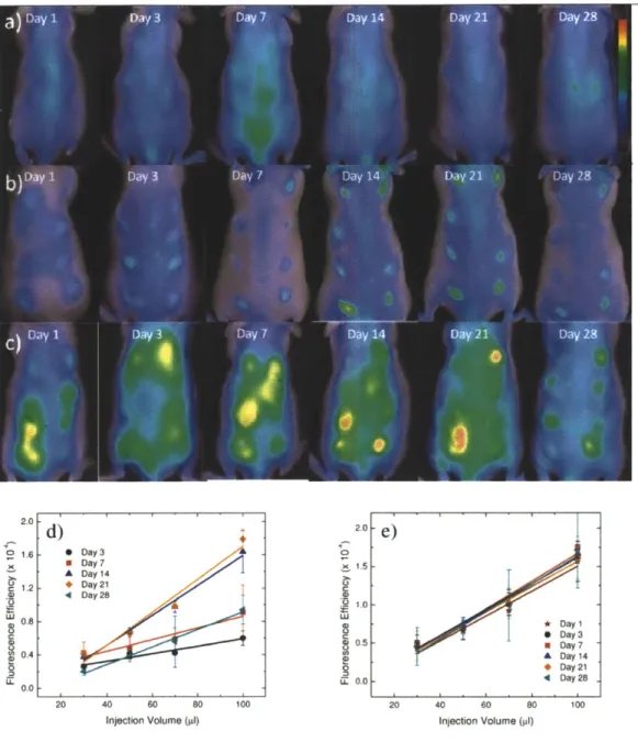
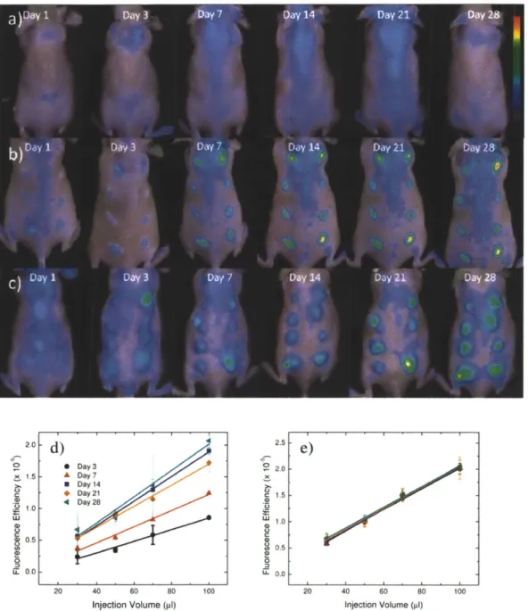
Qualitatively, for saline, the cathepsin activity and macrophage fluorescent signal appear to be very low with the exception of cathepsin activity on day 7. For polystyrene, cathepsin activity follows very similar trends wherein protease activity is detected on day one, peaks at three weeks, and begins to decline at four weeks. Recruitment of immune cells to alginate displays a different trend than polystyrene in which cathepsin activity remains constant from the first day to the fourth week. Macrophage recruitment for polystyrene reached a plateau at day seven. Alginate arrived at this plateau earlier, at the third day.
Quantitative performance criteria of methods are necessary in determining whether this technique is suitable in analyzing inflammatory responses. Detection limits are defined as the blank plus three times the standard deviation of the blank and limit of quantification (LOQ) is ten times the standard deviation of the blank. For macrophage detection, the detection limit is a fluorescence efficiency of 9.4
x 10-7 and the LOQ is a fluorescence efficiency of 2.3 x 10⁻⁶. ProSense-680 has a fluorescence efficiency
detection limit of 1.1x 10⁻⁵ and the fluorescence efficiency LOQ is 1.8 x 10⁻⁵. Quantification of
fluorescence efficiency of both cathepsin activity and macrophage recruitment is above the LOQ as shown in Figures 2.2 and 2.3. Cathepsin activity on the first day after injection of polystyrene was not above the LOQ and therefore not included in Figure 2.2D. Macrophage recruitment on day one for alginate and polystyrene were also below the LOQ and not included in Figure 2.3D and E.
2.4.3. Histology
Validation of the in vivo imaging technique for biocompatibility described required histologic analysis subsequent to each imaging time point. Several inflammation markers were quantified: PMNs, macrophages, and fibrosis. PMNs and macrophages were scored on the basis of zero being normal cell populations, one being scattered cells, two being numerous cells mostly populating the sides of the polymer, and three being the most severe where numerous cells surrounded the material. Quantified scores and representative images are shown in Figures 2.4 and 2.5, respectively. Minimal PMNs are seen infiltrating the injection site for saline whereas for alginate neutrophils completely surround the
saline, and agrees with previous results[65]. A more pronounced reaction occurs in response to
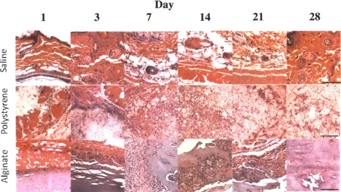
polystyrene and alginate. The macrophage signal for polystyrene reaches a plateau seven days post-injection, while that for alginate levels out at day three. The size of the polystyrene particles (3.0 pm) lends to being easily phagocytosed, which can be seen in Figure 2.5.
Fibrosis of the implants was also analyzed histologically in which a score of zero denotes no fibrosis, one signifies partial coverage with one to two layers of fibroblasts, two indicates a thicker layer nearly covering the implant, and three represents concentric fibrotic coverage of the polymer. As seen in Figure 2.4C, fibrosis for alginate and polystyrene gradually increases reaching a maximum at fourteen days. This observation is in line with previous findings[66] in which wound dressings of calcium alginate were grafted in porcine models and found fibrosis to reach a maximum at 14 days. The slight decrease in fibrosis scoring at day 28 might result from myofibroblasts contracting the wound as part of the healing process[67].
2.5.
DISCUSSION
Chemical signals responsible for invoking a response toward implanted biomaterials may include proteins from invading bacteria, clotting system peptides, complement products, and cytokines that have been released by macrophages located in the tissue near the implantation site[68]. Another group of chemical attractants are chemokines which recruit neutrophils and monocytes from the blood[69]. Macrophages derive from monocytes[70]. Macrophages and monocytes can phagocytose cellular debris and pathogens, and stimulate lymphocytes and other immune cells to respond to the pathogen.
Typically for acute inflammation, neutrophil recruitment peaks 1 - 2 days after implantation and gradually resolves after 7 - 10 days followed by macrophage migration at 1 - 2 days after injury[71]. Fibroblasts typically infiltrate at 2 - 3 days reaching a maximum population at 3 - 4 days[71]. Both macrophages and fibroblasts disperse after 5 - 9 days[71]. Chronic inflammation also begins with recruitment of neutrophils[72]. Additionally, protease levels are reported to be higher in chronic wounds[73]. Fibroblasts and macrophages become numerous one to two weeks after injury and diminish at six weeks[71, 74]. Histologic analysis and in vivo fluorescence imaging showed very similar trends in macrophage recruitment and also in comparing cathepsin activity derived from in vivo imaging
inflammation - macrophages and cathepsin activity - have been chosen to assess biocompatibility of various polymers.
Traditionally, the local pathological effect of a material on living tissue that is placed into an implant site is evaluated at both the gross level and the microscopic level. Various biological parameters such as cellular responses and histopathological changes are evaluated via ex vivo histology[52]. The throughput of histology is typically on the order of days to several weeks and involves steps such as fixation, embedding, processing, and staining. In vivo fluorescent images can be acquired in minutes and only require anesthetization of the subject, thus greatly reducing the time required to screen libraries of compounds. Recently, Sabaliauskas et al[76] have made advances in improving the throughput of histology by automating and digitizing data acquisition. Gersner et al[77] have developed laser scanning cytometry methods to quantify histological specimens, increasing the throughput of analysis. Specimen preparation still remains a costly, labor-intensive bottleneck in histology and, thus, in biocompatibility screening.
Aside from quantitative detection limit and LOQ, comparison of the dynamic ranges between histology and in vivo imaging is also necessary in determining the abilities of fluorescence imaging in assessing immune responses. In comparing histologic scores of polystyrene with fluorescence imaging for cathepsin activity (neutrophils), scores greater than 0.5 are above the detection limit, meaning that the injection sites are distinguishable from the background autofluorescence of the mouse. Histologic scores above 1 appear to correlate to fluorescence efficiencies above the LOQ for cathepsin activity (neutrophils). Comparing fluorescence imaging to histologic scores for macrophages leads to the conclusion that the detection limit and LOQ obtained for in vivo imaging corresponds to a histologic score of 1.5, indicating the possibility for false negatives in detecting macrophage infiltration and the necessity for histologic analysis. However, the use of amplification mechanisms such as use of fluorescent nanoparticles avidly taken up by macrophages[40] will likely enhance sensitivity. Although this technique is semi-quantitative owing to the poor depth penetration of visible light[37], in conjunction with histology it possesses the ability to transform the rapidity with which libraries of novel
histology. We anticipate that in vivo fluorescence imaging may therefore help address bottlenecks in analyzing biocompatibility of polymers and aid in understanding foreign body responses to biomaterials. In vivo fluorescence imaging also holds the advantage of monitoring temporal immune cell changes, thus eliminating mouse-to-mouse variations present when making a static histological assessment.
Figure 2.1: Subcutaneous Injection arrays.
Three array formats used for injecting saline and polymers subcutaneously in mice where A is 30 pil, B is
2.0
d)
20e
016 , Day 3 a Day 7 1.5 OaDay4 .A Day 14 + Day 21 D Day 28.0. -4 '.0 0. * DaylI 240 3 0.5 * Day 7 OA A Day 14 D ay 21 LL L 0. 0 41 Day 28 20 40 60 80 100 20 40 60 80 100Injection Volume (pl) Injection Volume (pl)
Figure 2.2: Time evolution of cathepsin activity in response to injected materials fluorescently imaged. In vivo fluorescence imaging using ProSense 680 for cathepsin activity at various time points for A) saline, B) polystyrene, and C) alginate. The scale bar ranges 0 - 6 x 10~ in fluorescence efficiency. The quantified fluorescence efficiencies of cathepsin activities are shown for D) polystyrene and E) alginate as the mean with standard deviation. Symbols represent data points and lines represent linear regressions.
2. 2.5 0 Day 3 2 0 x 1.5 A Day 7 8 Day 14 * Day21 1.5 4 Day 28 110 w1.0 0 0.5 0.5 o 0 UL 0.0 U 0.0 20 40 60 80 100 20 40 60 80 100
Injection Volume (al9) Injection Volume (pl)
Figure 2.3: Time evolution of macrophage response to injected materials fluorescently imaged.
In vivo fluorescence imaging of F4/80 pan macrophage antibody at various time points for A) saline, B) polystyrene, and C) alginate. The scale bar ranges 0 - 1.5 x 10~4 in fluorescence efficiency. The
quantified fluorescence efficiency of F4/80 pan macrophage responses are shown for D) polystyrene and
E) alginate as the mean with standard deviation. Symbols represent data points and lines represent
linear regressions.
f
C)0 5 10 15 20 Days Post-Injection 25 30 3.5 3.0 2.5 2.0 1.5 1.0 0.5 0.0 0 5 10 15 20 Days Post-Injection W 0 0 3.5 3.0 2.5 2.0 1.5 1.0 0.5 0.0 0 5 10 15 20 Days Post-Injection 25 30
Figure 2.4: Histological scores of materials subcutaneously injected.
Histological scores of A) neutrophils, B) macrophages, and C) fibrosis determined for tissue excised at various time points with injections of saline (0) , polystyrene (A), and alginate (m). Values shown are means with standard deviations.
a)
3.5 3.0 2.5 2.0 1.5 1.0 0.5 0.0 .2 I 25 30c)
-,1
~-T
b)Day
1
3
7
14
21
28
C rU
C
Figure 2.5: H&E staining of representative sections subcutaneously injected materials.
Representative sections stained with H&E are shown for saline, polystyrene, and alginate at various time points (1, 3, 7, 14, 21, and 28 days post-injection). (Magnification 20x, scale bar = 100 prm)
CHAPTER 3
-
IMAGING SPATIHYTEMPORAL EFFECTS OF A
CONTROLJED-RELEASE ANTI-INFIAMMATORY DRUG
The content of this chapter has been published in whole or in part in the following peer-reviewed journal article and incorporated into the following patent applications.
Dang TT Bratlie KM, Bogatyrev SR, Chen XY, Langer R, Anderson DG. "Spatiotemporal effects of a controlled release anti-inflammatory drug on the cellular dynamics of host response". Biomaterials 2011;32(19):4464-70
Anderson D.G, Langer R, and Dang TT. "Hybrid Microcapsules containing islets and anti-inflammatory drugs for diabetes therapy". U.S.S.N. 61/444206, filed on Feb. 2011.
Anderson D.G, Langer R, and Dang TT. "Hydrogel-encapsulated cells and anti-inflammatory drugs".
U.S.S.N. 13/400,382 and PCT/US2012/025806, filed on Feb 2012.
3.1. ABSTRACT
In general, biomaterials induce a non-specific host response when implanted in the body. This reaction has the potential to interfere with the function of the implanted materials. One method for controlling the host response is through local, controlled release of anti-inflammatory agents. Herein, we investigate the spatial and temporal effects of an anti-inflammatory drug on the cellular dynamics of the innate immune response to subcutaneously implanted poly(lactic-co-glycolic) (PLGA) microparticles. Noninvasive fluorescence imaging was used to investigate the influence of dexamethasone drug loading and release kinetics on the local and systemic inhibition of inflammatory cellular activities. Temporal monitoring of host response showed that inhibition of inflammatory proteases in the early phase was correlated with decreased cellular infiltration in the later phase of the foreign body response. We believe that using controlled-release anti-inflammatory platforms to modulate early cellular dynamics will be useful in reducing the foreign body response to implanted biomaterials and medical devices.
3.2. INTRODUCTION
One major challenge to clinical application of biomaterials and medical devices is their potential to induce a non-specific host response[10, 15, 49, 78-82]. This reaction involves the recruitment of early
sensors[15, 49, 87], neural probes[30], immunoisolated pancreatic islets[88-90] and biodegradable polymeric stents[79].
The incorporation of controlled-release delivery systems of anti-inflammatory drugs into medical devices has been proposed to mitigate host response and improve device durability [6, 27, 91-93]. This approach has shown promise in a number of clinical applications. For example, controlled elution of steroids from pace-maker leads has reduced fibrosis formation and enhanced long-term electrical communication between the leads and surrounding cardiac tissue[28]. However, similar attempts to improve the performance of other medical devices such as implanted glucose sensors [29] and immunoisolated islets for diabetes therapy have proven challenging [10]. There remains a substantial need to better understand the immunomodulatory effects of anti-inflammatory drugs on the host-tissue biology at the implant site[27]. Such knowledge can lead to better design of controlled-release drug delivery systems to improve the biocompatibility of implanted medical devices.
Researchers developing controlled-release drug formulations to mitigate host response have largely focused on decreasing the number of inflammatory cells infiltrating the host-device interface. Hickey et al. designed a mixed microsphere system containing dexamethasone, a steroidal anti-inflammatory drug, to achieve zero-order in vitro release kinetics and to suppress tissue response to thread-induced injuries in rats for up to one month[94, 95]. Recent studies on a hydrogel composite containing dexamethasone-loaded PLGA particles also suggested that sustained release of this drug may minimize the inflammatory reactions at the tissue-material interface [33, 96, 97]. While these studies have provided invaluable information, they only addressed the effects of these drug delivery systems via ex vivo analysis of the cell types, quantity and distribution in excised tissues. However, various factors in the design of controlled -release formulations such as drug selection, drug loading, particle sizes and corresponding release kinetics can dynamically affect a range of biological activities in the host response. The presence of anti-inflammatory drugs may alter not only the quantity and variety of immune cells recruited but also the kinetics of cellular activities such as the secretion of inflammatory enzymes or cell signaling pathways[98, 99]. In vivo cellular secretory products might affect the degradation rate of the polymeric matrix[11-13] used to encapsulate drugs, and are partly responsible for the discrepancy between in vitro and in vivo release kinetics[14]. Therefore, we hypothesize that
glycolic) (PLGA 50/50) microparticles with and without dexamethasone were subcutaneously injected in a six-spot array on the dorsal side of immunocompetent mice. Monitoring the in vivo activity of cathepsins, a class of inflammatory proteases, by noninvasive fluorescent imaging revealed that microparticles with low drug loading (1.3wt%) locally inhibited these enzymes, while high drug loading (26wt%) formulation resulted in systemic immunosuppression. The low dexamethasone loading (1.3wt%) was sufficient to attenuate the coverage of the implanted polymer by fibrotic cell layers. Temporal monitoring of the anti-inflammatory effect was carried out by in vivo imaging and ex vivo histological analysis.
3.3. MATERIALS AND METHODS
3.3.1. Fabrication and characterization of PLGA microparticles
Microparticles with or without dexamethasone were prepared using a single-emulsion method
[100] with biodegradable PLGA 50/50 (inherent viscosity of 0.95-1.2OdL/g) from Lactel (Pelham, AL).
Typically, a 5mL solution of PLGA and dexamethasone dissolved in dichloromethane, at concentrations of 40mg/ml and 2mg/ml respectively, was quickly added to a 25mL solution of 1% (w/v) polyvinyl
alcohol and homogenized for 60s at 5000rpm (Silverson L4R, Silverson Machines Ltd., Cheshire, England). The resulting suspension was quickly decanted into 75mL of deionized water and stirred for 30s prior to rotary evaporation (Buchi Rotavap, Buchi, Switzerland) for 3min. The suspension was washed five times by centrifugation at 3000rpm for 3min. The particles were collected by filtration using 0.2p.m filter, flash-frozen in liquid nitrogen, and lyophilized to dryness. Particle size distribution and morphology were examined by Scanning Electron Microscopy (JSM-6060, Jeol Ltd., Peabody, MA, USA). Fluorescence spectra of the PLGA polymer microparticles were collected by a Fluorolog-3 spectroflurometer (Horiba Yvon Jobin, Edison, NJ, USA). The dexamethasone loading of all microparticles was determined by dissolving 2mg of microspheres in 1mL of acetonitrile and comparing the resulting
UV absorbance at 234 nm to a standard curve of known concentrations of dexamethasone in
acetonitrile.
![Figure 1.3: Schematic illustration of an amperometric glucose sensor and potential sources of declining sensor signals [15].](https://thumb-eu.123doks.com/thumbv2/123doknet/14017471.457065/17.918.216.699.119.389/figure-schematic-illustration-amperometric-glucose-potential-sources-declining.webp)