The path to a better biomarker: application of a risk management
framework for the implementation of PD-L1 and TILs as
immuno-oncology biomarkers in breast cancer clinical trials and daily practice
Paula I Gonzalez-Ericsson1* ,Elisabeth S Stovgaard2,Luz F Sua3,Emily Reisenbichler4,Zuzana Kos5,Jodi M Carter6, Stefan Michiels7,John Le Quesne8,9,Torsten O Nielsen10,Anne-Vibeke Lænkholm11,Stephen B Fox12,13,
Julien Adam14,John MS Bartlett15,16,David L Rimm4,Cecily Quinn17,Dieter Peeters18,19,Maria V Dieci20,21, Anne Vincent-Salomon22,Ian Cree23 ,Akira I Hida24,Justin M Balko1,25,26,Harry R Haynes27,28 ,Isabel Frahm29, Gabriela Acosta-Haab30, Marcelo Balancin31 , Enrique Bellolio32, Wentao Yang33, Pawan Kirtani34,
Tomoharu Sugie35, Anna Ehinger36 , Carlos A Castaneda37, Marleen Kok38, Heather McArthur39,
Kalliopi Siziopikou40, Sunil Badve41, Susan Fineberg42, Allen Gown43, Giuseppe Viale44,45, Stuart J Schnitt46,47, Giancarlo Pruneri45,48, Frederique Penault-Llorca49,50, Stephen Hewitt51, E Aubrey Thompson52,
Kimberly H Allison53, William F Symmans54, Andrew M Bellizzi55, Edi Brogi56, David A Moore57, Denis Larsimont58, Deborah A Dillon46, Alexander Lazar54 , Huangchun Lien59, Matthew P Goetz60, Glenn Broeckx61 ,
Khalid El Bairi62 , Nadia Harbeck63, Ashley Cimino-Mathews64, Christos Sotiriou65, Sylvia Adams66, Shi-wei Liu67, Sibylle Loibl68, I-Chun Chen69, Sunil R Lakhani70, Jonathan W Juco71, Carsten Denkert72, Elizabeth F Blackley73, Sandra Demaria74, Roberto Leon-Ferre60, Oleg Gluz75, Dimitrios Zardavas76, Kenneth Emancipator71, Scott Ely77, Sherene Loi13,78, Roberto Salgado78,79, and Melinda Sanders1,26*, on behalf of the
International Immuno-Oncology Biomarker Working Group
1
Breast Cancer Research Program, Vanderbilt University Medical Center, Nashville, TN, USA 2
Department of Pathology, Herlev and Gentofte Hospital, University of Copenhagen, Herlev, Denmark 3
Department of Pathology and Laboratory Medicine, Fundación Valle del Lili, and Faculty of Health Sciences, Universidad ICESI, Cali, Colombia 4
Department of Pathology, Yale School of Medicine, New Haven, CT, USA 5
Department of Pathology, BC Cancer Agency, Vancouver, Canada 6
Department of Laboratory Medicine and Pathology, Mayo Clinic, Rochester, MN, USA 7
Biostatistics and Epidemiology Service, Centre de Recherche en Epidémiologie et Santé des Populations, Gustave Roussy, Université Paris-Sud, Villejuif, France
8
Leicester Cancer Research Centre, University of Leicester, Leicester, UK 9
MRC Toxicology Unit, University of Cambridge, Leicester, UK 10
Department of Pathology and Laboratory Medicine, University of British Columbia, Vancouver, Canada 11
Department of Surgical Pathology, Zealand University Hospital, Roskilde, Denmark 12
Department of Pathology, Peter MacCallum Cancer Centre, Melbourne, Australia 13
Sir Peter MacCallum Department of Oncology, University of Melbourne, Parkville, Australia 14
Department of Pathology, Gustave Roussy, Grand Paris, France 15
Ontario Institute for Cancer Research, Toronto, Canada 16
Edinburgh Cancer Research Centre, Institute of Genetics and Molecular Medicine, Edinburgh, UK 17
Department of Pathology, St Vincent’s University Hospital and University College Dublin, Dublin, Ireland 18
HistoGeneX NV, Antwerp, Belgium 19
AZ Sint-Maarten Hospital, Mechelen, Belgium 20
Department of Surgery, Oncology and Gastroenterology, University of Padova, Padova, Italy 21
Medical Oncology 2, Istituto Oncologico Veneto– IRCCS, Padova, Italy 22
Department of Pathology, Insitut Curie, Paris, France 23
International Agency for Research on Cancer (IARC), World Health Organization, Lyon, France 24
Department of Pathology, Matsuyama Shimin Hospital, Matsuyama, Japan 25
Department of Medicine, Vanderbilt University Medical Center, Nashville, TN, USA 26
Department of Pathology, Microbiology and Immunology, Vanderbilt University Medical Center, Nashville, TN, USA 27
Department of Cellular Pathology, North Bristol NHS Trust, Bristol, UK 28
Translational Health Sciences, University of Bristol, Bristol, UK 29
Department of Pathology, Sanatorio Mater Dei, Buenos Aires, Argentina 30
Department of Pathology, Hospital de Oncología Maria Curie, Buenos Aires, Argentina 31
Department of Pathology, Faculty of Medicine, University of S~ao Paulo, S~ao Paulo, Brazil 32
Department of Pathology, Universidad de La Frontera, Temuco, Chile 33
Department of Pathology, Fudan University Shanghai Cancer Centre, Shanghai, PR China 34
Department of Histopathology, Manipal Hospitals Dwarka, New Delhi, India 35
Breast Surgery, Kansai Medical University Hospital, Hirakata, Japan 36
Department of Clinical Genetics and Pathology, Skane University Hospital, Lund University, Lund, Sweden 37
Department of Medical Oncology, Instituto Nacional de Enfermedades Neoplásicas, Lima, Peru 38
Divisions of Medical Oncology, Tumor Biology & Immunology, The Netherlands Cancer Institute, Amsterdam, The Netherlands 39
Medical Oncology, Department of Medicine, Cedars-Sinai Medical Center, Los Angeles, CA, USA
Published online 9 April 2020 in Wiley Online Library
40 Department of Pathology, Breast Pathology Section, Northwestern University, Chicago, IL, USA 41
Department of Pathology and Laboratory Medicine, Indiana University, Indianapolis, IN, USA
42 Department of Pathology, Montefiore Medical Center and the Albert Einstein College of Medicine, Bronx, NY, USA 43
PhenoPath Laboratories, Seattle, WA, USA
44 Department of Pathology, Istituto Europeo di Oncologia IRCCS, Milan, Italy 45
University of Milan, Milan, Italy
46 Department of Pathology, Dana-Farber Cancer Institute, Boston, MA, USA 47
Department of Pathology, Brigham and Women’s Hospital and Harvard Medical School, Boston, MA, USA 48 Department of Pathology, IRCCS Fondazione Instituto Nazionale Tumori, Milan, Italy
49
Department of Biology and Pathology, Centre Jean Perrin, Clermont Ferrand, France 50 UMR INSERM 1240, Université Clermont Auvergne, Clermont Ferrand, France 51
Experimental Pathology Laboratory, Laboratory of Pathology, Center for Cancer Research, National Cancer Institute, National Institutes of Health, Bethesda, MD, USA
52
Department of Cancer Biology, Mayo Clinic, Jacksonville, FL, USA 53 Department of Pathology, Stanford University, Stanford, CA, USA 54
Department of Pathology, Division of Pathology and Laboratory Medicine, The University of Texas, MD Anderson Cancer Center, Houston, TX, USA 55 Department of Pathology, University of Iowa Hospitals and Clinics, Iowa City, IA, USA
56
Department of Pathology, Memorial Sloan Kettering Cancer Center, New York, NY, USA
57 CRUK Lung Cancer Centre of Excellence, UCL Cancer Institute, and Department of Cellular Pathology, UCLH, London, UK 58
Department of Pathology, Institut Jules Bordet, Université Libre de Bruxelles, Brussels, Belgium 59 Graduate Institute of Pathology, National Taiwan University, Taipei, Taiwan
60
Department of Oncology, Mayo Clinic, Rochester, MN, USA
61 Department of Pathology, University Hospital Antwerp, Edegem, Belgium 62
Cancer Biomarkers Working Group, Faculty of Medicine and Pharmacy, Mohamed Ist University, Oujda, Morocco 63 Breast Center, Department of OB&GYN and CCC (LMU), University of Munich, Munich, Germany
64
Department of Pathology and Oncology, The Johns Hopkins Hospital, Baltimore, MD, USA 65
Department of Medical Oncology, Institut Jules Bordet, Université Libre de Bruxelles, Brussels, Belgium 66
Perlmutter Cancer Center, New York University Medical School, New York, NY, USA 67
Sichuan Cancer Hospital, Chengdu, PR China 68
German Breast Group, Neu-Isenburg, Germany
69 Department of Medical Oncology, National Taiwan University Cancer Center, Taipei, Taiwan 70
The University of Queensland, Centre for Clinical Research, and Pathology Queensland, Royal Brisbane and Women’s Hospital, Herston, Australia 71 Translational Medicine, Merck & Co, Inc, Kenilworth, NJ, USA
72
Institute of Pathology, Universitätsklinikum Gießen und Marburg GmbH, Standort Marburg and Philipps-Universität Marburg, Marburg, Germany 73 Department of Medical Oncology, Peter MacCallum Cancer Centre, Melbourne, Australia
74
Department of Radiation Oncology, Department of Pathology and Laboratory Medicine, Weill Cornell Medicine, New York, NY, USA 75 Johanniter GmbH - Evangelisches Krankenhaus Bethesda Mönchengladbach, West German Study Group, Mönchengladbach, Germany 76
Oncology Clinical Development, Bristol-Myers Squibb, Princeton, NJ, USA 77 Translational Medicine, Bristol-Myers Squibb, Princeton, NJ, USA 78
Division of Research, Peter MacCallum Cancer Centre, Melbourne, Australia 79 Department of Pathology, GZA-ZNA Hospitals, Antwerp, Belgium
*Correspondence to: PI Gonzalez-Ericsson, Breast Cancer Research Program, Vanderbilt University, 1301 Medical Center Dr, TVC 4918, Nashville, TN 37232, USA. E-mail: paula.i.gonzalez.ericsson@vumc.org; or M Sanders, Department of Pathology, Microbiology and Immunology, and Breast Cancer Research Program, Vanderbilt University, 1301 Medical Center Dr, TVC 4918, Nashville, TN 37232, USA. E-mail: melinda.sanders@vumc.org
Abstract
Immune checkpoint inhibitor therapies targeting PD-1/PD-L1 are now the standard of care in oncology across several hematologic and solid tumor types, including triple negative breast cancer (TNBC). Patients with metastatic or locally advanced TNBC with PD-L1 expression on immune cells occupying≥1% of tumor area demonstrated survival benefit with the addition of atezolizumab to nab-paclitaxel. However, concerns regarding variability between immunohisto-chemical PD-L1 assay performance and inter-reader reproducibility have been raised. High tumor-infiltrating lym-phocytes (TILs) have also been associated with response to PD-1/PD-L1 inhibitors in patients with breast cancer (BC). TILs can be easily assessed on hematoxylin and eosin–stained slides and have shown reliable inter-reader repro-ducibility. As an established prognostic factor in early stage TNBC, TILs are soon anticipated to be reported in daily practice in many pathology laboratories worldwide. Because TILs and PD-L1 are parts of an immunological spectrum in BC, we propose the systematic implementation of combined PD-L1 and TIL analyses as a more comprehensive immuno-oncological biomarker for patient selection for PD-1/PD-L1 inhibition-based therapy in patients with BC. Although practical and regulatory considerations differ by jurisdiction, the pathology community has the respon-sibility to patients to implement assays that lead to optimal patient selection. We propose herewith a risk-management framework that may help mitigate the risks of suboptimal patient selection for immuno-therapeutic approaches in clinical trials and daily practice based on combined TILs/PD-L1 assessment in BC.
Keywords: PD-L1; TILs; breast cancer; biomarker risk-management; immunotherapy
Received 6 February 2020; Accepted 18 February 2020
Conflict of interest statement: ACM is a consultant for and reports research grant support from Bristol-Myers Squibb. AE is on the Roche advisory board and is a lecturer paid by Roche, Amgen, and Novartis. AIH reports honoraria from Chugai Pharmaceutical, Taiho Pharmaceutical, and Novartis Pharma. CQ is chair of the European Working Group for Breast Screening Pathology (EWGBSP), which has received funding from various companies for group meetings and also reports honoraria from Roche and Exact Sciences. DAM reports speaker fees from AstraZeneca. DAD is on the advisory board of Oncology Analytics, Inc., and consults for Novartis. DLR is on the advisory board for Amgen, AstraZeneca, Cell Signaling Technology, Cepheid, Daiichi Sankyo, GlaxoSmithKline, Konica Minolta, Merck, NanoString, Perkin Elmer, Roche, Ventana and Ultivue. He is a consultant for Biocept, NextCure, Odo-nate, and Sanofi and he is a founder and equity holder of PixelGear. He reports research support from AstraZeneca, Cepheid, Navdigate BioPharma, NextCure, Eli Lilly, and Ultivue and instrument support from Ventana, Akoya, Perkin Elmer, and NanoString. He reports travel honorarium from Bristol-Myers Squibb and royalties from Rarecyte. DZ is an employee of Bristol-Bristol-Myers Squibb with stock ownership. FPL is an advisor for AstraZeneca, Bayer, Bristol-Myers Squibb, Diaceutics, Elli Lilly, Illumina, MSD, and Roche, and reports research funding form AstraZeneca, Bayer, Bristol-Myers Squibb, MSD, and Roche. GV reports honoraria from Roche, Ventana, Agilent, MSD, Myers Squibb, and AstraZeneca. HM is a consultant for Amgen, Bristol-Myers Squibb, Celgene, Eli Lilly, Genentech/Roche, Immunomedics, Merck, OBI Pharma, Pfizer, Puma, Spectrum Pharmaceuticals, Syndax Pharmaceu-ticals, Peregrine, Calithera, Daiichi-Sankyo, and TapImmune, and has research supported by Bristol-Myers Squibb, MedImmune, LLC AstraZeneca, BTG, and Merck. . JA is an advisor for AstraZeneca, Bayer, Bristol-Myers Squibb, MSD, Roche, and Diaceutics, and reports research funding form MSD, Bayer, and Pierre Fabre. JB is a consultant for Insight Genetics Inc, BioNTech AG, Biotheranostics Inc, Pfizer, RNA Diagnostics Inc, and oncoX-change, and reports honoraria form NanoString Technologies, Oncology Education, and Biotheranostics Inc. He reports research funding from Thermo Fisher Scientific, Genoptix, Agendia, NanoString Technologies, Stratifyer GmbH, and Biotheranostics Inc, and travel expenses from Biotheranostics Inc and NanoString Technologies. He possesses patents regarding biomarker evaluation and gene signatures to predict response to treatment. JMB reports research support from Genentech/Roche, Bristol-Myers Squibb, and Incyte Corporation; has received consulting expert witness fees from Novartis, and is an inventor on provisional patents regarding immunotherapy targets and biomarkers in cancer. JJ is a full-time employee of Merck & Co., Inc. and owns stock in Merck & Co., Inc., Regeneron, and Illumina. KE is a full-time employee if Merck & Co., Inc. and owns stock in that company as well as Bayer AG and Johnson & Johnson. His wife is a full-time employee of Bristol-Myers Squibb and owns stock in that company. KS reports one-time honorarium from Roche Ventana. MK is on the Advisory board for Bristol-Myers Squibb and Daiichi (uncompensated) and her institute receives funding from BMS, Roche, and AstraZeneca. OG reports travel support from Roche, Celgene, and Daiichi, and is on honoraria advisory boards from Roche, Celgene, Novartis, Pfizer, Eli Lilly, GHI, NanoString, Amgen, and MSD. RLF reports travel Support from Immunomedics. RS reports research funding from Roche-Genentech, Puma Biotech-nology, and Merck; honoraria for consulting for Bristol-Myers Squibb; and travel funding from Roche Genentech, Merck, and AstraZeneca. SD reports past research grant support from Lytix Biopharma and Nanobiotix, and honorarium for consulting from EMD Serono and Mersana Therapeutics. SE is an employee of Bristol-Myers Squibb with stock ownership. SF served one time on an expert panel for Genomic Health. SL receives research funding to her institution from Novartis, Bristol-Myers Squibb, Merck, Roche-Genentech, Puma Biotechnology, Pfizer, AstraZeneca, Eli Lilly, and Seattle Genetics. She has acted as consultant (not compensated) to Seattle Genetics, Pfizer, Novartis, Bristol-Myers Squibb, Merck, AstraZeneca, and Roche-Genentech. She has acted as a consultant (paid to her institution) to Aduro Biotech, Novartis, and G1 Therapeutics. TON has a proprietary interest in the PAM50 sub-type classifier form Bioclassifer LLC, and NanoString Technologies. TS reports a speaker fee from AstraZeneca, Novartis, Chugai, Pfizer, Eisai, Takeda, Kyowa Kirin, Eli Lilly, MSD, and Genomic Health; and a research grant from Chugai, Pfizer, and Eisai. SBF is an Associate Editor of The Journal of Pathology.
Introduction
Immune checkpoint inhibitor (ICI) therapies targeting programmed cell death 1 (PD-1) and programmed death ligand 1 (PD-L1) are now the standard of care in oncology. Anti-PD-1 pembrolizumab (Keytruda, Merck & Co. Inc., Kenilworth, NJ, USA) and
nivolu-mab (Opdivo, Bristol-Myers Squibb Company,
New York, NY, USA), and anti-PD-L1 atezolizumab (Tecentriq, Genentech Inc, South San Francisco, CA,
USA), durvalumab (Imfinzi, AstraZeneca plc,
Cam-bridge, UK), and avelumab (Bavencio, Merck KGA, Darmstadt, Germany) have been approved to treat mul-tiple tumor types, in many countries. To date, atezolizu-mab specifically has been approved for triple-negative breast cancer (TNBC). At the same time,
immunohisto-chemistry (IHC)–based detection of PD-L1 expression
has been proposed as the predictive biomarker to select
patients that may benefit from these therapies. Five
pri-mary antibody clones have been developed in the form of assays paired with a specific staining platform.
PD-L1 22C3 (Agilent Technologies Inc., Santa Clara, CA, USA), 28-8 (Agilent Technologies Inc.), SP142 (Roche Tissue Diagnostics, Tucson, AZ, USA),
SP263 (Roche Tissue Diagnostics), and 73-10
(Agilent Technologies Inc.) have been used in clinical trials of the above-mentioned drugs, respectively. In addition, laboratory-developed tests (LDTs) using any of the above-mentioned primary antibodies or the E1L3N clone with different staining platforms are in use in research and clinical scenarios. Parallel to the
multiple assays, multiple scoring systems exist.
Table 1 shows technical details and defines scoring methods used for each antibody. Furthermore, different
cut-offs are used to define PD-L1 positivity for
differ-ent tumor types, whereas for certain indications
PD-L1 testing is not required for PD-1/PD-PD-L1 inhibition–
based therapy, from now on referred to as ICI. For several years the oncology and pathology com-munities have raised concerns about the reliability of IHC-based detection of PD-L1 to appropriately select patients for ICI. To date, although PD-L1 is currently
Table 1. Technical details, scoring system, and use on completed breast cancer clinical trials for each PD-L1 antibody Comm ercial diag nostic assays used in clinical trial s B iosimila r dia gnost ic ant ibodies u sed in clinic al practice Assay SP142 22C3 SP 263 73-3 28-8 E1 L3N (C ell Sig naling , Techno logy), CAL 10 (Zyt omed), QR1 (Qu artett), ZR3 (Cell Mark) Binding epi tope C-terminus cyt oplas mic domai n Discontinuo us segm ents on th e extrace llular doma in C-terminus cyt oplasmi c doma in C-terminus cyt oplasmi c do main Discontinuo us segm ents on th e extrace llular doma in E1 L3N: C-ter minus cyt oplasmi c doma in Platform Ventan a BenchM ark ULTRA Agilent Link 48 V entana Ben chMark ULTRA Agilent Link 48 Agilent Link 48 Any Scored cell type IC IC and TC IC or TC IC or TC TC Dep endin g o n score Scoring system ICA: PD-L1 +IC tumor area CPS: PD-L1 +IC + PD-L1 +TC TC TPS: PD-L1 +TC TC T% : PD-L 1 +TC TC ICIC% : PD-L 1 +IC IC T% : PD-L1 +TC TC ICIC% : PD-L1 +IC IC T% : PD-L1 +TC TC Dep endin g Partner drug Atezolizumab Pembro lizuma b Dur valumab Avelum ab Nivolum ab Any Breast can cer clinica l tria ls IMpass ion13 0 NCT0 137584 2 NCT0 163397 0 KATE-2 KEYNOT E-11 9 KEYNOT E-15 0 KEYNOT E-08 6 KEYNOT E-01 2 PANACE A KEYNOT E-17 3 KEYNOT E-55 2 TONIC (Niv oluma b) Ge parNu evo JAVELIN Non e CPS, combined positive score; IC, immune cells; TC, tumor cells; TPS, tumor positive score.
the only approved biomarker for these agents, it remains controversial given the complexities of its clinical use due to variability in assay performance of the PD-L1 IHC antibodies, spatial and temporal heterogeneity,
absence of a unified scoring system, and concerns about
inter-reader reproducibility for scoring PD-L1 on immune cell (ICs). Due to these inconsistencies, some
patients who could benefit might not receive treatment,
whereas others may be treated based on erroneous test results, exposing them to potential adverse side effects
with no drug benefit. In addition, because
PD-1/PD-L1 interaction is only one of many factors that may determine the clinical response to immunotherapeutics, it is unlikely that a single biomarker will sufficiently predict clinical outcomes in response to ICI. The use of composite biomarkers can provide biologically rele-vant information on multiple factors that determine response. In a meta-analysis, combined biomarker approaches such as PD-L1 IHC and tumor mutational
burden (TMB) and multiplex fluorescent
IHC-evaluating protein co-expression and spatial relation-ships, demonstrated an improved performance over
PD-L1 or TMB alone [1]. As guardians of patient’s
samples, pathologists partnered with clinicians, indus-try, and regulators must guide evidence-based inclusion of biomarkers in clinical trials and daily practice to ensure the best patient outcomes possible. Stromal
tumor-infiltrating lymphocytes (TILs) have also been
studied as a predictive biomarker of response to ICI for a variety of cancers including breast cancer (BC). TILs can be assessed on a simple hematoxylin and eosin (H&E) slide with reliable reproducibility among pathol-ogists when they adhere to the standardized method [2,3]. We propose PD-L1 and TILs as a more compre-hensive composite biomarker.
A good biomarker should be analytically valid, robust, reproducible, and clinically useful. To be incor-porated into daily practice, it must also be affordable and accessible to pathologists in both academic and community-hospital practices worldwide [4]. In this review, we propose a systematic implementation of combined PD-L1 and TIL analysis as a comprehensive immuno-oncological biomarker for patient selection for ICI in both clinical trials and daily practice. In support of this position, we outline the evolution of PD-L1 and TILs as biomarkers, from the analytical and clinical val-idation phases through clinical implementation, review the challenges we have encountered, and propose miti-gation approaches within a risk-management framework as previously published [5]. The collective of available evidence anticipates enhancement of patient selection and safety by the systematic implementation of com-bined PD-L1 and TIL analysis.
Technical validation phase: analytical validity of
PD-L1 IHC
Biomarker development starts with an initial discovery in pre-clinical studies, which we do not cover in this
review, followed by a validation phase in which the bio-marker is adapted to clinically applicable assay
plat-forms and subjected to analytical and clinical
validation [6]. For PD-L1 IHC, analytical validity refers to the accuracy and consistency of the technique to detect the presence of PD-L1 protein. To be able to ana-lyze the accuracy and consistency of the test we must first define the presence of PD-L1 protein. PD-L1 can be expressed on solid and hematologic tumor cells (TCs) and on ICs, including macrophages, dendritic cells, lymphocytes, and granulocytes [7,8]. PD-L1 is expressed in the cytoplasm and/or on the cell membrane.
A PD-L1-positive (PD-L1+) TC has been defined as
showing partial or complete membranous staining of any intensity [8–13]. Accompanying cytoplasmic stain-ing is often observed but ignored in TC. On the other hand, a PD-L1+ IC is one that shows membranous or cytoplasmic staining of any intensity. Cytoplasmic stain-ing may show a punctate or granular pattern, most com-monly observed with SP142 [11,12,14]. IC can be observed in aggregates or as single cells dispersed in the intratumoral or peritumoral stroma as well as admixed with TC [8,14].
Chromogenic IHC-based detection of PD-L1 has been largely concordant with other methods to detect
PD-L1 expression, such as immunofluorescence, mass
spectrometry, and RNA in situ hybridization
[9,15–18]. Each PD-L1 diagnostic kit has shown
preci-sion, reproducibility, and robustness when standard operating procedures and optimization of conditions
are followed [8,14,19–21]. Studies comparing PD-L1
assays performance on archival, routine clinical prac-tice, and clinical trial TNBC samples have shown dis-crepancies among SP142, SP263, and 22C3 assays.
PD-L1 positivity defined as the proportion of tumor
area occupied by PD-L1- positive immune cells
(ICA)≥1% with SP142 showed between 20 and
38, 10 and 35, and 7 and 19% fewer PD-L1+ cases
compared to SP263 ICA≥1% and 22C3 combined
pos-itive score (CPS)≥1 and ICA≥1%, respectively
[22–26]. Prevalence with each assay is shown in
Table 2. Similar findings were observed in previous
multi-institutional studies on archival clinical non–
small cell lung cancer (NSCLC) and urothelial carci-noma specimens, in which results between 22C3, 28-8, SP263, 73-3, and E1L3N assays were broadly comparable, whereas SP142 has shown lower PD-L1
expression on both TC and IC [9,10,12,13,16,38–43].
To investigate this discordance, a study mapped the antibody-binding sites for each antibody [44]. SP142, SP263, and E1L3N bind amino acid residues in the cytoplasmic tail of PD-L1 [14,44,45], whereas 22C3 and 28-8 target the extracellular domain [44,46]. 22C3 and 28-8 binding sites contain N-linked glyco-sylation sites, which may lead to variability in antigen
retrieval. N-glycosylation may also affect binding ef
fi-cacy of antibodies with cytoplasmic binding; differ-ences between mass spectrometry and E1L3N IHC were reported on melanoma samples with high glycan
modifications could interfere with recognition of bind-ing sites [17]. SP142 and SP263 bind to the same epi-tope [44]; hence the above-described discordance between these assays may be due to differences in
assay protocol leading to insufficient antibody
satura-tion. The visualization and amplification methods have
been shown to affect the extent and pattern of expres-sion of PD-L1 on IC and TC [47], at least partly explaining the discordance among assays.
Inter-observer reproducibility represents a major chal-lenge to the reliable assessment of any IHC assay; this is especially true for PD-L1. Although inter-pathologist reproducibility for the assessment of PD-L1 on TC is high, concordance has been lower for IC evaluation
across multiple tumor types [10,13,39], irrespective of
the assay. Scoring IC is more difficult from a
methodo-logical standpoint. Identification of IC may be
straight-forward in some cases, but complex in others, especially when attempting to differentiate between TC and intra-tumoral monocytic (macrophages/dendritic) cells, which cannot be easily distinguished on H&E. In addition, the four kits reportedly show different IC stain-ing patterns: 22C3, 28-8, and SP263 assays mainly stain macrophages and dendritic cells, whereas the SP142 assay, while staining a lower number of ICs, also
iden-tifies some lymphocyte-like cells [47]. Using SP142, the
majority of non-neoplastic cells were CD68+, whereas 5% were CD8+ [48]. Two multi-institutional studies,
Table 2. Prevalence of PD-L1 according to assay in breast cancer
Study Samples number and site SP142 SP263 22C3 Others Scottet al [22] 196 TNBC ICA≥ 1%:32% CPS≥ 1: 35% TC%≥ 1%: 11% ICA≥ 1%:54% CPS≥ 1: 64% TC%≥ 1%: 53% ICA≥ 1%:51% CPS≥ 1: 60% TC%≥ 1%: 50% 28-8 ICA≥ 1%:46% CPS≥ 1: 52% TC%≥ 1%: 35%
Noskeet al [23] 30 primary TNBC samples ICA≥ 1%: 50% ICA≥ 1%: 87% ICA≥ 1%: 57%
CPS≥ 1: 60% 28-8 ICA≥ 1%: 63%
Noskeet al [23] 104 primary TNBC samples ICA≥ 1%: 44% ICA≥ 1%: 82%
Reisenbichler et al [25] 68–76 primary TNBC samples ICA≥ 1%: 58% (n = 68) ICA≥ 1%: 78% (n = 76) IMpassion130 NCT02425891 [24]
614 primary and metastatic TNBC samples
ICA≥ 1%: 46% IC≥ 1%:75% CPS≥ 1: 81%
IMpassion130 NCT02425891 [24,27,28]
902 primary and metastatic TNBC samples
All: ICA≥1%:41%
primary: ICA≥1%:44%
metastatic: ICA≥1%:36%
All: TC%≥1%: 9% (900)
FDA SSED [14] 2744 primary and 50 metastatic TNBC samples
All: ICA≥1%:50%
primary: ICA≥1%:50%
metastatic: ICA≥1%:78%
Carteret al [29] 500 chemotherapy naïve TNBC ICA≥1%: 46%
TC%≥1%: 9% Downeset al [26] 30 BC ICA≥1%:47–50% CPS≥1: 53–63% E1L3N: ICA≥1%:53–63% CPS≥1: 53–67% NCT01633970 [30] 24 TNBC ICA≥1%: 50% TC≥1%:17% (of which 92% were ICA≥1%) NCT01375842 [7] 112 TNBC ICA≥1%: 78%* KEYNOTE-119 NCT02555657 [31] 622 TNBC CPS≥1: 65% CPS≥10: 31% CPS≥20: 18% KEYNOTE-012 NCT01848834 [32] 111 TNBC CPS≥1: 59% KEYNOTE-086 NCT02447003 [33]
170 primary and metastatic samples TNBC CPS≥1: 62% KEYNOTE-150 NCT02513472 [34] 107 TNBC CPS≥1: 46% JAVELIN NCT01772004 [35] 136 BC, 48 TNBC 73-3 All: IC≥10%: 9% TNBC: IC≥10%: 19% TONIC NCT02499367 [36] 70 metastatic TNBC samples IC≥1%: 86% IC≥5%: 67% GeparNuevo NCT02685059 [37] 158 TNBC ICIC%and/or TC%≥1%: 87%
CPS, combined positive score; FDA SSED, U.S. Food and Drug Administration summary of safety and effectiveness data; IC, immune cells; met, metastatic or non-primary sample; n, number of patients included in the analysis; prim, primary sample; TC, tumour cells.
including up to 19 pathologists, show moderate
agree-ment (interclass correlation coefficient [ICC]
0.560–0.805) between pathologists for SP142 assay on
TNBC samples [23,25]. Pathologists were trained on the evaluation of PD-L1 IHC and were required to pass
a proficiency test in one of these studies [23].
Agree-ment for other assays was slightly lower. Table 3 shows details of studies evaluating inter-observer reproduc-ibility on BC samples. Of interest, SP142 has been shown to have the highest concordance among readers
for PD-L1 IC≥1% in studies including other tumor
types [10–12], although the differences are not
statisti-cally significant. This may be because SP142 stains TC
with lower prevalence, allowing the IC staining to be
more easily identified.
Overall percent agreement (OPA) is the proportion of
samples that are classified the same by all observers. The
U.S. Food and Drug Administration (FDA) summary of safety and effectiveness data for SP142 showed an OPA of 91.1%; however, this study included only three pathologists [14]. In contrast, the study including 19 pathologists found an OPA of 41% with SP142. Recently, Reisenbichler et al [25] showed a new method for analysis of OPA as a function of the number of observers. The resulting graphs reaches a plateau at the number of observers required to provide realistic concor-dance estimate. If there is high concorconcor-dance, then the plot
will plateau at a high OPA with a small number of
observers. In contrast, OPA for PD-L1 ICA≥1%
decreased as the number of observers increased, reaching a plateau of 40% at nine observers. Results of real-world training conducted by Roche demonstrated an OPA of 98% between 903 pathologists from 75 countries
asses-sing 28 TNBC cases in a proficiency test; however, the
methodology for calculating OPA was not disclosed on the abstract [49]. On re-analysis of the National Com-prehensive Cancer Network (NCCN) study with lung
cancer samples, OPA between 13 pathologists
increased from 0% with a three-category score to 18%
using a two-category scale (IC≥1 and <1%), or even
67% if an outlier pathologist is excluded [38], showing that two categories are more reproducible. Moreover, low values, such as 1%, show lower inter-reader repro-ducibility [51].
Clinical validation phase: Clinical validity and
utility of PD-L1 IHC and TILs as predictive
biomarkers of response to PD-1/PD-L1 inhibitors
Clinical validation refers to how reliably the biomarker correlates with response to ICI and divides the patient
population into groups with divergent expected
Table 3. Studies evaluating inter-reader reproducibility on breast cancer samples
Study Assay and scoring Participating pathologists Samples evaluated Training Concordance Reisenbichleret al [25]
SP142 CDA ICA≥1% 19 68 primary TNBC No specific training for the
study.
ICC 0.560, OPA 41%
SP263 CDA ICA≥1% ICC 0.513
Noskeet al [23] SP142 CDA ICA≥1% 7 30 primary TNBC Trained on digital platform for
the evaluation of PD-L1 IC with SP142 and had to pass a proficiency exam.
ICC 0.805
SP263 CDA ICA≥1% ICC 0.616
22C3 CDA ICA≥1% ICC 0.605
28-8 CDA ICA≥1% ICC 0.460
FDA SSED [14] SP142 CDA ICA≥1% 3 60 TNBC Not specified. OPA 91.1%
Denniset al [49] SP142 CDA ICA≥1% 903 28 TNBC Regional trainer lead sessions
and digital platform training conducted by Roche International Pathologist Training program. A proficiency test was evaluated.
OPA 98%
Downeset al [26] SP142 CDA ICA≥1% 3 30 BC Not specified. ICC 0.956, OPA 98%
22C3 CDA CPS≥1 ICC 0.862, OPA 93% E1L3N LDT IC≥1% ICC 0.862, OPA 93% E1L3N LDT CPS≥1 ICC 0.815, OPA 91%
Solinaset al [50] E1L3N LDT IC≥1% 2 441 BC Not specified. ICC 0.10–0.58 for primary treatment naïve tumours,
ICC 0.94[0.84–0.97] for NAC treated, ICC 0.00 [−0.54–0.35] for relapses Overall percentage agreement (OPA) is calculated as the total number of times in which the readers agree, divide by the total number of readings. The OPA is expected to vary by classification difficulty and by the number of observers but does not take chance into account. Kappa does and should therefore be calculated as an associated measurement. Agreement measurements focus on the reliability of evaluations between different readers and do not require a standard reference, thus should not be confused with studies of accuracy. When using these measures of agreement, the FDA recommends to clearly state the calculations being performed. These calculations were not available for all the studies in Table 3 precluding fair comparison among studies.
CDA, commercial diagnostic assay; FDA SSED, U.S. Food and Drug Administration summary of safety and effectiveness data; ICC, interclass correlation coefficient; LDT, laboratory developed test; OPA, overall percent agreement.
Table 4. Studies evaluating clinical validity and utility of PD-L1 IHC and/or TILs as a predictive biomarker of response to PD 1/PD-L1 inhibitors in br east cancer Cl inical trial Drug Tum or ty pe (n) Bioma rker de tails (n) Predic tive capacity of PD-L1/TILs Adv anced sett ing IMpa ssion1 30 NC T02425 891 [24 ,27,28 ] Nab paclit axel +/ − atez olizuma b rando mized phas e III UnT x LA dv or mTNBC (902) PD-L1 (SP 142) was prospe ctively tested a t BTx (902) and used as a strati fi cation fact or for randomizati on. TILs we re eva luated retrospectively (460 an d 614). PD-L1 SP 263 and 22C 3 were performe d retrospectively o n BEP (614) post-h oc explora tory analysis. Improve d PFS (HR 0.6 2[0.49 –0.7 8]) and OS (HR 0.62[0.45 –0.8 6]) with the addit ion of atezolizu mab in PD-L 1+ tumo rs (SP1 42 ICA ≥ 1%). ORR 56 vers us 46% in the ITT populat ion and 59 vers us 43% in PD-L1+ tumo rs (p = 0.0 02). Better PFS (0.53[0. 38 –0.74]) an d O S (0.57[0.35 –0.9 2]) for TIL > 1 0 % PD-L 1 ≥ 1% popu lation (n = 460). PD-L 1+ cases sh owed high er me dian TILs (10%[ IQR:5 –20]) on BEP. Improve d PFS and OS (0.64 [0.53 –0.79]; 0.7 5 [0.59 –0.9 6]) with th e addition of atez olizuma b in SP26 3 (IC ≥ 1%) an d (0.68 [0.56 –0.82]; 0.78 [0.6 2– 0.9 9]) 22C3 (CPS ≥ 1) on BEP .Med ian PFS SP142 4.2 mont hs, 22C3 2.1 mont hs, SP263 2.2 mont hs, and media n O S SP142 9.4 mont hs, 22C3 2.4 mon ths, SP 263 3.3 mont hs. NC T01375 842[7 ] Ate zolizuma b single arm phas e Ib Pre Tx mTNBC (11 6) PD-L1 (SP 142) tested prospe ctively (116) . TICs (116) . PD-L 1 ICA ≥ 1% (ORR :12 vers us 0% ; HR: 0.5 5[0.33 –0.9 2]) and TICs > 10% (HR:0 .54[0.3 5– 0.83]) were asso ciated with better outco me. TICs > 1 0 % was indepe ndent ly associa ted with ORR, PFS and OS in mult ivariate analysis. PD-L 1 TC ≥ 1% wa s not asso ciated with res ponse. NC T01633 970[3 0] Ate zolizuma b + na b pac litaxel singl e arm phas e Ib UnT x (13) and PreTx (20) mTNB C (33 ) PD-L1 (SP 142) and TILs tested retro spect ively at BTx (23 and 20 ) and PostTx (11 an d 15, res pectively). No statistically sign ifi can t associa tion of basel ine PD-L 1 o r TILs with respon se. Numerica lly high er OR R (41. 7 versus 33.3% ) and longe r PFS (6.9 vers us 5.1mo ) and OS (21.9 vers us 11.4mo) in PD-L 1+ tumo r (IC A ≥ 1%). Numeric ally high er OS in TILs > 5%. Chang es in PD-L 1 o r TILs were not associa ted with clinic al respon se. K EYNOT E-119 NC T02555 657 [31 ,55] Ph ysician ’s choice che mo +/ − pembro lizuma b rando mized phas e III Pre Tx mTNBC (62 2) PD-L1 (22 C3) was prospe ctively tested a t BTx (622) and used as a strati fi cation fact or for randomizati on. No impro ved ou tcome in ITT po pulation or PD-L 1+ tumors (C PS ≥ 10 p = 0.0 57; CPS ≥ 1 p = 0.073 ). For CPS ≥ 20 HR OS: 0.58 [0.38 –0.88]. Better OS for TILs ≥ 5% (0.7 5[0.59 –0.9 6]) in th e pemb rolizu mab arm but not the che motherap y arm (1.46[1. 11 –1.92]). TILs and PD-L1 CPS mode rately correl ated (0.45). TILs (p = 0.0 04) and CPS (p = 0.0 9) were indepe ndent ly predict ive. K EYNOT E-150 NC T02513 472 [34 ] Pemb rolizu mab + eribuli n singl e arm ph ase Ib/II Pre Tx and UnT x mTNBC (10 7) PD-L1 (22 C3) ORR indepe ndent (30.6 vers us 22.4% ) o f PD-L 1 status (CPS ≥ 1).5 K EYNOT E-086 NC T02447 003 [33 ,56,57 ] Pemb rolizu mab singl e arm phas e II A: PreTx mTNBC (17 0) B: UnTx PD-L 1+ mTNBC (84) PD-L1 (22 C3) was prospe ctively tested a t BTx (254). ORR indepe ndent of PD-L 1 status (CPS ≥ 1) on cohort A (5.7 versus 4.7%). No diffe rence in PFS or OS betw een PD-L 1+ and PD-L1-. 21.4% ORR cohort B. (Continues)
Table 4. Continued Clinica l trial Drug Tumo r type (n) Bi omark er deta ils (n) Pr edictive capac ity of PD-L 1/TILs TILs were eval uated retro spect ively (193) . Bet ter OR R in pts with TILs > media n in coho rt A (6 vers us 2% ) a n d B (39 vers us 9% ) and combi ned cohort s (OR: 1.26[1 .03 –1.55]). Hi gher me dian Higher TILs in respo nders versus non-res ponde rs in coho rt A (10 versus 5%) and cohort B (50 vers us 15 %). KEYNOT E-012 NCT0 1848 834 [32] Pemb rolizuma b single arm phase Ib PreTx PD-L 1+ mTNBC (32) PD-L 1 (22C 3) wa s prosp ectively test ed at BTx (32) Incre asing ORR (p = 0.0 28) an d reduction in HR (p = 0.012) with inc reasing PD-L 1 exp ression. TONI C NCT0 2499 367 [36] Nivol umab +previous inductio n therap y (Rx/chemo) randomi zed phase II PreTx and UnTx mTNBC (67) PD-L 1 (22C 3) an d TILS at BTx , afte r induc tion an d Post Tx. H igher BTx TILs (me dian 12.5 vers us 6% ,p = 0.004 ) a n d PD-L 1 o n IC (me dian 15 versus 5%) on respo nders vers us non-respon ders. Bet ter PFS and OS was obse rved in PD-L1 IC ≥ 5% patients. No diffe rence was ob served betwe en PD-L 1 TC ≥ 1 and <1% po pulations. PANAC EA NCT0 2129 556 [58] Pemb rolizuma b + trastuz umab single arm phase Ib/II PreTx LAdv or mHER2+ BC Ib: PD-L1+ (6) II: PD-L 1+ & PD-L 1-(52 ) PD-L 1 (Qu alTek/ 22 C3) test ed prospe ctively at BTx (58). TILs we re eva luated retro spect ively (48). II: Higher ORR (15 vers us 0%) in PD-L1+ (CPS ≥ 1). Long er OS for PD-L1+ populat ion. H igher TILs lev els in objective respon ders (me dian ~25 vers us 1.5 % p = 0.006 ) and in PD-L 1+ populati on (p = 0.000 4). KATE-2 NCT0 2924 883 [59] T-DM 1+/ − atezolizu mab randomized phase II PreTx LAdv or mHE R2+ BC (20 2) PD-L 1 (SP1 42) test ed prosp ectively (20 2). PFS surviv al bene fi t (HR0.6 0[0.32 –1.1 1]) and nume rically highe r ORR (54 versus 33%) in PD-L 1+ tumo rs (IC A ≥ 1% ) with th e addit ion of atez olizu mab. JAVE LIN NCT0 1772 004 [35] Avelum ab sing le arm ph ase Ib PreTx LAdv or mBC (168) PD-L 1 (73-1 0) evalua ted prosp ectively o n IC a n d TC (168). Bet ter ORR in PD-L 1+ (IC ≥ 10%) BC (16.7 vers us 1.6 % p = 0.039, 22 .2 versus 2.6 % in TNBC). No associa tion betwe en ou tcome and PD-L 1+ (HR PFS :0.66[ 0.34 –1.2 6], OS: 0.62[0. 25 –1.54]). PD-L 1 o n TC show ed no association with res ponse. Neoa djuvan t setting KEYNOT E-552 NCT0 3036 488 [60] Neoa djuvan t paclitaxe l + carbop latin + AC/EC +/ − pembro lizuma b randomized phase III UnT x TNBC (602) 22 C3 pCR ach ieved irres pective PD-L 1 status (CPS ≥ 1) with the addit ion of pemb rolizu mab (68. 9 versus 45.3% ). KEYNOT E-173 NCT0 2622 074 [61,62 ] Neoa djuvan t pembro lizuma b + nab-paclitaxe l + /− carbop latin +/ -AC randomized phase Ib UnT x LAdv TNBC (60 ) PD-L 1 (22C 3) retro spect ively tested o n BTx (52). TILs we re retro spect ively eva luated on BTx (53) and OnTx (50) samples. H igher BTx (p = 0.0 28) an d OnT x TILs (p = 0.005 ) and BTx PD-L1 CPS (p = 0.021 ) were asso ciated with pCR. Respo nders ha d high er me dian pre (40 vers us 10 %) and OnTx (65 versus 22.5% ) TILs. Δ , change between baseline and after treatment; AC, doxorubicin + cyclophosphamide; BEP, biomarker evaluable population; BTx, baseline or pre-trea tment; CPS, combined positive score; EC, epirubicin + cyclophosphamide; HR, hazard ratio; IC, immune cells; IQR, interquartile range; LAdv, unresectable locally advanced; ITT, intention-to-treat population; mBC, metastatic breast cancer ,all subtypes; mTNBC, metastatic TNBC; n, number of patients included in the analysis; OnTx, on treatment; OR, odds ratio; ORR, objective response rates; OS, overall survival; PostTX, post-treatment; PreTx, previously treated; PFS, progression free sur vival; Rx, radiation; TC, tumor cells; TIC, tumor in fi ltrating immune cells (lymphocytes, macrophages, den-dritic cells and granulocytes) scored as a percentage of tumor area; UnTx, untreated.
outcomes. Clinical utility is a measure of whether clini-cal use of a test improves cliniclini-cal outcome and assists clinical decision-making [52]. The gold standard for evaluating biomarker clinical utility is the outcome of prospective randomized trials, which include biomarker evaluation in the study design, such that it is powered to specifically evaluate the benefit derived from the new
drug according to biomarker status [52–54]. However,
most randomized trials adopt a primary end point of drug
efficacy and do not employ a biomarker design. Table 4
shows the characteristics and results of clinical trials uti-lizing PD-L1 IHC and TILs as predictive biomarkers of response to ICI in BC.
Patients with newly diagnosed metastatic or locally
advanced PD-L1 ICA≥1% TNBC demonstrated
sur-vival benefit with the addition of the PD-L1 inhibitor
ate-zolizumab to nab-paclitaxel in the randomized phase III IMpassion130 trial in which all patients were prospec-tively tested for PD-L1 with SP142 [28]. Evaluation of progression-free survival (PFS) and overall survival (OS) in the PD-L1+ subgroup was one of the primary efficacy end points. Although the primary endpoint of OS for the intention-to-treat (ITT) population was not
reached, and although a pre-specified statistical testing
hierarchy prevented further formal analysis, OS was improved within the PD-L1+ subgroup with the addition of atezolizumab [28,63].
No improved outcome was observed for pre-treated metastatic TNBC patients with PD-1 inhibitor pembroli-zumab as monotherapy or compared to chemotherapy (treatment per physician choice: vinorelbine, capecita-bine, or gemcitabine) in the ITT population or PD-L1+
populations (PD-L1 CPS≥1 or CSP ≥10 with 22C3) on
the randomized phase III KEYNOTE-119 study [31]. Large randomized trials with survival end points, like the aforementioned, are generally required to establish the medical utility of a predictive biomarker. Neverthe-less, retrospective analysis of specimens collected from prospective trials may also establish biomarker clinical utility if appropriately designed and if archival tissue is available from enough patients to have adequate statisti-cal power [64]. An exploratory analysis with a cut-off of
CPS≥20 did show a longer benefit in OS with the
addi-tion of pembrolizumab to chemotherapy [31]. To further reliably establish clinical utility, these results should be validated in similar, but separate cohorts [64]. Likewise, response to pembrolizumab monotherapy or in combina-tion with chemotherapy was independent of PD-L1
sta-tus (CPS≥1) on a single-arm phase II KEYNOTE-086
and KEYNOTE-150 trials, respectively [33,34]. Of note, patients participating in these studies were
pre-trea-ted. TNBC patients with PD-L1 IC≥1% and IC ≥5%
showed improved survival outcomes with nivolumab after induction treatment on the phase II TONIC trial [36].
For patients with metastatic trastuzumab-resistant
HER2-positive (HER2+) BC, PD-L1 CPS≥1 was
predictive of response to the pembrolizumab plus
trastu-zumab combination in the single-arm phase II
PANACEA trial [58]. Conversely, on the phase II ran-domized KATE-2 trial, although the response was
numerically higher in patients with PD-L1 ICA≥1%
tumors, no significant benefit was observed with the
addition of atezolizumab to T-DM1 [59]. Notably, in an exploratory biomarker-analysis, the hazard ratio (HR) for OS was similar for PD-L1 as for TILs in this
trial, suggesting that both predict benefit from the
addi-tion of atezolizumab to T-DM1.
In the neoadjuvant setting, an increase in pathological complete response (pCR) rate observed with the addition of pembrolizumab to chemotherapy was independent of
PD-L1 status (CPS≥1) on the randomized phase III
KEYNOTE-552 trial [60]. Similarly, PD-L1 ICIC
%≥1% not only failed to predict pCR after the addition
of durvalumab to chemotherapy, but in fact was predic-tive of response in the chemotherapy-only arm on the phase II randomized GeparNuevo trial [37].
Exploratory analysis of the randomized phase III KEYNOTE-119 trial showed that patients with TILs higher than the median (5%) had better OS in the pem-brolizumab monotherapy arm but not in the chemother-apy arm [55]. TILs greater than the median were also shown to be predictive of response to single-agent pem-brolizumab regardless of PD-L1 status on retrospective biomarker analysis of the previously treated PD-L1 unselected cohort A of KEYNOTE-086 (median TILs 5%), but even more so within PD-L1+ treatment-naïve cases on cohort B (median TILs 17.5%) [57]. Further-more, patients with TNBC and HER2+ BC who responded to treatment with pembrolizumab alone and in combination with trastuzumab showed higher median TILs on the single arm phase II KEYNOTE-086 and PANACEA trials [57,58] and on the TONIC phase II
trial evaluating nivolumab after induction
treat-ment [36].
In the neoadjuvant setting, baseline TILs evaluated as
a continuous variable and stratified (<10, 11–59, ≥60%)
were predictive of pCR in both the durvalumab plus che-motherapy and cheche-motherapy plus placebo arms of GeparNuevo [37]. In addition, overall T-cell density was associated with pCR in response to pembrolizumab in the randomized phase II I-SPY 2 trial [65].
It is important to keep in mind that TILs have also proven predictive of response to neoadjuvant chemo-therapy (NAC) in patients with TNBC and HER2+ BC [66,67] and strongly prognostic of outcome in patients with early TNBC treated with standard
anthracycline-based adjuvant chemotherapy [68–70] on phase III and
pooled trials. In addition, in early stage treatment-naïve TNBC patients, high TIL-counts predict >98% 5-year survival, suggesting that the benefit of chemotherapy is probably very limited in this group [71,72]. PD-L1 base-line expression has also been positively associated with response to anthracycline-based NAC in hormone
receptor–positive BC [73] and TNBC [74]. However,
both PD-L1 and TILs are predictive of response to monotherapy ICI, proving predictive capacity beyond chemotherapy treatment.
Clinical implementation: Inclusion of PD-L1 and
TILs in clinical trials
Given the existing evidence, we propose systematic implementation of combined PD-L1 and TIL analyses as a comprehensive immuno-oncological integral bio-marker for patient selection for ICI in BC clinical trials. Because both have proven to be influential determinants
of response to ICI, the use of both markers as strati
fica-tion factors on randomized clinical trial designs could improve the balance of baseline characteristics among arms. Trial design should include PD-L1 and TIL ana-lyses in real time, pre-specifying the inclusion of both biomarkers in the protocol and ensuring well-powered biomarker clinical utility data that can be used for regu-latory submissions of both TILs and PDL1 as markers of
efficacy for immunotherapy. In addition, new protocols
can be written to conduct prospective–retrospective
bio-marker analysis on archival tissues from completed tri-als. All studies must be conducted and analyzed in a standardized manner per Reporting Recommendations for Tumor Marker Prognostic Studies (REMARK) cri-teria [75,76]. TILs should be scored as recommended by the International Immuno-oncology Biomarker Working Group (TIL-WG) [2,3] as a continuous vari-able with clinically relevant cut-offs in mind. A recent publication demonstrated the feasibility of the applica-tion of a web-based TIL scoring platform to enable the
use of TILs as a stratification factor in an
immunother-apy clinical trial for TNBC within a risk-management framework [77]. This pilot study proposes a standardize
workflow that can be used in future clinical trials.
In BC, both PD-L1 and TILs have shown higher expression in primary tumors samples than in metastases [2,24,57]. Nonetheless, PD-L1 expression on either pri-mary breast (HR PFS: 0.61[0.47–0.81]) or metastatic
lesion samples (HR OS: 0.55[0.32–0.93]) was predictive
of response to atezolizumab and nab-paclitaxel combi-nation [24]. Although the most recent sample may be more representative of the current immunologic status, evaluating all available samples on clinical trials would
provide useful data to define the most appropriate time
point for testing. Pre- and on-treatment TILs have been associated with response to ICI [61,62]. On-treatment biopsies could be included in protocols, since they may provide real-time information to help guide future treat-ment choices.
Furthermore, the existence of multiple scoring sys-tems for PD-L1 assays precludes the harmonization of assays and complicates reproducibility of scoring among pathologists. A single scoring system would allow a more accurate and direct comparison among assays and simplify scoring, likely facilitating adoption into clinical
practice. For BC patients, clinical benefit has been
corre-lated with PD-L1 expression on IC [7,27,35,36].
More-over, PD-L1 expression on macrophages was
associated with outcome in response to neoadjuvant dur-valumab [78]. Although PD-L1 expression on TCs with SP263 was predictive of response to durvalumab in the
neoadjuvant setting [37], in the advanced setting, expression on TCs evaluated by SP142 [7,24,27], 22C3 [36], and 73-10 [35] was not predictive. We
there-fore encourage reporting PD-L1 expression as IC, TC%/
tumor positive score (TPS), and CPS separately for all assays in clinical trials to assess which scoring system is most clinically relevant for each setting. Note that IC scored as proportion of tumor area occupied by PD-L1 expressing IC is not equivalent to IC as a percent of TC, given that most BCs contain distinct stromal areas in-between tumor areas; a score normalized by cross-sectional area produces lower scores than a score nor-malized by number of TCs.
We believe that the application of systematic criteria for combined PD-L1 and TIL analyses to future clinical trial designs will produce reliable data to better
under-stand which patients will benefit the most from ICI.
The resultant data could ultimately allow the conduction of a meta-analysis to provide clinically impactful data. Nevertheless, PD-L1 expression and IC presence are subject to dynamic regulation processes that are incom-pletely understood biologically. In addition, several
other factors also influence responses to ICI, including
tumor neoantigen load, IC composition, and expression of other costimulatory and inhibitory molecules.
Addi-tional biomarkers may help further refine patient
selec-tion. These potential biomarkers will likely be
predictive in a tumor type–specific dependent manner.
For instance, TMB has been showed to be a predictive biomarker of response to ICI across multiple cancers in retrospective studies [79]. However, mutational load is relatively low in BC. In addition, in TMB, estimates are variable across laboratories [80], with slower turn-around and higher cost compared to IHC. Class II major histocompatibility complex (MHC-II) tumor expression has been associated with response to ICI in breast [81] and other tumor types. Further investigation of these and other biomarkers in correlative studies in clinical tri-als is warranted, such as those evaluated by multiplex fluorescence IHC or gene-expression profiling.
Clinical implementation: Inclusion of PD-L1 and
TILs in daily practice
An analytically and clinically validated biomarker assay can be implemented into clinical care, but level 1 evi-dence is needed to change clinical practice. Results from randomized phase III IMpassion130 [28] led to the accelerated approval of atezolizumab and nab-paclitaxel as the standard treatment regimen for PD-L1+
(ICA≥1%) metastatic TNBC in many countries. Clinical
implementation of a biomarker requires three key ele-ments: Regulatory approval, reimbursement by health systems, and incorporation into clinical practice guide-lines [6]. Regulatory approval is different in every coun-try. Only the SP142 assay has been approved by regulatory agencies as the companion diagnostic test for the administration of atezolizumab and
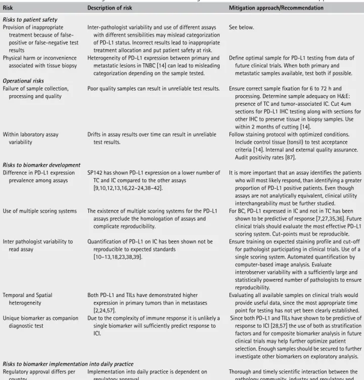
nab-Table 5. Risks associated with the integration of PD-L1 as immuno-oncological biomarkers for clinical trials and the daily practice
Risk Description of risk Mitigation approach/Recommendation Risks to patient safety
Provision of inappropriate treatment because of false-positive or false-negative test results
Inter-pathologist variability and use of different assays with different sensibilities may mislead categorization of PD-L1 status. Incorrect results lead to inappropriate treatment allocation and put patient safety at risk.
See below.
Physical harm or inconvenience associated with tissue biopsy
Heterogeneity of PD-L1 expression between primary and metastatic lesions in TNBC [14] can lead to misleading categorization depending on the sample tested.
Define optimal sample for PD-L1 testing from data of future clinical trials. When both primary and metastatic samples available, test both if possible. Operational risks
Failure of sample collection, processing and quality
Poor quality samples can result in unreliable test results. Ensure correct samplefixation for 6 to 72 h and processing. Determine sample adequacy on H&E: presence of TC and tumor-associated IC. Cut 4um sections for PD-L1 IHC testing along with sections for other IHC to preserve tissue in biopsy samples. Use within 2 months of cutting [14].
Within laboratory assay variability
Drifts in assay results over time can result in unreliable test results.
Follow staining protocol with optimized conditions. Include control tissue (tonsil) to test acceptance criteria [14]. Internal and external quality assurance. Audit positivity rates [87].
Risks to biomarker development Difference in PD-L1 expression
prevalence among assays
SP142 has shown PD-L1 expression on a lower number of TC and IC compared to the other assays
[9,10,12,13,16,22–24,38–42].
It is more important that an assay identifies the patients who will most likely respond, than identifying a greater proportion of PD-L1 positive patients. Even though assays are not analytically equivalent, clinical utility interchangeability must be further studied. Use of multiple scoring systems The existence of multiple scoring systems for the PD-L1
assays preclude the homologation of assays and complicate reproducibility.
For BC, PD-L1 expressed in IC and not in TC has been shown to be predictive of response [7,27,35,36]. Future clinical trials should evaluate the most effective PD-L1 scoring system. Cut-points must be reproducible. Inter pathologist variability to
read assay
Quantification of PD-L1 on IC has been shown not be reproducible to expected standards
[10–13,18,23,38,39].
Ensure training on expected staining profile and cut-off for pathologist participating in clinical trials. Use of a single scoring system. Automated quantification by computer-based image analysis. Evaluate
interobserver variability with a sufficiently large and statistically powered number of pathologists to ensure reproducibility.
Temporal and Spatial heterogeneity
Both PD-L1 and TILs have demonstrated higher expression in primary tumors than in metastases [2,24,57].
Evaluating all available samples on clinical trials would provide useful data, since the most appropriate time point for testing has not yet been clearly established. Unique biomarker as companion
diagnostic test
Due to the complexity of immune response it is unlikely a single biomarker will sufficiently predict response to ICI.
Since both PD-L1 and TILs have shown to be predictive of response to ICI [28,57] the use of both as stratification factors and for composite biomarker analysis in future clinical trials may help further optimize patient selection. Enough samples should be secured to further investigate other biomarkers on exploratory analysis. Risks to biomarker implementation into daily practice
Regulatory approval differs per country
Implementation into daily practice is dependent on regulatory approval.
Thorough and timely scientific interaction between the pathology community, industry and regulatory and national reimbursement agencies is needed. Biomarker accessibility and
affordability
PD-L1 testing is not yet covered by health insurance in many countries.
Thorough and timely scientific interaction between the pathology community, industry and regulatory and national reimbursement agencies is needed. Use of multiple PD-L1 assays for
a single analyte
With multiple PD-L1 assays available, pathology labs cannot be expected to have all tests available, causing variability in test results between laboratories.
Choice of assay will depend on regional regulations, availability of antibody, automated staining platform and optimized assay in currently in use. Consider LDTs. Outsource to reference laboratories.
Difference in PD-L1 expression prevalence among assays
SP142 has shown PD-L1 expression on a lower number of TC and IC compared to the other assays
[9,10,12,13,16,22–24,38–42].
It is more important that an assay identifies the patients who will most likely respond, than identifying a greater proportion of PD-L1+ patients. For BC SP142, SP263 and 22C3 have shown to identify patients that derive better outcome in response to atezolizumab and nab-paclitaxel [24].
Inter pathologist variability to read assay
Quantification of PD-L1 on IC has been shown not be reproducible to expected standards
[10–13,18,23,38,39].
Training on expected staining profile and cut-off. Interpretation guideline. Use of a single scoring system. Automated quantification by computer-based image analysis.
paclitaxel in countries such as the United States, Japan, Sweden, Peru, and Argentina. Whereas in certain counties in the European Union (EU), China, and Brazil, any PD-L1 assay can be used as long as it has been val-idated. In the EU, drugs are generally not regulatorily linked to a companion diagnostic test. The NCCN and other guidelines [82] include PD-L1 diagnostic testing as part of the workup for recurrent or metastatic TNBC as well as other tumor types. However, to date, in most countries, PD-L1 testing is not performed routinely on metastatic TNBC, but mainly upon oncologist request.
Following regulatory approval and incorporation into clinical practice guidelines, a biomarker must also be affordable and accessible to pathologists in both aca-demic and community-hospital practices worldwide to be successfully incorporated into daily practice. In Japan, where the SP142 assay is the approved compan-ion diagnostic test for TNBC, only this assay is covered by the health system. In the United States, the SP142 assay and LDTs are covered by health insurance. In Peru, PD-L1 testing is covered by prepaid health insur-ance but it is not yet covered by the public health system. In Argentina, Australia, Brazil, Chile, India, Morocco, and some countries in the EU, the test is not yet covered by the health system. In the UK, the National Institute for Health and Care Excellence (NICE), the UK regulatory
agency that evaluates drug efficacy, reported:
‘Atezoli-zumab with nab-paclitaxel […] does not meet NICE’s
criteria for inclusion in the Cancer Drugs Fund. This is because it does not have the potential to be cost effective at the current price, and there is no clear evidence that
further trial data would resolve the uncertainties’ [83].
Subsequently, each pathology laboratory faces chal-lenges including sample selection, sample processing, choice of assay, quality assurance, and interpretation to ensure correct implementation and consequent accurate patient selection. Table 5 summarizes these and previ-ously stated risks along with proposed mitigation approaches to ease the implementation of PD-L1 testing into clinical practice. It has been suggested that labs should test as many time points as are available such as to maximize patient eligibility for treatment. However,
such an approach will be costly without proven benefit
to the patient. It is also unclear whether insurance com-panies will pay for testing of multiple samples.
From a clinical perspective, it is imperative that an
assay identifies patients likely to respond to ICI, rather
than identifying a greater proportion of PD-L1+ patients.
The lower prevalence of PD-L1+ cases detected by the SP142 assay could potentially lead to fewer patients selected for therapy (false-negative tests), whereas use of SP263 or 22C3 could lead to greater patient eligibility at the expense of false-positive tests, unnecessarily
sub-jecting a subset of these patients to toxicity andfinancial
costs without clinical benefit. In an exploratory post hoc
analysis of IMpassion130, the PD-L1+ population iden-tified by each assay independently showed clinical
ben-efit with similar hazard ratio (HR) (HR [95% CI]:
SP142 ICA≥1%: PFS: 0.60 [0.47–0.78], OS: 0.74
[0.54–1.101]), 22C3 CPS ≥1: PFS: 0.68 [0.56–0.82],
OS: 0.78 [0.62–0.99], SP263 IC ≥1%: PFS: 0.64
[0.53–0.79], OS: 0.75 [0.59–0.96]) [24]. 22C3 and
SP263 identified a larger PD-L1+ population, of which
the SP142 positive cases are a subgroup. Of note, the biomarker evaluable population (BEP) included only 68% of the original ITT population, and although it may be adequately sized to reliably identify a larger treatment effect in the two-category test-positive patients, it could be underpowered to analyze a tripartite population of dual-assay analysis. OPA for analytical
concordance with SP142 (ICA≥1%) was 64% (22C3
CPS≥ 1) and 69% (SP263 IC ≥ 1%), demonstrating that
the assays are not equivalent [24]. Nevertheless, even if mostly driven by the SP142-positive subpopulation,
SP263 and 22C3 identified patients that showed
improved PFS and OS, making them clinically inter-changeable, since they identify populations with near-similar clinical outcomes [9]. Further studies such as this, done in partnership between academia, industry, and regulatory entities, need to be encouraged, prefera-bly before formal regulatory approval of an assay as a
companion diagnostic linked to a specific drug. In a
meta-analysis including samples from various tumor types, each diagnostic kit was found to better match with properly validated corresponding LDTs than with other diagnostic kit assays [43]. Although further studies are warranted, the use of LDTs is a reality in daily practice.
From a practical point of view, a single pathology lab-oratory cannot have all assays available. Labs perform-ing PD-L1 IHC testperform-ing for NSCLC already use other assays, most commonly 22C3 and SP263 assays or an LDT [38,40]. Developing and validating the SP142 assay could be an unwarranted burden for some labora-tories. SP142 and 22C3 commercial diagnostic assays are performed on different platforms, each a large capital
Table 5. Continued
Risk Description of risk Mitigation approach/Recommendation Unique biomarker as companion
diagnostic test
Due to the complexity of immune response it is unlikely a single biomarker will sufficiently predict response to ICI. The use of PD-L1+ IC score as a unique biomarker test maybe suboptimal in real world conditions.
Since TILs and PD-L1 are part of an immunological spectrum and PD-1/PD-L1 interaction is only one of many factors that may determine the clinical outcome of immunotherapeutic therapies, assessing both as a composite biomarker could be a better way to identify patients most likely to respond to ICI.
H&E, hematoxylin and eosin; IC, immune cells; IHC, immunohistochemistry; LDTs, laboratory developed test; PD-L1+ IC, proportion of tumor area covered by IC with discernible PD-L1 staining of any intensity expressed as a percentage; ICI, PD-1/PD-L1 inhibition based therapy; TC, tumor cell; TILs, tumor infiltrating lymphocytes; TNBC, triple negative breast cancer.
expenditure. In countries where regulatory agencies per-mit, PD-L1 could be performed as an LDT, if analyti-cally validated. For the SP142 antibody, similar PD-L1 expression was observed with different platforms [15], although using a different detection method has proven to impact assay performance [47]. In countries where the regulatory agencies mandate the use of the SP142 assay, smaller hospitals will likely need to outsource testing to a reference laboratory. To date, in most coun-tries, only a handful of large academic hospitals and ref-erence labs are performing PD-L1 testing for TNBC. The choice of assay should be an agreement between pathologist, oncologist, and patients, and be directed by good laboratory practices and common sense. Patient advocates need to be aware of how the choice of an assay
can influence treatment decisions.
For quality assurance purposes, tonsil-control tissue must be included as positive and negative controls alongside the clinical case to accept or reject the assay run. Tonsil tissue is recommended because it demon-strates granular punctate staining on lymphocytes arranged in aggregates and dispersed single-cell pat-terns, diffuse staining in the reticulated crypt epithelium,
and absence of staining on superficial squamous
epithe-lium [8]. A control sample staining close to the cut-off point is also recommended [87]. Unlike HER2, PD-L1
has no reflex alternative testing method that can be
employed to ascertain accuracy. In addition, because the different PD-L1 assays are not equivalent, they can-not be tested against each other for accuracy. Pathology laboratories must audit their PD-L1 positivity rates as part of internal quality assurance. Prevalence of PD-L1
+ (ICA>1%) TNBC with SP142 was 41% (44% on
pri-mary and 36% on metastatic samples) on IMpassion130 [24,28]. Other studies have shown a similar range of prevalence 32–58% on TNBC samples using SP142
ICA≥1% [14,22–25,28–30]; one study had an outlier
prevalence of 78%, in which thefirst 25 patients were
selected only if PD-L1+; then enrollment was extended to all patients [7]. However, PD-L1+ prevalence reaches
54–87 and 46–86% when using SP263 ICA≥1% and
22C3 CPS≥1, respectively [22–25,31–34]. Prevalence
of PD-L1+ on each of the cited studies is shown on Table 2. As part of an external quality assessment and validation, samples with known PD-L1 expression
should be tested and compared on proficiency tests. A
validated standardized PD-L1 Index Tissue Microarray [16] containing cell-line samples with known varying PD-L1 expression levels could be used for this purpose. For LDTs, laboratories must show results comparable to those obtained in clinical trials, with a diagnostic assay validated to predict potential response to a particular drug in a particular disease as a gold standard [84]. The Canadian Association of Pathologists has published a guide to ensure the quality of PD-L1 testing [85].
As discussed previously, inter-observer reproducibil-ity is one of the main pitfalls regarding PD-L1 validreproducibil-ity as a viable prognostic or predictive marker. These errors in patient selection not only put patients at risk, but also generate extra costs for health systems, generating issues
at the national regulatory level regarding reimbursement-criteria. Pathologists must be trained to interpret and score PD-L1 assays. Training material developed by assay manufacturers, including a digital training platform
with a proficiency test, can be accessed freely [86,88].
The value of training should be established in statistically rigorous studies that include post-training evaluation with proper decay time. In addition, pathologists must partici-pate in external quality assurance programs. A guideline for the interpretation of PD-L1 IHC developed by pathol-ogists for patholpathol-ogists, like those for TILs [2,3,89], ER [90], and HER2 [91], is needed. Such a guideline devel-oped by the International Association for the Study of Lung Cancer is available [92]. Even though reproducibil-ity among pathologists has been shown to be higher with two-category scoring [38], we believe the percentage of
PD-L1+ ICAshould be incorporated into the pathology
report in addition to a positive or negative PD-L1 deliberation.
Another tool available for pathologists that can improve reproducibility is digital image analysis of whole-slide images. Evaluation of TILs in solid tumors is a highly suitable application for computational
assess-ment; automated quantification by computer-based
image analysis provides accurate and reproducible results that can aid pathologists, especially for borderline cases surrounding the clinically relevant 1% cut-off that are challenging to distinguish by eye. In the basic retro-spective research realm, image analysis algorithms have shown better or comparable concordance between the automated algorithm score and the mean pathologist score than between pathologists [9,93]. Like any bio-marker, computer-based image analysis algorithms would need to be analytically and clinically validated with demonstrated clinical utility such that results are consistent with trial materials used to established cut-points for clinical decision-making and approved by cor-responding regulatory agencies before they can be applied in the daily practice. A recent publication
out-lines possible workflows and challenges for analytical
and clinical validation of computational TIL assessment [94], paving the path for its incorporation into clinical trials and daily practice.
In view of the considerable level Ib evidence for the prognostic value of TILs, the expert panels at St Gallen 2019 [95] and authors of the 2019 edition of the World
Health Organization Classification of Tumors of the
Breast recommended quantification of TILs in TNBC.
Internationally, some institutions have already begun incorporating TILs into pathology reports, paving the way for TIL counts to inform BC therapies. Going for-ward, a standardized format for reporting TIL counts, similar to those used to report hormone receptors, will need to be adopted. Given the inherent variability in TIL distribution and heterogeneity of sampling, we pro-pose that TIL counts should be scored in treatment-naïve and advanced-setting BC specimens, while in the clini-cal post-treatment setting TILs should be scored only on clinical trial samples according to established guide-lines [96]. TILs should be scored as recommend by the