HAL Id: hal-01961920
https://hal.archives-ouvertes.fr/hal-01961920
Submitted on 20 Dec 2018
HAL is a multi-disciplinary open access
archive for the deposit and dissemination of
sci-entific research documents, whether they are
pub-lished or not. The documents may come from
teaching and research institutions in France or
abroad, or from public or private research centers.
L’archive ouverte pluridisciplinaire HAL, est
destinée au dépôt et à la diffusion de documents
scientifiques de niveau recherche, publiés ou non,
émanant des établissements d’enseignement et de
recherche français ou étrangers, des laboratoires
publics ou privés.
A new polymer-based approach for in vivo transfection
in postnatal brain
C. Di Scala, M. Tessier, C. Sapet, F. Poulhès, F. Sicard, O. Zelphati, C.
Pellegrino
To cite this version:
C. Di Scala, M. Tessier, C. Sapet, F. Poulhès, F. Sicard, et al.. A new polymer-based approach for in
vivo transfection in postnatal brain. Journal of Neuroscience Methods, Elsevier, 2019, 311, pp.295-306.
�10.1016/j.jneumeth.2018.11.004�. �hal-01961920�
Contents lists available atScienceDirect
Journal of Neuroscience Methods
journal homepage:www.elsevier.com/locate/jneumeth
A new polymer-based approach for in vivo transfection in postnatal brain
C. Di Scala
a,1, M. Tessier
a,1, C. Sapet
b,1, F. Poulhes
b, F. Sicard
b, O. Zelphati
b,⁎, C. Pellegrino
a,⁎ aINMED, INSERM, Aix-Marseille Univ, 163 route de luminy, BP13, Marseille, FrancebOZ Biosciences, Parc Scientifique de Luminy, 163 Avenue de Luminy case 922, 13288 Marseille cedex 9, France
A R T I C L E I N F O
Keywords: Transfection In vivo Gene delivery Non viral gene therapy KCC2
Postnatal brain Rattus norvegicus
A B S T R A C T
Background: Gene delivery within the central nervous system at postnatal age is one of the most challenging tasks in neuroscience and currently only a few effective methods are available.
Comparison with existing methods: For postnatal central nervous system cells, viral approaches are commonly used for genetic engineering but they face several biosafety requirements for production and use making them less accessible to the community. Conversely, lipid-based methods are widely used in cell culture but face limitation in vivo mainly due to the inflammatory responses they induce. To this aspect, the use of a transgenic mouse line can represent a credible answer to the community working on rat models still requires an effective and successful solution to circumvent these difficulties.
New method: We describe a new polymer-based gene delivery system allowing persistent and robust in vivo transfection with low DNA amount, reduced inflammation and high diffusion. The expression profile along the brain, the stability, the diffusion of the DNA together with the quantity of cells transfected were evaluated through in vivo approaches.
Results: With a single low-volume injection, we targeted different cell types within the rat brain. We measured the diffusion rate ranging from 1 to 5 mm based on the injected volume, in the three-dimensions axis. Finally, we modified brain susceptibility to epileptic seizures using a specific knock-down of the neuronal specific po-tassium-chloride transporter 2.
Conclusions: This safe and easy system opens perspectives for non viral gene delivery in the rat brain with perspectives to study brain function in vivo.
1. Introduction
Transfection of mammalian cells is now a common technique in all laboratories over the world. Depending on the cell type and the brain structure several systems are in use, from the lipid-based approach in the hippocampus (Buerli et al., 2007) or in the dopaminergic neurons (Underhill et al., 2014) to the Magnetofection method adapted from a variety of primary adherent cells in vitro (Plank et al., 2011) such as primary neurons (Buerli et al., 2007; Underhill et al., 2014), neural stem cells (Sapet et al., 2011), microglial cells (Smolders et al., 2018) as well as for in vivo application (Plank et al., 2011;Ohashi et al., 2014; Titze de Almeida et al., 2018). In addition the electroporation-mediated approach is widely used for the motoneurons (Jacquier et al., 2006) or embryonic cortical structures (Ackman et al., 2009;Carabalona et al., 2011) or also retinal cells (Matsuda and Cepko, 2007). The main issue with all these techniques is the need to get sufficient cell density to assess the brain formation andfinally study the brain physiology. From
this perspective, up to now only viral particles offer the correct com-bination between the number of cells, the structure of interest and minimization of toxicity (Sapet et al., 2011; Stauffer et al., 2016). Nevertheless, the requirements of viral production as well as the ele-vated viral titer needed to proceed to in vivo infection represent clear limits in regards to the monitoring of the brain activity. Considering this concern, we developed together with OZ Biosciences a new delivery system allowing effective Central Nervous System (CNS) cell transfec-tion with reduced toxicity. The nanoparticles-based system has already been studied for brain use (Roy et al., 2008) but its effect on the brain physiology was restricted to local effect. Here we used a brand new polyplexes-based approach in which DNA is complexed to BrainFectIN reagent (OZ Biosciences). The obtained nanoparticles were firstly evaluated in their capacity to protect and deliver DNA inside cells, and then their efficiency in modifying a large cell population. We demon-strated, using shRNA-encoding plasmid strategy, that by knocking down KCC2 in the hippocampus it was possible to significantly modify
https://doi.org/10.1016/j.jneumeth.2018.11.004
Received 14 May 2018; Received in revised form 26 October 2018; Accepted 4 November 2018
⁎Corresponding authors at: INMED, INSERM 1249, 163 route de Luminy, 13273 Marseille 09, France.
E-mail addresses:ozelphati@ozbiosciences.com(O. Zelphati),christophe.pellegrino@inserm.fr(C. Pellegrino).
1These authors contributed equally to the work.
Journal of Neuroscience Methods 311 (2019) 295–306
Available online 05 November 2018
0165-0270/ © 2018 The Author(s). Published by Elsevier B.V. This is an open access article under the CC BY-NC-ND license (http://creativecommons.org/licenses/BY-NC-ND/4.0/).
the brain activity, and consequently highlighting a sustained brain hyperexcitability and susceptibility to seizures. Altogether BrainFectIN offers a potent effect with reduced toxicity and easy handling, opening new pathways for future studies aiming to modify cell populations within the CNS of postnatal animals.
2. Material and methods
All manipulations with animals were performed in agreement with the guidelines of the Animal Care and Use Committee of INSERM (Institut National de la Santé et de la Recherche Médicale) as project “Apafis#2797-2015112016427629”.
2.1. Polyplex formation
Polyplexes consisting of DNA and BrainFectIN reagent (OZ Biosciences, Marseille) were prepared in PBS and incubated at room temperature (RT) for the indicated period of time. When not specified, incubation time was 30 min. The initial DNA concentration was 1μg/μL resuspended in water. Ranges of ratios of 0.25:1 to 3:1 or 4:1 (respec-tively 0.25μL–3 μL or 4 μL per μg DNA) were used for complex and stability studies and ratio of 2:1 (2μL per μg DNA) was chosen for all other experiments. The capacity of BrainFectIN to form polyplexes with DNA was evaluated by electrophoresis on 1% agarose gel.
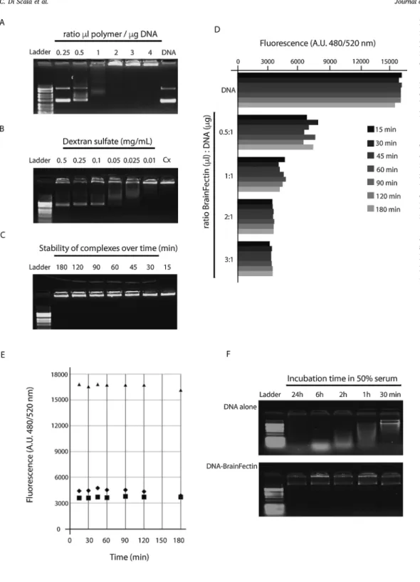
Fig. 1. Physico-chemical charater-ization of BrainFectIN/DNA com-plexes. The capacity of BrainFectIN to form complexes with DNA depending on the ratio of polymer-to-DNA used (A) and the release of DNA from poly-plexes (ratio 2:1) in presence of raising doses of Dextran sulfate (B) was studied by electrophoresis on 1% agarose gel. Polyplexes of BrainFectIN and DNA (2μL per μg DNA) were incubated from 15 to 180 min and the stability of complexes was then analyzed by 1% agarose gel electrophoresis (C) and by fluorescence (D and E). Fluorescent nucleic acid stain SYBR Gold was used to monitor DNA availability in solution complexed or not to different ratios of BrainFectIN (D) and to evaluate DNA release from polyplexes after 24 h (E). (F) BrainFectIN protection from de-gradation capacity at a 2:1 ratio was studied by comparing incubation of DNA alone (upper gel) or DNA com-plexed into polyplexes (lower gel) in 50% serum at 37 °C for 30 min to 24 h.
2.2. DNA release from complexes
After 30 min incubation at RT, doses of Dextran Sulfate (Sigma-Aldrich) ranging from 0.01 to 0.5 mg/mLfinal in PBS were added to complexes of BrainFectIN and DNA (ratio 2:1). The DNA release from polyplexes was monitored by electrophoresis on 1% agarose gel. 2.3. Stability of complexes
The stability of polyplexes wasfirst evaluated by electrophoresis on 1% agarose gel after 15 min to 3 h incubation at RT using a 2:1 ratio.
Complex stability and DNA release at ratios ranging from 0.5:1 to 3:1 were also studied by measuring the DNA accessibility tofluorescent nucleic acid binding reagent SYBR™ Gold (Thermo Fisher Scientific) on a fluorescence plate reader (CytoFluor Series 4000, Perceptive Biosystem) at 495/20 nm excitation and 530/20 nm emission and compared to DNA alone.
2.4. DNA protection from degradation
DNA alone or complexed to BrainFectIN (ratio 2:1) was incubated in 50% Foetal Calf Serum (FCS) at 37 °C from 30 min to 24 h. Enzyme
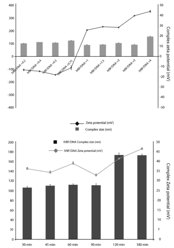
Fig. 2. Particle size and Zeta poten-tial ofinvivo BrainFectIN/DNA com-plexes in water. The formed nano-particles have been studied at different polymer/DNA ratio ranging from 0.2 to 4 in the same volume of water. The mean hydrodynamic particle size and charge measurements for vector/ pDNA complexes were performed using Dynamic Light Scattering (DLS) and Laser Doppler Velocimetry (LDV), re-spectively, using Malvern Nano ZS in-strument and DTS software (Malvern Instruments, UK) in a water of Grade 2. Each measurement has been recorded after 30 min of incubation.
Fig. 3. Evolution over time of the physico-chemical properties of the 2:1 complex of the BrainFectIN/DNA (2:1) complexes formed in water. The nanoparticles have been formed in water and been incubated for times ranging from 30 min to 3 h, before having been analyzed in DLS or LDV. The chosen Polymer/BrainFectIN ratio for these data is 2:1.
activity was then stopped by the addition of EDTA (0.5μM). Polyplexes were finally dissociated as previously described by the addition of Dextran sulfate (0.5 mg/mL) and DNA integrity was assessed by elec-trophoresis on 1% gel agarose.
2.5. Particle size and zeta potential measurements
The mean hydrodynamic particle size and charge measurements for vector/ pDNA complexes were performed using Dynamic Light
Scattering (DLS) and Laser Doppler Velocimetry (LDV), respectively, using Malvern Nano ZS instrument and DTS software (Malvern Instruments, UK) in a water of Grade 2. Various amounts of BrainFectIN reagent were diluted in 100μL of water and mixed with an equal vo-lume of the same water containing DNA. The measurements were car-ried out in automatic mode and the results are reported as mean +/-SEM of 3 runs. Each mean represents the average value of 30 mea-surements per run.
2.6. DNA extraction and purification
A pcDNA3.1-eGFP expressing-vector under cytomegalovirus pro-moter was transformed into Escherichia coli cells. The DNA extraction was performed using Nucleobond Xtra Midi/Maxi kit (Qiagen) ac-cording to the manufacturer’s instructions. Total DNA was measured by
spectrophotometry at 260 nm on a nanodrop (Implen,
Nanophotometer®). The DNA purity was assessed through the mea-surement of 260/280 nm ratio. DNA having 260/280 nm ratio below 1,9 were discarded from injection.
2.7. Stereotaxic procedure
Twenty days old Sprague-Dawley rats were housed in an enriched environment at INMED animal house facility, maintained in a 12 h light/12 h dark cycle environment with controlled temperature (23 ± 2 °C), food and water were given ad libitum. The stereotaxic procedure was performed using aseptic technique. Briefly, 30 min be-fore surgery, buprenorphine (0.03 mg/kg) was given intra-peritoneally (i.p) then rats were anesthetized using 4% isoflurane vaporized by air together with additional 0.3% oxygen enrichment. Surgical anesthesia was maintained the same way but using only 2% of isoflurane in air enriched with 0.3% oxygen while rats were positioned in a stereotaxic frame (David Kopf Instruments, Tujunga, CA). Body temperature was maintained at 37 ± 2 °C with a heating pad (Harvard Apparatus). Stereotaxic coordinates were chosen to allow injection above the hip-pocampus (Antero-Posterior axis: -2, laterality: -3, verticality: -2). Then the skull was drilled while leaving the dura intact. Using a micro in-jector (micropump 4, WPI) 0.5 to 2 microliters were then injected at 100 nL/min speed using NanoFil® D needle (WPI). The injection lasted maximum 15 min, and then the needle was left in place for the same duration period to prevent back-flow of cells. Altogether the total duration of the surgery did not overpass 40 min. The skin was sutured and animals placed in the post-operative room of the facility. Brains were then either perfused and fixed for immunochemistry or freshly dissected for Western blot analysis 3–10 days after the injection.
In control, eGFP plasmid was transfected using JetPEI (Polyplus, France) as recommended by the manufacturer protocol.
2.8. Immunohistochemistry
Rats were deeply anaesthetized with i.p injection of ketamine/xy-lazine then transcardiacally perfused with cold phosphate buffer saline
(PBS 0.01 M) prior to 3% paraformaldehyde solution (AntigenFix, Diapath). Brains were post-fixed overnight in 3% paraformaldehyde at 4 °C and then coronally sliced with a Leica VT1200S Vibratome (60μm thickness). Sections were permeabilized and blocked in PBS with 0.3% Triton X-100 and 5% normal goat serum (NGS) for 1 h at room tem-perature. Incubation with primary antibodies diluted in PBS with 5% NGS and 0.1% Triton X-100 was carried out at 4 °C overnight using GFP (G1544, Sigma-Aldrich), Gad67 (MAB5406, Merck), anti-Iba1 (W1W019-19741, Wako) and anti-GFAP (Z033429-2, Agilent Dako). After rinsing 3 times in PBS, slices were incubated with the corresponding Alexa Fluor 488-conjugated secondary antibodies di-luted in PBS (1/500, Thermo Fisher Scientific) for 2 h at room tem-perature and finally counterstained for 1 min with Hoechst 33,258 (10μg/mL in PBS, Sigma-Aldrich). Sections were then mounted onto Superfrost Plus glass slides in Fluoromount G mounting medium. For each section, images were taken using a confocal microscope with 4×, 10×, 20× or 40× objectives.
2.9. Western blot analysis
Animals were killed by decapitation after deep isoflurane an-esthesia. Hippocampi were quickly dissected out,flash-frozen in liquid nitrogen and stored at −80 °C until processed. Brain tissue was homogenized in RIPA buffer (50 mM Tris-HCl pH 8; 150 mM NaCl; SDS 0.1%; Deoxycholic Acid 0.5%; 1% Triton X-100), containing complete Protease/Phosphatase Inhibitor Tablet (Thermofisher) and loaded with Laemmli 3X loading buffer. The samples were separated in 4–15% SDS–PAGE gel (Criterion gel, Bio-Rad) and transferred to nitrocellulose membrane (Whatman). After blocking in Tris-buffered saline/ 0.1% tween/ 5% bovine serum albumin (BSA), membranes were exposed overnight at 4 °C to primary antibody diluted in blocking solution (Tris-buffered saline/ 0.1% tween/ 2.5% BSA) (anti-GFP, [NB600-308], Novus Biologicals). Secondary antibody (anti-mouse HRP, #MA1-10371, ThermoFisher Scientific) was applied 2 h at room temperature before chemiluminescence assay was performed using horseradish peroxidase-conjugated detection. Signals were revealed using ECL-plus Western blotting reagents (ECL-plus kit, Pierce Biotech) on the image analysis software G box (Syngene). Then, membranes were stripped using 50 mM DTT/ 2% SDS in 50 mM Tris-HCl, pH 7.0 for 30 min at 65 °C, blocked again in Tris-buffered saline/ 0.1% tween/ 5% BSA and finally probed with α-tubulin (#62204, Life technologies) or anti-β-tubulin (TUBB3 Poly18020, Biolegend) for normalization, signal de-tection and revelation were done following the same procedure as the one for primary antibody. Quantifications were performed using Gel Plot Analyzer plugin (ImageJ).
2.10. Seizure susceptibility test (pentylenetetrazol, PTZ)
To evaluate the susceptibility to seizures in GFP-injected versus shRNA-KCC2-injected rat, we performed PTZ injection 10 days after transfection (10dpt) at the age of 30 days old. PTZ (25 mg/kg; Sigma) was administered intra-peritoneally every 10 min until generalized
Fig. 4. A. Expression of eGFP at different depth of the hippocampus after injection of 1,5 μL of BrainFectIN-eGFP with a 2:1 ratio. (a) eGFP expression in the dorsal ipsilateral hippocampus (ventrally to the injection point).(b) eGFP expression in the ipsilateral hippocampus (near to the injection point). (c) eGFP expression in the ventral ipsilateral hippocampus (dorsally to the injection point. Scale bar = 100μm. Magnification 4 × . B. Expression of eGFP in the ipsilateral hippo-campus after injection of physiological serum + DNA for negative control or 1,5μL of BrainFectIN-eGFP with different ratio (1:1 ; 2:1 ; 3:1). Coordinates: AP : -2 ; L : -3 ; V :-4.(a) eGFP expression with negative control injection. (b) eGFP expression with an 1:1 ratio. (c) eGFP expression with a 2:1 ratio. (d) eGFP expression with an 3:1 ratio. CA1, Cornu Ammonis 1 hippocampal region; CA2, Cornu Ammonis 2 hippocampal region; CA3, Cornu Ammonis 3 hippocampal region ; DG, dentate gyrus. Scale bar = 100μm. Magnification 4 × . C. Influence of the injected volume on the efficiency of the transfection. (a) eGFP expression in the hippocampus after injection of 0,5μL of BrainFectIN-eGFP. Small amount of transfected cells. Black window: 20x magnification of the CA1 region. (b) eGFP expression in the hippocampus after injection of 1μL of BrainFectIN-eGFP. Small amount of transfected cells with low inflammation. Black window: 20x magnifi-cation of the CA1 region.(c) eGFP expression in the hippocampus after injection of 1,5μL of BrainFectIN-eGFP. Lots of cells are transfected with a bit of in-flammation. Black window: 20x magnification of the CA1 region. (d) eGFP expression in the hippocampus after injection of 2 μL of BrainFectIN-eGFP. Appearance of many cells transfected together with inflammation. Black windows: 20x magnification of the CA1 region, Scale bar = 20 μm. Magnification 4 × . Slices thickness is 60μm.
seizures occur. Under these conditions both the number of injection and time to induce generalized seizure were plotted (Petit et al., 2014).
2.11. Volumetry analysis
Coronal 60μm thick vibratome sections were stained for eGFP using anti GFP antibody (as indicated in the immunochemistry part),
digitized, acquired and analyzed using 10x objective for eGFP presence. Brain volume measurement was performed essentially as described (Schaible and Straub, 2014). Briefly, slices were cut between from −1.13 to −6.76 mm anterior to the Bregma, allowing to obtain be-tween 90 and 100 slices, the area of both hemispheres positive for eGFP was measured and volume was calculated by summation of areas multiplied by the number of positive sections.
2.12. Statistical analysis
All mean values are given with the standard error mean (sem). Normality was tested using a d’Agostino-Pearson test under Prism Software. Two-tailed Student’s test was used for testing statistical sig-nificance between two groups and Mann-Whitney test used when nor-mally test failed. Statistics were done using Prism software, Version 5.0 (GraphPad software, Inc., La Jolla, CA, USA) and represent as fol-lowing: * p < 0.05; ** p < 0.01 and *** p < 0.001; all results and statistical significance are reported in Appendix table S1.
3. Results
3.1. Polyplexes, complex formation & stability
BrainFectIN, a cationic amphiphilic block co-polymer, was synthe-sized and formulated by OZ Biosciences. A full physico-chemical characterization was performed before starting biological experiments. Several parameters related to transfection were studied such as the polymers’ capacities to complex, release and protect DNA from de-gradation. It was necessary to validate that BrainFectIN was able to condense and release DNA and that its use was totally compatible with the design of biological in vivo experiments. Our first set of results confirm that BrainFectIN complexes with DNA as polyplexes, which are completely retained in the agarose gel at a ratio of 2:1(Fig. 1A). Fur-thermore, DNA started to be released from the polyplexes in presence of 0.5 mg/mL Dextran Sulfate (Fig. 1B). This set of data confirms that the BrainFectIN polymer posseses the main properties required being an effective DNA vector.
During an in vivo biological assay, an important parameter is the kinetics of experiment; depending on the number of animals being in-jected for transfection, differences in incubation times may occur be-tween thefirst and the last injected complexes. It was thus necessary to check that incubation time neither impairs the capacity of BrainFectIN to form complexes with DNA, nor induces massive release of DNA. The stability of polyplexes was also investigated. Results by electrophoresis on agarose gel demonstrate that incubation time from 15 min to 3 h does not influence the complexion capacity of BrainFectIN with DNA at a 2:1 ratio (Fig. 1C) and that no DNA is released from the complexes during this period. The use of afluorescent nucleic acid binding reagent shows that DNA becomes less accessible tofluorescent stain as the ratio of BrainFectIN increased, confirming that DNA is totally complexed at a 2:1 ratio (Fig. 1D). Moreover, these results demonstrate also that complexes are stable for several hours, during a time window largely
compatible with our experimental design (Fig. 1D & E). More inter-estingly, the conditions in vivo expose DNA to serum nucleases that may limit transfection efficiency. Protection from degradation thus appears as a prerequisite for maximal transfection efficiency. By incubating polyplexes of BrainFectIN/DNA (ratio 2:1) from 30 min to 24 h in 50% FCS we assessed that DNA remains intact when complexed to Brain-FectIN (Fig. 1F) whereas DNA alone is rapidly degraded.
3.2. Polyplexes, physico-chemical characterization
In parallel to these studies, polyplexes were formed at various BrainFectIN/DNA ratios and characterized in terms of charge and Zeta potential (Fig. 2). These results highlight the high capacity of Brain-FectIN to complex and compact DNA even at really low ratio. Indeed, at a ratio of 0.2, we have been able to observe nanoparticles with a size remaining around 100 nm, indicating that an early compaction already takes place. Increasing the ratio of BrainFectIN has zero to little effect on the size of nanoparticles, leading to polyplexes with sizes ranging from 90.3 nm (ratio 2) to 115.2 nm (ratio 0.75). This range of particle size is compatible with in vivo application and should allow the nano-particles to enter the cell through non-clathrin-mediated endocytosis. The smallest observed particles are formed at ratio 2:1 confirming thisis the most suitable ratio for complete DNA complexation and compac-tion.
On the other hand, the evolution of the Zeta potential logically in-creases with the introduction of larger amount of BrainFectIN, re-maining negative at a ratio lower than 1 (values ranging from -17.9 mV to -10.9 mV). The inversion in sign takes place at polymer/DNA ratio between 0.75 and 1. Starting from there, increasing the amount of BrainFectIN from 1 to 2 versus DNA has only a marginal effect on the Zeta potential which remains around 29 mV. Another shift is observed starting at ratio 3, which sees the Zeta potential becoming more in-tensively positive. Again, the value observed at ratio 2 (28.4 mV), which is high enough to promote interactions between the polyplex and the cell’s membrane but still low enough to avoid excessive toxicity, places this ratio as the mostfitted for preliminary transfection test.
Fig. 3depicts the study of stability over time, of polyplexes formed in water with BrainFectIN and DNA at ratio 2, using DLS measurements. This must be considered together with the results observed inFig. 1, and leads to the same conclusions. Indeed at this ratio, the polyplexes exhibited the same physico-chemical properties during the first hour and a half (sizes around 105 nm and zeta potential around 30 mV) suggesting a good stability in water. Extending the test to 2 h slightly increases the values (which are 173 nm and 41.5 mV after 2 h of in-cubation), which however remained unchanged after 3 h incubation. This latter fact tends to prove that this slight change is more likely due to a complex reorganization after 2 h than a regular aggregation and does approve that BrainFectIN used with DNA at ratio 2 allows its great complexation, making it suitable for in vivo purposes.
Fig. 5. A. Expression of eGFP after injection of 1,5μL of BrainFectIN-eGFP with a 2:1 ratio in different areas showing the spreading of BrainFectIN-eGFP transfected cells, (a) in the ipsilateral cortex of the brain, (b) in the ipsilateral CA1 area, (c) in CA3 area, (d) in the contralateral CA1 area, (e) in the contralateral CA3 area,(f) in the contralateral dentate gyrus and (g) in the lateral thalamic nucleus. Scale bar = 200μm. Magnification 20 × . B. Western blot showing eGFP expression in the hippocampus of mice injected with 1,5μL of BrainFectIN-eGFP with a 2:1 ratio and with physiologic serum as a negative control. Example of different hippocampi extracted from 4 different experiments. Tissues were extracted 3 days after the injection. The ipsi and contralateral side were separated. Samples were resolved on a 4–15% SDS-PAGE gel and Western blotting was performed using an anti-eGFP antibody. 25 μg of protein were loaded on the gel. The position of eGFP is indicated at 25 kDa and normalized over the alpha tubulin marker.C. Relative expression of eGFP in ipsi and contralateral hippocampus of mice injected with 1,5μL of BrainFectIN-eGFP or injected with 1,5 μL of physiologic serum. The quantification was performed, 3 days post injection, using Gel Plot Analyzer plugin (ImageJ). eGFP protein expression wasfirst normalized on ubiquitous marker alpha tubulin then expressed in variation to the positive control condition. One way anova test is performed followed with a Dunn’s multiple comparisons test, and expressed as following **p < 001; ***p < 0001. Statistical analysis details can be found in the appendix table S1.D. Representation of Gad67 staining on eGFP positive cells. White circles highlighting the eGFP positive Gad67 negative cells and the yellow squares the Gad67 positive staining in the pyramidale layer of CA1. Merge image indicate the non-colocalization of the two stainings. Slice thickness is 60μm. Scale bar 50 μm.
3.3. In vivo use in rat brain
Our objective was first to test the potency of the polyplexes at reaching a region of interest within the brain. To ensure a correct lo-cation and to minimize damage we decided to use a stereotaxic-based approach. This method is largely used within the community and al-lowsspecific targeting of brain regions using low volumes. This method
also induces improved recovery, accurate injection site and enhanced reproducibility. First, we targeted the hippocampus by selecting co-ordinates above the Cornu Ammonis 1, in the lateral cortex of 20-days old rats. In ourfirst set of experiments, we harvested the brains 10 days after injection and showed a moderate level of auto-fluorescence mainly triggered by the use offixative (Roy et al., 2008). To circumvent this issue and to amplify the signal we decided to process all slices for
fluorescence using an antibody directed against eGFP, to enhance the signal and then confirm the specificity of the staining. After such treatment, we found positive eGFP cells all over the hippocampus, ventrally but also dorsally to the injection site in the lateral cortex (Fig. 4A). Over the 150 animals used in the study with a 2:1 ratio, 100% of the animals exhibitedfluorescent signals. This result was confirmed using different DNA ratios, and even though the number of eGFP po-sitive cells varied depending on the condition (Fig. 4B), cells were still visible in all cases.
In the light of these results, we then compared several BrainFectIN/ DNA ratios to inject; ratio 1:1, 2:1 and 3:1 were selected while afixed injection volume of 1,5μL was kept. As showed onFig. 4B c, the 2:1 ratio induced more transfected cells than the others (Fig. 4B b and d). We then looked at the influence of the injected volume on the efficacy of transfection. We started with an injected volume of 2μL, combined with the 100 nL/min injection speed. This combination of parameters produced some damage to the cortical structure. Taken together with the age of the animal, we defined this combination as non-suitable for in vivo purposes. We tested then 0,5; 1 and 1,5μL injection volume, while keeping the same injection speed. After evaluating the tissue damage, the inflammatory processes triggered by the injection and the efficacy, we determined that, in our hands, the 1,5μL volume exhibited the best compromise (Fig. 4B & C). Thereafter, we compared our polyplexes with one lipid-based product (DreamFect) available in the OZ Bios-ciences’ catalogue. We observed that the lipid based compound induced a significant inflammatory response that was maximal around the in-jection site and decreased together with the distance (data not shown). This inflammatory response is not acceptable when studying the brain function as it triggers changes in brain activity and protein expression (Robel et al., 2015) and is responsible for change in blood brain barrier permeability (Varatharaj and Galea, 2017). Consequently, such a re-sponse can cause irreversible damages to the brain, making it both non-relevant to rat study and unsuitable for human purpose. This study allowed us to set up the optimal volume to be injected and the optimal BrainFectIN/DNA ratio.
3.4. Quantification of the diffusion (volume comparison)
We investigated the diffusion capacity of the polyplexes by looking at the localization of the transfected cells. We showed inFig. 5A that there were eGFP positive cells in different structures of the brain such as the cortex and lateral thalamic nucleus, even if we injected the con-structs into the ipsilateral hippocampus.
In order to assess the immunostaining results, we performed a biochemical analysis using a Western blot. Results showed the presence of a eGFP positive band on the membrane confirming that the observed positive cells were indeed eGFP stained cells and not auto-fluorescence due to tissuefixative. Furthermore, we were able to confirm the pro-pagation of the polyplexes in the contralateral hemisphere as the eGFP positive band was also present, although at a lower intensity. This was confirmed again by the presence of the 25 kDa band specific for the
eGFP protein in the four examples given (Fig. 5B and C). The question of the specificity of the transfection was then raised. By im-munostaining of the hippocampus we confirmed that the eGFP positive cells were not only principal transfected neurons as seen on the pre-sented picture but also transfected interneurons. This observation was confirmed in the CA1 region of the hippocampus using specific im-munostaining against GAD67 to specifically detect interneurons, as shown inFig. 5D. This confirms the eGFP co-localization within the GAD67 positive neurons (Fig. 5D yellow square) and Gad67 negative cells (Fig. 5D white circle) but in which the dendritic perisomatic in-nervation is visible. Assessing the efficiency of this kind experiment is tightly linked to the diffusion of the transfection reagent in the tissue. To assess the diffusion parameter we performed a volumetric analysis of the eGFP-positive detectable cells after immunostaining in the different conditions tested ranging from 0.5 to 2μL of injection volume. To reach this goal, we cut 60μm thick slices that were stained for eGFP, the number of positive slices over the anterio-posterior axis was evaluated. For this experiment, a slice is considered positive when eGFP positive cells are observed by confocal microscopy. Each analysis was performed blind to avoid any over-estimation of the transfection efficacy. We showed that with 1.5μL injected volume together with a 2:1 ratio, positive cells were found 4 mm around the injection site in a 3D re-construction of the brain (Fig. 6A) and the diffusion rate was modulated depending on the injected volume. Finally, to ensure that this method does not trigger any inflammatory response that can be deleterious, we quantified glial fibrillary acidic protein (GFAP) expression by Western blot on the same samples used for the eGFP quantification (Fig. 6B and C). We showed an increase of the inflammation in the ipsilateral hip-pocampus (4.7-fold) whereas there were no significant changes in the contralateral side (2.0-fold increase). This was confirmed by im-munohistochemistry analyses showing an increase of the number of GFAP and Iba1 positive cells in the ipsilateral dentate gyrus in both negative and positive control (Fig. 6D a, c, e and g). InFig. 6D b and f that correspond to the contralateral dentate gyrus of negative and po-sitive control we showed no increase of the number of GFAP and Iba1 positive cells. However, inFig. 6D c, d, g and h, microglia cells could be observed presenting a ramified morphology, which is a sign of a resting state of microglia with a non-activated phenotype (Burda et al., 2015). 3.5. Efficacy and comparison to another polymer-based compound
So far, very few transfection reagents dedicated to in vivo/Brain applications are available on the market. To confirm the potential of our polyplexes we decided to compare the efficacy of the BrainFectIN to another commercially available polymer, the jetPEI. We injected sev-eral animals (N = 3) and checked for eGFP expression, keeping the coordinates unchanged but following the manufacturer recommenda-tion to avoid any biases. In our hand, even if some cells were visible with the jetPEI condition, the cell dispersion and the number of positive cells was strongly reduced compared to BrainFectIn condition (Fig. 6E). Additionally, when DNA alone was injected as a negative control, the
Fig. 6. A. Volumetric analysis on the injected brains. The diffusion is represented depending on the injected volume first and then is expressed, on the table, over the number of positive slices for eGFP together with the corresponding volume. Slice thickness is 60μm. B. Western blot 3 days post injection showing GFAP expression in the hippocampus of mice injected with 1,5μL of BrainFectIN-eGFP (BrainFectIN at a 2:1 ratio) or physiologic serum as positive and negative control respectively. Example of different hippocampi extracted from 4 different experiments. The position of GFAP is indicated at 55 kDa and normalized over the alpha tubulin marker.C. Relative expression of GFAP in ipsi and contralateral hippocampi of mice injected with 1,5μL of BrainFectIN complexed to eGFP or mixed with physiologic serum. The quantification was performed using Gel Plot Analyzer plugin (ImageJ). GFAP protein expression was first normalized on ubiquitous marker alpha tubulin then expressed in variation to the positive control condition. One-way Anova test is performed followed with a Dunn’s multiple comparisons test, and expressed as following *p < 005. Statistical analysis details can be found in the appendix table S1.D. 20x magnification pictures of the dentate gyrus of mice injected with 1,5μL of physiologic serum (negative control a, b, c and d) or BrainFectIN-eGFP (positive control e, f, g and h). Slices were stained with anti-Iba1 antibody (red), anti-GFAP antibody (green) or anti-GFP antibody (green or red).(a) GFAP staining in the ipsilateral dentate gyrus. (b) GFAP staining in contralateral dentate gyrus.(c) Iba1 staining in the ipsilateral dentate gyrus. (d) Iba1 staining in the contralateral dentate gyrus. (e) GFAP (green) and GFP (red) staining in the ipsilateral dentate gyrus.(f) GFAP and GFP (red) staining in contralateral dentate gyrus. (g) Iba1 (red) and GFP (green) staining in the ipsilateral dentate gyrus.(h) Iba1 (red) and GFP (green) staining in the contralateral dentate gyrus. Scale bar 8μm. E. Example of in vivo transfection performed using JetPEI compound. eGFP images, low and high magnification showing the few number of transfected cells together with vessels staining. Scale bar = 100 μm.
relative quantity of positive cells was tremendously decreased and the eGFP expression was also very low (data not shown).
3.6. Functional relevance of the polyplexes
The next part of the study aims (1) in establishing and demon-strating that this non-viral gene delivery system could be potent enough to affect brain function and (2) in validating its use to follow neuronal functioning. We choose to take advantage of the large cell diffusion
previously described in Fig. 6. After considering the literature, we considered this polymer as suitable to provide a relevant solution to brain physiology and brain study. Our experimental strategy was to knock-down the neuronal specific potassium chloride exporter KCC2 as this protein has been largely shown to be down regulated under pa-thologic conditions. Also it plays a major role in the changes of the GABAergic homeostatic processes, rendering brains hyperexcitable (Rivera et al., 1999;Medina et al., 2014;Pallud et al., 2014;Kourdougli et al., 2017). Moreover, impairment in KCC2 function (absence or down
regulation) has also been involved in different pathological conditions, such as temporal lobe epilepsy (Huberfeld et al., 2007;Bragin et al., 2009; Kourdougli et al., 2017), stroke (Pin-Barre et al., 2017) and spasticity (Mòdol et al., 2014). Others and us have proposed that after epileptic seizures the expression level of the KCC2 protein is largely modified within the hippocampus
(Huberfeld et al., 2007;Noebels et al., 2012;Kelley et al., 2016;Wu et al., 2016; Kourdougli et al., 2017). Our hypothesis supported by those observations was that genetically driven KCC2 disruption could facilitate the seizure induction by a convulsive agent acting on cell excitability. Accordingly, we transfected shRNA-encoding plasmid DNA against KCC2 using the same condition as previously described (1.5μL using 2:1 ratio). The construct used elsewhere were already validated in in vitro sets of experiments and showed potent action in reducing KCC2 protein expression (Pellegrino et al., 2011). Consequently, 10 days after transfection at the age of 30 days old, animals were subjected to pen-tylenetetrazol (PTZ) injection (Carabalona et al., 2011;Hu et al., 2017). PTZ is largely used as convulsing agent to test hippocampal function and impaired GABAergic function (Kourdougli et al., 2015; Riffault et al., 2016;Hu et al., 2017;Wang et al., 2017). As a result, we observed that shRNA-KCC2 treated animals exhibited significantly faster seizure induction in regards to both, the number of injection (Fig. 7A) and the time (Fig. 7B) needed to induce generalized seizure using a protocol in which 25 mg/kg of PTZ is injected IP every 10 min to the animals. Al-together these data highlight the efficacy in genetically modifying a large amount of cells over the brain with a functional consequence in brain machinery. To reinforce these data, we showed that after trans-fection of shRNA-KCC2, the expression of KCC2 protein was, reduced by 70% and 50% in the ipsilateral and the contralateral hippocampus, respectively (Fig. 7C and D). The GFAP quantification performed on the same samples showed a significant increase in the inflammatory re-sponse in the ipsilateral hippocampus (3.67-fold increase). This was not observed in the contralateral hippocampus (2.84-fold increase which is not statistically significant) (Fig. 7E and F).Fig. 7G showed both the
knockdown of KCC2 and the inflammatory response by
im-munochemistry analysis. Interestingly, this experiment showed that this polymer-based non-viral delivery system can be used as a tool to study the functionality of the entire network since the effect was not limited to the hippocampus. Indeed, both the effect and the action of PTZ is not restricted to the hippocampus which emphasizes again the efficacy and the widespread diffusion of the polyplexes formed with BrainFectIN and DNA as already shown inFig. 5.
4. Discussion
Here we describe a novel cationic amphiphilic block co-polymer-based gene delivery method to modify a large population of cells within the postnatal CNS cells. Our nucleic acid delivery system shows a robust and potent action in stabilizing, compacting and protecting the DNA against degradation and early release within the brain. Furthermore, the nanometric size of the polyplexes (complexes of polymer and DNA) allows them to enter the cell through an endocytotic pathway and is suitable for in vivo application. Within the stereotaxically injected brain, a large diffusion area of the transfected cells together with re-duced inflammatory processes was observed. Indeed, we showed a moderate increase of the inflammatory response overall mainly re-stricted to the ipsilateral hippocampus; this localization corresponds to the injection site and could be the result of the needle insertion into the brain in both positive and negative controls. Nonetheless, the in-flammatory process remains low and doesn’t spread to the contralateral side as shown by our biochemical study. This straightforward non-viral gene delivery system renders the method convenient without excessive biosafety level requirements. In addition, the cost of settin up this kind of experiment in a laboratory is highly accessible. Viral approaches, among which adenoviruses and to a lesser extent adeno-associated viruses (AAV) remain the most commonly used vectors for gene de-livery in the mature CNS (Ziello et al., 2010). These vectors however still encounter some issues that have so far hampered the development of successful gene therapy strategies. We can cite as example of these issues, transient expression and sub-optimal delivery, vector dis-semination causing strong immune response (Pandori et al., 2002), impossibility for cells lacking CAR receptors to be genetically modified (Leopold and Crystal, 2007), and dose dependent effects due to high vector titers (Xu et al., 2005). In this way, BrainFectIN through direct injection to the desired site of action allows minimization of side effects and bypassing of biosafety limitations. Untill now, the proposed ca-tionic amphiphilic polymer-based gene delivery method has reached the desired level of efficacy in contrast to, the lipid-based approach that triggers high inflammatory responses and to the virus-based approach that limits the number of modified cells. Of course, this new system should be now tested in different applications in order to evaluate its possible uses at different ages, notably at the adult stages. In summary, we have described a polymer-based gene delivery system suitable for brain application that showed a large diffusion scale combined with a high efficacy system. We showed that after one single injection at low volume, different cell population could be genetically modified. The stereotaxic strategy used here to inject polyplexes directly into specific area of the brain allows reliable, reproducible and precise injections at
Fig. 7. A. 10 days post injection, pentylenetetrazol (PTZ) experiment was performed. 25 mg/kg body weight of PTZ was injected every 10 min to the animals until generalized seizures occur. The histogram describes the number of injection needed to induce generalized seizures between shRNA-Scramble injected versus shRNA-KCC2 injected animals.B. Triggering of generalized seizures. The histogram represents the latency in minutes to induce generalized seizures in minutes between the same two groups of animals. *p < 005. Mann-Whitney test.C. 10 days post injection, Western blot showing KCC2 expression in the hippocampus of mice injected with 1,5μL of BrainFectIN complexed to shRNA-KCC2 with at 2:1 ratio or injected with 1,5 μL of physiologic serum as a positive control. Samples were resolved on a 4–15% SDS-PAGE gel and Western blotting was performed using an anti-KCC2 antibody. The position of KCC2 is indicated. β-tubulin was used as a housekeeping gene.D. Relative expression of KCC2 in ipsi and contralateral hippocampi of mice injected with 1,5μL of BrainFectIN complexed with shRNA-KCC2 or injected with 1,5μL of physiologic serum. The quantification was performed using Gel Plot Analyzer plugin (ImageJ). KCC2 protein expression wasfirst normalized on ubiquitous marker β3-tubulin, and then expressed in variation to the positive control condition. One-way Anova test is performed, followed with a Dunn’s multiple comparisons test and expressed as following *p < 005; **p < 001. E. Western Blot showing GFAP expression in the hippo-campus of mice injected with 1,5μL of BrainFectIN complexed to shRNA-KCC2 with a 2:1 ratio or injected with 1,5 μL of BrainFectIN mixed with physiologic serum as a negative control. Samples were resolved on a 4–15% SDS-PAGE gel and Western Blotting was performed using an anti-KCC2 and anti-GFAP antibodies. The position of GFAP is indicated. Alpha tubulin was used as housekeeping gene.F. Relative expression of GFAP in ipsi and contralateral hippocampi of mice injected with 1,5μL of BrainFectIN mixed with shRNA-KCC2 or injected with 1,5 μL of BrainFectIN mixed with physiologic serum. The quantification was performed using Gel Plot Analyzer plugin (ImageJ). GFAP protein expression wasfirst normalized on ubiquitous marker alpha tubulin, and then expressed in variation to the positive control condition. One-way Anova test was performed, followed with a Dunn’s multiple comparisons test and expressed as following *p < 005.G. 20x magnification pictures of the dentate gyrus of mice injected with 1,5 μL of BrainFectIN mixed with physiologic serum (as negative control a and b or BrainFectIN coupled with shRNA-KCC2 (c and d). Slices were stained with an anti-GFAP antibody (green) and anti-KCC2 antibody (red) (a) and(c) GFAP and KCC2 staining in the ipsilateral dentate gyrus. (b) and (d) GFAP and KCC2 staining in contralateral dentate gyrus. Scale bar 8μm. Statistical analysis details can be found in the appendix table S1.
low volume to specifically target different brain areas in order to in-crease cell transfection and also by using specific promoters. Altogether this non-viral gene delivery system offers the possibility to selectively modify cell sub-populations.
Potential conflicts of interest
C Sapet, F Poulhes, F Sicard and O Zelphati are employed by OZ
Biosciences SAS, which manufactures and commercializes the
BrainFectIN™ transfection reagent. The authors have no other relevant affiliations or financial involvement with any organization or entity with afinancial interest in or financial conflict with the subject matter or materials discussed in the manuscript apart from those disclosed. Acknowledgements
This work is supported by the French national agency for research, the Eranet Neuron III program ANR through CP & MT. This research was supported by the ICN and A*MIDEX programs of Aix-Marseille University (AMU) contract N° 2017-AMIDEX. This work has been car-ried out thanks to the support of the A*MIDEX project (n° ANR-11-IDEX-0001-02) funded by the « Investissements d’Avenir » French Government program, managed by the French National Research Agency (ANR).
Appendix A. Supplementary data
Supplementary material related to this article can be found, in the online version, at doi:https://doi.org/10.1016/j.jneumeth.2018.11. 004.
References
Ackman, J.B., Aniksztejn, L., Crépel, V., Becq, H., Pellegrino, C., Cardoso, C., et al., 2009. Abnormal network activity in a targeted genetic model of human double cortex. J. Neurosci. 29 (January (2)), 313–327.
Bragin, D.E., Sanderson, J.L., Peterson, S., Connor, J.A., Müller, W.S., 2009. Development of epileptiform excitability in the deep entorhinal cortex after status epilepticus. Eur. J. Neurosci. 30 (August (4)), 611–624.
Buerli, T., Pellegrino, C., Baer, K., Lardi-Studler, B., Chudotvorova, I., Fritschy, J.-M., et al., 2007. Efficient transfection of DNA or shRNA vectors into neurons using magnetofection. Nat. Protoc. 2 (12), 3090–3101.
Burda, J.E., Bernstein, A.M., Sofroniew, M.V., 2015. Astrocyte roles in traumatic brain injury. Exp. Neurol. (March), 28.
Carabalona, A., Beguin, S., Pallesi-Pocachard, E., Buhler, E., Pellegrino, C., Arnaud, K., et al., 2011. A glial origin for periventricular nodular heterotopia caused by impaired expression of Filamin-A. Hum. Mol. Genet. (November), 25.
Hu, D., Yu, Z.-L., Zhang, Y., Han, Y., Zhang, W., Lu, L., et al., 2017. Bumetanide treatment during early development rescues maternal separation-induced susceptibility to stress. Sci Rep. Nat. Publ. Group 7 (September (1)), 11878.
Huberfeld, G., Wittner, L., Clemenceau, S., Baulac, M., Kaila, K., Miles, R., et al., 2007. Perturbed chloride homeostasis and GABAergic signaling in human temporal lobe epilepsy. J. Neurosci. 27 (September (37)), 9866–9873.
Jacquier, A., Buhler, E., Schäfer, M.K.E., Bohl, D., Blanchard, S., Beclin, C., et al., 2006. Alsin/Rac1 signaling controls survival and growth of spinal motoneurons. Ann. Neurol. Wiley-Blackwell 60 (July (1)), 105–117.
Kelley, M.R., Deeb, T.Z., Brandon, N.J., Dunlop, J., Davies, P.A., Moss, S.J., 2016. Compromising KCC2 transporter activity enhances the development of continuous seizure activity. Neuropharmacology. (April), 21.
Kourdougli, N., Pellegrino, C., Renko, J.-M., Khirug, S., Chazal, G., Kukko-Lukjanov, T.-K., et al., 2017. Depolarizing GABA contributes to glutamatergic network rewiring in epilepsy. Ann. Neurol. (January), 11.
Kourdougli, N., Varpula, S., Chazal, G., Rivera, C., 2015. Detrimental effect of post Status Epilepticus treatment with ROCK inhibitor Y-27632 in a pilocarpine model of tem-poral lobe epilepsy. Front. Cell. Neurosci. 9, 413.
Leopold, P.L., Crystal, R.G., 2007. Intracellular trafficking of adenovirus: many means to many ends. Adv. Drug Deliv. Rev. 59 (August (8)), 810–821.
Matsuda, T., Cepko, C.L., 2007. Controlled expression of transgenes introduced by in vivo electroporation. Proc. Natl. Acad. Sci. U. S. A. 104 (January (3)), 1027–1032.
Medina, I., Friedel, P., Rivera, C., Kahle, K.T., Kourdougli, N., Uvarov, P., et al., 2014. Current view on the functional regulation of the neuronal K(+)-Cl(-) cotransporter KCC2. Front. Cell. Neurosci. 8, 27.
Mòdol, L., Mancuso, R., Alé, A., Francos-Quijorna, I., Navarro, X., 2014. Differential ef-fects on KCC2 expression and spasticity of ALS and traumatic injuries to moto-neurons. Front. Cell. Neurosci. 8, 7.
Noebels, J.L., Avoli, M., Rogawski, M.A., Olsen, R.W., Delgado-Escueta, A.V., Miles, R., et al., 2012. Chloride Homeostasis and GABA Signaling in Temporal Lobe Epilepsy, 4 ed. National Center for Biotechnology Information (US), Bethesda (MD).
Ohashi, K., Enomoto, T., Joki, Y., Shibata, R., Ogura, Y., Kataoka, Y., et al., 2014. Neuron-derived neurotrophic factor functions as a novel modulator that enhances endothelial cell function and revascularization processes. J. Biol. Chem. 289 (May (20)), 14132–14144.
Pallud, J., Le Van Quyen, M., Bielle, F., Pellegrino, C., Varlet, P., Labussiere, M., et al., 2014. Cortical GABAergic excitation contributes to epileptic activities around human glioma. Sci. Transl. Med. 6 (July (244)), 244ra89.
Pandori, M., Hobson, D., Sano, T., 2002. Adenovirus-microbead conjugates possess en-hanced infectivity: a new strategy for localized gene delivery. Virology. 299 (August (2)), 204–212.
Pellegrino, C., Gubkina, O., Schaefer, M., Becq, H., Ludwig, A., Mukhtarov, M., et al., 2011. Knocking down of the KCC2 in rat hippocampal neurons increases intracellular chloride concentration and compromises neuronal survival. J Physiol (Lond). 589 (May (Pt 10)), 2475–2496.
Petit, L.F., Jalabert, M., Buhler, E., Malvache, A., Peret, A., Chauvin, Y., et al., 2014. Normotopic cortex is the major contributor to epilepsy in experimental double cortex. Ann. Neurol. 76 (September (3)), 428–442.
Pin-Barre, C., Constans, A., Brisswalter, J., Pellegrino, C., Laurin, J., 2017. Effects of high-versus moderate-intensity training on neuroplasticity and functional recovery after focal ischemia. Stroke 48 (October (10)), 2855–2864.
Plank, C., Zelphati, O., Mykhaylyk, O., 2011. Magnetically enhanced nucleic acid de-livery. Ten years of magnetofection-progress and prospects. Adv. Drug Deliv. Rev. 63 (November (14-15)), 1300–1331.
Riffault, B., Kourdougli, N., Dumon, C., Ferrand, N., Buhler, E., Schaller, F., et al., 2016. Pro-brain-Derived neurotrophic factor (proBDNF)-Mediated p75NTR activation pro-motes depolarizing actions of GABA and increases susceptibility to epileptic seizures. Cereb. Cortex (December), 1.
Rivera, C., Voipio, J., Payne, J.A., Ruusuvuori, E., Lahtinen, H., Lamsa, K., et al., 1999. The K+/Cl- co-transporter KCC2 renders GABA hyperpolarizing during neuronal maturation. Nature 397 (January (6716)), 251–255.
Robel, S., Buckingham, S.C., Boni, J.L., Campbell, S.L., Danbolt, N.C., Riedemann, T., et al., 2015. Reactive astrogliosis causes the development of spontaneous seizures. J. Neurosci. 35 (February (8)), 3330–3345.
Roy, I., Stachowiak, M.K., Bergey, E.J., 2008. Nonviral gene transfection nanoparticles: function and applications in the brain. Nanomedicine 4 (June (2)), 89–97.
Sapet, C., Pellegrino, C., Laurent, N., Sicard, F., Zelphati, O., 2011. Magnetic nano-particles enhance adenovirus transduction in vitro and in vivo. Pharm. Res. 29 (December (5)), 1203–1218.
Schaible, H.-G., Straub, R.H., 2014. Function of the sympathetic supply in acute and chronic experimental joint inflammation. Auton. Neurosci. 182 (May), 55–64.
Smolders, S., Kessels, S., Smolders, S.M.-T., Poulhes, F., Zelphati, O., Sapet, C., et al., 2018. Magnetofection is superior to other chemical transfection methods in a mi-croglial cell line. J. Neurosci. Methods 1 (January (293)), 169–173.
Stauffer, W.R., Lak, A., Yang, A., Borel, M., Paulsen, O., Boyden, E.S., et al., 2016. Dopamine neuron-specific optogenetic stimulation in Rhesus macaques. Cell. 166 (September (6)), 1564–1566.
Titze de Almeida, S.S., Horst, C.H., Soto-Sánchez, C., Fernandez, E., Titze de Almeida, R., 2018. Delivery of miRNA-Targeted oligonucleotides in the rat striatum by magne-tofection with neuromag®. Mol. Multidiscip. Digit. Publ. Inst. 23 (July (7)), 1825.
Underhill, S.M., Wheeler, D.S., Li, M., Watts, S.D., Ingram, S.L., Amara, S.G., 2014. Amphetamine modulates excitatory neurotransmission through endocytosis of the glutamate transporter EAAT3 in dopamine neurons. Neuron 83 (July (2)), 404–416.
Varatharaj, A., Galea, I., 2017. The blood-brain barrier in systemic inflammation. Brain Behav. Immun. 60 (February), 1–12.
Wang, F., Wang, X., Shapiro, L.A., Cotrina, M.L., Liu, W., Wang, E.W., et al., 2017. NKCC1 up-regulation contributes to early post-traumatic seizures and increased post-trau-matic seizure susceptibility. Brain Struct. Funct. Springer Berlin Heidelberg 222 (April (3)), 1543–1556.
Wu, H., Che, X., Tang, J., Ma, F., Pan, K., Zhao, M., et al., 2016. The K(+)-Cl(-) co-transporter KCC2 and chloride homeostasis: potential therapeutic target in acute central nervous system injury. Mol. Neurobiol. 53 (May (4)), 2141–2151.
Xu, Z.-L., Mizuguchi, H., Sakurai, F., Koizumi, N., Hosono, T., Kawabata, K., et al., 2005. Approaches to improving the kinetics of adenovirus-delivered genes and gene pro-ducts. Adv. Drug Deliv. Rev. 57 (August (5)), 781–802.
Ziello, J.E., Huang, Y., Jovin, I.S., 2010. Cellular endocytosis and gene delivery. Mol. Med. 16 (May (5-6)), 222–229.