Determinants of Translational Efficiency
in Saccharomyces Cerevisiae
By Boris Zinshteyn B.A. Biochemistry M.S. Chemistry University of Pennsylvania, 2009Submitted to the Department of Biology In Partial Fulfillment of the Requirements for the Degree of
Doctor of Philosophy At the
MASSACHUSETTS INSTITUTE OF TECHNOLOGY February 2015
© 2015 Massachusetts Institute Of Technology. All rights reserved
Signature of Author: ________________________________________________
Department of Biology January 15, 2015 Certified by: ______________________________________________________
Wendy Gilbert Associate Professor of Biology Thesis Supervisor Accepted by: _____________________________________________________
Amy Keating Associate Professor of Biology Chair, Graduate Committee
Determinants of Translational Efficiency in Saccharomyces Cerevisiae By
Boris Zinshteyn
Submitted to the Department of Biology
On January 15, 2015 in Partial Fulfillment of the Requirements for the Degree of Doctor of Philosophy
Abstract
The goal of this thesis is to elucidate the mechanisms that govern translational
efficiency (TE) - the amount of protein produced from each molecule of mRNA. While the mechanisms regulating the TE of a few specific messages are well understood, the general contribution of translational control to differences in cellular protein levels is currently unclear. Recent advances have enabled the direct measurement of protein levels and translation rates genome-wide, and studies in multiple organisms have found varying degrees of translation regulation, both at steady state, and in response to stress or developmental cues. Despite this influx of high-throughput data, the mechanisms underlying the differences in gene-specific and condition-dependent TE remain largely unknown.
In this thesis, I describe the roles of two different components of the translational machinery in regulating translational efficiency. In Chapter 1, I discuss the features of mRNA coding sequences that can affect TE, thereby introducing Chapter 2, in which I investigate the role of a conserved anticodon tRNA modification in determining the rate of translation elongation and the phenotypic consequences of its loss for budding yeast. In Chapter 3, I discuss the regulation of translation initiation to introduce Chapter 4, in which I explore how the RNA binding specificity of the core translation factor, yeast eukaryotic initiation factor 4G (eIF4G), contributes to genome-wide competition between mRNAs. Finally, I will discuss future directions for this work.
Thesis Supervisor: Wendy Gilbert Title: Associate Professor of Biology
Acknowledgements
I would first like to thank my PhD advisor, Wendy Gilbert, for her guidance,
encouragement and enthusiasm throughout this long process. I must acknowledge my thesis committee, Uttam RajBhandary and Dave Bartel, for providing sound scientific and career advice, and for always agreeing to meet with me, even on short notice. I am also grateful to Allan Jacobson for serving on my defense committee.
To all members of the Gilbert Lab, past and present: you made this a wonderful place to spend time and do science. Thank you to Mary Kay Thompson and Joshua Arribere, for paving the way as the lab’s first grad students. A special thank you to Thomas Carlile, for being the best snoRNbAymate I could ask for. He saved me from both frostbite and eye injury, and answered all my questions about yeast many times over. Thank you Pavan Vaidyanathan, Maria Rojas-Duran, Kristen Bartoli, Audra Amasino, Julia Wang and Gina Mawla for all of the enjoyable scientific and social interactions we’ve had. I am indebted to my truly amazing friends and classmates at MIT, who rarely hesitate to take the time to talk over an experiment, show me a new technique, or play some board games.
This endeavor would not have even begun if not for the support of my family, who had the courage to leave everything they knew behind and seek out a better life. Thank you Yan, Larisa, Elena, Misha, Tatyana and Mark. You have been a source of great
inspiration and strength. I also thank my brother Daniel, who has engaged me in many hours of honest and unfiltered scientific discussion and debate.
Lastly, and most importantly, I must thank my beloved wife Laura for putting up with the odd hours, the missed dinners, and the terrible cell phone reception for all of these years; for being a finder of lost things, and the best partner I could ever ask for.
Biographical Note
EDUCATION
MIT, Department of Biology, Cambridge, MA
Ph.D. Candidate in Biology 2009-2015
University of Pennsylvania, Philadelphia, PA
B.A. with Distinction in Biochemistry 2009 Magna Cum Laude
Roy and Diana Vagelos Science Scholar Minor in Mathematics
Minor in Computer & Information Science
M.S. in Chemistry 2009
RESEARCH EXPERIENCE
MIT Department of Biology, Cambridge, MA
Graduate Student with Professor Wendy Gilbert 2010-2015 The determinants of translational efficiency in S. cerevisiae
The Wistar Institute, Philadelphia, PA
Research Assistant with Professor Kazuko Nishikura 2005-2009 The relationship between Adenosine Deaminases that Act on RNA and microRNA processing and function.
PUBLICATIONS
Carlile TM, Rojas-Duran MF, Zinshteyn B, Shin H, Bartoli KM, and Gilbert WV.
Pseudouridine profiling reveals widespread regulated mRNA pseudouridylation in yeast and human cells. Nature (2014) Nov 6;515(7525):143-6
Vaidyanathan, PP, Zinshteyn, B, Thompson, MK, and Gilbert, WV (2014). Protein kinase A regulates gene-specific translational adaptation in differentiating yeast. RNA. Zinshteyn B, Gilbert WV (2013) Loss of a Conserved tRNA Anticodon Modification Perturbs Cellular Signaling. PLoS Genet 9(8): e1003675.
doi:10.1371/journal.pgen.1003675
Valente L, Kawahara Y, Zinshteyn B, Iizasa H, Nishikura K. Posttranscriptional Gene Regulation by an Editor: ADAR and its Role in RNA Editing. Post-transcriptional Gene regulation, RNA processing in Eukaryotes. J Wu Ed. John Wiley & Sons, (2013). 41- Zinshteyn, B., and Nishikura, K. Adenosine-to-inosine RNA editing. Wiley Interdiscip Rev Syst Biol Med. 2009 Sep-Oct;1(2):202-9.
Kawahara Y*, Zinshteyn B*, Sethupathy P, Iizasa H, Hatzigeorgiou AG, Nishikura K. Redirection of Silencing Targets by Adenosine-to-Inosine Editing of miRNAs. Science 315: 1137-1140 (2007) [* these authors contributed equally]
Kawahara Y, Zinshteyn B, Chemdrimada TP, Shiekhattar R, Nishikura, K. RNA editing of the microRNA-151 precursor blocks cleavage by the Dicer-TRBP complex. EMBO Reports 8: 763-769 (2007)
TEACHING EXPERIENCE MIT, Cambridge, MA
Teaching Assistant, 7.91: Computational & Systems Biology Spring 2013 Teaching Assistant, 7.012: Introduction to Biology Fall 2010 AWARDS AND HONORS
Poster Prize 2014
MIT Biology, Building 68 retreat
Henry and Francis Keany Rickard Fund Fellowship 2013 MIT Office of the Dean for Graduate Education
Rose Award for Undergraduate Research 2009 University of Pennsylvania School of Arts and Sciences
PGFI Excellence in Genomics Undergraduate Award 2009 Penn Genome Frontiers Institute
John C. Makris Memorial Award 2009
University of Pennsylvania Department of Biochemistry
Phi-Beta-Kappa 2009
Vagelos Award for Undergraduate Research 2007 University of Pennsylvania
Table of Contents
Abstract 2 Acknowledgements 3 Biographical Note 4 Table of Contents 6 Thesis Overview 10 References 11Chapter 1: Coding Sequence Determinants of Translational Efficiency 13 The Mechanism of Translation Elongation in Eukaryotes 13 The Role of Translation Elongation in Determining TE 14 Global Regulation Of Elongation Rate By eEF2 Phosphorylation 16 The Role of tRNA Abundance in Modulating TE 17 The Role of tRNA Modifications in Determining Translation Elongation Rate 19
References 22
Chapter 2: Loss of a Conserved tRNA Anticodon Modification Perturbs Cellular Signaling 27
Abstract 27
Introduction 28
Results 30
Ribosome Footprint Profiling Reveals Features of Translation for Specific Codons 30 Loss of MSUM Genes Reduces Translation Rate at AAA, CAA, GAA Codons 36 mcm5s2U is Not Required for Wobble Decoding of AAG, CAG, and GAG Codons In Vivo 38 The Elongation Defects in MSUM Strains Appear Insufficient to Affect Protein Levels 40 The GCN4-Mediated Stress Response Is Activated in MSUM Strains 42
Induction of GCN4 Occurs Independently of GCN2 48
Disruption of The GCN Pathway Partially Suppresses Some MSUM Phenotypes 51
Discussion 54
Materials and Methods 58
Ribo-seq and RNA-seq 58
Read Mapping and Positional Assignment 59
Metacodon Plots and Bulk Occupancy Calculations 60
Single Codon Occupancy Metric 60
Hierarchical Clustering 61
Queuing Analysis 61
Gene Expression Analysis 62
β-galactosidase Assays 62
Quantitative RNA Analysis 63
Western Blotting 63
Automated Liquid Growth Assays 63
Acknowledgements 65
References 65
Chapter 3: Regulation of Translational Efficiency By Translation Initiation Factors 72 The Mechanism of Ribosome Recruitment on mRNAs 72 Phosphorylation of eIF2 Rapidly Inhibits Global Translation Initiation While Upstream Open Reading Frames Regulate the TE of Specific mRNAs 75 eIF4E Binding Proteins Inhibit Cap-Dependent Translation of Specific mRNAs 77 mRNA Competition for Limiting Initiation Factors Can Affect TE 78
References 79
Chapter 4: Intrinsic RNA-Binding Preferences of Eukaryotic Translation Initiation Factor eIF4G Contribute to Competitive Discrimination of Different mRNAs 83
Abstract 83
Introduction 84
Results 86
eIF4G1 binds oligo(U) sequences with high affinity 86
eIF4G1 preferentially binds unstructured oligo(U) sequences 89
Oligo(U) sequences are conserved and enriched in genes with regulatory functions 91 Oligo(U) motifs affect eIF4G-dependent translation in vitro and in vivo 93
Discussion 98
Experimental Procedures 101
RNA Library Synthesis 103
RNA Bind-n-Seq 103
Sequence Enrichment Analysis 104
RNA Folding Analysis 104
Conservation Analysis 105
Estimation of Cellular mRNA and eIF4G Content 105
In Vitro Translation 106
Electrophoretic Mobility Shift Assays 106
Acknowledgements 108
References 109
Chapter 5: Future Directions 131
Summary 131
The link between loss of the mcm5s2U tRNA modification and organismal phenotypes 132
Amino acid misincorporation and protein misfolding 132
Loss of tRNA modification could cause degradation of mRNA with extreme elongation stalls 133
A signaling response to hypomodified tRNA 133
Regulation of translation initiation by factor competition 134
Translation rates are likely to be determined by redundant interactions with multiple
initiation factors 134
eIF4G paralogs with different RNA binding properties could contribute to gene-specific
translational efficiencies. 135
References 135
Appendix I: Condition-specific Perturbation of Ribosome Footprints by Cycloheximide
Treatment 138
Abstract 138
Introduction 138
Results 140
CHX treatment leads to accumulation of ribosomes at start codons and in transcript leaders 140 Ribosome footprint accumulations at start codons are caused by ribosome run-on during
CHX treatment altered the codon-level distributions of ribosome footprints 144
CHX-dependent accumulation of ribosomes at uORFs 146
CHX omission leads to ribosome accumulation at stop codons 152
Discussion 153
Materials and Methods 155
Yeast Strains and Culture Conditions 155
Ribo-seq and RNA-seq 155
Data Analysis 156
uORF annotations 156
Ribosome release scores 156
Accession Numbers 157
Acknowledgements 157
Thesis Overview
Each step of eukaryotic gene expression – from transcription into messenger RNA (mRNA), RNA processing, export to the cytoplasm, and finally translation into protein – is regulated to ensure that cells have the right amount of each protein required to survive and function. The goal of this thesis is to elucidate the mechanisms that govern translational efficiency (TE) - the amount of protein produced from each molecule of mRNA.
While the mechanisms regulating the TE of a few specific messages are well understood, the general contribution of translational control to differences in cellular protein levels is currently unclear. Recent advances have enabled the direct
measurement of protein levels and translation rates genome-wide (Ghaemmaghami et al., 2003; Ingolia et al., 2009), and studies in multiple organisms have found varying degrees of translation regulation, both at steady state (Albert et al., 2014; Li et al., 2014; McManus et al., 2014; Quax et al., 2013; Stadler and Fire, 2011), and in response to stress or developmental cues (Brar et al., 2012; Guo et al., 2010; Ingolia et al., 2011; Stadler and Fire, 2013; Stumpf et al., 2013; Subtelny et al., 2014; Vaidyanathan et al., 2014). Despite this influx of high-throughput data, the mechanisms underlying the differences in gene-specific and condition-dependent TE remain largely unknown.
In this thesis, I describe the roles of both elongation and initiation rates in
regulating TE. In Chapter 1, I discuss the features of mRNA coding sequences that can affect TE, thereby introducing Chapter 2, in which I investigate the role of a conserved anticodon tRNA modification in determining the rate of translation elongation and the
phenotypic consequences of its loss for budding yeast. In Chapter 3, I discuss the regulation of translation initiation to introduce Chapter 4, in which I explore how the RNA binding specificity of the core translation factor, yeast eukaryotic initiation factor 4G (eIF4G), contributes to genome-wide competition between mRNAs. Finally, in chapter 5 I will discuss future directions for this work.
References
Albert, F.W., Muzzey, D., Weissman, J.S., and Kruglyak, L. (2014). Genetic influences on translation in yeast. PLoS Genet 10, e1004692.
Brar, G.A., Yassour, M., Friedman, N., Regev, A., Ingolia, N.T., and Weissman, J.S. (2012). High-Resolution View of the Yeast Meiotic Program Revealed by Ribosome Profiling. Science 335, 552–557.
Ghaemmaghami, S., Huh, W.-K., Bower, K., Howson, R.W., Belle, A., Dephoure, N., O'Shea, E.K., and Weissman, J.S. (2003). Global analysis of protein expression in yeast. Nature 425, 737–741.
Guo, H., Ingolia, N.T., Weissman, J.S., and Bartel, D.P. (2010). Mammalian microRNAs predominantly act to decrease target mRNA levels. Nature 466, 835–840.
Ingolia, N.T., Ghaemmaghami, S., Newman, J.R.S., and Weissman, J.S. (2009). Genome-wide analysis in vivo of translation with nucleotide resolution using ribosome profiling. Science 324, 218–223.
Ingolia, N.T., Lareau, L.F., and Weissman, J.S. (2011). Ribosome profiling of mouse embryonic stem cells reveals the complexity and dynamics of mammalian proteomes. Cell 147, 789–802.
Li, G.-W., Burkhardt, D., Gross, C., and Weissman, J.S. (2014). Quantifying absolute protein synthesis rates reveals principles underlying allocation of cellular resources. Cell 157, 624–635.
McManus, C.J., May, G.E., Spealman, P., and Shteyman, A. (2014). Ribosome profiling reveals post-transcriptional buffering of divergent gene expression in yeast. Genome Research 24, 422–430.
Quax, T.E.F., Wolf, Y.I., Koehorst, J.J., Wurtzel, O., van der Oost, R., Ran, W., Blombach, F., Makarova, K.S., Brouns, S.J.J., Forster, A.C., et al. (2013). Differential Translation Tunes Uneven Production of Operon-Encoded Proteins. Cell Reports 4, 938–944.
Stadler, M., and Fire, A. (2011). Wobble base-pairing slows in vivo translation elongation in metazoans. RNA 17, 2063–2073.
Stadler, M., and Fire, A. (2013). Conserved Translatome Remodeling in Nematode Species Executing a Shared Developmental Transition. PLoS Genet 9, e1003739. Stumpf, C.R., Moreno, M.V., Olshen, A.B., Taylor, B.S., and Ruggero, D. (2013). Short Article. Molecular Cell 52, 574–582.
Subtelny, A.O., Eichhorn, S.W., Chen, G.R., Sive, H., and Bartel, D.P. (2014). Poly(A)-tail profiling reveals anembryonic switch in translational control. Nature 508, 66-71 Vaidyanathan, P.P., Zinshteyn, B., Thompson, M.K., and Gilbert, W.V. (2014). Protein kinase A regulates gene-specific translational adaptation in differentiating yeast. RNA 20, 912–922.
Chapter 1: Coding Sequence Determinants of Translational
Efficiency
Differential translational efficiencies (TEs) of mRNAs are determined by the nucleotide sequences of the mRNAs themselves, and the interaction of these
sequences with the translational machinery and regulatory proteins. All mRNAs have a 5’ transcript leader (TL) and 3’ untranslated region (UTR), which are often bound by regulatory factors that can affect the translatability or stability of the mRNA. In contrast, the primary role of the coding sequence is to determine the amino acid sequence of the encoded protein, but the exact nucleotides used to encode that sequence have major effects on the amount of functional protein produced. In this chapter, I will focus on how events during translation elongation influence the TE of an mRNA.
The Mechanism of Translation Elongation in Eukaryotes
Compared to eukaryotic translation initiation (see Chapter 3), elongation is a fairly simple process, requiring only two universally conserved elongation factors: eEF1A and eEF2. The elongation phase (Figure 1.1) begins once a ribosome has been assembled on the start codon at the end of translation initiation. During elongation, the ribosome must select the transfer RNA (tRNA) that correctly matches the codon in its acceptor (A) site while avoiding incorporation of non-cognate and near-cognate tRNAs, which are present in great excess. The free charged tRNAs are in complex with eEF1A, and recognition of the proper tRNA causes ribosome-stimulated hydrolysis of the
eEF1A-associated GTP, releasing eEF1A and allowing the tRNA be fully
accommodated in the A site. The ribosome then catalyzes the transfer of the growing polypeptide chain to the A-site tRNA, and performs a 3-nucleotide translocation along the mRNA. Translocation is catalyzed by eEF2 binding and GTP hydrolysis and places the next codon into the A-site. The cycle is repeated until a stop codon is encountered, leading to peptide release and ribosome recycling (Dever and Green, 2012).
Figure 1.1: Mechanism of eukaryotic translation elongation.
This reaction scheme highlights the role of tRNA in the translation elongation cycle. Additional detail can be found in (Dever and Green, 2012; Kapp and Lorsch, 2004).
The Role of Translation Elongation in Determining TE
The effect on TE of inhibiting translation elongation depends on the relative rates of initiation and elongation for an mRNA. Low doses of elongation inhibitors do not affect overall synthesis of rabbit β-globin (Lodish, 1971) or reovirus proteins in infected human cells (Walden et al., 1981), indicating that elongation is not rate-limiting for these messages. Genome-wide experiments in yeast (Arava et al., 2003) and mouse
packed on the majority of mRNAs that any single elongation cycle would need to be many fold slower in order to make elongation rate-limiting (Figure 1.2A). The exception to this rule are messages on which elongation occurs slowly at the 5’ end due to rare codon usage (Chu et al., 2013) (see below), stable RNA structure (Doma and Parker, 2006), depletion or inhibition of elongation factors (Carlberg et al., 1990; Gutierrez et al., 2013), or stalling induced by peptide sequences, chaperones and regulatory proteins (Ingolia et al., 2011; Liu et al., 2013; Shalgi et al., 2012; Woolstenhulme et al., 2013). Slow elongation specifically at the start of an open reading frame can prevent the ribosome from vacating the start codon, effectively blocking initiation (Chu et al., 2013) (Figure 1.2B). Inhibition of initiation could also occur if an extreme ribosome pause later in the message was sufficiently long to cause queuing of ribosomes back to the start codon (Figure 1.2C).
Due to the wide range of initiation and elongation rates, different messages are likely to be initiation-limited to different extents and thus show different sensitivities to elongation inhibitors. Specifically, a message that is initiated highly efficiently would need a smaller decrease in elongation rate to reduce protein output than an inefficiently-initiated message. Consistent with this view, global inhibition of translation elongation preferentially inhibits production of specific proteins (Walden and Thach, 1986; Walden et al., 1981).
Figure 1.2: Models for rate-limiting translation elongation
(A) Initiation-limited translation leads to sparsely spaced ribosomes. This is thought to be the case for most yeast messages in rich media (Arava et al., 2003). Start and stop codons are indicated by green ovals and red octagons, respectively.
(B) Ribosomes are slow to leave the start codon, preventing new initiation events. (C) An extremely slow elongation event in the middle of the ORF causes ribosomes to queue all the way to the start codon, preventing new initiation events.
Global Regulation Of Elongation Rate By eEF2 Phosphorylation
The best-characterized mechanism for regulating elongation is byphosphorylation of eEF2, which prevents this GTPase from binding ribosomes and thereby reduces the global rate of elongation in response to changes in pH or calcium in mammalian cells (Dorovkov et al., 2002; Ryazanov et al., 1988). This rate reduction is sufficient to render elongation rate-limiting and reduce overall protein output. eEF2 phosphorylation is thought to affect elongation of all mRNAs equally. In contrast, the choice of codons used to encode a particular amino acid sequence can have gene-specific effects on elongation rate.
The Role of tRNA Abundance in Modulating TE
Abundant and well-translated genes show a profound preference for use of specific codons when there is a choice of multiple codons for the same amino acid, a phenomenon referred to as codon bias. These codon preferences are often strikingly correlated with the abundance of the cognate tRNA (Gingold et al., 2014; Ikemura, 1981a; Tuller et al., 2010). Moreover, codon bias can dramatically affect protein
production in certain contexts. Yields of recombinant protein are often greatly increased by optimization of the codon composition of the mRNA to match the codon bias of the host organism (Gustafsson et al., 2004; Plotkin and Kudla, 2010), suggesting that that elongation is rate-limiting for translation of these messages. Studies of frame-shifting efficiency, which is inversely proportional to elongation speed, showed that rare codons are on average decoded more slowly than preferred codons (Curran, 1989), raising the possibility that elongation is rate-limiting on some genes due to slow elongation at rare codons. Direct measurement of protein synthesis rates by incorporation of radiolabelled amino acids showed that genes with rare codons are elongated more slowly when transcribed at unnaturally high levels in E. coli (similar to methods used for recombinant protein production); however, these differences between codons disappeared at more physiological levels of transcription (Pedersen, 1984). Thus, the potential impact of codon bias on elongation rates and protein yields from ordinary cellular mRNAs is unclear.
An alternative explanation for codon bias is that highly expressed messages have co-evolved with the tRNA repertoire of the cell to prevent depletion of the cellular
tRNA pool (Ikemura, 1981b). Detailed analysis of reporter gene expression (Kudla et al., 2009; Qian et al., 2012) has shown that the primary effect of overexpression of an mRNA with rare codons is depletion of the ribosome or tRNA pools, which reduces protein output from all genes sharing the rare codons, and decreases the overall fitness of the host organism. This fitness effect is even detectable when preferred codons are overused in highly expressed mRNAs (Qian et al., 2012). These results suggest that codon usage and tRNA abundance has co-evolved with mRNA levels to prevent depletion of shared components of the translation machinery, and not primarily for fast translation of specific codons.
Different sub-steps of the elongation cycle may be rate-limiting for different codons, thereby causing codon-specific differences in the impact of mechanisms that regulate overall elongation rate. Earlier in vitro work found the tRNA binding and accommodation steps to be rate-limiting (Ledoux and Uhlenbeck, 2008; Pape et al., 1998), but recent studies with improved reaction conditions (Johansson et al., 2011) suggest that the chemistry of peptide bond formation may be rate-limiting for a subset of codons. Modern ribosome footprint profiling methods, which allow the detection of
ribosome positions along mRNAs, have so far detected only a modest negative correlation between tRNA abundance and translation time of the cognate codon on cellular messages (Gardin et al., 2014), an effect far less than the several order of magnitude range predicted from models of codon adaptation (Chu et al., 2013; Tuller et al., 2010). It is possible that a technical limitation of the footprint profiling technique prevents detection of larger effects of tRNA abundance, but other studies have
successfully detected robust ribosome pausing in conditions of amino acid starvation (Gerashchenko and Gladyshev, 2014; Guydosh and Green, 2014; Li et al., 2012) and severe tRNA defects (Ishimura et al., 2014). In the latter case, ribosome accumulations were observed only in the absence of a quality control factor, suggesting that paused ribosomes are cleared in vivo, potentially preventing their detection in most experiments (Shoemaker and Green, 2012). In any case, it appears likely that factors other than cognate tRNA abundance determine the translation rate of specific codons. One of these factors is likely to be post-transcriptional modification of the cognate tRNAs for each codon.
The Role of tRNA Modifications in Determining Translation Elongation
Rate
tRNAs play a central role in determining the speed and accuracy of translation elongation. Each tRNA must be recognized by only one of twenty or more aminoacyl-tRNA synthetases to ensure attachment of the correct amino acid. It must next base pair with one or more cognate codons, while rejecting a great excess of near-cognate and non-cognate codons (Agris et al., 2007; Crick, 1966). All tRNAs have a characteristic L-shaped structure (Ladner et al., 1975; Quigley and Rich, 1976) (Figure 1.3B) that must fit into the ribosome to catalyze peptidyl transfer. In order to enable specific and efficient decoding within the constraints of this common structure, tRNAs are extensively post-transcriptionally modified (Figure 1.3) – more than one hundred modified tRNA
nucleotides are currently known, and the average yeast tRNA contains about a dozen distinct modifications (Agris et al., 2007; Phizicky and Hopper, 2010). Some, like the
thymidine and pseudouridine of the TψC loop, are found in every tRNA. Others, like the mcm5s2U nucleotide (Figure 1.3A, Chapter 2), are specific to a subset of tRNA species. The greatest variety of modified nucleotides is found in the anticodon loop and stem (Phizicky and Hopper, 2010), which are required for specific base-pairing interactions with the mRNA and for recognition by some tRNA synthetases (Rould et al., 1989; Ruff et al., 1991).
Extensive biochemical and genetic characterization has shown that tRNA
modifications play crucial roles in tRNA charging (Madore et al., 1999; Sen and Ghosh, 1976), tRNA stability (Alexandrov et al., 2006), ribosome binding (Ashraf et al., 1999; Rezgui et al., 2013), decoding speed (Krüger et al., 1998), and fidelity (Johansson et al., 2008; Yarian et al., 2002). However, despite extensive evolutionary conservation, the majority of tRNA modifications are not required for yeast growth under laboratory conditions (Phizicky and Hopper, 2010), indicating that the effects of most individual tRNA modifications are likely to be modest in vivo, or only manifest under specific growth conditions. Indeed, numerous tRNA modifications are regulated in response to cellular stimuli, and may regulate translation of specific genes in response to stress (Chan et al., 2010; Patil et al., 2012; Söll and RajBhandary, 1995).
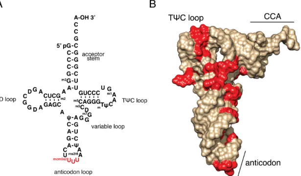
Figure 1.3: All tRNAs maintain a common structure, but are differentiated by multiple post-transcriptional modifications.
(A) Primary and secondary structure of mammalian tRNA lys. This sequence is unique to this tRNA, but the secondary structure is universal. Common elements are labeled, and the anticodon nucleotides are colored red. Note that the anticodon contains the mcm5s2U nucleotide, which is studied in Chapter 2.
(B) Crystal structure of the same tRNA in (A), showing the distinctive bent structure. Modified nucleotides are colored red. Sequence and structure data are from PDB ID 1FIR (Bénas et al., 2000). Structure visualization generated with UCSF Chimera (Pettersen et al., 2004).
Previous attempts to determine the in vivo effect of tRNA modifications on translation elongation rate have used artificial reporters with high mRNA expression or long stretches of cognate codons (Begley et al., 2007; Krüger et al., 1998), as it was not previously possible to measure elongation rates on individual native mRNAs. In Chapter 2, I use modern ribosome profiling methods to investigate the role in translation
budding yeast in numerous stress conditions. I find that loss of this modification causes slowed elongation at specific codons in native yeast mRNAs, as well as activation of stress response signaling.
References
Agris, P.F., Vendeix, F.A.P., and Graham, W.D. (2007). tRNA's wobble decoding of the genome: 40 years of modification. Journal of Molecular Biology 366, 1–13.
Alexandrov, A., Chernyakov, I., Gu, W., Hiley, S.L., Hughes, T.R., Grayhack, E.J., and Phizicky, E.M. (2006). Rapid tRNA decay can result from lack of nonessential
modifications. Molecular Cell 21, 87–96.
Arava, Y., Wang, Y., Storey, J.D., Liu, C.L., Brown, P.O., and Herschlag, D. (2003). Genome-wide analysis of mRNA translation profiles in Saccharomyces cerevisiae. Proc. Natl. Acad. Sci. U.S.A. 100, 3889–3894.
Ashraf, S.S., Sochacka, E., Cain, R., Guenther, R., Malkiewicz, A., and Agris, P.F. (1999). Single atom modification (O-->S) of tRNA confers ribosome binding. RNA 5, 188–194.
Begley, U., Dyavaiah, M., Patil, A., Rooney, J.P., DiRenzo, D., Young, C.M., Conklin, D.S., Zitomer, R.S., and Begley, T.J. (2007). Trm9-catalyzed tRNA modifications link translation to the DNA damage response. Molecular Cell 28, 860–870.
Bénas, P., Bec, G., Keith, G., Marquet, R., Ehresmann, C., Ehresmann, B., and Dumas, P. (2000). The crystal structure of HIV reverse-transcription primer tRNA(Lys,3) shows a canonical anticodon loop. RNA 6, 1347–1355.
Carlberg, U., Nilsson, A., and Nygård, O. (1990). Functional properties of phosphorylated elongation factor 2. Eur. J. Biochem. 191, 639–645.
Chan, C.T.Y., Dyavaiah, M., DeMott, M.S., Taghizadeh, K., Dedon, P.C., and Begley, T.J. (2010). A quantitative systems approach reveals dynamic control of tRNA
modifications during cellular stress. PLoS Genet 6, e1001247.
Chu, D., Kazana, E., Bellanger, N., Singh, T., Tuite, M.F., and Haar, von der, T. (2013). Translation elongation can control translation initiation on eukaryotic mRNAs. Embo J. 33, 21–34.
Crick, F.H. (1966). Codon--anticodon pairing: the wobble hypothesis. Journal of Molecular Biology 19, 548–555.
Curran, J.F., and Yarus, M. (1989). Rates of aminoacyl-tRNA selection at 29 sense codons in vivo. Journal of Molecular Biology 209, 65–77.
Dever, T.E., and Green, R. (2012). The elongation, termination, and recycling phases of translation in eukaryotes. Cold Spring Harbor Perspectives in Biology 4, a013706.
Doma, M.K., and Parker, R. (2006). Endonucleolytic cleavage of eukaryotic mRNAs with stalls in translation elongation. Nature 440, 561–564.
Dorovkov, M.V., Pavur, K.S., Petrov, A.N., and Ryazanov, A.G. (2002). Regulation of Elongation Factor-2 Kinase by pH †. Biochemistry 41, 13444–13450.
Gardin, J., Yeasmin, R., Yurovsky, A., Cai, Y., Skiena, S., and Futcher, B. (2014). Measurement of average decoding rates of the 61 sense codons in vivo. Elife 3. Gerashchenko, M.V., and Gladyshev, V.N. (2014). Translation inhibitors cause abnormalities in ribosome profiling experiments. Nucleic Acids Research.
Gingold, H., Tehler, D., Christoffersen, N.R., Nielsen, M.M., Asmar, F., Kooistra, S.M., Christophersen, N.S., Christensen, L.L., Borre, M., Sørensen, K.D., et al. (2014). A Dual Programfor Translation Regulationin Cellular Proliferation and Differentiation. Cell 158, 1281–1292.
Gustafsson, C., Govindarajan, S., and Minshull, J. (2004). Codon bias and heterologous protein expression. Trends Biotechnol. 22, 346–353.
Gutierrez, E., Shin, B.-S., Woolstenhulme, C.J., Kim, J.-R., Saini, P., Buskirk, A.R., and Dever, T.E. (2013). eIF5A promotes translation of polyproline motifs. Molecular Cell 51, 35–45.
Guydosh, N.R., and Green, R. (2014). Dom34 Rescues Ribosomes in 3' Untranslated Regions. Cell 156, 950–962.
Ikemura, T. (1981a). Correlation between the abundance of Escherichia coli transfer RNAs and the occurrence of the respective codons in its protein genes: a proposal for a synonymous codon choice that is optimal for the E. coli translational system. Journal of Molecular Biology 151, 389–409.
Ikemura, T. (1981b). Correlation between the abundance of Escherichia coli transfer RNAs and the occurrence of the respective codons in its protein genes. Journal of Molecular Biology 146, 1–21.
Ingolia, N.T., Lareau, L.F., and Weissman, J.S. (2011). Ribosome profiling of mouse embryonic stem cells reveals the complexity and dynamics of mammalian proteomes. Cell 147, 789–802.
Nishimura, Y., Chuang, J.H., and Ackerman, S.L. (2014). Ribosome stalling induced by mutation of a CNS-specific tRNA causes neurodegeneration. Science 345, 455–459. Johansson, M., Ieong, K.-W., Trobro, S., Strazewski, P., Åqvist, J., Pavlov, M.Y., and Ehrenberg, M. (2011). pH-sensitivity of the ribosomal peptidyl transfer reaction
dependent on the identity of the A-site aminoacyl-tRNA. Proceedings of the National Academy of Sciences 108, 79–84.
Johansson, M.J.O., Esberg, A., Huang, B., Björk, G.R., and Byström, A.S. (2008). Eukaryotic wobble uridine modifications promote a functionally redundant decoding system. Mol Cell Biol 28, 3301–3312.
Kapp, L.D., and Lorsch, J.R. (2004). The molecular mechanics of eukaryotic translation. Annu. Rev. Biochem. 73, 657–704.
Krüger, M.K., Pedersen, S., Hagervall, T.G., and Sørensen, M.A. (1998). The
modification of the wobble base of tRNAGlu modulates the translation rate of glutamic acid codons in vivo. Journal of Molecular Biology 284, 621–631.
Kudla, G., Murray, A.W., Tollervey, D., and Plotkin, J.B. (2009). Coding-Sequence Determinants of Gene Expression in Escherichia coli. Science 324, 255–258.
Ladner, J.E., Jack, A., Robertus, J.D., Brown, R.S., Rhodes, D., Clark, B.F., and Klug, A. (1975). Structure of yeast phenylalanine transfer RNA at 2.5 A resolution. Proc. Natl. Acad. Sci. U.S.A. 72, 4414–4418.
Ledoux, S., and Uhlenbeck, O.C. (2008). Different aa-tRNAs Are Selected Uniformly on the Ribosome. Molecular Cell 31, 114–123.
Li, G.-W., Oh, E., and Weissman, J.S. (2012). The anti-Shine-Dalgarno sequence drives translational pausing and codon choice in bacteria. Nature 484, 538–541.
Liu, B., Han, Y., and Qian, S.-B. (2013). Cotranslational response to proteotoxic stress by elongation pausing of ribosomes. Molecular Cell 49, 453–463.
Lodish, H.F. (1971). Alpha and beta globin messenger ribonucleic acid. Different amounts and rates of initiation of translation. J. Biol. Chem. 246, 7131–7138.
Madore, E., Florentz, C., Giegé, R., Sekine, S., Yokoyama, S., and Lapointe, J. (1999). Effect of modified nucleotides on Escherichia coli tRNAGlu structure and on its
aminoacylation by glutamyl-tRNA synthetase. Predominant and distinct roles of the mnm5 and s2 modifications of U34. Eur. J. Biochem. 266, 1128–1135.
Pape, T., Wintermeyer, W., and Rodnina, M.V. (1998). Complete kinetic mechanism of elongation factor Tu-dependent binding of aminoacyl-tRNA to the A site of the E. coli ribosome. EMBO J. 17, 7490–7497.
Patil, A., Dyavaiah, M., Joseph, F., Rooney, J.P., Chan, C.T.Y., Dedon, P.C., and Begley, T.J. (2012). Increased tRNA modification and gene-specific codon usage regulate cell cycle progression during the DNA damage response. Cell Cycle 11, 3656– 3665.
Pedersen, S. (1984). Escherichia coli ribosomes translate in vivo with variable rate. Embo J. 3, 2895–2898.
Pettersen, E.F., Goddard, T.D., Huang, C.C., Couch, G.S., Greenblatt, D.M., Meng, E.C., and Ferrin, T.E. (2004). UCSF Chimera--a visualization system for exploratory research and analysis. J Comput Chem 25, 1605–1612.
Phizicky, E.M., and Hopper, A.K. (2010). tRNA biology charges to the front. Genes & Development 24, 1832–1860.
Plotkin, J.B., and Kudla, G. (2010). Synonymous but not the same: the causes and consequences of codon bias. Nature Publishing Group 12, 32–42.
Qian, W., Yang, J.-R., Pearson, N.M., Maclean, C., and Zhang, J. (2012). Balanced Codon Usage Optimizes Eukaryotic Translational Efficiency. PLoS Genet 8, e1002603. Quigley, G.J., and Rich, A. (1976). Structural domains of transfer RNA molecules. Science 194, 796–806.
Rezgui, V.A.N., Tyagi, K., Ranjan, N., Konevega, A.L., Mittelstaet, J., Rodnina, M.V., Peter, M., and Pedrioli, P.G.A. (2013). tRNA tKUUU, tQUUG, and tEUUC wobble
position modifications fine-tune protein translation by promoting ribosome A-site binding. Proceedings of the National Academy of Sciences 110, 12289–12294.
Rould, M.A., Perona, J.J., Söll, D., and Steitz, T.A. (1989). Structure of E. coli
glutaminyl-tRNA synthetase complexed with tRNA(Gln) and ATP at 2.8 A resolution. Science 246, 1135–1142.
Ruff, M., Krishnaswamy, S., Boeglin, M., Poterszman, A., Mitschler, A., Podjarny, A., Rees, B., Thierry, J.C., and Moras, D. (1991). Class II aminoacyl transfer RNA synthetases: crystal structure of yeast aspartyl-tRNA synthetase complexed with tRNA(Asp). Science 252, 1682–1689.
Ryazanov, A.G., Shestakova, E.A., and Natapov, P.G. (1988). Phosphorylation of elongation factor 2 by EF-2 kinase affects rate of translation. Nature 334, 170–173. Sen, G.C., and Ghosh, H.P. (1976). Role of modified nucleosides in tRNA: effect of modification of the 2-thiouridine derivative located at the 5'-end of the anticodon of yeast transfer RNA Lys2. Nucleic Acids Research 3, 523–535.
Widespread regulation of translation by elongation pausing in heat shock. Molecular Cell 49, 439–452.
Shoemaker, C.J., and Green, R. (2012). Translation drives mRNA quality control. Nature Structural & Molecular Biology 19, 594–601.
Söll, D., and RajBhandary, U. (1995). tRNA (Amer Society for Microbiology).
Tuller, T., Carmi, A., Vestsigian, K., Navon, S., Dorfan, Y., Zaborske, J., Pan, T., Dahan, O., Furman, I., and Pilpel, Y. (2010). An evolutionarily conserved mechanism for
controlling the efficiency of protein translation. Cell 141, 344–354.
Walden, W.E., and Thach, R.E. (1986). Translational control of gene expression in a normal fibroblast. Characterization of a subclass of mRNAs with unusual kinetic properties. Biochemistry 25, 2033–2041.
Walden, W.E., Godefroy-Colburn, T., and Thach, R.E. (1981). The role of mRNA competition in regulating translation. I. Demonstration of competition in vivo. J. Biol. Chem. 256, 11739–11746.
Woolstenhulme, C.J., Parajuli, S., Healey, D.W., Valverde, D.P., Petersen, E.N., Starosta, A.L., Guydosh, N.R., Johnson, W.E., Wilson, D.N., and Buskirk, A.R. (2013). Nascent peptides that block protein synthesis in bacteria. Proceedings of the National Academy of Sciences 110, E878–E887.
Yarian, C., Townsend, H., Czestkowski, W., Sochacka, E., Malkiewicz, A.J., Guenther, R., Miskiewicz, A., and Agris, P.F. (2002). Accurate translation of the genetic code depends on tRNA modified nucleosides. J. Biol. Chem. 277, 16391–16395.
Chapter 2: Loss of a Conserved tRNA Anticodon
Modification Perturbs Cellular Signaling
*Abstract
Transfer RNA (tRNA) modifications enhance the efficiency, specificity and fidelity of translation in all organisms. The anticodon modification mcm5s2U34 is required for normal growth and stress resistance in yeast; mutants lacking this modification have numerous phenotypes. Mutations in the homologous human genes are linked to neurological disease. The yeast phenotypes can be ameliorated by overexpression of specific tRNAs, suggesting that the modifications are necessary for efficient translation of specific codons. We determined the in vivo ribosome distributions at single codon resolution in yeast strains lacking mcm5s2U. We found accumulations at AAA, CAA, and GAA codons, suggesting that translation is slow when these codons are in the
ribosomal A site, but these changes appeared too small to affect protein output. Instead, we observed activation of the GCN4-mediated stress response by a non-canonical pathway. Thus, loss of mcm5s2U causes global effects on gene expression due to perturbation of cellular signaling.
*
This research was originally published in PLoS Genetics, and has been edited for presentation here. Zinshteyn B, Gilbert WV (2013) Loss of a Conserved tRNA Anticodon Modification Perturbs Cellular Signaling. PLoS Genet 9(8): e1003675.
Introduction
Transfer RNAs (tRNAs) from all domains of life contain numerous
post-transcriptional modifications, many of which are highly conserved. These modifications enhance the efficiency, specificity and fidelity of translation(Agris et al., 2007;
Johansson and Byström, 2005; Phizicky and Hopper, 2010). In the budding yeast
Saccharomyces cerevisiae, three tRNAs are modified by
addition of 5-methoxycarbonylmethyl (mcm5) and 2-thio (s2) groups to uridine at the 5' nucleotide of the tRNA anticodon (U34), resulting in an mcm5s2U nucleotide. The mcm5s2U modification (MSUM) and many of the responsible modifying enzymes are conserved across eukaryotes, having been identified in fungi (Huang et al., 2005; Leidel et al., 2009), plants(Mehlgarten et al., 2010), worms (Chen et al., 2009) and mammals (Chan et al., 1982). Despite widespread conservation, and extensive biochemical characterization, the physiological role of MSUM is unknown.
Genes required for MSUM are unusual among tRNA modification genes in the number and severity of their mutant phenotypes. Most yeast strains lacking tRNA modifications are viable and show no growth impairment (Johansson and Byström, 2005; Phizicky and Hopper, 2010), but S. cerevisiae and C. elegans double mutants lacking both mcm5 and s2 are not viable (Björk et al., 2007; Chen et al., 2009). In yeast, single mutants lacking either mcm5 or s2 have numerous phenotypes including
temperature sensitivity, various chemical stress sensitivities, exocytosis defects, and transcriptional defects (Esberg et al., 2006; Krogan and Greenblatt, 2001). In C. elegans, mutants of the Elongator complex (comprised of elp1 through elp6), which is
(tRNAUUULys , tRNA UUG
G ln, tRNA UUC
required to produce the mcm5 modification, display neurological defects (Chen et al., 2009). In humans, mutations in IBKAP, the elp1 homolog, cause familial dysautonomia (FD)(Slaugenhaupt et al., 2001), and mutations in elp4 are associated with Rolandic epilepsy (Strug et al., 2009).
The molecular connection between these cellular/organismal phenotypes and the lack of specific tRNA anticodon modifications is currently unknown. Loss of either mcm5 or s2 impairs reading of both Watson-Crick (VAA) and wobble (VAG) cognate codons by the modified tRNAs (Johansson et al., 2008; Krüger et al., 1998), and chemical removal or modification of the s2 moiety leads to a reduction in the rate of tRNA charging in vitro (Sen and Ghosh, 1976; Seno et al., 1974). The MSUM phenotypes were originally attributed to a proposed role of the Elongator complex in transcriptional elongation (Otero et al., 1999) before its function in tRNA modification was discovered (Huang et al., 2005). However, the phenotypes of yeast MSUM mutants, including the lethality in mutants lacking both mcm5 and s2, can be suppressed by overexpression of unmodified versions of two tRNAs that normally contain mcm5s2U – and (Esberg et al., 2006). These observations indicate that at least a subset of the yeast cellular phenotypes are tied to tRNA function. It has been argued that loss of MSUM leads to codon-specific translation defects leading to insufficient protein production, either from many genes, or from a few genes required to carry out particular cellular processes or stress responses, but this hypothesis has not been directly tested.
In this study, we examined codon level ribosome distributions genome-wide using ribosome footprint profiling (Ribo-seq). We found that loss of mcm5 or s2 leads to
tRNAUUGG ln tRNA UUU
slow translation elongation specifically at codons that Watson-Crick pair with MSUM tRNAs, but the magnitude of these changes appeared insufficient to affect protein output. Surprisingly, all of the MSUM strains showed gene expression signatures consistent with activation of the Gcn4p-mediated stress response pathway. We
demonstrate that disruption of this pathway suppresses the MSUM mutant phenotypes independently of tRNA concentration.
Results
Ribosome Footprint Profiling Reveals Features of Translation for Specific
Codons
We set out to determine whether MSUM mutants display codon-specific
translation defects. Translational activity genome-wide was determined using Ribo-seq, which consists of isolating and sequencing ribosome-protected mRNA fragments from RNase-treated whole-cell lysates (Ingolia et al., 2009). This method reveals ribosome positions at single nucleotide resolution, and thus has the potential to identify
translational defects affecting single codons (Ingolia et al., 2009; Stadler and Fire, 2011). Wild type (WT) yeast, as well as strains lacking the s2 moiety (ncs2Δ, ncs6Δ, and uba4Δ), or mcm5 (elp3Δ) (Figure 2.1A), were profiled by Ribo-seq, as well as RNA-seq. To assess the impact of these modifications on translation, the ribosome dwell time at specific codons was determined as follows. The positions of the A, P and E site codons within ribosome footprints of various lengths (25-31 nt) were determined by examining the 5’ ends of footprints mapping to start codons, where initiating ribosomes
are expected to contain start codons in their P sites (Figure 2.1B)(Kapp and Lorsch, 2004). Next, to determine the genome-wide average ribosome dwell time for a given codon (Figure 2.1C, left), all instances of that codon in the genome were aligned, and 5’ ends of reads mapping to the surrounding positions (Figure 2.1C) were summed (see Materials and Methods). The resulting metacodon plots show the relative number of ribosome footprints, and thus the relative amount of time the ribosome spends at each position, as the codon moves through the A, P and E sites. Codon identity is not expected to affect translation from the outer sites (±1, ±2), so the entire plot was
normalized to the height of these peaks. The height of each peak is the bulk occupancy for that codon in that ribosomal site, similar to a previously described metric (Stadler and Fire, 2011). The metacodon distributions for ATG and stop codons indicated that the reads were properly assigned to the ribosomal sites (Figure 2.1C, right). We observed distinct and reproducible patterns of ribosome density for different codons in WT yeast (Figure 2.1C, 2.2A,B), consistent with the single-nucleotide resolution of this technique.
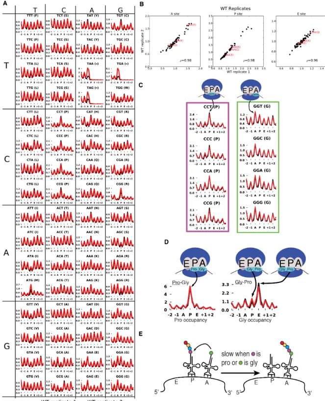
The metacodon plots of WT yeast provided insights into the determinants of translation rate for specific codons. Notably, all four proline codons spent over 2-fold more time in the P site than the average codon, while glycine codons spent ~40-50% more time in the A site (Figure 2.2C). This effect was additive for Pro-Gly pairs in the P and A sites, but not if the codon order was reversed (Figure 2.2D), indicating that the effects of Pro and Gly were specific to the P and A sites, respectively. This proline effect is reminiscent of the proline/glycine pausing recently discovered in bacteria lacking
elongation factor P (Doerfel et al., 2013; Ude et al., 2013; Woolstenhulme et al., 2013). The observed effects were consistent with in vitro data which showed that peptidyl transfer can be rate limiting for A-site glycine and proline codon translation at
physiological pH (Johansson et al., 2011), and that proline induces particularly slow peptide bond formation when it is at the carboxyl terminus of the growing peptide chain (Pavlov et al., 2009) (Figure 2.2E). These results suggest that peptidyl transfer is rate limiting for certain Pro and Gly codons in yeast cells as well.
Experiments in recombinant systems have led to the strong expectation that translation times for codons should be inversely proportional to the concentrations of their cognate tRNAs (Pedersen, 1984; Tuller et al., 2010). To investigate potential sources of the distinctive metacodon distributions we observed, we performed unsupervised hierarchical clustering on them (Figure 2.3A). This analysis clustered many codons together based on their encoded amino acid or the first two nucleotides of the codon. Notably, codons did not cluster by tRNA adaptation index (tAI), a proxy for cognate tRNA abundance (Tuller et al., 2010). More directly, the bulk occupancies did not show a negative correlation with tAI in the A site (Figure 2.3B). There was also no correlation of codon occupancy with tRNA abundance measurements, genomic copy number, or a more recent codon usage metric which accounts for tRNA competition (Pechmann and Frydman, 2012) (data not shown). These results demonstrate that translation rates for particular yeast codons are not determined by the cellular concentrations of their cognate tRNAs, consistent with findings from Ribo-seq
experiments in mice and bacteria (Ingolia et al., 2011; Li et al., 2012) and from protein synthesis reporters (containing codon repeats) in yeast (Letzring et al., 2010).
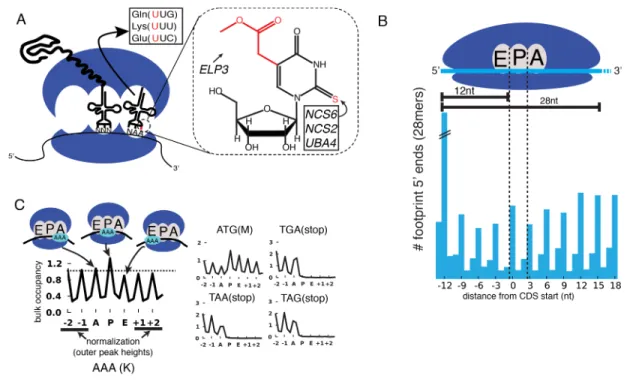
Figure 2.1: Method for bulk analysis of codon occupancy in MSUM strains. (A) (left) mcm5s2U is found at the 5’ nucleotide of the anticodon in three yeast tRNAs. (right) The structure of mcm5s2U, and the subset of modification genes whose mutants were profiled in this study are indicated.
(B) (top) Anatomy of a ribosome footprint, with P-site offset for 28mer reads indicated. (bottom) Metaplot of WT ribosome footprint reads summed across all start codons. The peak of upstream reads corresponds to ribosomes with start codons in their P site. The location of this peak is used to determine the location of A, P and E sites for each read length.
(C) (left) Explanation of metacodon plots. Similar to panel B, all in-frame instances of a given codon in the genome are aligned, and the reads mapping around those positions are summed. The resulting plot is then offset by the P-site distance, and normalized to the average peak height of the outer sites (±1, ±2). The peak heights for each site are the bulk codon occupancies, a proxy for the amount of time the ribosome spends with a given codon in each site, compared to its neighbors. (right) ATG codons and stop codons display the expected distributions with this metric. All plots are from WT yeast.
Figure 2.2: Metacodon plots provide information on translation kinetics at the codon level.
(A) Full set of metacodon plots, with superimposed WT replicates.
(B) Reproducibility of bulk codon occupancy metric. Spearman correlations are indicated.
(D) Metacodon plots for Pro-Gly and Gly-Pro pairs.
(E) Model for Pro and Gly metacodon plots. Peptidyl transfer is slow when Pro is in the P site, or Gly is in the A site, possibly making peptidyl-transfer rate-limiting for
translocation, especially for Pro-Gly pairs.
Figure 2.3: Codon occupancy is not determined by codon adaptation.
(A)Unsupervised hierarchical clustering of WT metacodon plots. Codons for the same amino acid that cluster together have been colored. The tRNA adaptation index (tAI) for each codon is indicated in red. The tAI is a proxy for cognate tRNA abundance.
(B) Correlations between WT codon occupancy and tAI for codons in each ribosome site.
Loss of MSUM Genes Reduces Translation Rate at AAA, CAA, GAA
Codons
Having established the ability to detect differences in the translation of different codons, we next examined changes in codon-specific translation in the MSUM strains. Bulk occupancy for each codon in each ribosomal site (the height of the peaks in the metacodon plots) was determined for each mutant. All of the strains lacking the s2 modification showed increases in ribosome density corresponding to CAA and AAA in the A site, while the elp3Δ strain showed an increase in the CAA and GAA codons (Figure 2.4A). The magnitude of the changes was largest when the affected codon was found in the ribosomal A-site. The magnitude and direction of change for the GAA codon was variable between mutants lacking the same modification, and even between biological replicates (Figure 2.4A), indicative of some underlying biological or technical noise in this measurement. Nonetheless, in all but one replicate, the largest increases in each mutant were for codons decoded by Watson-Crick pairing with MSUM tRNAs.
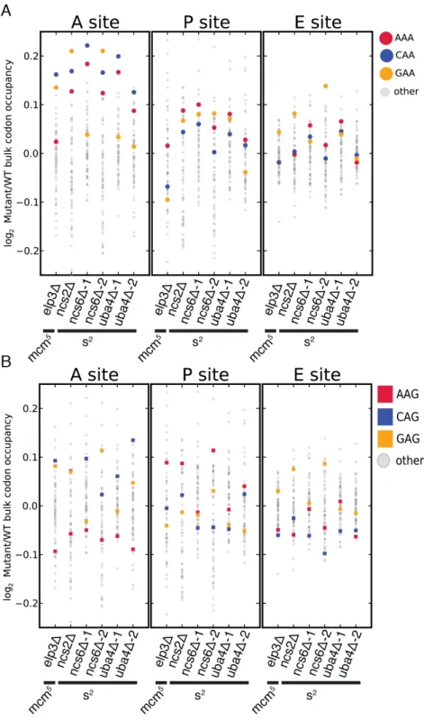
Figure 2.4: Genetic ablation of mcm5 or s2 leads to ribosome accumulation at specific codons.
(A and B) Changes in bulk codon occupancy in MSUM mutants. Both plots are the same, with different codons highlighted. Independent biological replicates were done for ncs6Δ and uba4Δ. All mutants are compared to a WT sample prepared and processed simultaneously.
mcm
5s
2U is Not Required for Wobble Decoding of AAG, CAG, and GAG
Codons In Vivo
MSUM is necessary for wobble decoding of G-ending codons in strains that lack other cognate tRNAs(Johansson et al., 2008), but it is not clear whether the modified tRNAs contribute to decoding in the WT state where these other tRNAs are present. In our datasets AAG, CAG, and GAG codons showed smaller increases in bulk occupancy (and some net decreases) compared to their A-ending counterparts, suggesting that MSUM is mainly required for translation of VAA codons (Figure 2.4B). In order to assess the statistical significance of these changes, a metric for ribosome dwell time at
individual codons was developed (Figure 2.5A). This metric normalizes the read counts at a particular codon by the mean read density of the open reading frame that contains it. The genome-wide distributions for all instances of each codon were compared between mutant and WT strains using the K-S test (Figure 2.5B, C). Due to the noise inherent in read sampling, many codons showed statistically significant changes. However, the VAA codons had p values many orders of magnitude smaller than all other codons, particularly in the ncs6Δ and uba4Δ datasets, which were from pooled biological replicates (Figure 2.5C). The pooled datasets provided data for
approximately twice as many codons and may have averaged out biological and technical noise. Consistent with our analysis of bulk codon occupancy, the effect of MSUM loss was strongest in the A site for all 3 VAA codons. We did not see a
corresponding statistical significance for the VAG codons (Figure 2.5C), indicating that mcm5s2U does not significantly contribute to the decoding of these codons in vivo. This
result does not contradict previous evidence that the modifications are required for translation of VAG codons by wobble pairing (Johansson et al., 2008), but indicates that tRNAsUUB contribute minimally to the translation of VAG codons in vivo, where tRNAsCUB
with Watson-Crick complementarity are available.
Figure 2.5: A single-codon occupancy metric shows that ribosome footprint accumulations at AAA, CAA, and GAA are statistically significant
(A) Description of the single codon occupancy metric. The occupancy for a given codon in a given site is the number of in-frame reads for that codon in that site, compared to the average in-frame read density for the parent gene.
(B) Cumulative distributions of single-codon occupancy for select codons in ncs6Δ and uba4Δ.
(C) Heatmap of K-S test p-values for all sense codons in all mutants. For ncs6Δ and uba4Δ, mutant and WT replicates were pooled to improve the accuracy of the metric.
The Elongation Defects in MSUM Strains Appear Insufficient to Affect
Protein Levels
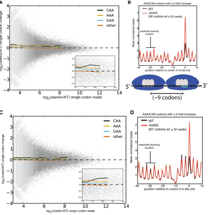
Despite the statistical significance of the increased ribosome dwell times at VAA codons in MSUM mutants, the magnitude of the changes does not seem to be large enough to generally affect protein output. Initiation, not elongation, is the rate-limiting step of eukaryotic translation in most circumstances (Lodish and Jacobsen, 1972; Walden et al., 1981), and the mean ribosome density is only 1 per 164 nts (Arava et al., 2003). Given this sparse spacing of ribosomes on yeast mRNAs, transcripts with mean ribosome density would require an elongation delay greater than the average translation time of 50 codons in order for an MSUM mutation to make elongation rate limiting. The most densely populated messages would require a 20-fold elongation delay. The average bulk increase observed for VAA codons was less than 17% (Figure 2.4D), and the largest confidently assigned (≥32 reads) single-codon change was less than 5-fold (Figure 2.6A,C). In the event of an elongation delay long enough to affect protein output, ribosome queuing should occur behind AAA and CAA codons with increased
occupancy. However, no queuing was observed (Figure 2.6B, D). Codons with more read coverage display smaller changes than codons with low read coverage, indicating that the range of this metric is not being limited by sequencing depth (Figure 2.6A, 2.6C). We also did not observe increased ribosome density at stretches of 2 or more VAA codons (data not shown). These results were consistent with the polysome gradient profiles of the MSUM strains, which were indistinguishable from WT (data not shown), indicating that translation elongation in bulk was unaffected.
Figure 2.6: Single codon occupancy changes may be insufficient to affect protein output.
(A) Fold changes for all single codons in uba4Δ are plotted against their read density in grey. Colored lines are the mean fold changes for the specified codons over read-coverage bins of width 0.2 (log2 scaled). “Other” is a pool of all non-VAA codons.
(B) Metaplot of ribosome footprint density around all AAA and CAA codons with ≥2-fold change in uba4Δ, and ≥32 reads in both datasets. Reads at each position were
normalized by the total number of reads for the parent gene, and averaged across all host genes that overlap that position. The plot is offset such that 0 corresponds to having the codon in the A site. The expected location of a ribosome queuing event is
indicated, and a diagram of such an event is shown below. The dip in ribosome footprint density at -10 is a computational artifact, due to an inability to determine read lengths of poly-adenylated fragments when they end in one or more adenosines.
(C, D)Same as in (A) and (B), but for ncs6Δ.
The GCN4-Mediated Stress Response Is Activated in MSUM Strains
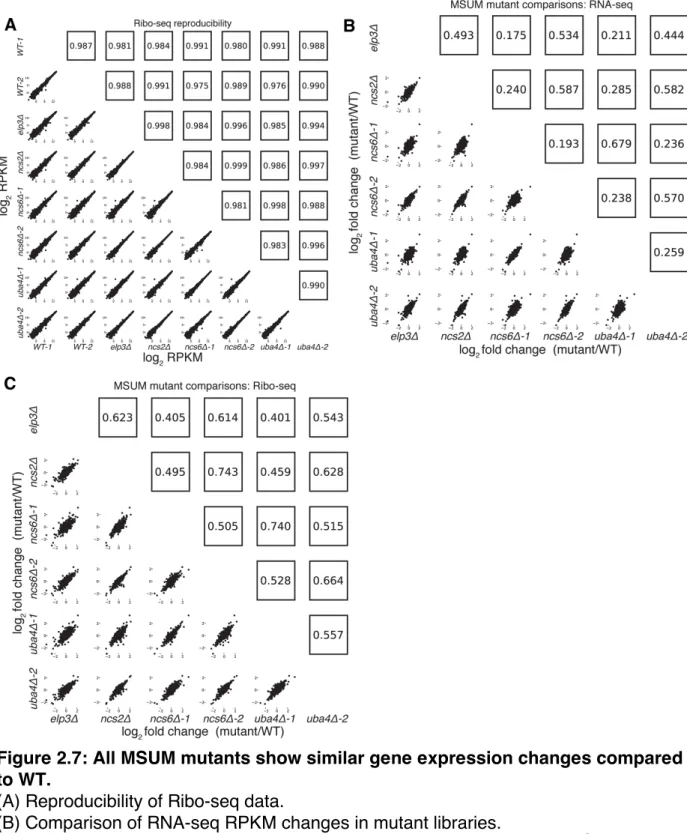
In search of an alternative explanation for MSUM mutant phenotypes, we examined global ribosome footprint densities and transcript levels for perturbations in the MSUM mutant strains. Consistent with previous reports (Brar et al., 2012; Ingolia et al., 2009), gene expression values from Ribo-seq were highly reproducible (Figure 2.7A). Furthermore, all of the mutant strains showed similar RNA-seq and Ribo-seq changes when compared to WT strains (Figure 2.7B,C), indicating that these gene expression changes are likely to be downstream of a common defect. Replicate data for ncs6Δ and uba4Δ enabled us to assess the significance of particular changes using
counting statistics (Robinson et al., 2009). This analysis identified a set of genes with significant changes in ribosome footprint densities, which were largely shared between ncs6Δ and uba4Δ (Figure 2.8A-C, 2.7B,C). The changes in ribosome footprint density
were correlated with changes in transcript levels (pearson r=0.59 for ncs6Δ, 0.64 for uba4Δ), indicating that these gene expression changes were largely due to changes in
the mRNA pool (Figure 2.8A,B). Intriguingly, a significant fraction (24/68) of the affected genes are known targets of the GCN4 transcription factor (Natarajan et al., 2001)
(Figure 2.8 A-C). To investigate the specificity of the observed induction of GCN4 targets in MSUM mutants, we examined the behavior of GCN4 targets in 1,924 yeast microarray studies using data from the SPELL curated yeast microarray compendium.
This compendium includes experiments sampling a broad range of environmental and genetic perturbations (Hibbs et al., 2007). We determined the significance of overlap between GCN4 targets and the set of upregulated (≥2-fold) genes in each of these 1,924 datasets. Notably, the overlap between GCN4 targets and induced genes in MSUM strains was more statistically significant than the overlap between GCN4 targets and induced genes in 82% of the SPELL datasets. The datasets with a higher degree of overlap consisted mostly (at least 276/343) of gene deletions and stress conditions in which GCN4 is known to play a role (e.g. heat, nutritional perturbation, osmotic stress and DNA damage) (Available online as Table S4 with the version of this work at
http://www.plosgenetics.org/). Furthermore, GCN4 targets as a whole showed increased ribosome footprint density in MSUM strains (Figure 2.8D). We further confirmed this enrichment for functional GCN4 targets by examining the predicted Gcn4p binding affinity of the promoters for the affected genes (Nutiu et al., 2011). The promoter
regions of the upregulated genes were enriched for Gcn4p binding motifs (Figure 2.8E). Using the same sets of upregulated genes from the SPELL compendium as above, less than 6% of these upregulated gene sets had a mean predicted Gcn4p occupancy
greater than the genes upregulated in the MSUM strains (Table S4, available online). Thus, GCN4 target genes are transcriptionally upregulated in all MSUM strains.
To provide context for these gene expression changes, the same analyses were performed on Ribo-seq data from yeast subjected to amino acid (AA) starvation, a well-characterized GCN4-inducing condition (Ingolia et al., 2009). In this dataset, 20 minutes of amino acid starvation leads to a 4-fold increase in ribosome footprints on the GCN4
ORF. A larger number of genes displayed changes in AA starvation compared to MSUM ablation, and GCN4 targets as a group had larger fold changes (median 2.0-fold
induction vs. 1.2 and 1.1-fold for uba4Δ and ncs6Δ respectively). (Figure 2.9A, 2.9C). However, a smaller fraction of the significantly changing genes are GCN4 targets (13% in AA-starved cells, vs 29% and 30% for uba4Δ and ncs6Δ respectively) (Figure 2.8C, 2.9B). Furthermore, the starvation-induced genes had a smaller enrichment for
predicted Gcn4p occupancy in their promoters compared to genes upregulated in the MSUM strains (Figure 2.8E). The limited induction of high-affinity Gcn4p targets in MSUM mutants is consistent with a weak but specific activation of the GCN4 pathway.
Figure 2.7: All MSUM mutants show similar gene expression changes compared to WT.
(A) Reproducibility of Ribo-seq data.
(B) Comparison of RNA-seq RPKM changes in mutant libraries.
(C) Comparison of Ribo-seq RPKM changes in mutant libraries. Pearson r2 are presented.
Figure 2.8: MSUM strains show the gene-expression signatures of GCN4 activation
(A) Comparison of RNA-seq and Ribo-seq RPKM changes in uba4Δ. GCN4 targets and statistically significant Ribo-seq changes are indicated. Values are the means of 2 biological replicates. Pearson R values shown.
(C) Venn diagram of overlap between GCN4 functional targets (blue) and significant Ribo-seq RPKM changes in uba4Δ (pink) and ncs6Δ (green). The significance of the overlap was computed using the hypergeometric distribution.
(D) Cumulative distribution plots of fold Ribo-seq changes for GCN4 targets (solid lines) compared to all other genes (dashed lines) in uba4Δ (top) and ncs6Δ. P values are from a KS test of GCN4 targets against the rest of the genome.
(E) Mean±SEM of predicted Gcn4p occupancy for groups of genes from panel B and figure S5, as determined by high-throughput in vitro binding assays (Nutiu et al., 2011). Bars are colored to match groups in panel B. P values are from t-tests comparing the indicated gene set against all genes in the genome.
Figure 2.9: Amino acid starvation causes a stronger but less specific activation of GCN4 targets than MSUM ablation.
(A) Comparison of RNA-seq and Ribo-seq RPKM changes in amino acid (AA) starved yeast (data from (Ingolia et al., 2009)). GCN4 targets and statistically significant Ribo-seq changes are indicated. Values are the means of 2 biological replicates.
(B) Venn diagram of overlap between GCN4 functional targets (blue) and significant Ribo-seq changes upon AA starvation. The significance of the overlap was computed using the hypergeometric distribution.
(C) Cumulative distribution plots of fold Ribo-seq changes for GCN4 targets (solid line) compared to all other genes (dashed line). P values are from a KS test of GCN4 targets against the rest of the genome.
Induction of GCN4 Occurs Independently of GCN2
We next sought to identify the mechanism of GCN4 pathway induction in MSUM strains. GCN4 is known to be translationally regulated in response to a variety of insults, most notably by amino acid starvation (Hinnebusch, 2005). Translational repression of GCN4 is mediated by four upstream open reading frames (uORFs), which prevent
ribosomes from initiating on the protein-coding ORF. Conditions that decrease the efficiency of re-initiation allow some ribosomes to scan through the uORFs and initiate at the GCN4 ORF. All four MSUM mutants showed ~2-fold translational upregulation of GCN4, as evidenced by increased ribosome footprint density in the ORF with no
increase in mRNA levels (Figure 2.10A).
A reporter construct containing the transcript leader of GCN4 fused to lacZ verified that the uORF-containing leader was sufficient to recapitulate the translational induction observed in MSUM strains (Figure 2.10B). The magnitude of this induction (2-4 fold) is consistent with a weak activation of the GCN pathway, as a 3hr shift to SC-Ura, and a constitutive GCN2 allele (Ramirez et al., 1992) induced GCN4-lacz 7-fold and 50-fold, respectively. The best-characterized pathway of inducing GCN4 involves the activation of the Gcn2p kinase by uncharged tRNA, leading to phosphorylation of eukaryotic initiation factor 2α (eIF2α) and reduced efficiency of initiation and re-initiation. We therefore tested the effect of gcn2Δ on GCN4 induction by MSUM mutants.
Surprisingly, GCN4-lacZ was still induced in MSUM strains lacking GCN2 (Figure
2.10C). In addition, basal eIF2α phosphorylation levels were not increased in the MSUM strains, consistent with a GCN2-independent mechanism (Figure 2.10D). Thus, GCN4 translational induction in MSUM strains occurs by a non-canonical pathway.
In addition to the canonical GCN2-dependent response, some tRNA charging and modification defects have been shown to cause induction of GCN4 by a GCN2-independent mechanism (Daugeron et al., 2011; de Aldana et al., 1994; Qiu et al., 2000). MSUM mutations may affect charging. In vitro experiments have shown that loss of the s2 moiety of MSUM tRNAs reduces the efficiency of tRNA charging (Sen and Ghosh, 1976; Seno et al., 1974), although steady state tRNA charging levels are unaltered in MSUM mutants (Johansson et al., 2008). We reasoned that a kinetic
defect in tRNA charging could lead to compensatory increases in tRNA synthetase gene expression (Frugier et al., 2005), which could suppress steady-state charging defects. We examined synthetase expression by unsupervised hierarchical clustering of mRNA abundance changes in all of the mutant strains. GlnRS, LysRS, GluRS and AspRS formed a cluster of increased expression in the MSUM mutants (Figure 2.11). Three of these synthetases (Gln, Lys, and Glu) have MSUM tRNAs as substrates. The specific upregulation of this set of tRNA synthetases, along with the global activation of GCN4 targets, suggests that MSUM mutants have adjusted their cellular state to cope with the loss of the mcm5s2U modification (see Discussion).