HAL Id: hal-03027855
https://hal.archives-ouvertes.fr/hal-03027855
Submitted on 27 Nov 2020HAL is a multi-disciplinary open access archive for the deposit and dissemination of sci-entific research documents, whether they are pub-lished or not. The documents may come from teaching and research institutions in France or abroad, or from public or private research centers.
L’archive ouverte pluridisciplinaire HAL, est destinée au dépôt et à la diffusion de documents scientifiques de niveau recherche, publiés ou non, émanant des établissements d’enseignement et de recherche français ou étrangers, des laboratoires publics ou privés.
Functional heterogeneity in the pineal projection
neurons of zebrafish
Dora Sapède, Clair Chaigne, Patrick Blader, Elise Cau
To cite this version:
Dora Sapède, Clair Chaigne, Patrick Blader, Elise Cau. Functional heterogeneity in the pineal projection neurons of zebrafish. Molecular and Cellular Neuroscience, Elsevier, 2020, 103, �10.1016/j.mcn.2020.103468�. �hal-03027855�
Functional heterogeneity in the pineal projection neurons of zebrafish
Dora Sapède1,2, Clair Chaigne1, Patrick Blader1 and Elise Cau1*
1Centre de Biologie du Développement (CBD, UMR5547), Centre de Biologie
Intégrative (CBI, FR 3743), Université de Toulouse, CNRS, UPS, France
2 Present address IRMB, Université de Montpellier, INSERM, Montpellier, France. *Corresponding author: elise.cau@univ-tlse3.fr
Molecular and Cellular Neuroscience, Elsevier, 2020, 103,
⟨10.1016/j.mcn.2020.103468⟩
Key words: melanopsin, ipRGCs, pineal gland, photoreceptor
Highlights :
-A subpopulation of pineal projection neurons expresses the melanopsin gene opn4xa -This new pineal cell type shares a developmental program with other projection neurons
-The pineal gland contains both opn4xa- LIGHT OFF and opn4xa+ LIGHT ON projection neurons
Summary
The zebrafish pineal organ is a photoreceptive structure containing two main neuronal populations (photoreceptors and projections neurons). Here we describe a subpopulation of projection neurons that expresses the melanopsin gene, opn4xa. This new pineal cell type, that displays characteristics of both projection neurons and
with classical non-photosensitive projection neurons (PN). Functionally, however, whereas classical, opn4xa–negativePNs display an achromatic LIGHT OFF response, the novel cell type we describe exhibit a LIGHT ON character that is elicited by green and blue light. Taken together, our data suggest a previously unanticipated heterogeneity in the projection neuron population in the zebrafish pineal organ raising the question of the importance of these differences in pineal function.
Introduction
The zebrafish pineal gland is a neuroendocrine structure of the dorsal diencephalon that plays a key role in mediating the effects of the circadian clock on sleep/wake rhythms. An important part of this activity is controlled by pineal photoreceptors (PhRs) that secrete the sleep-promoting hormone melatonin (Ben-Moshe Livne et al., 2016; Gandhi et al., 2015). The PhR population is heterogeneous and contains three described subtypes: cells expressing red cone opsin (15 cells at 48-54 hpf), those expressing exorhodopsin (20 cells at 48-54 hpf; Cau et al., 2019; Clanton et al., 2013) and cells coexpressing parietopsin (a green-sensitive opsin) and the UV-sensitive opsin, parapinopsin1 (6 cells at 48-54 hpf; Cau et al., 2019; Koyanagi et al., 2015; Wada et al., 2018). In addition to PhRs, the pineal gland contains projection neurons (PNs, 20 cells at 48 hpf) which project to the ventral diencephalon (Wilson and Easter, 1991) and a population of AgRP2+ cells that do not express neuronal markers nor exhibit a neuronal morphology, and share molecular characteristics with retinal-pigment epithelium cells (Shainer et al., 2017, 2019).
While a significant amount is known concerning the subpopulations of pineal PhRs and their function (Ben-Moshe Livne et al., 2016; Cau et al., 2019; Clanton et al.,
2013; Gandhi et al., 2015; Koyanagi et al., 2015; Wada et al., 2018), much remains to be discovered concerning a possible corresponding heterogeneity in the PN population. PN functions have not been investigated. Electrophysiological experiments in lampreys and goldfishes suggest that PNs receive and integrate inputs from the PhRs (Meissl et al., 1986; Uchida et al., 1992). Most projection neurons are thought to function in a LIGHT OFF fashion. Indeed, electrophysiological experiments performed in the rainbow trout suggest that teleost pineal projection neurons fire constantly in the dark and exhibit a LIGHT OFF response. This response is elicited at all visible wavelengths, although maximum firing occurs at green wavelengths. While it is not clear whether individual projection neurons (PNs) respond to all wavelengths, nevertheless, this response was referred to as ‘achromatic’ as it is elicited by a wide range of wavelengths (Dodt, 1963; Morita, 1966). In contrast, in the same species, few projection neurons show a chromatic response. This response is excitatory at medium and long wavelengths and inhibitory in the violet-UV range (Morita, 1966). PN achromatic LIGHT OFF responses were also recorded in other species such as frog, pike and turtle (Falcón and Meissl, 1981; Meissl and Ueck, 1980; Morita and Dodt, 1965) while chromatic LIGHT ON responses were described in lizards, frogs and pikes (Dodt and Heerd, 1962; Dodt and Meissl, 1982; Meissl and Donley, 1980). These results suggest heterogeneity in the population of pineal projection neurons in a number of species. However, whether a similar heterogeneity exists in zebrafish is unclear, as are the molecular mechanisms that permit PN responses to changes in brightness and wavelength.
In the main photosensitive structure of the vertebrate, the retina, there is a clear heterogeneity in the population of projection neurons. Indeed, based on morphological,
Retinal Ganglion Cells, or RGCs, have been described so far in the mouse (see Sanes and Masland, 2015 for a review). Strikingly, among these subpopulations, a subset of the RGCS has been shown to be directly photosensitive, owing to the expression of melanopsin, a blue-green photosensitive pigment. These intrinsically photosensitive RGCs (ipRGCs), integrate light information from rods and cones, as well as from their innate photosensitivity, to regulate photo-entrainment of the circadian system (Lucas et al., 2012). Therefore, ipRGCs form a class of atypical photoreceptors that can both directly sense light and project to distinct brain areas to influence circadian rhythms, suggesting they function both as photoreceptors and projection neurons.
In this paper, we describe that the expression of the zebrafish melanopsin gene
opn4xa is restricted to a subset of pineal PNs and analyse their development. The
development of PhRs and PNs require specific signalling pathways: the Bone Morphogenetic Protein (BMP) pathway is necessary and sufficient to trigger PhR fate as well as to activate the Notch pathway in these cells, which inhibits a PN fate (Cau and Blader, 2009; Cau et al., 2008; Quillien et al., 2011; Sapède and Cau, 2013). Analysis of embryos deficient for the BMP or the Notch signalling pathways suggest that Notch inhibits the production of opn4xa+ cells, as it does with the rest of the PN population, and that the BMP pathway is dispensable for opn4xa+ cell development. These results suggest that opn4xa+ cells and classical PNs share a common basic developmental program. Finally, monitoring of the induction of the immediate early gene c-fos in different illumination regimes highlights two distinct activities in the PN population. A subpopulation of opn4xa- cells show a LIGHT OFF response when the embryos are adapted in white, blue, red or green light. In contrast, opn4xa+ PNs exhibit
a LIGHT ON response following a pulse of white, blue or green light. We thus propose a parallel between opn4xa+ PNs and the ipRGCs of the retina.
Results
A subset of pineal projection neurons expresses the melanopsin gene opn4xa
To describe photoreceptor heterogeneity in the pineal, we previously analysed the expression of classical opsin photo-pigments (Cau et al., 2019). We also considered the expression of the five zebrafish melanopsin genes and found a small population of opn4xa+ cells at the left and right borders of the pineal organ (Fig. 1A) (Matos-Cruz et al., 2011). Combining in situ hybridisation for opn4xa with immunostaining against GFP in the transgenic lines Tg(aanat2:GFP)y8 and
Tg(elavl3:EGFP)knu3, which label PhR and PN respectively (Fig 1B, (Cau et al., 2008)), revealed that opn4xa expression is not detected in classical PhR (Fig. 1C,D,E) but in PNs (Fig. 1F,G,H). opn4xa expression can be first detected at 22-23 hours post-fertilisation (hpf) in 1-2 elavl3:EGFP-positive cells. At 23 hpf an average of 4 opn4xa+ cells are observed and this number remains stable to at least 10 days post-fertilisation (dpf; Fig. 1L). To further characterise opn4xa+ cells, we looked for different markers displaying similarly restricted expression within the pineal organ. We found that the gene encoding the Wnt transcriptional effector Tcf7 (Veien et al., 2005) is expressed in a small subset of PNs (Fig S1 A-C). We took advantage of a tcf7 enhancer trap line
Et(T2KHG)nns8(Nagayoshi et al., 2008) to characterise tcf7 expression in the pineal further. We first confirmed that expression of GFP from the Et(T2KHG)nns8line co-localiseswith endogenous tcf7 expression in PNs (Fig. S1 A-C). We next assessed co-labelling of the Et(T2KHG)nns8 enhancer trap line with opn4xa mRNA at stages ranging from 24 hpf to 4 dpf. Up to 30 hpf, the majority of GFP+ cells were also opn4xa-positive
a lower level of GFP appeared in the pineal of Et(T2KHG)nns8 that do not express
opn4xa (Fig. S1 D-G). Altogether, we describe a subpopulation of PNs expressing opn4xa that can be labelled specifically with the Et(T2KHG)nns8 enhancer trap line up to 30 hpf.
Specification of the opn4xa+ pineal fate does not require Wnt signalling
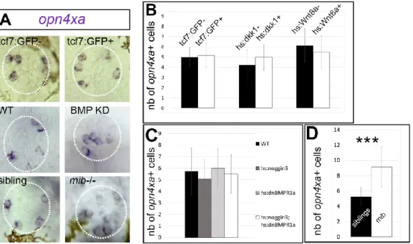
The fact that the tcf7 enhancer trap line Et(T2KHG)nns8is expressedin opn4xa+ cells prompted us to investigate whether Wnt signalling was involved in the specification of the opn4xa+ fate. First, we tested the expression of opn4xa in
Et(T2KHG)nns8 homozygous embryos as the enhancer trap insertion abolishes expression of the endogenous tcf7 gene (Nagayoshi et al., 2008). Pineal opn4xa+ cells were observed in normal numbers in all GFP+ embryos coming from Et(T2KHG)nns8 +/- incrosses suggesting that tcf7 is not required fortheir specification (Fig 2 A,B) as one third of GFP+ embryos are expected to be homozygous for the insertion. To look at a possible redundancy with other Wnt effectors, we analysed the expression of tcf7l2 and lef1 in the pineal and compared it with the Et(T2KHG)nns8 enhancer trap. Only the former is expressed in the pineal but did not show co-expression with the tcf7 enhancer trap (Figure S2). Finally, we modulated Wnt activity using conditional overexpression of Dkk1 or Wnt8 in heat-shocked Tg(hsp70l:dkk1b-GFP)w32 and
Tg(hsp70:wnt8-GFP)w34 embryos,respectively (Stoick-Cooper et al., 2007). Heat-shocked embryos carrying the Tg(hsp70l:dkk1b-GFP)w32 transgene show normal expression of opn4xa (Fig 2B) suggesting that Wnt activity is dispensable for the specification of opn4xa+ cells. Similarly, heat–shocked embryos transgenic for the Tg(hsp70:wnt8-GFP)w34 transgene show normal numbers of opn4xa + cells suggesting that Wnt activity is not
sufficient to drive the opn4xa+ fate. We conclude that Wnt signalling does not play a role during specification of the opn4xa+ fate.
Specification of opn4xa+ cells does not require BMP signalling and is
inhibited by Notch signalling
Classical PhRs are specified by a combination of Notch and BMP pathway activity with BMP activity being necessary and sufficient to activate the PhR fate and Notch being required to inhibit the inappropriate PN program (Cau et al., 2008; Quillien et al., 2011). To understand whether opn4xa+ cells are closer to PNs or PhRs developmentally speaking, we analysed the effects of reducing BMP or Notch activities on the specification of these cells.
We used Tg(hsp70l:dnXla.Bmpr1a-GFP)w30 ; Tg(hsp70l:nog3)fr14 double transgenic embryos heat-shocked at 14hpf to reduce BMP signalling, a condition that we have previously shown reduces the number of classical PhRs (Quillien et al., 2011). Under these conditions, the number of opn4xa+ cells was similar compared to wildtype embryos (Fig 2A, C); pineal opn4xa+ cells in BMP loss-of-function embryos exhibit an aberrant medial location which is likely due to the absence of classical photoreceptors normally found in the centre of the pineal gland (Fig2A). The effects of impairing Notch activity was analysed in the mindbomb (mib) mutant (mibta52b (Itoh et al., 2003)). We found that mib embryos exhibit twice the number of opn4xa+ cells at 48 hpf compared to controls (Fig 2A,D). Altogether these results suggest that pineal opn4xa+ cells behave as classical pineal PNs with respect to their requirement for BMP and Notch activity.
In contrast to classical PNs, opn4xa + projection neurons respond to light
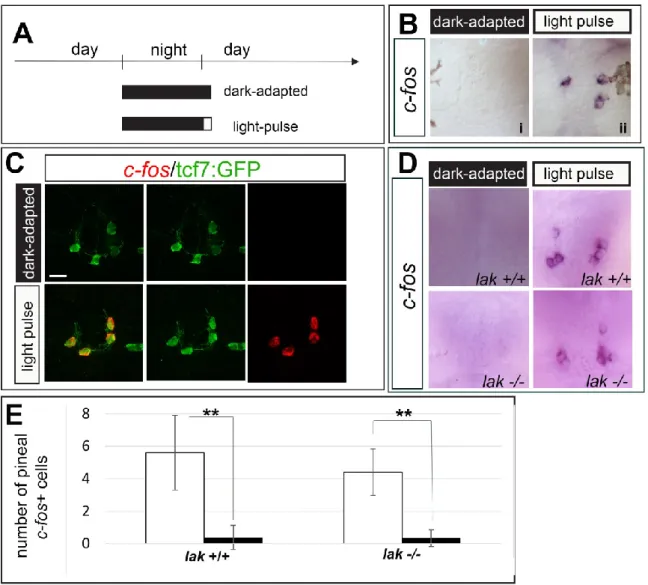
in a LIGHT ON fashion
Electrophysiological experiments performed in the rainbow trout, pike, frog and turtle have shown that projection neurons are continuously activated in the dark suggesting that they function in a LIGHT OFF mode (Dodt, 1963; Falcón and Meissl, 1981; Meissl and Ueck, 1980; Morita, 1966; Morita and Dodt, 1965). To assess whether opn4xa+ PNs are also LIGHT OFF, we analysed the expression of c-fos after a 30 minute dark-pulse in 48 hpf light-adapted embryos (Fig 3A); c-fos is an immediate early gene that is extensively used as a read out of neuronal activation (reviewed in Guzowski et al., 2005). While light-adapted embryos fixed at the same circadian time show no c-fos expression in the pineal (Fig 3Bi), a pulse of dark induced expression of
c-fos expression in 8.36±2.13 cells per embryo, which are located laterally in the pineal
(from n=14 embryos) (Fig 3Bii). The same experiment performed inEt(T2KHG)nns8 or
Tg(elavl3:EGFP)knu3 embryos showed that these LIGHT OFF cells are tcf7:GFP- but elavl3:EGFP+ and thus correspond to classical, opn4xa- projection neurons (Fig 3C,D). To address if opn4xa+ PNs are LIGHT ON, next we examined the effect of a light pulse in dark-adapted embryos (Fig 4A). We found that a 30 minute pulse of white light delivered to 48 hpf dark-adapted embryos induced c-fos expression in 3±1.41 cells (from n=17 embryos) located laterally within the pineal organ (Fig 4Bii) in cells that strongly express the Et(T2KHG)nns8 transgene (Fig 4C); c-fos transcript was not detected in the pineal of non-light stimulated embryos fixed at the same circadian time (Fig 4Bi). Similar results were obtained in 27 hpf and 3 dpf dark-adapted embryos (Fig S3). In mammals, the pineal gland is connected to the retina via a multi-synaptic pathway (Sapède and Cau, 2013).To rule out that the pineal LIGHT ON response we describe is caused by photosensitivity in the retina, we assessed the presence of this
response in lakritz (lak) mutant which lack neurotransmission from the retina owing to a complete absence of RGCs (Kay et al., 2001). The pineal LIGHT ON response is similar in lak+/+ and lak-/- embryos at 48 hpf suggesting that the retina is not involved in this activity (Fig 4D,E).
Taken together, our results indicate heterogeneity in the light-response within the PN population with opn4xa+ PNs functioning in a LIGHT ON fashion and classical PNs in a LIGHT OFF mode.
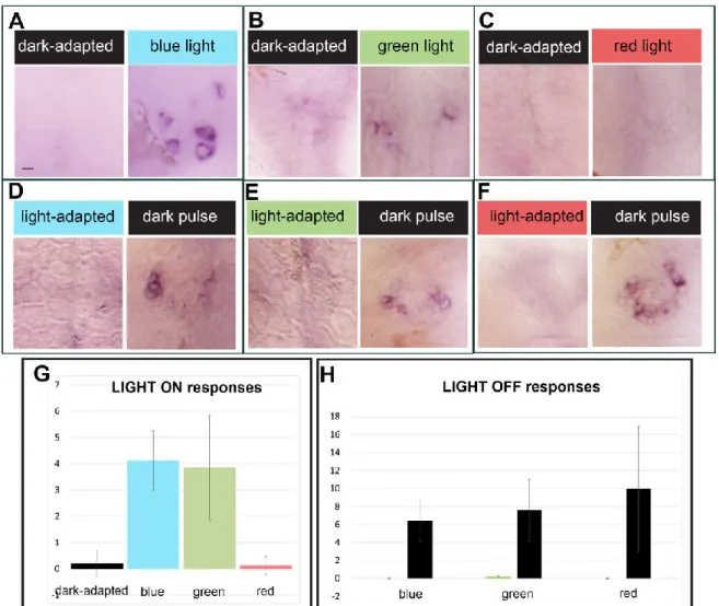
LIGHT ON and LIGHT OFF responses are elicited by different range of
wavelengths
Apart from Opn4xa, which is sensitive to blue-green light (Davies et al., 2011), the classical PhR in the pineal express opsin photo-pigments (exorhodopsin, red cone
opsin and parietopsin) that are sensitive to green and red light (Cau et al., 2019). We
next examined whether LIGHT ON in blue, red or green wavelengths were involved in the induction of c-fos expression. We observed that 30 minute light pulses in the blue and green range induce c-fos expression while similar pulses of red light could not induce a LIGHT ON response (Fig 5 A-C, G). Given that the absorbance spectrum of the Opn4xa photo-pigment overlaps with the emission of the green and blue LEDs used in this study, we propose that this LIGHT ON response reflects the direct photosensitivity of opn4xa+ photo-pigment. In marked contrast with the results obtained for the LIGHT ON response, LIGHT OFF was elicited when the embryos were adapted in either red, blue or green light (Figure 5 D-F, H) suggesting that this response is elicited by a wide-range of wavelengths and can, therefore, be referred to as achromatic.
Our results suggest that classical, opn4xa– projection neurons function in a LIGHT OFF mode and most likely transduce the light input received by classical PhRs. In contrast, opn4xa+ projection neurons are activated by light and most likely directly photosensitive.
Discussion
In this paper, we describe a new population of cells in the zebrafish pineal gland. These cells express the green-blue photosensitive pigment Opn4xa and share developmental characteristics with classical, non-photosensitive projection neurons. Intriguingly, while classical PNs exhibit a LIGHT OFF response, a LIGHT ON response is observed in
opn4xa+ PN upon illumination. These results suggest a previously unanticipated
cellular diversity in the projection neuron population within the pineal gland. In the following section, we discuss the specification and potential function of the opn4xa+ PNs.
Projection neurons and photoreceptors of the pineal gland differ in their requirement for Notch and BMP signalling pathways: photoreceptors require BMP activation (while projection neurons do not) and BMP is in turn needed to activate the Notch pathway in these cells, which inhibits the projection neurons fate (Cau et al., 2008; Quillien et al., 2011). This led us to postulate that decoupling of the activation of BMP and Notch activity could represent an attractive mechanism to further diversify cell fates. Indeed, activation of the BMP signalling pathway without activation of Notch should lead to the generation of cells with a mixed photoreceptor/projection neuron identity (Cau and Blader, 2009). In contrast with this hypothesis, opn4xa+ cells, that exhibits both characteristics of photoreceptors and projection neurons, do not require BMP activity suggesting that they are more closely related to projection neurons than
to classical photoreceptors. Similarly, Notch activity inhibits the production of both
opn4xa– and opn4xa+ projection neurons to the same extent. Therefore, neither BMP
nor Notch activities contribute to specifying opn4xa+ versus opn4xa– PN fates.
Since opn4xa+ and classical projection neurons show similar behaviour upon BMP or Notch loss of function, what are the signals that could specify these distinct identities? The Wnt effector tcf7 is specifically expressed in opn4xa+ PN and so is the
Et(T2KHG)nns8 enhancer trap line at early stages. Nevertheless, tcf7 mutants show a normal number of opn4xa+ pineal cells. Moreover, neither gain nor loss of Wnt activity affects the number of opn4xa+ cells, which suggests that Wnt activity is neither necessary nor sufficient for specification of opn4xa expression. It remains to be addressed whether tcf7 (and/or Wnt signalling) is required to specify other aspects of the phenotype of these atypical PNs. Despite the absence of an observable function for tcf7, the Et(T2KHG)nns8 enhancer trap line provides a useful marker for opn4xa + cells at early stages which will help us in analysing the projection pattern of these cells. Our analysis confirms previous results obtained in another teleost, the rainbow trout, as well as in numerous other species showing that an important part of the projection neuron population function in a LIGHT OFF manner (Dodt, 1963; Falcón and Meissl, 1981; Meissl and Ueck, 1980; Morita, 1966; Morita and Dodt, 1965). Within the projection neuron population only opn4xa+ PNs function in a LIGHT ON fashion. Given that this activity is only seen using blue and green light and not red light, it likely requires the function of Opn4xa, a photo-pigment with an absorbance peak at 470 nm (Davies et al., 2011). This result contrasts with the observation that a LIGHT ON (excitatory response) is observed at medium to long wavelengths in the rainbow trout (Morita, 1966). A surprising degree of variation is indeed observed in the wavelengths
light triggers the excitatory response in lizards and frogs while red light elicits this response in the pike. Similarly, the chromatic inhibitory response is stronger in the ultraviolet range in fish and frogs but mainly due to blue light in lizards (see Dodt and Meissl, (1982) for a review). It would be interesting to understand whether the LIGHT ON chromatic response observed in lizard, frog and pike require a direct photosensitivity from specific projection neurons. Alternatively, in these species, pineal photoreceptors could elicit a dual excitatory/inhibitory action on their targets similar to that described for retinal photoreceptors, which trigger a different response in bipolar cells depending on the type of glutamate receptor they express (Euler et al., 1996; Masu et al., 1995; de la Villa et al., 1995).
Are there other LIGHT ON projection neurons in the zebrafish pineal gland? Adaptation with white light allowed us to identify an average 8 LIGHT OFF PNs for around 3 LIGHT ON PNs. Since at 48 hpf the pineal gland contains 20 PNs, either some of the PN responses have not been identified or the LIGHT OFF cells do not systematically activate c-fos following a 30 min dark pulse, in the present testing conditions. It thus remains possible that there are other unidentified projection neurons that respond in a LIGHT ON fashion for instance in response to UV light.
While a complete description of the neurotransmitter/neuromodulator content of all zebrafish pineal neurons is lacking, glycinergic cells with a photoreceptor morphology have been observed in the pineal gland (Marquart et al., 2015; Moly et al., 2014)). These observations suggest that at least a proportion of pineal photoreceptors exert an inhibitory action on projection neurons, therefore providing a possible mechanism for the LIGHT OFF response we describe. Alternatively, photoreceptors could constantly produce an excitatory neurotransmitter in the dark, which would be blocked by light leading to an interruption of projection neuron activity through a
“disfacilitation mechanism” (Uchida et al., 1992). A more detailed analysis of the neurotransmitter/neuromodulator content of all pineal neurons, and opn4xa+ PNs in particular, would be useful. Finally, the fact that the LIGHT OFF response is observed independently of the wavelength at which the embryos are adapted suggests that LIGHT OFF projection neurons receive inputs from several types of pineal photoreceptors.
Although direct photosensitivity of opn4xa+ PNs remains to be confirmed, they share many characteristics with retinal ipRGCS raising intriguing questions regarding the functions of these pineal cells. Indeed, in mammals ipRGCs exhibit crucial roles during the control of circadian, wake/sleep rhythms as well as in mediating the direct effects of illumination on physiology and behaviour (see Lazzerini Ospri et al., (2017); Lucas et al.,(2012) for reviews)). In particular, ipRGCs transmit light information, via their own intrinsic photosensitivity as well as inputs from rod and cone PhRs, to the mammalian ‘master clock’ the suprachiasmatic nucleus (Hannibal and Fahrenkrug, 2004; Hattar et al., 2002, 2006). It is unclear whether a suprachiasmatic nucleus is present in the larval zebrafish and whether opn4xa+ retinal or pineal cells project onto such a structure. On the other hand, the zebrafish pineal gland has long been thought to play a ‘master clock’ role owing to its direct photosensitivity, its capacity to generate intrinsic rhythms of melatonin synthesis/secretion and the fact that disrupting the function of the molecular clock in pineal PhRs affect circadian rhythms of locomotor activity (Ben-Moshe Livne et al., 2016; Bolliet et al., 1997; Cahill, 1996, 1997; Falcón et al., 1989). Could opn4xa+ PNs play a role alongside classical pineal PhRs in the function of the circadian system? Analysis of an opn4xa mutant allele combined with retina-specific and/or pineal specific transgenic tools would help us understand
Experimental Procedures
Zebrafish lines and developmental conditions
All animals were handled in the CBI fish facility, which is certified by the French Ministry of Agriculture (approval number A3155510). The project was approved by the French Ministry of Teaching and Research (agreement number APAFIS#3653-2016011512005922).
Embryos were reared at 28 degrees in a 14 hours Light/ 10 hours Dark cycle and staged according to standard protocols. All the transgenic and mutant lines have been described previously: Tg(aanat2:GFP)y8 (Gothilf et al., 2002), Tg(elavl3:EGFP)knu3 (Park et al., 2000), Et(T2KHG)nns8 (Nagayoshi et al., 2008), Tg(hsp70l:dkk1b-GFP)w32
and Tg(hsp70:wnt8-GFP)w34 (Stoick-Cooper et al., 2007),
Tg(hsp70l:dnXla.Rbpj-MYC)vu21 and Tg(hsp70l:nog3)fr14 (Chocron et al., 2007), mibta52b (Itoh et al., 2003),
lakritz (Kay et al., 2001). Conditions of heat-shock were: Tg(hsp70l:dnXla.Rbpj-MYC)vu21 and Tg(hsp70l:nog3)fr14 : 30 minutes at 39.5°C; Tg(hsp70l:dkk1b-GFP)w32
and Tg(hsp70:wnt8-GFP)w34 : 1 hour at 38°C. The protocol for genotyping lakritz embryos is available upon request.
Application of different light regimes
To assess LIGHT ON responses, two different protocols were used. In the first protocol, the light was turned on at 9 am and the embryos fixed at 9.30 am. In the second one, the embryos were dark adapted for 2 hours (from 9.30 to 11.30) before a 30 mn light pulse and a fixation at 12 am. We did not notice a difference in the responses between these two protocols. On the other hand, for the assessment of the
LIGHT OFF responses, the embryos were light-adapted from 9 am to 12 am after which light was turned off and the embryos were fixed 30 mns after. The comparison of adapted and pulsed embryos fixed at the same circadian time allowed us to conclude that the observed c-fos expression was caused by the pulse rather than by circadian rhythms.
Illumination with white light was performed using a regular neon lamp (80 lux). Illumination with blue, green and red light was performed in a MWP unit (Zantiks) equipped with standard blue, green and red LEDs. The characteristics of the light emitted from these LEDS is:
Blue light: 92 lux, max= 449nm, half-band width: 25,4nm Green light: 109 lux, max= 512 nm, half-band width: 33,07 nm Red light: 34 lux, max= 627 nm, half-band width: 21,67 nm
Scripts controlling the different light regimes are available upon request.
Immunohistochemistry and in situ hybridisation
Immunohistochemistry and in situ hybridisation were performed as previously described and fluorescent in situ hybridisation were revealed using either Fast Red (Sigma) or the TSA Plus System (TSA-Fluorescein, Perkin Elmer; Cau et al., 2019). Immunohistochemistry against Tcf7l2 was performed as described in Hüsken et al., (2014) using an anti-Human TCF3,4 antibody (1:400, Biomol, clone 0.T.148).
Probes used in this study were: opn4xa (Matos-Cruz et al., 2011), c-fos (Ellis et al., 2012), lef1 and tcf7 (Veien et al., 2005).
Fluorescent cells were counted using the ImageJ software on confocal data sets with 2 m interval optical planes. Statistical tests and number of embryos used are stated in each figure and/or figure legend.
Acknowledgements
We thank S. Mazères for his help with the spectral analysis of LED light, R. Dorsky for providing the Et(T2KHG)nns8, Tg(hsp70l:dkk1b-GFP)w32 and Tg(hsp70:wnt8-GFP)w34 transgenic lines, and M. Halpern and K. Soanes for sharing probes. We are indebted to members of the Blader team for helpful discussions. This work was supported by the Centre National de la Recherche Scientifique (CNRS); the Institut National de la Santé et de la Recherche Médicale (INSERM); Université de Toulouse III (UPS); Fondation pour la Recherche Médicale (FRM; DEQ20131029166); Fédération pour la Recherche sur le Cerveau (FRC); Association pour la Recherche sur le Cancer (ARC); Association Rétina France and the Ministère de la Recherche. We would like to thank Brice Ronsin, Stéphanie Bosch and the Toulouse RIO Imaging platform; as well as Stéphane Relexans, Aurore Laire and Richard Brimicombe for taking care of the fish.
Declaration of competing interest
The authors declare that no competing interests exist.
References
Ben-Moshe Livne, Z., Alon, S., Vallone, D., Bayleyen, Y., Tovin, A., Shainer, I., Nisembaum, L.G., Aviram, I., Smadja-Storz, S., Fuentes, M., et al. (2016).
Genetically Blocking the Zebrafish Pineal Clock Affects Circadian Behavior. PLoS Genet. 12, e1006445.
Bolliet, V., Bégay, V., Taragnat, C., Ravault, J.P., Collin, J.P., and Falcón, J. (1997). Photoreceptor cells of the pike pineal organ as cellular circadian oscillators. Eur. J. Neurosci. 9, 643–653.
Cahill, G.M. (1996). Circadian regulation of melatonin production in cultured zebrafish pineal and retina. Brain Res. 708, 177–181.
Cahill, G.M. (1997). Circadian melatonin rhythms in cultured zebrafish pineals are not affected by catecholamine receptor agonists. Gen. Comp. Endocrinol. 105, 270–275. Cau, E., and Blader, P. (2009). Notch activity in the nervous system: to switch or not switch? Neural Dev. 4, 36.
Cau, E., Quillien, A., and Blader, P. (2008). Notch resolves mixed neural identities in the zebrafish epiphysis. Development. 135, 2391–2401.
Cau, E., Ronsin, B., Bessière, L., and Blader, P. (2019). A Notch-mediated, temporal asymmetry in BMP pathway activation promotes photoreceptor subtype
diversification. PLoS Biol. 17, e2006250.
Chocron, S., Verhoeven, M.C., Rentzsch, F., Hammerschmidt, M., and Bakkers, J. (2007). Zebrafish Bmp4 regulates left-right asymmetry at two distinct developmental time points. Dev. Biol. 305, 577–588.
Clanton, J.A., Hope, K.D., and Gamse, J.T. (2013). Fgf signaling governs cell fate in the zebrafish pineal complex. Development. 140, 323–332.
Davies, W.I.L., Zheng, L., Hughes, S., Tamai, T.K., Turton, M., Halford, S., Foster, R.G., Whitmore, D., and Hankins, M.W. (2011). Functional diversity of melanopsins and their global expression in the teleost retina. Cell. Mol. Life Sci. CMLS 68, 4115– 4132.
Dodt, E. (1963). PHOTOSENSITIVITY OF THE PINEAL ORGAN IN THE TELEOST, SALMO IRIDEUS (GIBBONS). Experientia 19, 642–643.
Dodt, E., and Heerd, E. (1962). Mode of action of pineal nerve fibers in frogs. J. Neurophysiol. 25, 405–429.
Dodt, E., and Meissl, H. (1982). The pineal and parietal organs of lower vertebrates. Experientia 38, 996–1000.
Ellis, L.D., Seibert, J., and Soanes, K.H. (2012). Distinct models of induced hyperactivity in zebrafish larvae. Brain Res. 1449, 46–59.
Euler, T., Schneider, H., and Wässle, H. (1996). Glutamate responses of bipolar cells in a slice preparation of the rat retina. J. Neurosci. Off. J. Soc. Neurosci. 16, 2934– 2944.
Falcón, J., and Meissl, H. (1981). The Photosensory Function of the Pineal Organ of the Pike (Esox lucius L.) Correlation Between Structure and Function. J. Comp. Physiol. A 144, 127–137.
Falcón, J., Marmillon, J.B., Claustrat, B., and Collin, J.P. (1989). Regulation of melatonin secretion in a photoreceptive pineal organ: an in vitro study in the pike. J. Neurosci. Off. J. Soc. Neurosci. 9, 1943–1950.
Gandhi, A.V., Mosser, E.A., Oikonomou, G., and Prober, D.A. (2015). Melatonin is required for the circadian regulation of sleep. Neuron 85, 1193–1199.
Gothilf, Y., Toyama, R., Coon, S.L., Du, S.-J., Dawid, I.B., and Klein, D.C. (2002). Pineal-specific expression of green fluorescent protein under the control of the serotonin-N-acetyltransferase gene regulatory regions in transgenic zebrafish. Dev. Dyn. Off. Publ. Am. Assoc. Anat. 225, 241–249.
Guzowski, J.F., Timlin, J.A., Roysam, B., McNaughton, B.L., Worley, P.F., and Barnes, C.A. (2005). Mapping behaviorally relevant neural circuits with immediate-early gene expression. Curr. Opin. Neurobiol. 15, 599–606.
Hannibal, J., and Fahrenkrug, J. (2004). Melanopsin containing retinal ganglion cells are light responsive from birth. Neuroreport 15, 2317–2320.
Hattar, S., Liao, H.W., Takao, M., Berson, D.M., and Yau, K.W. (2002). Melanopsin-containing retinal ganglion cells: architecture, projections, and intrinsic
Hattar, S., Kumar, M., Park, A., Tong, P., Tung, J., Yau, K.-W., and Berson, D.M. (2006). Central projections of melanopsin-expressing retinal ganglion cells in the mouse. J. Comp. Neurol. 497, 326–349.
Hüsken, U., Stickney, H.L., Gestri, G., Bianco, I.H., Faro, A., Young, R.M.,
Roussigne, M., Hawkins, T.A., Beretta, C.A., Brinkmann, I., et al. (2014). Tcf7l2 is required for left-right asymmetric differentiation of habenular neurons. Curr. Biol. CB
24, 2217–2227.
Itoh, M., Kim, C.-H., Palardy, G., Oda, T., Jiang, Y.-J., Maust, D., Yeo, S.-Y., Lorick, K., Wright, G.J., Ariza-McNaughton, L., et al. (2003). Mind bomb is a ubiquitin ligase that is essential for efficient activation of Notch signaling by Delta. Dev. Cell 4, 67–82. Kay, J.N., Finger-Baier, K.C., Roeser, T., Staub, W., and Baier, H. (2001). Retinal Ganglion Cell Genesis Requires lakritz, a Zebrafish atonal Homolog. Neuron 30, 725–736.
Koyanagi, M., Wada, S., Kawano-Yamashita, E., Hara, Y., Kuraku, S., Kosaka, S., Kawakami, K., Tamotsu, S., Tsukamoto, H., Shichida, Y., et al. (2015). Diversification of non-visual photopigment parapinopsin in spectral sensitivity for diverse pineal functions. BMC Biol. 13, 73.
Lazzerini Ospri, L., Prusky, G., and Hattar, S. (2017). Mood, the Circadian System, and Melanopsin Retinal Ganglion Cells. Annu. Rev. Neurosci. 40, 539–556.
Lucas, R.J., Lall, G.S., Allen, A.E., and Brown, T.M. (2012). How rod, cone, and melanopsin photoreceptors come together to enlighten the mammalian circadian clock. Prog. Brain Res. 199, 1–18.
Marquart, G.D., Tabor, K.M., Brown, M., Strykowski, J.L., Varshney, G.K., LaFave, M.C., Mueller, T., Burgess, S.M., Higashijima, S.-I., and Burgess, H.A. (2015). A 3D Searchable Database of Transgenic Zebrafish Gal4 and Cre Lines for Functional Neuroanatomy Studies. Front. Neural Circuits 9, 78.
Masu, M., Iwakabe, H., Tagawa, Y., Miyoshi, T., Yamashita, M., Fukuda, Y., Sasaki, H., Hiroi, K., Nakamura, Y., and Shigemoto, R. (1995). Specific deficit of the ON response in visual transmission by targeted disruption of the mGluR6 gene. Cell 80, 757–765.
Matos-Cruz, V., Blasic, J., Nickle, B., Robinson, P.R., Hattar, S., and Halpern, M.E. (2011). Unexpected diversity and photoperiod dependence of the zebrafish
melanopsin system. PloS One 6, e25111.
Meissl, H., and Donley, C.S. (1980). Change of threshold after light-adaptation of the chromatic response of the frog’s pineal organ (Stirnorgan). Vision Res. 20, 379–383. Meissl, H., and Ueck, M. (1980). Extraocular Photoreception of the Pineal Gland of the Aquatic Turtle Pseudemys scripta elegans. J. Comp. Physiol. A 140, 173–179. Meissl, H., Nakamura, T., and Thiele, G. (1986). Neural response mechanisms in the photoreceptive pineal organ of goldfish. Comp. Biochem. Physiol. A 84, 467–473. Moly, P.K., Ikenaga, T., Kamihagi, C., Islam, A.F.M.T., and Hatta, K. (2014).
Identification of initially appearing glycine-immunoreactive neurons in the embryonic zebrafish brain. Dev. Neurobiol. 74, 616–632.
Morita, Y. (1966). [Lead pattern of the pineal neuron of the rainbow trout (Salmo irideus) by illumination of the diencephalon]. Pflugers Arch. Gesamte Physiol. Menschen Tiere 289, 155–167.
Morita, Y., and Dodt, E. (1965). Nervous activity of the frog’s epiphysis cerebri in relation to illumination. Experientia 21, 221–222.
Nagayoshi, S., Hayashi, E., Abe, G., Osato, N., Asakawa, K., Urasaki, A., Horikawa, K., Ikeo, K., Takeda, H., and Kawakami, K. (2008). Insertional mutagenesis by the
Tol2 transposon-mediated enhancer trap approach generated mutations in two developmental genes: tcf7 and synembryn-like. Development. 135, 159–169. Park, H.C., Kim, C.H., Bae, Y.K., Yeo, S.Y., Kim, S.H., Hong, S.K., Shin, J., Yoo, K.W., Hibi, M., Hirano, T., et al. (2000). Analysis of upstream elements in the HuC promoter leads to the establishment of transgenic zebrafish with fluorescent neurons. Dev. Biol. 227, 279–293.
Quillien, A., Blanco-Sanchez, B., Halluin, C., Moore, J.C., Lawson, N.D., Blader, P., and Cau, E. (2011). BMP signaling orchestrates photoreceptor specification in the zebrafish pineal gland in collaboration with Notch. Development. 138, 2293–2302. Sanes, J.R., and Masland, R.H. (2015). The types of retinal ganglion cells: current status and implications for neuronal classification. Annu. Rev. Neurosci. 38, 221–246. Sapède, D., and Cau, E. (2013). The pineal gland from development to function. Curr. Top. Dev. Biol. 106, 171–215.
Shainer, I., Buchshtab, A., Hawkins, T.A., Wilson, S.W., Cone, R.D., and Gothilf, Y. (2017). Novel hypophysiotropic AgRP2 neurons and pineal cells revealed by BAC transgenesis in zebrafish. Sci. Rep. 7, 44777.
Shainer, I., Michel, M., Marquart, G.D., Bhandiwad, A.A., Zmora, N., Ben-Moshe Livne, Z., Zohar, Y., Hazak, A., Mazon, Y., Förster, D., et al. (2019). Agouti-Related Protein 2 Is a New Player in the Teleost Stress Response System. Curr. Biol. CB 29, 2009–2019.e7.
Stoick-Cooper, C.L., Weidinger, G., Riehle, K.J., Hubbert, C., Major, M.B., Fausto, N., and Moon, R.T. (2007). Distinct Wnt signaling pathways have opposing roles in appendage regeneration. Development. 134, 479–489.
Uchida, K., Nakamura, T., and Morita, Y. (1992). Signal transmission from pineal photoreceptors to luminosity-type ganglion cells in the lamprey, Lampetra japonica. Neuroscience 47, 241–247.
Veien, E.S., Grierson, M.J., Saund, R.S., and Dorsky, R.I. (2005). Expression pattern of zebrafish tcf7 suggests unexplored domains of Wnt/beta-catenin activity. Dev. Dyn. Off. Publ. Am. Assoc. Anat. 233, 233–239.
de la Villa, P., Kurahashi, T., and Kaneko, A. (1995). L-glutamate-induced responses and cGMP-activated channels in three subtypes of retinal bipolar cells dissociated from the cat. J. Neurosci. Off. J. Soc. Neurosci. 15, 3571–3582.
Wada, S., Shen, B., Kawano-Yamashita, E., Nagata, T., Hibi, M., Tamotsu, S.,
Koyanagi, M., and Terakita, A. (2018). Color opponency with a single kind of bistable opsin in the zebrafish pineal organ. Proc. Natl. Acad. Sci. U. S. A. 115, 11310–
11315.
Wilson, S.W., and Easter, S.S. (1991). Stereotyped pathway selection by growth cones of early epiphysial neurons in the embryonic zebrafish. Development. 112, 723–746.
Figure 1: opn4xa is expressed in a restricted population of projection neurons
within the pineal gland.
(A) in situ hybridisation at 48hpf showing pineal opn4xa+ cells. The pineal is highlighted with a white dotted circle.
(B) Confocal projection showing the relative distribution of PNs (red, labelled with an anti-HuC/D antibody) and PhRs (green, labelled with an anti-GFP in a Tg(aanat2:GFP)y8 background) at 48 hpf. Topro (cyan) labels cell nuclei.
(C-K) Co-expression of opn4xa mRNA (in red) on the one hand with the Tg(aanat2:GFP)y8 (‘aanat2:GFP’, C,D,E), the Tg(elavl3:EGFP)knu3 (‘elavl3:EGFP’, F,G,H) and the Et(T2KHG)nns8 (‘tcf7:GFP’,I,J,K transgenes (in green).
(A-K) Anterior is upwards. Scale bars: 10 µm.
(L)Time course of opn4xa expression in the developing pineal organ. Average numbers of opn4xa+ cells are quantified at different time points indicated in hours post-fertilisation (hpf). Values are mean ±SD, n=4, 5, 3, 6, 9, 26, 6 and 6, respectively.
Figure 2: Effect of manipulating Notch, BMP and Wnt pathways on the
opn4xa+ fate
(A) Representative pictures of in situ hybridisations for opn4xa in the pineal organ upon BMP, Notch and Wnt modulation (as indicated) and their corresponding controls. (B-D) Average numbers of pineal opn4xa+ positive cells upon manipulation of Wnt (B), BMP (C) or Notch signalling activity (D). Values indicated on the graphs are mean ±SD (error bars). Two tailed Mann Whitney non parametric tests, were used for B and D. *P<0.05; ***P<0.001.
(B) Effect of gain or loss of Wnt activity on the number of pineal opn4xa+ cells: Embryos from Et(T2KHG)nns8 +/- incrosses were assessed for GFP fluorescence; countings were performed at 30hpf. The size of the GFP- and GFP+ populations (‘tcf7:GFP-’ and ‘tcf7:GFP+’) were respectively n=8 and n=31. Embryos carrying the
Tg(hsp70l:dkk1b-GFP)w32 transgene after an heat shock at 18 hpf, were compared with negative siblings
at 48 hpf (n=13 for the ‘hs:dkk1-’ and n=11 for the ‘hs:dkk1+’). Embryos carrying the Tg(hsp70:wnt8-GFP)w34 transgene (n=14) after an heat shock at 14 hpf, were compared to siblings (n=14) at 48 hpf.
(C) Effect of a reduction in Notch activity: Control embryos (n=30) were compared to homozygous mindbomb (mib-/-; n=7) mutants at 48 hpf
(D) Effect of a reduced BMP signalling: Embryos transgenic for
Tg(hsp70l:dnXla.Bmpr1a-GFP)w30 (hs:dnBMPR1a, n=12) or Tg(hsp70l:nog3)fr14 (hs:noggin3, n=30) or both (n=18) were compared to siblings (n=7). Kruskal-Wallis test with Dunn's multiple comparisons test, P=0.44; not significant.
Figure 3: Identification of a population of opn4xa- PNs functioning in a LIGHT
OFF mode:
(A) Experimental paradigm for the ‘dark pulse versus light-adapted' comparison Embryos were light-adapted for three hours before being submitted to a 30 min dark pulse (‘dark pulse’) or maintained in the light (‘light-adapted’)
(B) Representative pictures of c-fos induction in the pineal organ after application of the light regimes described in A). i) light-adapted, ii) dark-pulse
(C-E) Induction of c-fos in the different light regimes in embryos carrying the
Et(T2KHG)nns8 (‘tcf7:GFP’, C) or the Tg(elavl3:EGFP)knu3 (‘elavl3:EGFP’, D) transgenes (in green). Scale bars: 10 µm. All embryos shown are 48 hpf.
Figure 4: opn4xa+ PNs function in a LIGHT ON mode:
(A) Experimental paradigm for the ‘light-pulse versus dark-adapted' comparison Embryos were dark-adapted for more than 14 hours and submitted to a 30 min light pulse (‘light pulse’ ) or maintained in the dark (‘dark-adapted’) before fixation.
(B) Representative pictures of c-fos induction in the pineal organ after application of the light regimes described in A). i) dark-adapted, ii) light-pulse
(C) Induction of c-fos in the different light regimes in embryos carrying the
Et(T2KHG)nns8 (‘tcf7:GFP’) transgene (in green). Scale bars: 10 µm.
(D) Induction of c-fos upon a 30mn light pulse in lak +/+ and lak -/- embryos.
(E) Quantification of the number of pineal c-fos+ cells in light-pulsed (white bars) and dark-adapted (dark bars) lak+/+ and lak-/- embryos at 48 hpf. Two tailed Mann Whitney non parametric tests, were used *P<0.05. Values are mean ±SD, n=5,8,10 and 6, respectively.
Figure 5: Different spectral sensitivities for the LIGHT ON and the LIGHT OFF
responses within the pineal gland:
(A-F) Representative pictures of c-fos induction in the pineal organ after application of light pulses in the blue, green or red range compared to dark adapted embryos (A-C) or after light adaptation with blue, green or red light followed by a dark pulse (‘dark pulse’) or maintained in the same lighting conditions (‘light-adapted’) (D-F)
(G, H) Quantification of the number of cells activating c-fos after a light pulse (G, LIGHT ON responses) or a dark pulse (H, LIGHT OFF responses). G) Numbers of embryos : n= 41, 8, 20,16 respectively
Scale bar: 10 µm.
Supplemental figure legends:
Supplemental Figure 1: Characterisation of the expression driven by the
Et(T2KHG)nns8 (‘tcf7:GFP’) enhancer trap in the pineal gland :
(A-F) Comparison of tcf7 (A-C) or opn4xa (D-F) expression (in red, in situ hybridisation) with GFP expression from the Et(T2KHG)nns8 (‘tcf7:GFP’) transgene (in green, immunohistochemistry). White arrowheads highlight co-expressing cells. White arrow show a weak tcf7:GFP+ opn4xa – cell. Scale bars: 10 µm. (G) Comparison of the number of cells expressing tcf7:GFP strongly or weakly and opn4xa. Means ± S.D are shown.
Supplemental Figure 2: Comparison of the expression of tcf7l2 and lef1 with the
Et(T2KHG)nns8 (‘tcf7:GFP’) enhancer trap in the pineal gland
(A-F) Expression of two other TCF encoding genes: tcf7l2 (A-C) or lef1 (D-F) (in red, in situ hybridisation) with expression from the Et(T2KHG)nns8 (‘tcf7:GFP’) transgene (in
green, immunohistochemistry). (G) Double immunostaining with anti-GFP (green) and anti-TCF7l2 (red) in pineal neurons.
Supplemental Figure 3 : Induction of c-fos expression following a light pulse in
the pineal gland at 27 hpf and 3 dpf.
Expression of c-fos (in red, in situ hybridisation) compared with expression from the
Et(T2KHG)nns8 (‘tcf7:GFP’) transgene (in green, immunohistochemistry) at 27hpf