HAL Id: hal-02322646
https://hal.archives-ouvertes.fr/hal-02322646
Submitted on 11 Dec 2020
HAL is a multi-disciplinary open access
archive for the deposit and dissemination of
sci-entific research documents, whether they are
pub-lished or not. The documents may come from
teaching and research institutions in France or
abroad, or from public or private research centers.
L’archive ouverte pluridisciplinaire HAL, est
destinée au dépôt et à la diffusion de documents
scientifiques de niveau recherche, publiés ou non,
émanant des établissements d’enseignement et de
recherche français ou étrangers, des laboratoires
publics ou privés.
Refinement of the Primate Corticospinal Pathway
During Prenatal Development
Ana Rita Ribeiro Gomes, Etienne Olivier, Herbert Killackey, Pascale Giroud,
Michel Berland, Kenneth Knoblauch, Colette Dehay, Henry Kennedy
To cite this version:
Ana Rita Ribeiro Gomes, Etienne Olivier, Herbert Killackey, Pascale Giroud, Michel Berland, et al..
Refinement of the Primate Corticospinal Pathway During Prenatal Development. Cerebral Cortex,
Oxford University Press (OUP), 2020, 30 (2), pp.656-671. �10.1093/cercor/bhz116�. �hal-02322646�
© The Author(s) 2019. Published by Oxford University Press.
This is an Open Access article distributed under the terms of the Creative Commons Attribution Non-Commercial License (http://creativecommons.org/ licenses/by-nc/4.0/), which permits non-commercial re-use, distribution, and reproduction in any medium, provided the original work is properly cited. For commercial re-use, please contact [email protected]
doi: 10.1093/cercor/bhz116
Advance Access Publication Date: 25 July 2019 Original Article
O R I G I N A L A R T I C L E
Refinement of the Primate Corticospinal Pathway
During Prenatal Development
Ana Rita Ribeiro Gomes
1
, Etienne Olivier
2,†
, Herbert P. Killackey
3
,
Pascale Giroud
1
, Michel Berland
1
, Kenneth Knoblauch
1
, Colette Dehay
1
and
Henry Kennedy
1,4
1
Univ Lyon, Université Claude Bernard Lyon 1, Inserm, Stem Cell and Brain Research Institute U1208, Bron,
France,
2Institute of Neuroscience, Université Catholique de Louvain, Belgium,
3Department of Neurobiology
and Behavior, University of California, Irvine, CA, USA and
4Institute of Neuroscience, Key Laboratory of
Primate Neurobiology, Chinese Academy of Sciences, Shanghai 200031, China
Address correspondence to Ana Rita Ribeiro Gomes, Colette Dehay, Henry Kennedy, Stem Cell and Brain Research Institute, Inserm U1208, 18 avenue Doyen Lépine, 69500 Bron, France. Emails: [email protected]; [email protected]; [email protected]
†Deceased 9 March 2016.
Abstract
Perturbation of the developmental refinement of the corticospinal (CS) pathway leads to motor disorders. While non-primate developmental refinement is well documented, in primates invasive investigations of the developing CS pathway have been confined to neonatal and postnatal stages when refinement is relatively modest. Here, we investigated the developmental changes in the distribution of CS projection neurons in cynomolgus monkey (Macaca fascicularis). Injections of retrograde tracer at cervical levels of the spinal cord at embryonic day (E) 95 and E105 show that: (i) areal distribution of back-labeled neurons is more extensive than in the neonate and dense labeling is found in prefrontal, limbic, temporal, and occipital cortex; (ii) distributions of contralateral and ipsilateral projecting CS neurons are comparable in terms of location and numbers of labeled neurons, in contrast to the adult where the contralateral projection is an order of magnitude higher than the ipsilateral projection. Findings from one largely restricted injection suggest a hitherto
unsuspected early innervation of the gray matter. In the fetus there was in addition dense labeling in the central nucleus of the amygdala, the hypothalamus, the subthalamic nucleus, and the adjacent region of the zona incerta, subcortical structures with only minor projections in the adult control.
Key words: anatomy, cortex, macaque, spinal cord, tract tracing
Introduction
The brain exerts control over movement of the body through descending telencephalic, hypothalamic, diencephalic, mid-brain, and hindbrain pathways (Kuypers 1982, Nudo and Masterton 1988,Lemon 2008). Among these parallel-descending routes, the corticospinal (CS) pathway provides the fastest and
most direct influence of the cerebral cortex over different aspects of planning, execution, and control of voluntary movements (Dum and Strick 1996,2002;Lemon 2008).
Certain features of the adult and developing CS system are conserved across species. In the adult the CS tract originates in layer 5 of the somatomotor regions of the frontal and parietal
Refinement of the Primate Corticospinal Pathway During Prenatal Development Gomes et al. 657
lobes. The axons of the large CS projection neurons descend ipsilaterally through the internal capsule, and in the lower brain-stem, most of the CS fibers decussate in the medullary pyra-mids, invading the contralateral spinal cord. Descending in the spinal funiculi, the CS fibers invade the spinal gray matter, establishing functional synapses with the interneurons, and, in some primates, motoneurons, located in different antero-posterior segments of the spinal cord. During development there is evidence of common maturational events involving large-scale elimination of CS projections (Stanfield 1992, Luo and O’Leary 2005,Martin 2005). Firstly, there is evidence of a refine-ment of the spatial extent of CS fiber innervation of the spinal gray matter as development progresses (hamster:Reh and Kalil 1981; cat:Alisky et al. 1992,Li and Martin 2000; opossum:Cabana and Martin 1985; macaque:Kuypers 1962,Armand et al. 1997). Secondly, there is a widespread distribution of CS projection neurons in the cortex at early stages of development and a subsequent elimination of CS axons leading to the substantially more restricted pattern of CS projection neurons observed in the adult (mouse:Kamiyama et al. 2015; rat:Stanfield et al. 1982, Leong 1983,Bates and Killackey 1984,Schreyer and Jones 1988; ferret:Meissirel et al. 1993; opossum:Cabana and Martin 1984; macaque:Galea and Darian-Smith 1995).
Across species, the adult CS pathway exhibit important structural differences, particularly between primates and non-primates. In primates there is a species-specific increase in the number and size of the CS fibers and in the percentage of fibers projecting to the ipsilateral spinal cord, either by not crossing in the pyramidal decussation or by re-crossing the midline in the spinal cord. Importantly, the primate CS pathway is characterized by monosynaptic projections from the cortex to the motoneurons in the gray matter of the spinal cord (Lemon 2008,Welniarz et al. 2017). This well-developed feature in Old World monkeys, great apes, and man is thought to be essential for the acquisition of specialized motor functions. In particular, cortico-motoneuronal connections are key for the development of unique levels of manual dexterity including the capacity to achieve varying degrees of independent finger movements (IFMs) (Lemon 2008).
Regarding the developmental refinement of the CS pathway, previous studies in primates mostly addressed the maturation of axonal terminations, correlating the development of fine motor skills such as IFM, with the postnatal increase of motoneuron pool innervation by the cortex (macaque:Lawrence and Hopkins 1976,Armand et al. 1997,Olivier et al. 1997). At present there is a paucity of studies using invasive techniques addressing the early widespread distribution of cortical neurons project-ing to the spinal cord. To our knowledge, there is only one such study addressing this issue in primates, specifically in the macaque. By examining the topographical changes in the distribution of CS projection neurons during the first postnatal year,Galea and Darian-Smith (1995) showed a 3-fold reduction in the number of CS projection neurons and a 50% reduc-tion of the cortical territory where these cells were located. While these findings confirm the presence of a wider projec-tion prior to adult maturaprojec-tion, the results obtained differed significantly from those obtained in non-primates. Whereas in the neonate macaque the early projections (ipsilateral and contralateral) are overall restricted to the same frontal, parietal, and insular regions that project to the spinal cord in the adult, tract tracing studies in the rat show that during development there are cortical neurons throughout the neocortex projecting to the spinal cord regions (Stanfield et al. 1982,Leong 1983,
Bates and Killackey 1984,Schreyer and Jones 1988,Joosten and van Eden 1989).
These observations raise the question of whether 1) the relatively spatially confined pattern observed perinatally in the macaque reflects a primate-specific feature, suggesting that in primates the initial transient projection is considerably more restricted than the projection observed during early develop-ment in other species or 2) alternatively, extensive transient projections do exist, but are largely eliminated before birth, and hence not observed in the postnatal period.
The development of other cortical projection systems in primate could appear to support the idea of an early restricted patterned connectivity in this order. The complete absence dur-ing primate development of widespread callosal connections between the primary visual cortices (Dehay et al. 1988b,Chalupa and Killackey 1989) indicates the possibility of a high degree of connectional specificity during primate development. Just how generalized is this connectional specificity is an open question, particularly pertinent to the CS system in which a wealth of perinatal clinical issues could implicate complications in devel-opmental refinement of the CS pathway. For instance, there is clinical evidence suggesting the presence of exuberant CS projections; trans-magnetic stimulation of the occipital cortex of an infant at 14 and 48 months who had a prenatal stroke elicited motor movements with the same characteristics as those elicited by stimulation of sensorimotor cortex (Basu et al. 2010). These observations suggest either that the infant suffered from an anomalous projection or that it had not undergone the normal developmental refinement of a transient pathway.
In the present study we have investigated the development of the CS pathway in the prenatal macaque. Use of retro-grade tracer injections in the cervical spinal cord revealed a widespread distribution of CS projection neurons from both contralateral and ipsilateral hemispheres. In one instance the injection site was largely restricted to the spinal gray matter and appeared to have spared the descending fiber pathways in the spinal cord. The results from this injection suggest that invasion of the gray matter could be more extensive than previously suspected. In addition we found extensive labeling of subcortical structures in the fetus that is much restricted during later development.
Materials and Methods
The present study is based on observations in two fetuses and one adult control (Table 1). Surgical and histology procedures were in accordance with European requirements 86/609/EEC. Injections of Retrograde Tracers in the Fetus
Timed pregnant cynomolgus monkeys (Macaca fascicularis) received atropine (1.25 mg, i.m.), dexamethasone (4 mg, i.m.), isoxsuprine (2.5 mg, i.m.), and chlorpromazine (2 mg/kg, i.m.) surgical premedication. They were prepared for surgery under ketamine hydrochloride (20 mg/kg, i.m.) anesthesia. Following intubation, anesthesia was continued with 1–2% halothane in a N20/02 mixture (70/30). The heart rate was monitored, and the expired CO2maintained between 4.5% and 6%. Body temperature was maintained using a thermostatically controlled heating blanket. On embryonic day 95 (E95) and E105 and using sterile procedures, a midline abdominal incision was made and uterotomy performed. The fetal head and neck was exposed, and the tracer Fast Blue (FB, 0.3–0.6 μL of aqueous
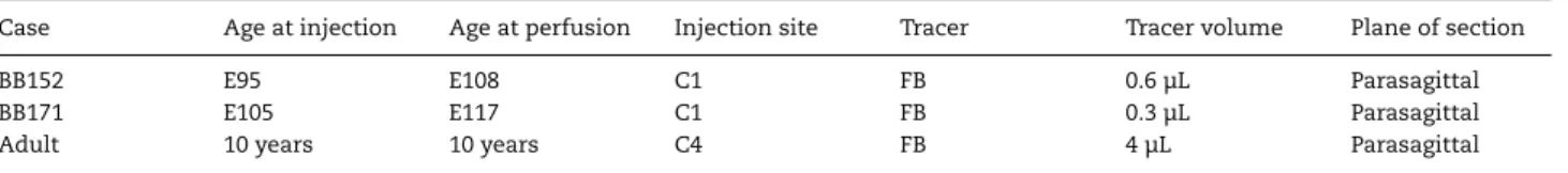
Table 1 Summary of experimental cases
Case Age at injection Age at perfusion Injection site Tracer Tracer volume Plane of section
BB152 E95 E108 C1 FB 0.6 μL Parasagittal
BB171 E105 E117 C1 FB 0.3 μL Parasagittal
Adult 10 years 10 years C4 FB 4 μL Parasagittal
Note: E, embryonic day; FB, Fast Blue.
solutions at 3%) was injected in the lateral cervical spinal cord under visual inspection with a Zeiss binocular surgical microscope control and using glass micropipettes. The fetus was replaced in the uterus, and the incisions were closed. The mother was returned to her cage and was given an analgesic (visceralgine, 1.25 mg, i.m.) twice daily for 2 days.
The pregnant monkey received postoperative medication consisting of a muscular relaxant (ixosuprine chlorhydrate) and an analgesic (tiemonium methylsulfate). Fetuses were delivered by caesarian section after a 12- to 13-day survival period. Animals were deeply anesthetized before being perfused transcardially with 200 mL of 2.7% saline, 1–3 L of a 0.5% glutaraldehyde and 8% paraformaldehyde mixture in 0.1 M phosphate buffer, 0.5 L of 8% sucrose, 0.5 L of 20% sucrose, and 0.5 L of 30% sucrose in phosphate buffer.
Injections of Retrograde Tracers in the Adult Control Identical medication, anesthesia, and monitoring procedures were used as described above. Tracer injection was placed using a Zeiss binocular surgical microscope. The injection was made by means of an Hamilton syringe using a stereotaxic frame. Following injection, muscle and skin were stitched back into position.
A 14-day survival period following tracer injection allowed optimal retrograde labeling of neurons projecting to the injec-tion site. Subsequently the adult animal was anesthetized with ketamine (20 mg/kg, i.m.) followed by a lethal dose of Nembu-tal (60 mg/kg, i.p.) and perfused through the heart with a 8% paraformaldehyde and 0.05% glutaraldehyde solution. After fix-ation, perfusion was continued with 10%, 20%, and 30% sucrose solutions to provide cryoprotection of the brain.
Data Acquisition
Following cryoprotection the brains and spinal cords of the fetuses and the brain of the adult control were immediately removed and blocked. Forty micrometers thick sections were cut on a freezing microtome and mounted in saline onto gelatinized slides. The brains were cut in the parasagittal plane and the spinal cords horizontally. One in 3 sections of the adult brain and 1 in 4 sections in the fetuses were retained to explore CS neuron distribution. Sections were examined in UV light with oil-immersion objectives using a Leitz fluorescence microscope equipped with a D-filter set (355–425 nm). High-precision maps were made using Mercator software running on Exploranova technology, coupled to the microscope stage. At least 1 in every 10 sections (1.6 mm apart in the fetuses and 1.2 mm in the adult) was observed, and labeled neurons were charted across the cortex and subcortical structures. The frequency of charting was increased to 1 in every 2 section in order to ensure a complete sampling of layer 5 in midline structures given the parasagittal plane of section. Controlled high frequency
sam-pling gives stable neuron counts despite curvature of the cortex and heterogeneity of neuron distribution in the projection zones of individual areas (Vezoli et al. 2004,Markov et al. 2014b).
Selections of sections were processed for cytochrome oxidase (CO) and acetylcholine esterase (AChE) using published proce-dures (Hardy et al. 1976,Silverman and Tootell 1987). Character-istics of neurons labeled with FB are described byBentivoglio et al. (1980). Area and/or regional limits were reported on the charts of labeled neurons (Supplementary Table S1). Whenever possible, labeled neurons were attributed to areas of our pub-lished atlas (Supplementary Table S1) based on landmarks and histology (Markov et al. 2014a).
Quantification of Projections and Statistical Analysis All statistical analyses were performed in the R statistical environment (R Development Core Team 2016). We derived the strength or weight of a projection from a given region by the number of labeled neurons in that region. Because the number of labeled cells depends on the amount of tracer injected in each spinal cord, referring to the total number of neurons of each projection is not appropriate. Hence, we calculated a normalized weight index for each projection using the fraction of labeled neurons (FLN). As previously defined (Markov et al. 2011), the FLN is the proportion of cells located in a given source region with respect to the total number of labeled neurons in the cortex. At all ages, FLN values were computed using the interpolated number of neurons in each region, which is obtained by inferring the numbers of labeled neurons on fluorescence sections that were not examined.
Experimental Findings
Injections of tracer were made to one side of the spinal cord so that we could determine the frequency of labeled CS neurons separately for the ipsilateral and contralateral hemispheres. Injections were made in two fetuses: the youngest was injected at E95 and perfused at E108, and the elder was injected at E105 and perfused at E117 (Table 1). In the description of the results we shall refer to these 2 cases by the age at perfusion. In a first instance we address the histological location of the injection sites. Next, in order to determine the areal location of CS projecting neurons in the fetuses we describe how we identified the areal borders with respect to the evolving sulci pattern. In order to obtain an accurate understanding of the changing areal pattern of CS neurons we made counts of labeled neurons, which we first describe in the adult before proceeding to report our findings in the fetuses. Finally, we briefly describe labeled spinal cord projection neurons in the subcortical struc-tures in fetuses, which are considerably more numerous than in the adult counterpart structures, suggesting that they constitute bona fide exuberant projections.
Refinement of the Primate Corticospinal Pathway During Prenatal Development Gomes et al. 659
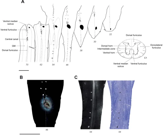
Figure 1. Fluorescent micrograph of E117 injection site. (A) Horizontal sections of the upper spinal cord with the uptake zone of the Fast Blue injection site indicated
in black; (B) Fluorescent micrograph showing the uptake zone outlined in red in section 46 in panel A; (C) high-exposure micrograph caudal to the injection site in section 54 in panel A, showing labeled fibers limited to the injected side of the spinal cord (seeFig. S1for further detail). Black and white arrows highlight the blood vessels present on the midline of the spinal cord. Scale bar: 2 mm.
Location of the Injection Sites
Injections targeted the right side of the spinal cord near the midline in order to maximally involve the spinal gray matter. Injections of FB gave rise to a dense and restricted tracer deposit. We have a relatively precise understanding of the uptake zone of FB (i.e., the spatial extent of the region in which tracer is taken up by axons and transported back to cell bodies) (Bullier and Kennedy 1983,Bullier et al. 1984). The uptake zone corresponds to a region of dense extracellular dye (Fig. 1B). This made it possible to accurately determine the full extent of the effective injection site in the E108 and E117 fetuses, which in both cases was immediately below the pyramidal decussation.
In the E117 fetus the uptake zone involved the spinal inter-mediate and ventral gray matter and the dorsal funiculus (Fig. 1). Importantly, this injection spared the white matter compart-ments known as the dorsolateral and ventral funiculi where the descending fiber pathways are located. Hence, we hypothesize that the labeling from this injection results from uptake and transport from axons that have invaded the gray matter of the spinal cord (seeDiscussion).
Full reconstruction of the E117 injection site showed a mini-mal 150-micron intrusion of the uptake zone to the gray matter of the left side of the cord (see panels 38 and 46 inFig. 1A,B).
This figure shows that there is a conspicuous labeling of the dorsal funiculus running posterior to the injection site, which importantly was observed to be restricted to the right side of the spinal cord (Fig. 1C). We therefore concluded that the minute involvement of the left cord was insignificant as it failed to label the equivalent longitudinal tract on the left side of the spinal cord (Fig. 1CandSupplementary Fig. S1). These observa-tions suggest that the tracer uptake in this injection was very largely restricted to the right hand gray matter of the spinal cord. Inspection of the injection site in the E108 fetus inFigure 2 reveals an extended uptake zone in the right pyramidal tract involving the gray matter. However, there was also an unex-pected, limited, secondary uptake zone restricted to the gray matter–white matter boundary of the left side of the spinal cord (Fig. 2). In the adult, FB was injected at the level of cervical vertebrae C4. In this case, no histology of the injection site in the spinal cord was available, and its location was inferred from the distribution of labeled CS neurons (seeDiscussion). Regional and Presumptive Areal Boundaries with Respect to the Developing Sulci Pattern
In order to determine the developmental changes of CS neurons in the two fetuses and compare this with the adult distribution,
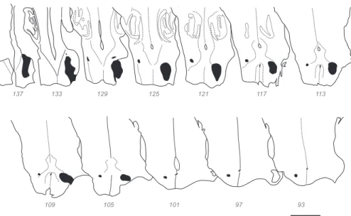
Figure 2. E108 Injection site. Horizontal sections of the upper spinal cord and medulla with the uptake zone of the injection site indicated in black. Scale bar: 2 mm.
it was necessary to parcellate the cerebral cortex across ages into comparable regions with respect to stable landmarks. In the fetal macaque there is a gradual transition from a lissencephalic to a gyrencephalic brain (Sawada et al. 2012). Hence, reference to cortical gyri and sulci as landmarks for areal and regional parcellation is relatively limited particularly at early developmental stages.
At E108 there are 5 clearly identifiable sulci (Fig. 3A,Band left panels inFigs. 5and6): the lateral (ls), the parieto-occipital (pos), the superior temporal (sts), and the circular and the hippocam-pal sulci. Previous studies reported the earliest manifestations of these sulci being between E70 and E100 (Sawada et al. 2012). In addition to the aforementioned sulci, at E108 we were able to detect morphological indications of the presumptive calcarine (cas), central (cs), arcuate (ars), inferior occipital (ios), occip-itotemporal (ots), and anterior middle temporal sulci (amts), which previously had been reported to appear at E80, E90, E90, E90, E120, and E120 (Sawada et al. 2012), respectively.
The older E117 fetus exhibited a significantly more mature sulci pattern, as nearly all the sulci found in the adult could be identified at this stage (Fig. 3C,Dand middle panels inFigs. 5and 6). In addition to those observed in the youngest fetus, at E117 we could identify the lunate (lus), the intraparietal (ips), and the principal sulci (ps), which are reported to appear between E100 and E110 (Sawada et al. 2012). Sulci that were only very weakly formed at E108, such as the central and arcuate sulci, were considerably more marked in the older fetus.
Cortical Landmarks and Cytoarchitecture
Figure 3shows exemplar sections displaying the distribution of CS neurons contralateral to the injection site (i.e., in the left cortical hemisphere) following an injection in the right cervical cord in the fetuses as well as in the adult control. Red dots represent individual FB back-labeled cells. Here and in follow-ing figures the cortex in all 3 brains was subdivided into 8 regions. Colored lines beneath the gray matter–white matter border code the regional location of labeled neurons: prefrontal (red), frontal (green), insular (gray), temporal (orange), parietal (black), occipital (blue), cingulate (pink), and limbic (yellow) (see Supplementary Table S1for individual area allocation).
The adult brain was parcellated according to a 91-area atlas that combines histological criteria with defined landmarks (Markov et al. 2014a). Because the present study uses a parasagittal plane of section, we converted the atlas from the coronal plane into the parasagittal plane using a 7 T MRI scan of the contralateral hemisphere used to create theMarkov et al. (2014a) atlas. ITK-SNAP software (http://www.itksnap.org) made it possible to create the virtual parasagittal plane displaying the appropriate area locations. Histological boundaries were confirmed using Nissl-, CO-, and AChE-stained sections.
Boundary locations in the developing brains were checked using Nissl- (not shown) and AChE-stained sections (Fig. 3). In the youngest fetus the primary visual cortex is readily apparent as a region of very low levels of AChE activity. At more anterior levels presumptive auditory cortex is relatively strongly labeled by AChE. In the E117 fetus, the transition from parietal to frontal cortex in the central sulcus coincided with a decrease in the density and thickness of layer 4 and AChE activity in the frontal cortex. The transition from frontal to prefrontal cortex in the arcuate sulcus was accompanied by an increase in AChE expres-sion in the prefrontal cortex, a feature also perceived in the youngest fetus. The transition from temporal to occipital cortex was indicated by an increase in the thickness of layer 4 and an increase of AChE activity in the occipital cortex. The anterior insular cortex in the posterior orbitofrontal cortex was identified by a decrease in layer 4 thickness and lower AChE activity when compared with neighboring regions. The remaining boundaries were determined using sulcal landmarks.
By assigning the back-labeled cells to individual regions, we were able to quantify the relative proportion of contralateral and ipsilateral CS neurons in the different regions across ages and hemispheres (Fig. 7).
Contralaterally Projecting Neurons
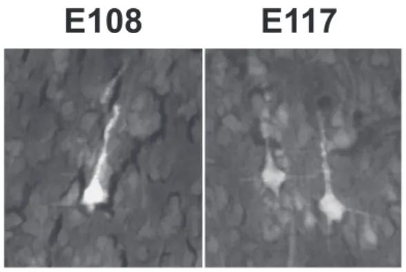
At all ages, the FB back-labeled cells in the cortex are layer 5 pyramidal neurons. Exemplar fetal CS neurons are shown in Figure 4.
In the adult, CS neurons projecting contralaterally are located principally in the frontal lobe and to a lesser extent in the
Refinement of the Primate Corticospinal Pathway During Prenatal Development Gomes et al. 661
Figure 3. Regional boundaries at E108, E117 and in the adult. (A, C, and E) Distribution of retrograde labeled neurons (red dots) in the left hemisphere in parasagittal
sections at equivalent lateral–medial levels following injection in the right cervical spinal cord. Colored lines beneath the white matter indicate the different regions (red, prefrontal; green, frontal; gray, insula; orange, temporal; black, parietal; blue, occipital; pink, cingulate; yellow, limbic). (B, D, and F) Presumptive regional and areal boundaries identified with AChE histochemistry in E108 and E117 and CO histochemistry in the adult. Note the well-defined boundaries of primary visual (V1) and auditory (Core) cortex. For areal abbreviations in this and subsequent figures, seeTable S1. Scale bar: 2 mm.
Figure 4. Exemplar pyramidal-shaped CS back-labeled neurons in layer 5 of the
motor cortex (presumptive F1) in the E108 and E117 fetuses. Scale bar: 20 μm.
parietal and in the cingulate regions (Fig. 5, right column, and Fig. 7). In the frontal cortex CS neurons are found in the primary motor cortex (area F1), and in all 6 subdivisions of the premotor region within the lateral dorsal (areas F2 and F7), lateral ventral (areas F4 and F5), and mesial (areas F3 and F6) cortex where they are restricted to the arm, leg, and trunk representations (Cure and Rasmussen 1954). The maximum density of CS neurons is on the lip of the central sulcus containing area F1 and the representation of movements of the fingers (Fig. 5C,D). In adult parietal cortex CS neurons are observed in areas 3a, 3b, 1/2, 5, 7A/7B, AIP, MIP, LIP, 7op, and SII (Fig. 5A-E).
The peak levels of CS neurons in the adult primary somatosensory area are reported to be located within the representation of the forelimb (wrist and arm) (Kaas et al. 1979). CS neurons are also observed in the insular cortex (Fig. 5B,C) throughout its granular subdivision, principally in the circular sulcus containing the representation of body parts (Robinson and Burton 1980). In addition, CS neurons are observed in multiple limb representations in the cingulate cortex in areas 23c, 24c, and 24d (He et al. 1995) (Fig. 5E,F).
During development there is a massive reduction in the cortical territory containing labeled CS neurons (Fig. 5). In the youngest fetus, outside of the occipital and frontal poles, labeled CS neurons form a near continuous territory spanning many regions lacking CS neurons in the adult. The older fetus showed a small reduction in the spatial extent of CS neurons, which, nevertheless, continue to have a considerably wider distribution compared with that observed in the adult.
Figure 5 distinguishes the distribution of early formed CS neurons, which, according to their location, are destined to be lost in development (marked in red), from those CS neurons that are located in regions where CS neurons are found in the adult (marked in black). This figure reveals the changing distribution of CS neurons projecting to the contralateral spinal cord during development. Those regions that in the adult will contain CS neurons (i.e., black dots inFigure 5) exhibit a continuous band of CS neurons during fetal development in contrast to the dis-continuous distribution observed in the adult. The regions that project transiently (i.e., red dots) during development, namely temporal (orange line), occipital (blue line), prefrontal (red line), and limbic (yellow line) regions, exhibit extensive labeling.
Although the adult temporal cortex virtually sends no pro-jections to the spinal cord (excepting 2 cells in Tpt; Fig. 5B), transient CS projections from the temporal cortex were numer-ous during development. At E108 and E117, CS neurons were observed in many subdivisions of the temporal cortex including
the posterior bank of the lateral sulcus where the primary auditory cortex (area A1) and belt auditory cortex is located in the adult (Fig. 5A-C). In addition, CS neurons in the fetuses were observed in the anterior bank of the superior temporal sulcus, within the superior temporal polysensory area (area STP). In the youngest fetus, transient CS neurons in the temporal cortex were more extensive and included 1) the temporal pole (Fig. 5B); 2) the fundus and posterior bank of the superior temporal sulcus, where presumptive areas TEa/m, TEOm, IPa, FST, MT, and V4t are housed (Fig. 5A-C); 3) the inferior temporal cortex, presumably within areas TE and TH/TF (Fig. 5A,B,D); and finally 4) in the subiculum where sparse labeling was observed (not shown). Overall the temporal cortex contributes 11.7% of the contralat-erally labeled CS neurons at E108 and 5.8% at E117.
The occipital cortex is devoid of CS neurons in the adult and only very sparse projections are present in the region at E108 and virtually none at E117 (Fig. 5). At E108, occipital CS neurons are present in presumptive areas V4 and V2 (Fig. 5B,C) and a few labeled neurons are observed in presumptive con-tralateral V3 (Fig. 5D). At E117, occasional labeled CS neurons are located in presumptive V4 (Fig. 5D). Note that at E108 and E117 no back-labeled CS neurons were found in the primary visual area, area V1. Overall occipital cortex contributes 0.3% of labeled CS neurons in the contralateral hemisphere at E108 and 0.02% at E117.
In the prefrontal cortex there are numerous CS neurons during development that disappear entirely in the adult brain. Between E108 and E117 there is a reduction in the extent of CS neurons (Fig. 5D,E). At E117, only a sparse projection is observed immediately anterior to the frontal–prefrontal boundary in the arcuate sulcus (Fig. 5B-D), in presumptive areas 44, 45B, and FEF. Overall prefrontal cortex contributes 2.3% of contralaterally labeled CS neurons at E108 and 0.33% at E117.
Limbic CS neurons arise from presumptive areas 23a/b, 24a/b, and 32/25 within the cingulate gyrus, in both E108 and E117 (Fig. 5F). Additional CS neurons are observed in presumptive retrosplenial cortex, areas 29/30, at E108 (Fig. 5F). Overall limbic cortex contributes 7.03% of the labeled CS neurons in the con-tralateral hemisphere at E108, and 1.14% at E117.
Ipsilaterally Projecting Neurons
Figure 6 shows the distribution of ipsilateral CS neurons at approximately the same levels as those in Figure 5. Adult ipsilateral CS neurons are located in those cortical areas containing contralateral labeled neurons, but the ipsilateral labeled neurons are found in a more restricted territory (compareFigs. 5Dand6D). As for the contralateral hemisphere, the main source of adult ipsilateral CS neurons is the frontal cortex. Adult parietal cortex has only sparse CS neurons, only slightly more numerous at medial levels (Fig. 6D) and very few CS neurons in the insula (not shown) and cingulate cortices (Fig. 6E,F).
The numbers and distribution of contralateral and ipsilateral CS neurons are much more comparable in the fetus compared with that observed in the adult indicating that there is only a minimal lateralization of the CS tract during early development (Fig. 7and Table 2). Although in the adult 20.1% of the back-labeled neurons were back-labeled in the ipsilateral hemisphere, in the E108 and in the E117 the ipsilateral projection represented 49.2% and 43.5%, respectively, of the cells (Table 2). There is a small contamination of the left spinal cord in the youngest E108 fetus (Fig. 2), which however is not expected to influence
Refinement of the Primate Corticospinal Pathway During Prenatal Development Gomes et al. 663
Figure 5. Developmental refinement of contralateral CS projecting neurons. Sections of left hemisphere at equivalent medio-lateral levels across all three ages
showing the areal distribution of CS pathway projection neurons. Labeled neurons in cortical regions that maintain CS neurons in the adult are in black, and labeled neurons in regions that in the adult lose their projection to the spinal cord are in red. Colored lines indicate different region (see color code inFig. 3). Scale bar: 5 mm.
the ipsilateral projecting CS neuron distribution. Nevertheless because of this contamination we rely principally on the older
fetus for an account of the differences in the ipsilateral and contralateral distributions of CS neurons (Fig. 6).
Figure 6. Developmental refinement of ipsilateral CS projecting neurons. Sections of the right hemisphere at equivalent medio-lateral levels across all three ages
showing the areal distribution of CS pathway projection neurons. Labeled neurons in cortical regions that maintain CS neurons in the adult are in black; labeled neurons in regions that lose their projection to the spinal cord are in red. Colored lines indicate different regions (see colored code inFig. 3). Scale bar: 5 mm.
In the E117 fetus, there is a continuous band of back-labeled cells spanning the full extent of the frontal (green), parietal (black), insula (gray), and cingulate (yellow) regions. In the tem-poral cortex, CS neurons project from 1) the entire auditory
subdivisions located within the posterior bank of the lateral sulcus (Fig. 6A–C), 2) the presumptive temporal pole (Fig. 6B), and 3) the STP subdivisions within the anterior bank of the superior temporal sulcus. In the prefrontal cortex, ipsilateral CS
Refinement of the Primate Corticospinal Pathway During Prenatal Development Gomes et al. 665
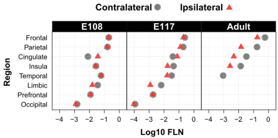
Figure 7. Relative strength of contralateral and ipsilateral projections across ages. Note the relative similarity of projection weights across all ages, the increasing
differences in weights of contralateral and ipsilateral projections with age and the complete loss of the prefrontal, occipital and limbic projections.
Table 2 Number and percentage of neurons in the contralateral and
ipsilateral CS pathway across ages
Age Hemisphere Number of neurons %
E108 Contralateral 300 324 50,8 E108 Ipsilateral 290 994 49,2 E117 Contralateral 118 422 56,5 E117 Ipsilateral 91 222 43,5 Adult Contralateral 17 496 79,9 Adult Ipsilateral 4407 20,1
neurons are present at lateral (Fig. 6A), medial (Fig. 6B-D), and midline (Fig. 6F) levels, in presumptive areas 44, 45B, FEF, and 8B/9. In the occipital cortex, no labeled CS neurons were found in area V1, and only very sparse labeling was observed in the vicinity of the occipital boundary (Fig. 6C). In the limbic region, CS neurons were observed in the cingulate gyrus, in presumptive areas 23a/b, 24a/b, and 25/32 (not shown).
Overall, at all ages and across ipsilateral and contraleral hemispheres, the highest density of CS neurons was always found in the motor cortex, closely followed by the parietal cortex (Fig. 7).
Subcorticospinal Pathways
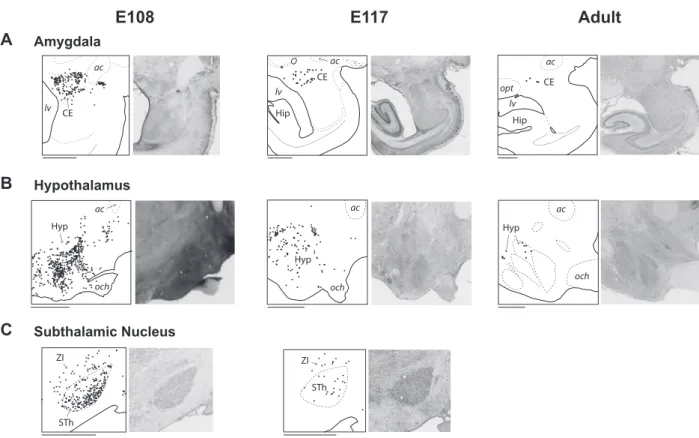
In the present study we found abundant ipsilateral and contralateral subcortical projections to the spinal cord with suggestive evidence of a developmental decrease in projections from the amygdala, hypothalamus and subthalamic nucleus (Fig. 8). In the contralateral hemisphere, at all ages nearly all of back-labeled amygdospinal neurons originated from the central nucleus (Fig. 8A). The projection from the hypothalamus (Fig. 8B) was centered in the dorsal and posterior hypothalamic areas, but during development this projection was much broader, encompassing many hypothalamic subdivisions. The projection from the subthalamic nucleus to the cervical spinal cord (Fig. 8C) was only observed in the fetuses, and no labeled neuron was observed in this nucleus in the adult. The distribution of the ipsilateral subcorticospinal projections is similar (not shown).
Discussion
The progressive acquisition of manual dexterity in primates occurs in parallel with substantial changes in the underlying neuronal circuit indicating a link between functional and
struc-tural maturation during the first year of postnatal life (macaque: Kuypers 1962,Lawrence and Hopkins 1976,Galea and Darian– Smith 1995,Armand et al. 1997,Olivier et al. 1997). Despite the evolutionary importance of the CS pathway in primates, its early development has been substantially less investigated in comparison with non-primates. The 6 main findings of the present study aimed at expanding our understanding of in utero primate CS pathway development are as follows. 1) In the prena-tal macaque the somata of CS neurons in the contralateral and ipsilateral hemispheres are restricted to layer 5. 2) The highest density of labeled CS neurons throughout development and in the adult is observed in the putative motor cortex. 3) CS neurons are much more broadly distributed in the fetus, encompassing regions that are devoid of such neurons in the neonate and adult. 4) While the adult CS pathway is strongly lateralized, during development there are comparable CS neurons in corresponding ipsilateral and contralateral cortical territories. 5) In parallel to the maturation of cortical projections to the spinal cord, there is substantial subcorticospinal maturation, whereby many projections to the spinal cord are eliminated from the amygdala, hypothalamus, and subthalamic nuclei. 6) The primary locus of the uptake zone in the E117 fetus does not encroach on the descending CS tract. Nevertheless this injection centered on the gray matter of a single segment of the spinal cord leads to labeled neurons throughout the full extent of the cortex. This observation indicates an extremely widespread invasion by the descending axons of the CS tract in to the gray matter of the spinal cord.
Technical considerations
We have to rely on the visual identification of the uptake zone in order to infer the effective location of the injection site. Earlier investigations of the uptake zone were carried out in the cortex; however we believe that similar results would be obtained for the spinal cord (Kennedy and Bullier 1985, Condé 1987). An indication that this is indeed the case is provided in the elder fetus. Here the injection goes right up to the midline and is accompanied by an intense longitudinal labeling posterior to the injection site uniquely observed in the right dorsal funiculus. This observation indicates that the uptake zone was effectively restricted to the right side of the spinal cord given that the longitudinal labeling was only seen on this side (Fig. 1 and SupplementaryFig. S1). This argues in favor of the FB uptake zone being largely restricted to the immediate vicinity of the
Figure 8. Subcorticospinal projections at E108, at E117, and in the adult. Parasagittal sections of the left hemisphere showing the distribution of contralateral subcortical
neurons (black dots) projecting to the spinal cord from the amygdala (A), the hypothalamus (B), and the subthalamic nucleus (C). Abbreviations: ac, anterior commissure; CE, central nucleus of the amygdala; Hip, hippocampus; Hyp, hypothalamus; lv, lateral ventricle; och, optic chiasm; opt, optic tract; STh, subthalamic nucleus; ZI, zona incerta. Scale bar: 2 mm.
needle tract as observed for cortical injections (Kennedy and Bullier 1985,Condé 1987).
The injection in the adult was carried out as in the fetus and intended to be restricted to one side of the spinal cord. In both fetuses and adult, injections were made under direct visual control using a high-power binocular surgical microscope. The surgery is considerably easier in the adult macaque and the quality of the visual control superior to that of the fetus. In the fetuses both injections were overall located as intended, the small uptake zone in the left cord in the E108 fetus likely caused by an error in handling of the micropipette subsequent to the correct injection.
In the adult the injection site material was not available for histological examination, so we have to rely on the labeling pat-tern to infer the location of the injection. There are significant differences in the topography of CS neurons reported in the primary motor and premotor areas following injections to the upper and lower cord levels (Liu and Chambers 1964;Kuypers and Brinkman 1970;He et al. 1993;Galea and Darian-Smith 1994, 1995). When the uptake zone of the retrograde tracer includes the dorsolateral column, the cortical territory projecting to the injection site is expanded and includes regions that project not only to the level where the injection is located but also to the lower levels of the spinal cord. In our adult material, cortical ter-ritories known to project to segments of the spinal cord posterior to the intended injected site were seen to contain many FB back-labeled neurons (Fig. 5E,F). Comparison withLiu and Chambers (1964),Kuypers and Brinkman (1970),He et al. (1993), andGalea and Darian-Smith (1994,1995) suggests that the involvement of the dorsolateral funiculus is extensive. Further, a reduced
number of CS neurons in the ipsilateral hemisphere is a strong indication that the injection was restricted to one side of the spinal cord, agreeing with the reduced ipsilateral CS projec-tion described previously in the literature (Liu and Chambers 1964;Galea and Darian-Smith 1994,1995;Dum and Strick 1996; Lacroix et al. 2004).
There is a Massive CS Projection in the Immature Brain The results from the present study show a massive cortical projection in the immature macaque brain to the cervical cord. This finding is in agreement with previous studies in non-primates. In rodents (rat:Leong 1983,Bates and Killackey 1984, Schreyer and Jones 1988), carnivores (ferret: Meissirel et al. 1993), and marsupials (opossum: Cabana and Martin 1984), injections of retrograde tracers in the CS tract lead to the labeling of CS neurons distributed over a much broader cortical territory during development than in the mature animal, including transient projections from the prefrontal and occipital cortex.
Additionally, the results in the oldest fetus of the present study are of particular interest. Here, widespread projections were found following a discrete injection in the gray matter without encroaching on the dorsolateral funiculus and the ventral funiculus, where the descending CS tracts are housed. The widespread distribution of labeled CS neurons in this fetus suggests the existence of extensive transient axonal branching in the gray matter during development. This finding would seem to be at odds with the notion that there is a topological organization of the CS projection and that there is a
Refinement of the Primate Corticospinal Pathway During Prenatal Development Gomes et al. 667
progressive innervation of the spinal gray matter (Donatelle 1977). In the rodent model there is a controversy concerning CS axons transiently entering into the spinal gray matter in regions where they are not present in the adult (Stanfield 1992). A number of earlier studies in the rat report failure to label CS axons entering the spinal gray matter from cortical regions that do not have CS neurons in the adult (transient CS axons) (Joosten and van Eden 1989,Joosten and Bar 1999), while others have found instances of innervation of spinal gray matter by transient CS axons (Stanfield and O’Leary 1985,O’Leary and Stanfield 1986). A recent retrograde study in mouse shows widespread projections to the gray matter of the cervical level of the spinal cord during CS pathway development (Kamiyama et al. 2015).
There are 2 series of observations that suggest that transient CS axons in primates may indeed enter into the spinal gray matter. Firstly, during development there is the possibility that individual CS fibers branch at multiple spinal levels, so that individual CS neurons in the adult could extend their axons to multiple spinal segments (macaque:Shinoda et al. 1981,He et al. 1993). Evidence of this occurs in cat where extensive spinal axonal collateralization with pruning of connections in latter stages is a feature of developing CS axons (Theriault and Tatton 1989). Secondly, there is some evidence of extensive innervation of the spinal gray matter observed in the postnatal monkey shortly after birth (Kuypers 1962,Armand et al. 1997). Likewise, innervation of the spinal gray matter by CS projecting neurons has been suggested in postmortem studies in preterm human (Eyre et al. 2000).
Whether individual CS neurons innervate the spinal gray matter at multiple levels of the cord is extremely important for understanding the developmental refinement of the CS path-way. One could speculate that developmental pruning in the gray matter could lead to greater flexibility of the developmental outcome. Transient projections could be involved in activity-dependent maturation of the spinal motor centers during a critical period of prenatal development, and the disruption of this process could lead to CS pathway perturbations that are observed following cortical lesions, such as those in cerebral palsy (Eyre et al. 2000,Eyre 2007). Note, the notion that tran-sient projections fail to penetrate the gray matter has been argued in the cortex (Innocenti 1981); however, subsequently there has been irrefutable evidence in that such gray matter connections clearly exist (Dehay et al. 1984,1988a). On this issue of innervation of the cord the present results based on the location of the injection site in the E117 fetus and the apparent non-involvement of the dorsolateral funiculus are very indirect. However, our observations point to the need for further studies on the development of the CS pathway using modern antero-grade tracer injection in diverse regions of the cortex in areas that do and do not maintain permanent CS projections in the adult. Exploring the extent of prenatal gray matter innervation in primates will address the extent to which the specificity of the developmental refinement of the primate CS pathway resides in the details of its timing and/or in the pattern of gray matter innervation.
Is There a Homogeneous Distribution of CS Neurons in the Prenatal Brain?
To address this issue we need to allocate labeled neurons to particular areas or regions. At present there is no available atlas for the developing macaque cortex, and only a handful of pub-lications have investigated macaque area identification during
fetal development (Goldman and Galkin 1978;Dehay et al. 1988b, 1989,1993;Barone et al. 1994;Dehay et al. 1996). This led us to combine cytoarchitectonics with sulci and gyri landmarks in the E108 and E117 to define in the developing cortex 8 different cortical territories that can be readily identified in the adult cortex (prefrontal, frontal, insular, temporal, parietal, occipital, cingulate, and limbic). Within these regions, as early as E108 the boundaries of the primary visual and auditory areas can be detected.
Having identified these cortical regions across all fetal and adult stages we are able to determine the developmental decrease in CS neuron number. This showed that the pattern of labeling across regions remains remarkably stable (Fig. 7). Overall, during development and in the adult macaque, the strongest CS projection arises from the frontal cortex and the second strongest from the parietal cortex, followed by the cingulate and insular cortices (Fig. 7). This demonstrates that, prenatally, the regions projecting to the spinal cord are already well defined in terms of differences in numbers of CS neurons. Exuberant Bilateral CS Projections
In the present study we show that in primates, as for the rodent, carnivores, and marsupials, there is a developmental narrowing of the extent of the cortex projecting to the spinal cord (Stanfield et al. 1982,Leong 1983,Bates and Killackey 1984,Cabana and Martin 1984,O’Leary and Stanfield 1986,Sharkey et al. 1986, Joosten et al. 1987,Meissirel et al. 1993). This is not only true for the regions that project in the adult and neonate (He et al. 1993; Galea and Darian-Smith 1994,1995;He et al. 1995) but also from regions that do not have CS neurons postnatally. These include the prefrontal, occipital, limbic, and temporal cortices. This exuberance was found bilaterally. While in the adult macaque there is a dominant contralateral CS projection, this does not appear to be the case prenatally.
During development of the CS pathway in the macaque we observed an extensive ipsilateral projection to the cervical cord. Further, the distribution pattern of back-labeled neurons is comparable across the ipsilateral and contralateral hemispheres (Fig 5and6and Table 2). Most studies investigating CS path-way development in non-primates employed anterograde tracer injections in the cortex and investigated the spatial distribution of the terminal labeling at different spinal levels. Studies in cat show that there are extensive bilateral terminations of CS axons with fairly similar terminal distribution patterns (Alisky et al. 1992,Li and Martin 2000). While the density of labeling in the ipsilateral and contralateral gray matter exhibits an important degree of variability (ipsilateral label densities ranging from 11% to more than 50%), the topography of ipsilateral labeling is relatively consistent (Li and Martin 2000). Hence, unilateral injections of retrograde tracer in kittens could produce relatively similar patterns of labeled neurons in the ipsilateral and con-tralateral hemispheres, in agreement with the findings of the present study.
Exuberant Bilateral Subcorticospinal Projections. A number of studies have examined non-cortical origins of projections to the spinal cord in the adult macaque by 1) lesion-ing descendlesion-ing fiber pathways, 2) injectlesion-ing anterograde trac-ers in different subcortical structures, and 3) injecting retro-grade tracers in the spinal cord (e.g.,Kneisley et al. 1978). These studies mainly focused on the projections from the midbrain
and hindbrain (reviewed inLemon 2008), namely those from the magnocellular red nucleus (rubrospinal), the superior culliculus (tectospinal), the vestibular nuclei (vestibulospinal), and the pontine and medullary reticular formation (reticulospinal).
Additional projections were found from other subcortical structures in the adult brain, but subject to a much less intensive investigation. There are reports of very weak, uniquely ipsi-lateral amygdalospinal projections from the central nucleus of amygdala (Mizuno et al. 1985) and similarly weak and ipsilateral projections from the hypothalamus (Kneisley et al. 1978,Barbas et al. 2003). Rare subcorticospinal projecting neurons have been observed in the subthalamic nucleus (Mizuno et al. 1988).
In the present study we observed subcorticospinal projecting neurons bilaterally in the central amygdala and in the hypotha-lamus of the adult and during development, and we found in the fetuses that these projections appeared considerably more extensive than in the adult (Figure 8). In addition, we observed a transient projection from the subthalamic nucleus to the spinal cord; no labeled neuron was found in the subthalamic nucleus in our adult case. To our knowledge these pathways were not described during development in other species. Future studies would be needed to determine if these pathways are present in non-primates and if they undergo reorganization following early lesions, as observed for CS projections.
Prenatal Lesions and Aberrant Ipsilateral CS Projections Experimental manipulations of the CS system in rodent sug-gest an important degree of plasticity of the projections from the cortex to the spinal cord. These findings are potentially important for clinical observations. First, occipital neurons that are transplanted at early stages of development to the frontal cortex retain their transient projection to the spinal cord (rat: O’Leary and Stanfield 1985). Second, lesions of the sensorimotor cortex during early development lead to an increase in the ipsi-lateral projections from the non-lesioned cortex, with a similar contralateral projection pattern (rat:Reinoso and Castro 1989). While these findings have not been replicated in primate, lesion studies in perinatal human point to similar processes. Prenatal lesions in humans lead to hypertrophy of the CS tract arising from the undamaged (non-affected) hemisphere, which projects to both sides of the spinal cord and innervates motoneuronal pools (Eyre 2007).
The results of our study demonstrate that during primate development there is a bilateral cortical projection to the spinal cord and further that both sets of projections have relatively similar strengths. It could be speculated that the increased ipsilateral projection in human (Eyre 2007) results from the maintenance of the transient ipsilateral projection that we have identified in the present study. A lesion to the cortex may lead to impairments of the normal activity and motor experience-dependent mechanisms of axonal pruning, with subsequent imbalance of ipsilateral pruning (Martin 2005,Eyre 2007). Relevance of the Present Findings for Clinical Medicine The present observations are a reminder of the classic literature on the formation of the adult patterns of connectivity via the extensive role of selective elimination of connections during development of the nervous system (Changeux et al. 1973, Rakic 1976, Innocenti et al. 1977, Cowan et al. 1984, Easter et al. 1985, Finlay et al. 1987, Killackey 1990, O’Leary 1992, Bourgeois et al. 1994, LaMantia and Rakic 1994). These
observations on the role of axonal pruning in the formation of the adult connectivity patterns need to be considered in the light of potentially specific features of primate neurodevelopment. In specific subcortical and cortical pathways of the visual system, the magnitude of transient connectivity has been shown to be less prominent in primates than in other species (Dehay et al. 1988b,Chalupa and Dreher 1991,Kennedy and Dehay 1993,Snider et al. 1998). Primate-specific features can bear high relevance for understanding the medical outcomes subsequent to neurodevelopmental perturbations in humans (Rakic 2009,Magrou et al. 2018). The potential of the macaque model for understanding health issues with respect to spinal cord injury is well established (Courtine et al. 2007). However, in addition to the difficulties originating from species-specific motor system organization, drawing firm conclusions from the extensive literature on CS tract development is problematic. This is largely due to variations in the location of the injection sites and differences in the tracer used and the developmental stages examined. There is clearly a need for a more detailed understanding of the specificity of the CS tract across species. For instance, the recent description of a transient projection of the cortex to motoneuron pools in the mouse (Maeda et al. 2016, Gu et al. 2017,Murabe et al. 2018), cautions against simplification of primate specific features. These considerations point to the need for an in-depth study of the development of the CS pathway in the macaque model. This is of particular importance in the framework of clinical studies, which address the medical outcome of disturbed development of the CS pathway in human (Eyre 2007,Welniarz et al. 2017).
Supplementary Material
Supplementary materialis available at Cerebral Cortex online.
Funding
LABEX CORTEX 11-LABX-0042) of Université de Lyon (ANR-11-IDEX-0007) operated by the French National Research Agency (ANR) (H.K., C.D.); 15-CE32-0016, CORNET (H.K.); ANR-17-NEUC-0004, A2P2MC (H.K.); ANR-17-HBPR-0003, CORTICITY (H.K.); ANR-14-CE13-0036, Primacor (C.D.); Chinese Academy of Sciences President’s International Fellowship Initiative. Grant No. 2018VBA0011 (H.K.); FRC “Rotary-Espoir en Tête” 2018 (H.K. and C.D.); Fondation pour la Recherche Médicale, Equipe FRM-DEQ20160334943 (C.D.)
Authors’ Contributions
Surgery PG, MB, CD, HK; Histological processing ARRG, PG, CD; Data acquisition ARRG, EO, HK; Data analysis and discussion/in-terpretation ARRG, EO, KK, CD, HK; Wrote the paper ARRG, EO, KK, CD, HK; All authors revised successive versions of the manuscript.
References
Alisky JM, Swink TD, Tolbert DL. 1992. The postnatal spatial and temporal development of corticospinal projections in cats.
Exp Brain Res. 88:265–276.
Armand J, Olivier E, Edgley SA, Lemon RN. 1997. Postnatal devel-opment of corticospinal projections from motor cortex to the cervical enlargement in the macaque monkey. J Neurosci. 17:251–266.
Refinement of the Primate Corticospinal Pathway During Prenatal Development Gomes et al. 669
Barbas H, Saha S, Rempel-Clower N, Ghashghaei T. 2003. Serial pathways from primate prefrontal cortex to autonomic areas may influence emotional expression. BMC Neurosci. 4:25. Barone P, Dehay C, Berland M, Kennedy H. 1994.
Developmen-tal changes in the distribution of acetylcholinesterase in the extrastriate visual cortex of the monkey. Dev Brain Res. 77:290–294.
Basu A, Graziadio S, Smith M, Clowry GJ, Cioni G, Eyre JA. 2010. Developmental plasticity connects visual cortex to motoneu-rons after stroke. Ann Neurol. 67:132–136.
Bates CA, Killackey HP. 1984. The emergence of a discretely distributed pattern of corticospinal projection neurons. Dev
Brain Res. 13:265–273.
Bentivoglio M, Kuypers HG, Catsman-Berrevoets CE, Loewe H, Dann O. 1980. Two new fluorescent retrograde neuronal trac-ers which are transported over long distances. Neurosci Lett. 18:25–30.
Bourgeois JP, Goldman-Rakic PS, Rakic P. 1994. Synaptogenesis in the prefrontal cortex of rhesus monkeys. Cereb Cortex. 4:78–96.
Bullier J, Kennedy H. 1983. Projection of the lateral geniculate nucleus onto cortical area V2 in the macaque monkey. Exp
Brain Res. 53:168–172.
Bullier J, Kennedy H, Salinger W. 1984. Bifurcation of subcortical afferents to visual areas 17,18 and 19 in the cat cortex. J Comp
Neurol. 228:309–328.
Cabana T, Martin GF. 1984. Developmental sequence in the origin of descending spinal pathways. Studies using retro-grade transport techniques in the north American opossum (Didelphis virginiana). Brain Res. 317:247–263.
Cabana T, Martin GF. 1985. Corticospinal development in the north-american opossum: evidence for a sequence in the growth of cortical axons in the spinal cord and for transient projections. Develop Brain Res. 23:69–80.
Chalupa LM, Dreher B. 1991. High precision systems require high precision “blueprints”: a new view regarding the formation of connections in the mammalian visual system. J Cognitive
Neurosci. 3:209–219.
Chalupa LM, Killackey HP. 1989. Process elimination underlies ontogenetic change in the distribution of callosal projection neurons in the postcentral gyrus of fetal rhesus monkey. Proc
Natl Acad Sci U S A. 86:1076–1076.
Changeux JP, Courrege P, Danchin A. 1973. A theory of the epigenesis of neuronal networks by selective stabilization of synapses. Proc Natl Acad Sci U S A. 70:2974–2978.
Condé F. 1987. Further studies on the use of the fluorescent tracers fast blue and diamidino yellow: effective uptake area and cellular storage sites. J Neurosci Meth. 21:31–43.
Courtine G, Bunge MB, Fawcett JW, Grossman RG, Kaas JH, Lemon R, Maier I, Martin J, Nudo RJ, Ramon-Cueto A et al. 2007. Can experiments in nonhuman primates expedite the translation of treatments for spinal cord injury in humans? Nat Med. 13:561–566.
Cowan WM, Fawcett JW, O’Leary DDM, Stanfield BB. 1984. Regressive events in neurogenesis. Science. 225:1258–1265. Cure C, Rasmussen T. 1954. Effects of altering the
parame-ters of electrical stimulating currents upon motor responses from the precentral gyrus of Macaca mulatta. Brain. 77: 18–33.
Dehay C, Bullier J, Kennedy H. 1984. Transient projections from the fronto-parietal and temporal cortex to areas 17, 18 and 19 in the kitten. Exp Brain Res. 57:208–212.
Dehay C, Giroud P, Berland M, Killackey H, Kennedy H. 1996. Contribution of thalamic input to the specification of cytoar-chitectonic cortical fields in the primate: effects of bilateral enucleation in the fetal monkey on the boundaries, dimen-sions, and gyrification of striate and extrastriate cortex. J
Comp Neurol. 367:70–89.
Dehay C, Giroud P, Berland M, Smart I, Kennedy H. 1993. Modu-lation of the cell cycle contributes to the parcelModu-lation of the primate visual cortex. Nature. 366:464–466.
Dehay C, Horsburgh G, Berland M, Killackey H, Kennedy H. 1989. Maturation and connectivity of the visual cortex in mon-key is altered by prenatal removal of retinal input. Nature. 337:265–267.
Dehay C, Kennedy H, Bullier J. 1988a. Characterization of tran-sient cortical projections from auditory, somatosensory, and motor cortices to visual areas 17, 18, and 19 in the kitten.
J Comp Neurol. 272:68–89.
Dehay C, Kennedy H, Bullier J, Berland M. 1988b. Absence of interhemispheric connections of area 17 during development in the monkey. Nature. 331:348–350.
Donatelle JM. 1977. Growth of the corticospinal tract and the development of placing reactions in the postnatal rat. J Comp
Neurol. 175:207–231.
Dum RP, Strick PL. 1996. Spinal cord terminations of the medial wall motor areas in macaque monkeys. J Neurosci. 16:6513–6525.
Dum RP, Strick PL. 2002. Motor areas in the frontal lobe of the primate. Physiol Behav. 77:677–682.
Easter SS, Purves D, Rakic P, Spitzer NC. 1985. The changing view of neural specificity. Science. 230:507–511.
Eyre JA. 2007. Corticospinal tract development and its plasticity after perinatal injury. Neurosci Biobehav Rev. 31:1136–1149. Eyre JA, Miller S, Clowry GJ, Conway EA, Watts C. 2000. Functional
corticospinal projections are established prenatally in the human foetus permitting involvement in the development of spinal motor centres. Brain. 123Pt 1:51–64.
Finlay BL, Wikler KC, Sengelaub DR. 1987. Regressive events in brain development and scenarios for vertebrate brain evolu-tion. Brain Behav Evol. 30:102–117.
Galea MP, Darian-Smith I. 1994. Multiple corticospinal neuron populations in the macaque monkey are specified by their unique cortical origins, spinal terminations, and connec-tions. Cereb Cortex. 4:166–194.
Galea MP, Darian-Smith I. 1995. Postnatal maturation of the direct corticospinal projections in the macaque monkey.
Cereb Cortex. 5:518–540.
Goldman PS, Galkin TW. 1978. Prenatal removal of frontal association cortex in the fetal rhesus monkey: anatomical and functional consequences in postnatal life. Brain Res. 152:451–485.
Gu Z, Kalambogias J, Yoshioka S, Han W, Li Z, Kawasawa YI, Pochareddy S, Li Z, Liu F, Xu X et al. 2017. Control of species-dependent cortico-motoneuronal connections underlying manual dexterity. Science. 357:400–404.
Hardy H, Heimer L, Switzer R, Watkins D. 1976. Simultane-ous demonstration of horseradish peroxidase and acetyl-cholinesterase. Neuroscience Letters. 3:1–5.
He SQ, Dum RP, Strick PL. 1993. Topographic organization of cor-ticospinal projections from the frontal lobe: motor areas on the lateral surface of the hemisphere. J Neurosci. 13:952–980. He SQ, Dum RP, Strick PL. 1995. Topographic organization
of corticospinal projections from the frontal lobe: motor
areas on the medial surface of the hemisphere. J Neurosci. 15:3284–3306.
Innocenti GM. 1981. Growth and reshaping of axons in the estab-lishment of visual callosal connections. Science. 212:824–827. Innocenti GM, Fiore L, Caminiti R. 1977. Exuberant projection into the corpus callosum from the visual cortex of newborn cats.
Neurosci Lett. 4:237–242.
Joosten EA, Bar DP. 1999. Axon guidance of outgrowing corti-cospinal fibres in the rat. J Anat. 194(Pt 1):15–32.
Joosten EA, Gribnau AA, Dederen PJ. 1987. An anterograde tracer study of the developing corticospinal tract in the rat: three components. Brain Res. 433:121–130.
Joosten EA, van Eden CG. 1989. An anterograde tracer study on the development of corticospinal projections from the medial prefrontal cortex in the rat. Brain Res Dev Brain Res. 45:313–319.
Kaas JH, Nelson RJ, Sur M, Lin CS, Merzenich MM. 1979. Multiple representations of the body within the primary somatosen-sory cortex of primates. Science. 204:521–523.
Kamiyama T, Kameda H, Murabe N, Fukuda S, Yoshioka N, Mizukami H, Ozawa K, Sakurai M. 2015. Corticospinal tract development and spinal cord innervation differ between cer-vical and lumbar targets. J Neurosci. 35:1181–1191.
Kennedy H, Bullier J. 1985. A double-labeling investigation of the afferent connectivity to cortical areas V1 and V2 of the macaque monkey. J Neurosci. 5:2815–2830.
Kennedy H, Dehay C. 1993. Cortical specification of mice and men. Cereb Cortex. 3:27–35.
Killackey HP. 1990. Neocortical expansion: an attempt towards relating phylogeny and ontogeny. J Cognit Neurosci. 2:1–17. Kneisley LW, Biber MP, LaVail JH. 1978. A study of the origin
of brain stem projections to monkey spinal cord using the retrograde transport method. Exp Neurol. 60:116–139. Kuypers HG. 1962. Corticospinal connections: postnatal
develop-ment in the rhesus monkey. Science. 138:678–680.
Kuypers HG. 1982. A new look at the organization of the motor system. Prog Brain Res. 57:381–403.
Kuypers HG, Brinkman J. 1970. Precentral projections to different parts of the spinal intermediate zone in the rhesus monkey.
Brain Res. 24:29–48.
Lacroix S, Havton LA, McKay H, Yang H, Brant A, Roberts J, Tuszynski MH. 2004. Bilateral corticospinal projections arise from each motor cortex in the macaque monkey: a quantita-tive study. J Comp Neurol. 473:147–161.
LaMantia AS, Rakic P. 1994. Axon overproduction and elimina-tion in the anterior commissure of the developing rhesus monkey. J Comp Neurol. 340:328–336.
Lawrence DG, Hopkins DA. 1976. The development of motor control in the rhesus monkey: evidence concerning the role of corticomotoneuronal connections. Brain. 99:235–254. Lemon RN. 2008. Descending pathways in motor control. Annu
Rev Neurosci. 31:195–218.
Leong SK. 1983. Localizing the corticospinal neurons in neonatal, developing and mature albino rat. Brain Res. 265:1–9. Li Q, Martin JH. 2000. Postnatal development of differential
pro-jections from the caudal and rostral motor cortex subregions.
Exp Brain Res. 134:187–198.
Liu CN, Chambers WW. 1964. An experimental study of the cortico-spinal system in the monkey (Macaca mulatta). The spinal pathways and preterminal distribution of degenerating fibers following discrete lesions of the pre- and postcentral gyri and bulbar pyramid. J Comp Neurol. 123:257–283.
Luo L, O’Leary DD. 2005. Axon retraction and degeneration in development and disease. Annu Rev Neurosci. 28:127–156. Maeda H, Fukuda S, Kameda H, Murabe N, Isoo N, Mizukami H,
Ozawa K, Sakurai M. 2016. Corticospinal axons make direct synaptic connections with spinal motoneurons innervating forearm muscles early during postnatal development in the rat. J Physiol. 594:189–205.
Magrou L, Barone P, Markov NT, Killackey HP, Giroud P, Berland M, Knoblauch K, Dehay C, Kennedy H. 2018. How areal specification shapes the local and Interareal circuits in a macaque model of congenital blindness. Cereb Cortex. 28: 3017–3034.
Markov NT, Ercsey-Ravasz MM, Ribeiro Gomes AR, Lamy C, Magrou L, Vezoli J, Misery P, Falchier A, Quilodran R, Gariel MA
et al. 2014a. A weighted and directed interareal
connec-tivity matrix for macaque cerebral cortex. Cereb Cortex. 24: 17–36.
Markov NT, Misery P, Falchier A, Lamy C, Vezoli J, Quilodran R, Gariel MA, Giroud P, Ercsey-Ravasz M, Pilaz LJ et al. 2011. Weight consistency specifies regularities of macaque cortical networks. Cereb Cortex. 21:1254–1272.
Markov NT, Vezoli J, Chameau P, Falchier A, Quilodran R, Huissoud C, Lamy C, Misery P, Giroud P, Barone P et al. 2014b. The anatomy of hierarchy: feedforward and feed-back pathways in macaque visual cortex. J Comp Neurol. 522: 225–259.
Martin JH. 2005. The corticospinal system: from development to motor control. Neuroscientist. 11:161–173.
Meissirel C, Dehay C, Kennedy H. 1993. Transient cortical path-ways in the pyramidal tract of the neonatal ferret. J Comp
Neurol. 338:193–213.
Mizuno N, Takahashi O, Satoda T, Matsushima R. 1985. Amyg-dalospinal projections in the macaque monkey. Neurosci Lett. 53:327–330.
Mizuno N, Ueyama T, Itoh K, Satoda T, Tashiro T, Shigemoto R. 1988. Direct projections from the subthalamic nucleus of Luys to the spinal cord in the Japanese monkey. Neurosci Lett. 89:13–18.
Murabe N, Mori T, Fukuda S, Isoo N, Ohno T, Mizukami H, Ozawa K, Yoshimura Y, Sakurai M. 2018. Higher primate-like direct corticomotoneuronal connections are transiently formed in a juvenile subprimate mammal. Sci Rep. 8:16536.
Nudo RJ, Masterton RB. 1988. Descending pathways to the spinal cord: a comparative study of 22 mammals. J Comp Neurol. 277:53–79.
O’Leary DDM. 1992. Development of connectional diversity and specificity in the mammalian brain by the pruning of collat-eral projections. Curr Opin Neurobiol. 2:70–77.
O’Leary DDM, Stanfield BB. 1985. Occipital cortical neurons with transient pyramidal tract axons extend and maintain col-laterals to subcortical but not intracortical targets. Brain Res. 336:326–333.
O’Leary DDM, Stanfield BB. 1986. A transient pyramidal tract projection from the visual cortex in the hamster and its removal by selective collateral elimination. Dev Brain Res. 27:87–99.
Olivier E, Edgley SA, Armand J, Lemon RN. 1997. An electro-physiological study of the postnatal development of the corticospinal system in the macaque monkey. J Neurosci. 17: 267–276.
R Development Core Team. 2016. R: A Language and Environ-ment for Statistical Computing. Vienna, Austria: R Founda-tion for Statistical Computing. http://www.R-project.org.