HAL Id: hal-02974726
https://hal.archives-ouvertes.fr/hal-02974726
Submitted on 25 Nov 2020HAL is a multi-disciplinary open access
archive for the deposit and dissemination of sci-entific research documents, whether they are pub-lished or not. The documents may come from teaching and research institutions in France or abroad, or from public or private research centers.
L’archive ouverte pluridisciplinaire HAL, est destinée au dépôt et à la diffusion de documents scientifiques de niveau recherche, publiés ou non, émanant des établissements d’enseignement et de recherche français ou étrangers, des laboratoires publics ou privés.
Chemo-Enzymatic Synthesis of S. mansoni O-Glycans
and Their Evaluation as Ligands for C-Type Lectin
Receptors MGL, DC-SIGN, and DC-SIGNR
Julie Pham, Alvaro Hernandez, Anna Cioce, Silvia Achilli, Giulio Goti,
Corinne Vivès, Michel Thepaut, Anna Bernardi, Franck Fieschi,
Niels-Christian Reichardt
To cite this version:
Julie Pham, Alvaro Hernandez, Anna Cioce, Silvia Achilli, Giulio Goti, et al.. Chemo-Enzymatic Synthesis of S. mansoni O-Glycans and Their Evaluation as Ligands for C-Type Lectin Receptors MGL, DC-SIGN, and DC-SIGNR. Chemistry - A European Journal, Wiley-VCH Verlag, 2020, 26 (56), pp.12818-12830. �10.1002/chem.202000291�. �hal-02974726�
Chemo-enzymatic Synthesis of S. mansoni O-glycans and their Evaluation as Ligands for C-type Lectin Receptors MGL, DC-SIGN and DC-SIGNR
Julie Pham1, Alvaro Hernandez1,2, Anna Cioce1, Silvia Achilli3,4, Giulio Goti5, Corinne Vivès3,
Michel Thepaut3, Anna Bernardi5, Franck Fieschi3, Niels Reichardt*1,6,7
1Glycotechnology Group, CIC biomaGUNE, Paseo Miramón 182, 20014 San Sebastian, Spain 2Asparia Glycomics S.L., Mikeletegi 83, 20009 San Sebastian , Spain
3Univ. Grenoble Alpes, CNRS, CEA, Institut de Biologie Structurale, 38100 Grenoble, France 4Present address: DCM, UMR 5250, Université Grenoble Alpes, CNRS, 38000 Grenoble
(France)
5Dipartimento di Chimica, Università degli Studi di Milano, via Golgi 19, 20133 Milano, Italy 6 CIBER-BBN, Paseo Miramón 182, 20014 San Sebastian, Spain
7Basque Research and Technology Alliance (BRTA), Paseo Miramón 182, 20014 San
Sebastian, Spain
nreichardt@cicbiomagune.es
Abstract
Due to their interactions with C-type lectin receptors (CLRs), glycans from the helminth Schistosoma mansoni represent promising leads for treatment of autoimmune diseases, allergies or cancer. We chemo-enzymatically synthesized nine O-glycans based on the two predominant O-glycan cores observed in the infectious stages of schistosomiasis, the mucin core 2 and the S. mansoni core. The O-glycans were fucosylated next to a selection of N-glycans directly on a microarray slide using a recombinant fucosyltransferase and GDP-fucose or GDP-6-azidoGDP-fucose as donor. Binding assays with fluorescently labelled human CLRs DC-SIGN, DC-SIGNR and MGL revealed the novel O-glycan O8 as the best ligand for
MGL from our panel. Significant binding to DC-SIGN was also found for azido-fucosylated glycans. Contrasting binding specificities were observed between the monovalent carbohydrate recognition domain (CRD) and the tetravalent extracellular domain (ECD) of DC-SIGNR.
Introduction
S. mansoni glycans play an important role in host infection by helminths and mounting evidence suggests that they contribute to the parasite evasion of the host immune system by hijacking C-type lectin receptors (CLRs). CLRs are crucial components in the immune system acting as pattern recognition receptors (PRRs).[1,2] Their interaction with pathogen
associated molecular patterns (PAMPs) at the pathogen surface can lead to a series of signalling events ultimately resulting in a skewed immune response.[3,4] The glycans on the
surface of S. mansoni are thought to be responsible for the characteristic TH2/Treg response
observed during chronic schistosomiasis.[5] Concomitantly, reduced disease related
inflammation was observed in autoimmune pathologies such as multiple sclerosis, rheumatoid arthritis, type I diabetes and inflammatory bowel diseases for patients infected with helminths or treated with parasite extracts.[6]
Although yet to be fully assigned, the S. mansoni glycome displays a rich array of immunogenic glycan epitopes such as LeX and LDNF which are known to interact with host
glycans. Indeed, the S. mansoni specific O-glycan core, which is predominantly found in the cercariae, closely resembles an O-glycan core found in the intermediate snail host Biomphalaria glabrata.[7,8] On the other hand, the mucin core 2, which is abundantly found
in mammalian host mucosal tissues, is predominantly found in the helminth eggs.[9] Thus, S.
mansoni is strongly suspected to employ glycan mimicry as a strategy to subvert the host immune system to allow parasitic survival.[10]
The immunomodulatory properties of S. mansoni O-glycans place them as potentially interesting lead compounds for the development of carbohydrate-based drugs to treat immuno-compromised patients with autoimmune diseases, allergies or cancer. However, most studies have focused on parasite N-glycans or the antigenic motifs alone and only few describe the relevance of parasite O-glycans and their interactions with CLRs.[11,12] This is
mainly due to the challenge of isolating pure compounds in sufficient amounts for functional and diagnostic tests.[13–15]
The aim of our study was to investigate the interaction of S. mansoni O-glycans with key CLRs and identify lead structures for improved targeting of immune cells via specific CLR-glycan interactions. To this end, we report, here for the first time, the chemoenzymatic synthesis of several S. mansoni O-glycans and evaluate their interactions with fluorescently labelled human CLRs DC-SIGN, DC-SIGNR and MGL.
Results and Discussion
The retrosynthetic analysis of the O-glycan cores O1 and O2 (Scheme 1) suggested a
convergent strategy involving three building blocks. After conjugation of the GalNAc donor
1 with a protected amine linker 2, glycosylation in O-3 with galactose donor 7 would
provide the Galβ1-3GalNAc disaccharide 5 common to both cores. A second glycosylation in
O-6 with galactose (Gal) 7 or N-acetylglucosamine (GlcNAc) donor 8 would furnish the S.
Scheme 1. Retrosynthetic analysis for the S. mansoni core and core 2 trisaccharides O1 and O2
To facilitate the immobilisation of final compounds as ligands on glycan arrays or to carrier proteins, the known galactosamine trichloroacetimidate donor 1[16] (Scheme 1) was
conjugated with N-benzyl-benzylcarbamate protected aminopentyl linker 2[17] to obtain
glycoconjugate 3 in 73%. Treatment of 3 with a guanidine/guanidium solution
quantitatively hydrolyzed all acetates without affecting the base-sensitive N-Troc group.[18]
Subsequent acetal protection of O-4 and O-6 with benzaldehyde dimethyl acetal under acid catalysis provided 4 in 70%. Glycosylation of the remaining free O-3 with the galactose
imidate 7 under TMSOTf catalysis then afforded disaccharide 5 in 62% yield, as a common
intermediate in the synthesis of both core structures. Reductive ring opening of the benzylidene acetal in 5 with BH3·THF and catalytic TMSOTf furnished the 4-benzyl ether 6 in
a moderate 61% yield. Finally, glycosylation at O-6 either with galactose donor 7 or GlcNAc
Scheme 2. Convergent synthesis of the partially protected S. mansoni and mucin 2 cores 11
and 12. Reagents and conditions: a)Linker 2, TMSOTf, DCM, -40°C, 73%; b)i) G/GHNO3,
MeOH/DCM; ii) PhCH(OMe)2, cat. CSA, DCM, 70%; c) Donor, TMSOTf, DCM, -40°C, 62%; d)
1M BH3.THF, TMSOTf, DCM, 61%; e) Donor 7, TMSOTf, DCM, -41°C, 40%; f) Donor 8,
TMSOTf, DCM, -40°C, 73%; g)i)1M TBAF, THF, reflux; ii) Ac2O, pyridine; iii) 0.5M NaOMe,
MeOH, 70-90%
For the enzymatic diversification of the core structures with recombinant glycosyltransferases both O-glycan cores were partially deprotected. The benzyl protection was left untouched at this stage of the synthesis to aid in the separation of products by HPLC after enzymatic modifications. Deprotection of the N-Troc functionality under standard conditions with LiOH or zinc/acetic acid amalgam failed to provide clean products but refluxing the compounds 9 and 10 in 1M TBAF in THF afforded the clean S. mansoni
Both core structures were then diversified by enzymatic glycosylations to access the Gal1-4GlcNAc (LN) and GalNAc1-Gal1-4GlcNAc (LDN) motifs typically observed in the cercarial and egg glycomes of S. mansoni. Towards this end, we employed three recombinant glycosyltransferases, a bovine beta-1,4-galactosyltransferase (β4Gal-T1), a double mutant (Y289LC342T) conferring beta-1,4-N-galactosaminyltransferase (DMGalT1) activity and a Neisseira meningitidis beta-1,3-N-acetylglucosaminyltransferase (LgtA_X) construct prepared exclusively for this purpose (scheme 3).[23–25] The new LgtA_X construct contains
two polyhistidine tags and a thioredoxin domain to increase LgtA purity and solubility, respectively.
Scheme 3.Enzymatic diversification of cores structures 11 and 12 yielding glycans 13 (57%), 14 (28%), 15 (49%), 16 (61%) and 17(59%).
The general substrate and donor specificity of LgtA, including a comparison with the human homologue B3GnT2, has previously been described by Blixt et al. Employing N-glycan G5
(Figure 1) we confirmed the enzymatic activity of LgtA_X to be similar to LgtA in solution, i.e. for the elongation of terminal galactose with GlcNAc. To evaluate the usefulness of the
enzyme for diversification of the S. mansoni core type trisaccharide we assessed the enzyme selectivity and activity on trisaccharide 11. Incubation of the O-glycan core with 2
potential acceptor sites afforded 57% of a β1-6 monosubstituted product 13, next to only
7% of the bis-substituted product.[26]Errore. Il segnalibro non è definito.
Before enzymatic introduction of an acid-labile fucose residue we decided to remove remaining benzyl and Cbz protecting groups by hydrogenation. Protected glycans 11-17
were dissolved in a mixture of H2O/MeOH/AcOH, and hydrogenated under Pd/C(10%)
catalysis under atmospheric pressure of H2(g), which afforded the deprotected final glycans
O1-O7 in 40-100% yield (Figure 1).[27]
For the synthesis of the proposed Galβ1-4[Fucα1-3]GlcNAc (LeX)-type and
GalNAcβ1-4[Fucα1-3]GlcNAc (LDNF) structures we employed a recombinant α1,3-fucosyltransferase from Caenorhabditis elegans (CeFUT6) and a commercial Heliobacter pylori α-1,3 fucosyltransferase (HP-FucT). CeFUT6 has a known Lewis-X fucosylation activity which was employed in this study to yield O8 from O7 in 73% yield.[28] HP-FucT has a broad substrate
and donor specificity including the C-6 azido-fucose surrogate (FucZ).[29,30] Introducing an
azide function as a biorthogonal handle in the molecule opened the possibility of generating glycomimetics by copper-catalyzed cycloaddition with alkynes to afford a library of novel glycomimetics with potentially improved affinity towards CLRs.[31–34]
We decided to employ a microarray platform for the rapid preparation of fucosylated and azido-fucosylated compounds. Performing the reactions in parallel and on a microscale decreases the need of expensive azido-donor by several orders of magnitude compared to the solution phase synthesis of individual compounds. The synthetic parasite O-glycans O1-O8 were printed alongside a set of N-glycans which were chosen as additional fucose
acceptors from a glycan library available in our laboratory onto NHS activated indium tin oxide (ITO) slides (Figure 1) .[35,36] The bio-functionality of the printed glycans was confirmed
by incubating the array with the plant lectin peanut agglutinin (PNA) ECD recognizing Galβ1-3GalNAc residues (see SI, Figure S4) [37,38]
Figure 1. a) Selection of ligands immobilized on the microarray; b) surface functionalization
of the ITO slide
Initially, we screened donor and HP-FucT enzyme concentrations to optimize on-chip fucosylation for printed glycans. Yields for the enzymatic on-chip glycosylation were determined by MALDI-TOF MS as ratios of the sum of product signal intensities (glycan#-F for fucosylation and glycan#-Z for azido-fucosylation) to remaining starting material signal intensities (Figure 2, A). Plotting the product ratios for the different glycans showed no increase in product formation beyond an enzyme concentration of 189 mU ml-1 (Figure 2, B)
possibly due to enzyme precipitation at higher concentration.
Repeated glycosylation with freshly prepared enzyme solution and 1mM of GDP-fucose produced higher product conversion than a single exposure with 2mM donor concentration (Figure 2, C). While for glycosylation with the natural GDP-fucose donor, two incubation cycles were sufficient to reach a maximum conversion between 65-90%, similar conversion yields employing the GDP- 6-azido-fucose required 3 cycles.
Figure 2. Optimisation of reaction conditions for on-chip fucosylation; a) MALDI-TOF MS of
fucosylation of O7; b) Effect of enzyme concentration; c) Fucose donor concentration; D)
Azido fucose donor concentration on product conversion
Product conversion yields showed a certain degree of structure-dependent variability but were altogether above 80% for the fucose addition and above 75% for the azido-fucose addition (see SI, Figure S5).
As a general note to the reader unfamiliar with glycan array binding experiments, the binding data between different arrays were only compared in relative terms i.e. by comparing ligand binding profiles. Apart from the underlying binding affinity between carbohydrate ligand and lectin, the absolute fluorescence intensities given as RFU values depend on the degree of protein labelling, the incubation time, the washing conditions and image acquisition parameters among others. Some of these parameters are more easily controlled than others and efforts are under way in many labs to improve reproducibility of glycan arrays to allow for a comparison of data between arrays and different immobilisation platforms. Unless an internal reference is used to normalise fluorescence values between different slides or arrays, or extreme measures are put in to place to reproduce incubation and analysis conditions for all slides, absolute fluorescence values between different arrays should not be compared.
The effect of the spatial organization of carbohydrate binding domains on glycan interaction was first assessed by comparing the fluorescent binding profiles of the monovalent CRD and the tetrameric ECD of DC-SIGNR on the unmodified array (Figure 3). DC-SIGNR is an endothelial tetrameric C-type lectin receptor that binds to high mannose and complex glycans found on surface glycoproteins of ebola, HIV or hepatis virus.[39] It has
also been suggested as an entry path for S. mansoni larvae that begin their migration towards the intestine and liver.[40] The glycan binding profile of DC-SIGNR has been
previously studied by glycan array and frontal affinity chromatography. Strong binders include high mannose and hybrid structures and to a lesser extent galactosylated complex glycans with terminal GlcNAc residues.[41–43] Lacking high mannose and hybrid structures on
our array, we found strongest binding to the ECD domain for glycans with terminal GlcNAc (G4, O2 and O3). The CRD showed preferential binding towards glycans with a S. mansoni
core and galactose residues G10, G14 and O4, while mucin core derivatives O2 and O3
which strongly bound the ECD construct did not show interaction with the isolated
carbohydrate recognition domain CRD. In addition, and contrary to the ECD, fucosylated
glycans (O8, G11, G13 and G7) displayed enhanced lectin binding compared to their
Figure 3. Relative fluorescence intensities and fluorescence image for the unmodified array
after incubation with DC-SIGNR CRD (10μg/ml, orange bars) and DC-SIGNR ECD (50µg/ml, blue bars). Each bar in the histogram represents the average of fluorescence from 3 spots which were normalized to G10 and the propagation of error is shown as an error bar.
The observed differences in binding profiles towards monovalent CRD and tetravalent ECD receptor organization are striking and have been observed previously for other systems.[44]
Based on our array binding data we can only speculate on the underlying molecular basis determining the observed change in specificity. Rapid rebinding to multiple equivalent binding sites in close proximity could improve binding to some structures, while others could face additional binding penalties due to steric clashes in the tetrameric lectin organization. In addition, chelation could favor binding to compounds which are capable of interacting with more than one binding site. This observation could have practical implications for lectin binding studies as for simplicity and practical reasons these are often
0 25 50 75 100 125 Monovalent CRD Relative glycan recognition (%)
0 100 200 300 400 G1 G2 G3 G4 G5 G6 G7 G8 G9 G10 G11 G12 G13 G14 G15 G16 O1 O2 O3 O4 O5 O6 O7 O8 Tetravalent ECD
Relative glycan recognition (%)
carried out on the monomeric CRD, which is easier to express and purify. The natural presentation of DC-SIGNR, however, is determined by the organization via the stem region into defined tetramers. Any development of multivalent inhibitors should therefore take the natural receptor organization into account. DC-SIGNR, unlike DC-SIGN, binds to neoglycoconjugates presenting all Lewis antigens except Lewis X epitopes.[42] We therefore
turned our attention to DC-SIGN. Recently, we reported complementary binding selectivity towards one of the isomeric N glycans G3/G8 and G4/G9 by DC-SIGN and DC-SIGNR.[43]
DC-SIGN is found on dendritic cells and macrophages as a tetrameric lectin and displays specificity for mannose, fucose and GlcNAc residues. N-glycans bearing LeX and LDNF
epitopes in the soluble egg antigens have been shown to bind to DC-SIGN and inhibit DC activation.[45,46] Incubation of the unmodified initial array with fluorescently labeled
DC-SIGN ECD showed strongest binding to glycans presenting LeX (G10, O8) or LDNF (G11-G13)
glycan determinants but not to the bis-core fucosylated N-glycan G7 (Figure 4) possibly due
to the impact of core fucosylation on glycan conformation and hence lectin recognition.[47]
Glycans G3, G4 and O3 presenting a free GlcNAc residue on the 6-arm barely showed
interaction with the lectin. However, in a previous screen on a glycan array with different surface architecture they have been shown to bind to DC-SIGN with similar strength as LDNF structures.[43] Among the strongest binders in our previously reported screen were
bi-antennary glycans with terminal GlcNAc residues and high mannose structures, not included in the present array.
Figure 4. Binding profile of the array immobilized glycans with DC-SIGN ECD (10μg/ml).
Histogram bars represent the average fluorescence from 3 spots and error bars represent the standard deviation.
After on-chip fucosylation with HP-FucT, DC-SIGN bound to all enzymatically extended structures, though the spot-to-spot variability increased considerably, possibly due to partial removal of the non-covalently attached glycoconjugates during prolonged washing procedures (Figure 5). 0 5 10 15 20 G1 G2 G3 G4 G5 G6 G7 G8 G9 G10 G11 G12 G13 G14 G15 G16 O1 O3 O4 O5 O2 O6 O7 O8 RF U/ 10 00
Figure 5. Glycan binding of glycan array with tetravalent DC-SIGN ECD (10µg/ml) after
on-chip fucosylation with HP-FucT. Each bar in the histogram represents the average of fluorescence from 3 spots and the standard deviation is given as an error bar.
Nevertheless, the population of fucosylated O-glycans in our array bound with intensity similar to the fucosylated N-glycans, making them equally as valuable in the analyses of CLR-glycan interaction, as previously stated by Van Diepen et al. [48]
Lewis X binding to DC-SIGN CRD is thought to be favoured by Van der Waals interactions between the fucose C-2 and the neighbouring V351.[49] However, given the limitations in
glycan-lectin interaction predictions, it was difficult to predict whether the azide modification at C-6 would affect glycan binding.[50,51] Despite also showing an increased
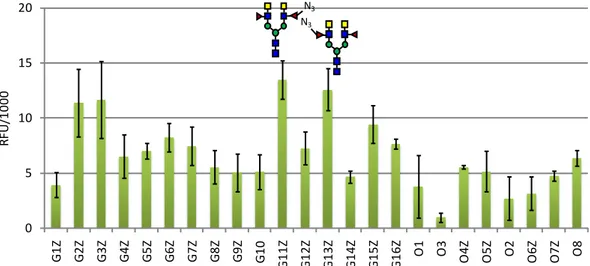
degree of variability, DC-SIGN bound to all azido-fucosylated structures of our array, indicating that the C-6 modification does not significantly contribute to glycan-CLR interaction (Figure 6). Additionally, it was interesting to note that glycans G11Z and G13Z
containing an LDNF and azido-LDNF epitope were observed to be the highest binders.
Figure 6. Glycan binding profile for DC-SIGN ECD (10μg/ml) after modification of the glycan
array with the C6-azido-fucose. Each bar in the histogram represents the average fluorescence for 3 replicate spots and the standard deviations are given as an error bar.
0 5 10 15 20 G1 F G2 F G3 F G4 F G5 F G6 F G7 F G8 F G9 F G1 0 G1 1F G12 G1 3F G1 4F G1 5F G1 6F O1 O3 O4F O5F O2 O6F O7F O8 RF U/ 10 00 0 5 10 15 20 G1 Z G2 Z G3 Z G4 Z G5 Z G6 Z G7 Z G8 Z G9 Z G1 0 G1 1Z G1 2Z G1 3Z G1 4Z G1 5Z G1 6Z O1 O3 O 4Z O5Z O2 O6Z O7Z O8 RF U/ 10 00 N3 N3
Finally, we looked at macrophage galactose lectin (MGL) binding to our collection of immobilised O- and N-glycans. MGL is a PRR expressed at the cell surface of macrophages and dendritic cells that recognizes predominantly terminal GalNAc residues found on Tn antigen glycosylated mucins overexpressed on cancer cells or the GalNAcβ1-4GlcNAc (LDN) disaccharide motif of the parasite S.mansoni.[52] Targeting MGL with high affinity glycans or
glycomimetics has been suggested as a strategy for macrophage targeting in cancer immune therapy and drug delivery.[53,54] In line with its reported binding specificity, we
found strong interactions of MGL with all glycans presenting terminal GalNAc residues of the unmodified array (Figure 7). We observed a low degree of oligosaccharide context dependent alteration of the interaction of GalNac residues with MGL. A similar binding strength was seen for the isolated LDN epitope G16 and glycans presenting the LDN motif
on one or both antennae.[43] Glycan O5 with an asymmetrically extended S. mansoni core,
presenting a single GalNAc residue, was the strongest binding ligand on the array.
Figure 7. Glycan binding profile with MGL ECD (10μg/ml). Histogram bars represent the
average fluorescence from 3 spots and the standard deviation is given as an error bar.
Molecular modelling studies suggest that the presence of a α1,3 fucose residue would disrupt the chelation of the GalNAc 3-OH with calcium at the binding site but our MGL binding profile results does not support this generalized rule.[55] Although lower binding was
observed for the LDNF glycan G12 than for the isolated LDN epitope G16, both
mono-fucosylated bi-antennary glycans G11 and G13 showed higher fluorescence intensities than
the non-fucosylated compound G6. In addition, we were surprised to observe binding of
the LeX epitope G10 with a similar binding strength as for the LDNF epitope G12, neither of
which was anticipated to bind MGL as fucosylation should inhibit binding. These results suggest that fucosylation may indeed, induce alternate binding modes for non-GalNAc ligands in MGL CRD.
On-chip enzymatic fucosylation led to a generally reduced binding to MGL with the exception of O5F and G2F which maintained high affinities (Figure 8, a).
0 5 10 15 20 25 30 G1 G2 G3 G4 G5 G6 G7 G8 G9 G10 G11 G12 G13 G14 G15 G16 O1 O3 O4 O5 O2 O6 O7 O8 RF U/ 10 00
Figure 8. Fluorescence intensities for a) fucosylated y and b) the azido-fucosylated glycan
array after incubation with MGL ECD (10μg/ml). Histogram bars represent the average fluorescence from 3 spots and the standard deviation is given as an error bar.
Despite the spot-to-spot variability increasing considerably here too, the comparison of relative binding profiles showed that the elongation with 6-azido-fucose decrease binding to most of the immobilized glycans with the exception of G2Z, G4Z and G6Z which
remained relatively unchanged (Figure 8, b). Conclusions
We have chemo-enzymatically synthesized eight novel O-glycans based on the S. mansoni specific core and the mammalian mucin core 2. The unexpected regioselectivity for the 6-arm of the S. mansoni core, observed for the recombinant glycosyltransferase LgtA_X, provided a simple entry into products with enzymatic elongations on the 6-arm. The eight synthesized O-glycans were printed alongside a number of N-glycans and were enzymatically modified in good to excellent yields with fucose and non-natural 6-azido-fucose. Glycan array based preliminary binding studies with the human C-type lectin receptors MGL, DC-SIGNR and DC-SIGN showed interesting novel insights into the fine specificity of CLR-glycan binding. This study highlights the potential of employing glycan arrays to identify leads for the further development of glycomimetics with improved CLR
targeting properties. In addition, our comparison of the binding profiles of CRD and ECD domains of DC-SIGNR underscores the importance of multivalent receptor presentation for inducing binding with an additional level of glycan selectivity.
Experimental Part Chemical Synthesis
5-(benzyl(benzyloxycarbonyl)amino)pentyl3,4,6-tri-O-acetyl-2-deoxy-2-((2,2,2-trichloro-ethoxycarbonylamino) β-D-galactopyranoside 3
To a solution of 1[16](11.8 g, 18.9 mmol) in dry DCM (30ml) on activated molecular sieves
and under argon was added a solution of N-benzyl-N-(5-hydroxypentyl)carbamate (7.42 g, 22.7 mmol) 2 in dry DCM (200 ml). The solution was placed at 0°C before TMSOTf (0.690 ml,
3.8 mmol) was added dropwise. The reaction was allowed to warm to RT and stirred for 2hours after which TLC showed full consumption of starting material. The reaction mixture was quenched with Et3N and filtered through celite. The crude sample was purified by flash
column chromatography (1540% EtOAc:Tol) to yield 3 as a white gum, 11 g, 73%. 1H NMR
(500 MHz, CDCl3) δ 7.41 – 7.23 (m, 9H, Ar), 7.17 (d, J = 8.1 Hz, 1H, Ar), 5.36 (d, J = 3.3 Hz, 1H,
H-4), 5.25 – 5.09 (m, 3H, H-3, CH2Bn), 4.79 – 4.59 (m, 2H, CH2Troc), 4.59 – 4.40 (m, 3H,
H-1,CH2Bn), 4.14 (qd, J = 11.2, 6.7 Hz, 2H, 2H-6), 3.94 – 3.70 (m, 3H, H-5, H-2, CHlinker), 3.50 –
3.25 (m, 1H, CHlinker), 3.23 – 3.12 (m, 2H, CH2), 2.14 (s, 3H, OCH3), 2.05 (s, 3H, OCH3), 1.99 (s,
3H, OCH3), 1.59 – 1.43 (m, 4H, 2CH2linker), 1.39 – 1.18 (m, 2H, CH2linker).13C NMR (126 MHz,
CDCl3) δ 170.45, 170.37, 156.77, 156.30, 154.38, 137.79, 136.74, 128.57, 128.44, 127.92,
127.81, 127.73, 127.35, 127.20, 101.25, 95.67, 74.30, 70.50, 69.86, 69.73, 67.21, 66.76, 61.48, 52.73, 50.48, 50.37, 47.28, 46.10, 29.04, 28.66, 27.76, 26.99, 22.96, 20.69, 20.65. HRMS (MALDI-Tof): m/z calcd. for C35H43Cl3N2O12 [M+Na]+ : 811.1778, found: 811.1728.
[α]D20 = -11.8° (c=1, CHCl3)
5-(benzyl(benzyloxycarbonyl)amino)pentyl4,6-O-benzylidene-2-deoxy-2-((2,2,2-trichloro-ethoxycarbonylamino) β-D-galactopyranoside 4
Compound 3 (10.2 g, 12.9 mmol) was dissolved in G/GHNO3[18](700 ml) and the solution
was stirred at RT for 15 mins until full consumption of starting material was observed by TLC. The reaction mixture was concentrated in vacuo then washed thoroughly with DCM and filtered through celite. The filtrate was concentrated to yield a pale pink residue. This was dissolved in dry acetonitrile (100 ml) under argon and benzaldehyde dimethyl acetal (3.4 ml, 2.25 mmol) and camphor sulfonic acid (600 mg, 2.6 mmol) were added. The reaction was stirred at RT overnight after which successful conversion was observed by TLC. Et3N was added and the reaction mixture was concentrated in vacuo. The resulting residue
was purified by flash column chromatography (310% MeOH:DCM) to yield 4 as a white
solid, 6.78 g, 70%. 1H NMR (500 MHz, CDCl 3) δ 7.51 (d, J = 5.2 Hz, 2H, Ar), 7.42 – 7.22 (m, 11H, Ar), 7.17 (d, J = 7.0 Hz, 2H, Ar), 5.57 (s, 1H, CHPh), 5.23 – 5.13 (m, 2H, CH2Ph), 4.72 (d, J = 11.7 Hz, 2H, CH2Troc), 4.50 (m, 3H, H-1, CH2Bn), 4.32 (d, J = 12.4 Hz, 1H, H-6a), 4.20 (d, J = 3.6 Hz, 1H, H-4), 4.08 (dd, J = 12.4, 1.9 Hz, 1H, H-6b), 4.03 – 3.82 (m, 2H, H-3, CH2), 3.65(m, 1H, H-2), 3.5 (s, 1H, H-5), 3.43-3.25(m, 1H, CHlinker), 3.25-3.15(m, 2H, CH2), 1.62 – 1.41 (m, 2H, 2CH2linker), 1.39 – 1.16 (m, 2H, CH2linker).13C NMR (126 MHz, CDCl3) δ 152.34, 151.88, 150.76, 150.49, 134.39, 134.36, 134.34, 134.15, 133.47, 133.32, 126.34, 125.72, 125.69, 125.60,
125.43, 125.16, 125.10, 124.98, 124.88, 124.57, 124.54, 124.39, 123.71, 99.78, 99.53, 99.33, 94.60, 75.14, 75.07, 74.49, 70.66, 70.37, 69.68, 69.62, 69.52, 67.75, 67.72, 66.93, 56.43, 51.84, 51.71, 48.87, 47.78, 31.72, 31.33, 30.52, 29.75, 26.02, 25.90. HRMS (MALDI-Tof) m/z calcd. for C36H41Cl3N2O9 [M+Na]+: 773.1774, found: 773. 1772. [α]D20= -3.5° (c=1,
CHCl3)
5-(benzyl (benzyloxycarbonyl)amino) pentyl 2,3,4,6-tetra-O-acetyl-β-D-galactopyranosyl- (1→3)-4,6-O-benzylidene-2-deoxy-2-((2,2,2-trichloroethoxycarbonylamino)β-D-galacto-pyranoside 5
To a solution of 4 (6.26 g, 8.3mmol) and 7[56](5.0 g, 10.1 mmol) in dry DCM (120 ml) on
activated molecular sieves at -40°C and under argon was added TMSOTf (0.301 ml, 1.7 mmol) and the reaction was stirred at -20 for 1hr as monitored by TLC. Triethylamine was added and the reaction was filtered through celite then concentrated to be purified by FCC (2070% EtOAc:Hex) yielding 5 as a white foam, 5.6 g, 62%. 1H NMR (500 MHz, CDCl3) δ
7.55 (d, J = 7.6 Hz, 2H, Ar), 7.40 – 7.20 (m, 12H, Ar), 7.16 (d, J = 7.3 Hz, 1H, Ar), 5.57 (s, 1H, CHPh), 5.36 (d, J = 3.4, 1.2 Hz, 1H, H-4’), 5.21 (dd, 1H, H-2’), 5.16 (s, 2H, CO2CH2Bn), 4.97 (dd, J
= 10.4, 3.5 Hz, 1H, H-3’), 4.90 – 4.80 (m, 2H, H-1, CHCCl3), 4.77 (d, J = 8.0 Hz, 1H, H-1’), 4.65
– 4.55 (m, 1H, CHCCl3), 4.47 (s, 3H, H-3, NCH2Bn), 4.34 – 4.28 (m, 2H, H-6a, H-4), 4.15 (ddd,
2H, 2H-6’), 4.06 (dd, J = 12.4, 1.8 Hz, 1H, H-6b), 3.92 (s, 1H, CHlinker), 3.87 (td, 1H, H-5’), 3.53
(s, 1H, H-2), 3.44 (s, 1H, H-5), 3.39 (s, 1H, CHlinker), 3.23 (s, 2H, CH2linker), 2.16 (s, 3H, Ac), 2.04
(d, J = 5.7 Hz, 6H, 2CH3), 1.97 (s, 3H, CH3), 1.52 (d, J = 28.7 Hz, 4H, CH2linker), 1.36 – 1.23 (m,
2H, CH2linker).13C NMR (126 MHz, CDCl3) δ 170.31, 170.08, 169.34, 156.77, 156.22, 154.05,
137.86, 136.86, 136.74, 128.84, 128.55, 128.47, 128.11, 127.93, 127.80, 127.30, 127.19, 126.26, 101.86, 100.69, 99.74, 95.61, 76.01, 74.23, 70.87, 70.82, 69.69, 69.41, 69.19, 68.87, 67.17, 67.08, 66.45, 61.56, 53.82, 50.35, 47.26, 45.95, 29.68, 29.08, 28.81, 27.77, 27.27, 23.31, 23.06, 20.71, 20.56. HRMS [M+Na]+ (MALDI-Tof) m/z calcd. for C50H63Cl3N2O18
[M+Na]+: 1103.2724 found: 1103.2716. [α]
D20= +13.9° (c=1, CHCl3)
5-(benzyl (benzyloxycarbonyl)amino) pentyl 2,3,4,6-tetra-O-acetyl-β-D-galactopyranosyl-
(1→3)-4-O-benzyl-2-deoxy-2-((2,2,2-trichloroethoxycarbonylamino)β-D-galacto-pyranoside 6
To a solution of 5 (5.6 g, 5.2 mmol) in dry DCM (35ml) under Ar at 0°C was added 1M
BH3.THF (20.7 ml, 20.7 mmol) dropwise. The solution was allowed to cool before adding
TMSOTf (0.467 ml, 2.59 mmol) dropwise and the reaction was stirred at 0°C under Ar for 1.5 hours after which full conversion was observed by TLC. The ice bath was removed and the solution was quenched with a solution of MeOH:Et3N (10:1) until effervescence ceased.
The reaction mixture was concentrated in vacuo and purified by FCC (50100% EtOAc:Hex) to yield 6 as a white solid, 3.4 g, 61%. 1H NMR (500 MHz, CDCl3) δ 7.43 (d, J = 6.8 Hz, 2H, Ar),
7.39 – 7.13 (m, 13H, Ar), 5.43 – 5.38 (S, 1H, H-4’), 5.27 (dd, J = 10.5, 8.0 Hz, 1H, H-2’), 5.17 (d, J = 10.9 Hz, 2H, OCH2Bn), 4.99 (dd, J = 10.5, 3.4 Hz, 1H, H-3’), 4.92 (d, J = 11.8 Hz, 1H,
4-OCH2Bn), 4.84 – 4.64 (m, 5H, CH2Troc, 4-OCH2Bn, H-1, H-1’), 4.47 (d, J = 8.8 Hz, 3H, H-3, NCH2Bn
), 4.17 (ddq, J = 15.9, 11.1, 6.1, 4.7 Hz, 2H, H-6’), 3.98 – 3.65 (m, 4H, H5’, H-4, CHlinker, H-6a),
3.53 – 3.14 (m, 6H, H-6b, H-2, H-5, CHlinker, CH2linker), 2.14 (s, 3H, CH3), 2.10 (s, 3H, CH3), 2.02
(s, 3H, CH3), 1.99 (s, 3H, CH3), 1.53 (d, J = 18.6 Hz, 4H, 2CH2linker), 1.31 (s, 2H, CH2linker). 13C
128.55 , 128.46 , 128.35 , 128.03 , 127.94 , 127.79 , 127.30 , 127.19 , 102.45 , 99.95 , 95.60 , 78.73 , 74.53 , 74.42 , 74.22 , 73.97 , 73.85 , 70.74 , 70.68 , 69.88 , 69.53 , 69.04 , 67.20 , 67.09 , 61.79 , 61.27 , 54.89 , 50.37 , 47.24 , 45.97 , 29.12 , 28.75 , 27.78 , 27.19 , 23.28 , 23.07 , 20.72 , 20.63 , 20.56 . HRMS (MALDI-Tof) m/z calcd. for C50H61Cl3N2O18 [M+Na]+:
1105.2881, found: 1105.2839. [α]D20= -18.4° (c=1, CHCl3)
5-(benzyl (benzyloxycarbonyl)amino) pentyl 2,3,4,6-tetra-O-acetyl-β-D-galactopyranosyl-(1→3)-[2,3,4,6-tetra-O-acetyl-β-D-galactopyranosyl-(1→6)]-4-O-benzyl-2-deoxy-2- ((2,2,2-trichloroethoxycarbonylamino) β-D-galactopyranoside 9
To a solution of 6 (470 mg, 0.434 mmol) and 7 (256 mg, 0.520 mmol) in dry DCM (5ml) on
activated molecular sieves, under argon and at -60°C was added TMSOTf (12 μl, 65.1 μmol) dropwise. The reaction was stirred between -40°C and -20°C for 1hr as monitored by TLC. Triethylamine was added and the reaction was filtered through celite then concentrated in vacuo before purification by FCC (40100% EtOAc:Hex) to yield 9 as a white foam, 252 mg,
41%. 1H NMR (500 MHz, CDCl
3) δ 7.43 – 7.21 (m, 14H, Ar), 7.16 (d, J = 7.4 Hz,1H, Ar), 5.99 (s,
0.5H, NHTroc), 5.42 – 5.35 (m, 2H, H-4’, H-4”), 5.31 (d, J = 10.4 Hz, 0.5H, NHTroc), 5.24 (dd, J =
10.5, 7.9 Hz, 1H, H-2’), 5.21 – 5.11 (m, 3H, H-2’, OCH2Bn), 5.01 – 4.89 (m, 3H, H-3, H-3’,
CHTroc), 4.83 – 4.62 (m, 6H, H-1, H-1’, 4-OCH2Bn, CHTroc), 4.58 – 4.40 (m, 4H, H-1”, H-3,
NCH2Bn), 4.21 – 4.09 (m, 4H, H-6’, H-6”), 3.96 – 3.61 (m, 6H, 2H-6, H-4, H-5’, H-5”, CHlinker), 3.60 – 3.54 (m, 1H, H-5), 3.52 – 3.14 (m, 4H, H-2, 3CHlinker), 2.15 – 1.95 (m, 24H, 8CH3), 1.52 (d, J = 26.0 Hz, 4H, 2CH2linker), 1.36 – 1.26 (m, 2H, CH2linker).13C NMR (126 MHz, CDCl3) δ 170.49, 170.43, 170.34, 170.27, 170.22, 170.19, 169.57, 169.30, 156.35, 154.23, 138.35, 137.96, 129.07, 128.66, 128.58, 128.28, 128.07, 127.97, 127.90, 127.42, 127.30, 102.46, 101.35, 99.88, 78.38, 75.15, 74.36, 70.95, 70.82, 70.78, 70.75, 69.14, 69.06, 67.29, 67.15, 67.03, 61.32, 61.08, 55.02, 50.50, 47.45, 29.81, 29.24, 28.84, 27.94, 27.42, 23.60, 23.28, 20.94, 20.85, 20.79, 20.76, 20.68. HRMS (MALDI-Tof) m/z calcd. for C64H79Cl3N2O27 [M+Na]+:
1435.383, found: 1435.3923. [α]D20= -11.3° (c=1, CHCl3)
5-(benzyl (benzyloxycarbonyl)amino) pentyl 2,3,4,6-tetra-O-acetyl-β-D-galactopyranosyl- (1→3)-[3,4,6-tri-O-acetyl-2-deoxy-2-((2,2,2-trichloroethoxycarbonylamino)-β-D-gluco- pyranosyl-(1→6)]-4-O-benzyl-2-deoxy-2-((2,2,2-trichloroethoxycarbonylamino)β-D-galactopyranoside 10
To a solution of 6 (420 mg, 0.387 mmol) and with the imidate 8 (291 mg, 0.465 mmol) in dry
DCM (5ml) on activated molecular sieves, under argon and at -60°C was added TMSOTf (10.5 µL, 58.1 µmol). The reaction was stirred between -40°C and -20°C for 1hr until full reaction was observed as monitored by TLC. Triethylamine was added and the reaction was filtered through celite. The reaction crude was then concentrated in vacuo and purified by FCC (4060% EtOAc:Hex) yielding 10 as a white solid, 438 mg, 73%. 1H NMR (500 MHz,
CDCl3) δ 7.42 – 7.13 (m, 15H, Ar), 5.98 (s, 0.5H, N-HTroc), 5.45 (s, 0.5H, N-HTroc), 5.39 (s, 1H,
H-4’), 5.30 – 5.13 (m, 4H, H-2’, H-3”, OCH2Bn), 5.06 (t, J = 9.6 Hz, 1H, H-4”), 4.98 (dd, J = 10.5,
3.5 Hz, 1H, H-3’), 4.91 (d, J = 11.3 Hz, 1H, CHTroc), 4.84 – 4.59 (m, 6H, CHTroc, 4-OCH2Bn
,H-1,H-1’,H-1”) 4.46 (dt, J = 22.7, 12.8 Hz, 3H, NCH2Bn, H-3), 4.27 (dd, J = 12.3, 4.7 Hz, 2H, H-6’a
,H-6”a), 4.12 (ddd, J = 26.1, 11.8, 4.5 Hz, 2H, H-6’b,H6”b), 3.96 – 3.25 (m, 10H, 4,5,5’,
H-5”, 2H-6, CH2linker, H-2, H-2”), 3.19 (s, 2H, CH2linker), 2.16 – 1.95 (m, 21H, 7CH3), 1.65 – 1.44
170.22, 170.12, 169.52, 156.32, 154.11, 138.38, 137.83, 136.67, 128.90, 128.58, 128.49, 128.18, 127.97, 127.79, 127.34, 127.23, 102.31, 101.03, 100.07, 95.63, 78.22, 75.09, 74.40, 74.23, 72.18, 71.82, 70.79, 70.70, 69.00, 68.50, 67.24, 67.09, 62.03, 61.18, 56.38, 54.80, 50.39, 47.35, 28.83, 27.81, 27.20, 23.37, 20.75, 20.68, 20.65, 20.59. HRMS (MALDI-Tof) m/z calcd. for C65H79Cl6N3O27 [M+Na]+: 1566.2928, found: 1566.2886. [α]D20= -6.2° (c=1, CHCl3)
5-(benzyl(benzyloxycarbonyl)amino)pentylβ-D-galactopyranosyl-(1→3)-[β-D-galacto-pyranosyl-(1→6)]-4-O-benzyl-2-deoxy-2-acetamido-β-D-galactopyranoside 11
To a solution of 9 (154 mg, 0.109 mmol) in THF (3ml) was added TBAF (1M in THF, 0.163
ml, 0.163 mmol) dropwise. The yellow reaction mixture was refluxed for 1.5 hours after which it was placed on ice and quenched with MeOH. The reaction mixture was concentrated to dryness in vacuo before being redissolved in anhydrous pyridine (1 ml). Acetic anhydride (0.3 ml, 3.27 mmol) was added and the reaction was left to stir overnight. MeOH was added and the reaction mixture was concentrated in vacuo. The crude was purified by FCC (50100% EtOAc:Hex) to yield 5- (benzyl (benzyloxycarbonyl)amino) pentyl
2,3,4,6-tetra-O-acetyl-β-D-galactopyranosyl-(1→3)-[2,3,4,6-tetra-O-acetyl-β-D-galactopyranosyl-(1→6)]-4-O-benzyl-2-deoxy-2- acetamido-β-D-galactopyranoside a pale yellow solid, 125 mg, 90%.1H NMR (500 MHz, Chloroform-d) δ 7.43 – 7.13 (m, 15H,Ar), 5.38
(dd, J = 12.7, 3.5, 1.1 Hz, 2H, H-4’,H-4”), 5.24 (t, J = 9.2 Hz, 1H,H-2’), 5.19 – 5.11 (m, 3H, OCH2Bn, H-2”), 5.02 (dd, J = 10.4, 3.4 Hz, 1H, H-3’), 4.95 (dd, J = 10.5, 3.4 Hz, 1H, H-3”), 4.88
(dd, 2H, H-1, 4-OCHBn), 4.75 – 4.63 (m, 3H, H-1’, H-3, 4-OCHBn), 4.60 – 4.38 (m, 3H,
H-1”,NCH2Bn), 4.22 – 4.09 (m, 4H, 2H-6’, 2H-6”), 3.96 – 3.55 (m, 7H, H-4, H-5, H-5’, H-5”, 2H-6,
CHlinker), 3.43 – 3.28 (m, 2H, H-2, CHlinker), 3.16 (s, 2H, CH2linker), 2.15 – 1.93 (m, 27H, 9CH3),
1.54 (s, 4H, 2CH2linker), 1.37 – 1.21 (m, 2H, CH2linker).13C NMR (126 MHz, CDCl3) δ 170.37,
170.31, 170.25, 170.17, 170.10, 170.07, 169.18, 138.26, 137.74, 101.95, 101.18, 99.26, 77.88, 75.12, 74.13, 70.83, 70.58, 69.88, 69.21, 68.91, 67.18, 67.06, 67.02, 61.26, 60.98, 55.21, 50.30, 47.29, 45.91, 29.63, 29.10, 28.71, 27.41, 23.59, 23.12, 20.81, 20.64, 20.61, 20.54. HRMS (MALDI-Tof) m/z calcd. for C63H80N2O26 [M+Na]+: 1303.4893, found:
1303.5106, [α]D20=-20,5(c=1 CHCl3). The product (120 mg, 94 mmol) was redissolved in dry
MeOH (4 ml) and 0.5M NaOMe (1.5 ml, 750 µmol) was added. The RM was stirred at RT for 1 hour after which it was quenched with Amberlite®IR 120 (H). The filtrate was concentrated, purified by Sephadex LH-20 (D=3.5 cm, H=45 cm, MeOH) and lyophilized to yield 11 as a white powder, 87.5 mg, 99%. 1H NMR (500 MHz, Methanol-d4) δ 7.46 – 7.16
(m, 15H,Ar), 5.15 (d, J = 16.3 Hz, 2H, OCH2Bn), 4.99 (d, J = 11.6 Hz, 1H, 4-OCHBn), 4.70 (d, J =
11.6 Hz, 1H, 4-OCHBn), 4.50 (s, 2H, NCH2Bn), 4.47 (s, 1H, H-1), 4.30 (d, J = 7.6 Hz, 1H, H-1’),
4.24 (d, J = 7.5 Hz, 1H, H-1”), 4.08 (dd, J = 15.4, 6.2 Hz, 2H, H-2, H-4), 3.90 (dd, J = 10.9, 2.9 Hz, 1H, H-3), 3.87 – 3.69 (m, 10H, H-4’, H-4”, 2H-6, 2H-6’, 2H-6”, H-5, CHlinker), 3.58 (dd, J =
9.7, 7.6 Hz, 1H, H-2’), 3.48 (tdd, J = 20.9, 10.2, 7.3 Hz, 7H, H-2”, H-3’, H-3”, H-5’,H-5”,H-2”, CHlinker), 3.22 (dd, J = 14.3, 6.9 Hz, 2H, CH2linker), 1.97 – 1.89 (m, 3H, CH3), 1.57 – 1.42 (m, 4H,
2CH2linker), 1.28 – 1.21 (m, 2H, CH2linker). 13C NMR (126 MHz, MeOD) δ 174.30, 140.51,
129.59, 129.55, 129.05, 128.36, 107.18, 105.20, 102.59, 81.67, 77.15, 76.96, 76.54, 75.63, 75.13, 74.97, 74.56, 72.63, 72.54, 70.28, 70.09, 69.83, 68.47, 62.85, 62.26, 53.74, 49.85, 30.24, 24.24, 23.27. HRMS (MALDI-Tof) m/z calcd. for C47H64N2O23 [M+Na]+: 967.4052,
5-(benzyl(benzyloxycarbonyl)amino)pentylβ-D-galactopyranosyl-(1→3)-[2-deoxy-2- acetamido-β-D-glucopyranosyl-(1→6)]-4-O-benzyl-2-deoxy-2-acetamido-β-D-galactopyranoside 12
To a solution of 10 (390 mg, 0.253 mmol) in THF (6ml) was added TBAF (1M in THF, 0.607
ml, 0.607 mmol) dropwise. The yellow reaction mixture was refluxed for 1.5 hours after which it was placed on ice and quenched with MeOH. The reaction mixture was concentrated to dryness in vacuo before being redissolved in anhydrous pyridine (3ml). Acetic anhydride (0.6 ml, 6.32 mmol) was added and the reaction was left to stir overnight. MeOH was added and the reaction mixture was concentrated in vacuo. The crude was purified by FCC (05% MeOH:DCM) to yield 5- (benzyl (benzyloxycarbonyl)amino) pentyl
2,3,4,6-tetra-O-acetyl-β-D-galactopyranosyl-(1→3)-[3,4,6-tri-O-acetyl-2-deoxy-2-
acetamido-β-D-glucopyranosyl-(1→6)]-4-O-benzyl-2-deoxy-2-acetamido-β-D-galactopyranoside as a brown solid, 226 mg, 70%. 1H NMR (500 MHz, CDCl3) d 7.43 – 7.13
(m, 15H, Ar), 5.39 (d, J = 3.5 Hz, 1H, H-4’), 5.26 – 5.10 (m, 4H, H-2’, H-3”, OCH2Bn), 5.08 – 4.98 (m, 2H, H-4”, H-3’), 4.87 (d, J = 11.4 Hz, 1H, 4-OCHBn), 4.82 (d, J = 8.3 Hz, 1H, H-1), 4.64 (dt, J = 27.6, 9.0 Hz, 4H, 4-OCHBn, H-1’, H-1”, H-3), 4.49 (m, J = 17.7, 16.6 Hz, 2H,NCH2Bn), 4.30 – 4.03 (m, 4H, 2H-6’, 2H-6”), 3.97 – 3.71 (m, 5H, H-2”, H6a, H-4, H-5’, CHlinker), 3.70 – 3.57 (m, 3H, H6b, H-5”, H-5), 3.52 – 3.25 (m, 3H, H-2, CH2linker), 3.24 – 3.11 (m, 2H, CH2linker), 2.15 – 1.84 (m, 24H), 1.64 – 1.42 (m, 4H, CH2linker), 1.36 – 1.26 (m, 2H, CH2Linker).13C NMR (126 MHz, CDCl3) δ 170.88, 170.75, 170.22, 170.12, 169.44, 169.23, 138.52, 137.76, 128.86, 128.60, 128.51, 128.12, 127.72, 127.68, 127.38, 127.27, 101.87, 100.95, 99.53, 75.34, 74.33, 73.63, 72.62, 71.76, 70.88, 70.77, 69.66, 69.26, 68.96, 68.53, 67.23, 67.18, 62.08, 61.17, 54.88, 50.35, 47.32, 28.82, 27.34, 23.66, 23.30, 20.85, 20.79, 20.75, 20.71, 20.68, 20.65, 20.59. HRMS (MALDI-Tof) m/z calcd. for C63H81N3O25 [M+Na]+: 1302.5057, found: 1302.5106,
[α]D20=-18.2° (c= 1, CHCl3). The product (218 mg, 0.170 mmol) was redissolved in dry
MeOH(9 ml) and 0.5M NaOMe (2.5 ml, 1.24 mmol) was added. The RM was stirred at RT for 1 hour after which it was quenched with Amberlite®IR 120 (H). The filtrate was concentrated, purified by Sephadex LH-20 (D=3.5cm, H=45cm, MeOH) and lyophilized to yield 12 as a white powder, 153 mg, 100%.1H NMR (500 MHz, Methanol-d4) δ 7.45 – 7.40
(m, 2H, Ar), 7.40 – 7.20 (m, 12H, Ar), 7.18 (s, 1H, Ar), 5.15 (d, J = 15.8 Hz, 2H, OCH2Bn), 4.98
(d, J = 11.6 Hz, 1H, 4-OCHBn), 4.67 (d, J = 11.5 Hz, 1H, 4-OCHBn), 4.50 (s, 2H, NCH2Bn), 4.41
(brs, 1H, H-1), 4.36 (d, J = 8.4 Hz, 1H, H-1”), 4.30 (d, J = 7.6 Hz, 1H, H-1’), 4.06 (brs, 1H, H-2), 4.02 (d, J = 3.1 Hz, 1H, H-4), 3.91 – 3.73 (m, 6H,H6’a, H6”b,H6c, H-3, H-4’), 3.71 – 3.54 (m,
5H,H6’a, H6”b,H6c, H-5, H-2’, H-2”, CHLinker), 3.54 – 3.50 (m, 1H, 5’), 3.49 – 3.33 (m, 3H,
H-3’, H-3”, CHLinker), 3.24 (m, 3H, H-5’, CH2Linker), 1.92 (d, 6H, 2CH), 1.52 (s, 4H, 2CH2linker), 1.29
(s, 2H, CH2linker). 13C NMR (126 MHz, MeOD) δ 174.24, 173.63, 158.36, 157.84, 140.36,
139.09, 137.95, 129.55, 129.48, 129.03, 128.88, 128.63, 128.36, 128.28, 107.00, 102.61, 102.42, 81.45, 77.79, 77.30, 76.93, 75.98, 75.65, 74.75, 74.47, 72.57, 71.99, 70.24, 70.12, 69.74, 68.29, 62.82, 62.67, 57.18, 53.64, 51.42, 51.22, 49.85, 47.44, 30.14, 28.85, 28.35, 24.19, 23.28, 23.20. HRMS (MALDI-Tof) m/z calcd. for C49H67N3O18 [M+Na]+: 1008.4317,
Acknowledgements
J.P., S.A., A.C., N.C.R, and F.F. were supported by the EU Horizon 2020 Research and Innovation Program (Marie Sklodowska-Curie Grant 642870, ETN-Immunoshape). N.C.R additionally acknowledges funding from the Ministry of Science and Education (MINECO) Grant No. CTQ2017-90039-R and RTC-2017-6126-1 and the Maria de Maeztu Units of Excellence Program from the Spanish State Research Agency-Grant No.MDM-2017-0720.F.F.acknowledge also the French Agence Nationale de la Recherche (ANR) PIA for Glyco@Alps (ANR- 15-IDEX-02).The Multistep Protein Purification Platform (MP3) was exploited for human DC-SIGN/R and MGL ECD production with support from FRISBI (ANR-10-INSB-05-02) and GRAL (ANR-10-LABX-49-01) within the Grenoble Partnership for Structural Biology.
References
[1] J. C. Paulson, O. Blixt, B. E. Collins, Nat. Chem. Biol. 2006, 2, 238–248.
[2] J. C. Hoving, G. J. Wilson, G. D. Brown, Cell. Microbiol. 2014, 16, 185–194.
[3] B. Miguel, M. Celeste, M. Teresa, Protein Kinases 2012, DOI 10.5772/37771.
[4] E. J. Pearce, A. S. MacDonald, Nat. Rev. Immunol. 2002, 2, 499–511.
[5] R. R. White, K. Artavanis-Tsakonas, Virulence 2012, 3, 668–677.
[6] L. M. Kuijk, I. Van Die, IUBMB Life 2010, 62, 303–312.
[7] H. Stepan, M. Pabst, F. Altmann, H. Geyer, R. Geyer, E. Staudacher, Glycoconj. J. 2012,
29, 189–198.
[8] E. Staudacher, Molecules 2015, 20, 10622–10640.
[9] C. H. Smit, A. Van Diepen, D. L. Nguyen, M. Wuhrer, K. F. Hoffmann, A. M. Deelder, C. H. Hokke, Mol. Cell. Proteomics 2015, 14, 1750–1769.
[10] T. P. Yoshino, X. J. Wu, L. A. Gonzalez, C. H. Hokke, Exp. Parasitol. 2013, 133, 28–36.
[11] A. Van Diepen, C. H. Smit, L. Van Egmond, N. B. Kabatereine, A. P. De, D. W. Dunne, C. H. Hokke, PLoS Negl. Trop. Dis. 2012, 6, 1–10.
[12] N. S. Prasanphanich, A. E. Luyai, X. Song, J. Heimburg-Molinaro, M. Mandalasi, M. Mickum, D. F. Smith, A. K. Nyame, R. D. Cummings, Glycobiology 2014, 24, 619–637.
[13] P. H. Jensen, D. Kolarich, N. H. Packer, FEBS J. 2010, 277, 81–94.
[14] S. Mulagapati, V. Koppolu, T. S. Raju, Biochemistry 2017, 56, 1218–1226.
[15] K. Yamada, S. Hyodo, M. Kinoshita, T. Hayakawa, K. Kakehi, Anal. Chem. 2010, 82,
7436–7443.
[16] B. Sun, A. V. Pukin, G. M. Visser, H. Zuilhof, Tetrahedron Lett. 2006, 47, 7371–7374.
Chem. Commun. 2011, 47, 2390–2392.
[18] U. Ellervik, G. Magnusson, Tetrahedron Lett. 1997, 38, 1627–1628.
[19] D. Benito-Alifonso, R. A. Jones, A. T. Tran, H. Woodward, N. Smith, M. C. Galan, Beilstein J. Org. Chem. 2013, 9, 1867–1872.
[20] T. B. Windholz, D. B. R. Johnston, Tetrahedron Lett. 1967, 8, 2555–2557.
[21] C. Huang, N. Wang, K. Fujiki, Y. Otsuka, M. Akamatsu, Y. Fujimoto, K. Fukase, J. Carbohydr. Chem. 2010, 29, 289–298.
[22] H. Liu, Y. Zhang, R. Wei, G. Andolina, X. Li, J. Am. Chem. Soc. 2017, 139, 13420–13428.
[23] E. E. Boeggeman, B. Ramakrishnan, P. K. Qasba, Protein Expr. Purif. 2003, 30, 219–229.
[24] Z. S. Kawar, I. Van Die, R. D. Cummings, J. Biol. Chem. 2002, 277, 34924–34932.
[25] O. Blixt, N. Razi, Methods Enzymol. 2006, 415, 137–53.
[26] K. Naruchi, T. Hamamoto, M. Kurogochi, H. Hinou, H. Shimizu, T. Matsushita, N. Fujitani, H. Kondo, S. I. Nishimura, J. Org. Chem. 2006, 71, 9609–9621.
[27] M. A. Oberli, M. L. Hecht, P. Bindschädler, A. Adibekian, T. Adam, P. H. Seeberger, Chem. Biol. 2011, 18, 580–588.
[28] S. Yan, S. Serna, N. C. Reichardt, K. Paschinger, I. B. H. Wilson, J. Biol. Chem. 2013, 288,
21015–21028.
[29] S. Yan, S. Serna, N.-C. Reichardt, K. Paschinger, I. B. H. Wilson, J. Biol. Chem. 2013, 288,
DOI 10.1074/jbc.M113.479147.
[30] D. Soriano del Amo, W. Wang, C. Besanceney, T. Zheng, Y. He, B. Gerwe, R. D. Seidel, P. Wu, Carbohydr. Res. 2010, 345, 1107–1113.
[31] C. D. Rillahan, E. Schwartz, R. McBride, V. V. Fokin, J. C. Paulson, Angew. Chem. Int. Ed.
2012, 51, 11014–11018.
[32] C. D. Rillahan, E. Schwartz, C. Rademacher, R. McBride, J. Rangarajan, V. V. Fokin, J. C. Paulson, ACS Chem. Biol. 2013, 8, 1417–1422.
[33] J. Tejler, F. Skogman, H. Leffler, U. J. Nilsson, Carbohydr. Res. 2007, 342, 1869–1875.
[34] T. Zheng, H. Jiang, M. Gros, D. Soriano del Amo, S. Sundaram, G. Lauvau, F. Marlow, Y. Liu, P. Stanley, P. Wu, Angew. Chem. Int. Ed. 2011, 50, 4113–4118.
[35] K. Brzezicka, U. Vogel, S. Serna, T. Johannssen, B. Lepenies, N. C. Reichardt, ACS Chem. Biol. 2016, 11, 2347–2356.
[36] A. Beloqui, J. Calvo, S. Serna, S. Yan, I. B. H. Wilson, M. Martin-Lomas, N. C. Reichardt, Angew. Chem. Int. Ed. 2013, 52, 7477–7481.
[37] B. Ultrastructure, J. Biol. Chem. 1975, 250, 8518–8523.
[38] M. J. Swamy, D. Gupta, S. K. Mahanta, A. Surolia, Carbohydr. Res. 1991, 213, 59–67.
Microbiol. Rev. 2014, 38, 598–632.
[40] B. Alberts, A. Johnson, J. Lewis, M. Raff, K. Roberts, P. Walter, Molecular Biology of the Cell, Garland Science, New York, 2002.
[41] R. Yabe, H. Tateno, J. Hirabayashi, FEBS J. 2010, 3, 4010–4026.
[42] Y. Guo, H. Feinberg, E. Conroy, D. A. Mitchell, R. Alvarez, O. Blixt, M. E. Taylor, W. I. Weis, K. Drickamer, Nat. Struct. Mol. Biol. 2004, 11, 591–598.
[43] B. Echeverria, F. Fieschi, J. Pham, S. Achilli, C. H. Hokke, N.-C. Reichardt, M. Thépaut, C. Vivès, S. Serna, ACS Chem. Biol. 2018, 13, 2269–2279.
[44] P. J. Coombs, R. Harrison, S. Pemberton, A. Quintero-Martinez, S. Parry, S. M. Haslam, A. Dell, M. E. Taylor, K. Drickamer, J. Mol. Biol. 2010, 396, 685–696.
[45] M. H. J. Meevissen, N. N. Driessen, H. H. Smits, R. Versteegh, S. J. van Vliet, Y. van Kooyk, G. Schramm, A. M. Deelder, H. Haas, M. Yazdanbakhsh, et al., Int. J. Parasitol.
2012, 42, 269–277.
[46] E. Van Liempt, S. J. Van Vliet, A. Engering, J. J. Garc, C. M. C. Bank, M. Sanchez-hernandez, Y. Van Kooyk, I. Van Die, Mol. Immunol. 2007, 44, 2605–2615.
[47] C. Unverzagt, S. André, J. Seifert, S. Kojima, C. Fink, G. Srikrishna, H. Freeze, K. Kayser, H.-J. Gabius, J. Med. Chem. 2002, 45, 478–91.
[48] A. van Diepen, A. J. van der Plas, R. P. Kozak, L. Royle, D. W. Dunne, C. H. Hokke, Int. J. Parasitol. 2015, DOI 10.1016/j.ijpara.2015.02.008.
[49] E. Van Liempt, A. Imberty, C. M. C. Bank, S. J. Van Vliet, Y. Van Kooyk, T. B. H. Geijtenbeek, I. Van Die, 2004, 279, 33161–33167.
[50] M. Thépaut, C. Guzzi, I. Sutkeviciute, S. Sattin, R. Ribeiro-Viana, N. Varga, E. Chabrol, J. Rojo, A. Bernardi, J. Angulo, et al., J. Am. Chem. Soc. 2013, 135, 2518–2529.
[51] C. Guzzi, F. Doro, J. Reina, M. Th, A. Bernardi, J. Rojo, P. M. Nieto, 2011, 2, 7705–7712.
[52] S. J. van Vliet, E. van Liempt, E. Saeland, C. A. Aarnoudse, B. Appelmelk, T. Irimura, T. B. H. Geijtenbeek, O. Blixt, R. Alvarez, I. van Die, et al., Int. Immunol. 2005, 17, 661–669.
[53] B. Lepenies, J. Lee, S. Sonkaria, Adv. Drug Deliv. Rev. 2013, DOI
10.1016/j.addr.2013.05.007.
[54] L. L. Eggink, K. F. Roby, R. Cote, J. Kenneth Hoober, J. Immunother. Cancer 2018, 6, 1–
16.
[55] S. A. Jégouzo, A. Quintero-Martínez, X. Ouyang, Á. Dos Santos, M. E. Taylor, K. Drickamer, Glycobiology 2013, 23, 853–864.
[56] H. Yu, X. Chen, Org. Lett. 2006, 8, 2393–2396.
TOC text : A series of parasite O-glycans structures have been prepared by chemoenzymatic synthesis and their affinity towards several C-type lectins screened using glycan arrays
TOC graphic
Please choose most suitable:
Toc 1
Toc 2