Imagerie médicale
Texte intégral
Figure
![Fig. I.2 – Anatomie du poumon [15].](https://thumb-eu.123doks.com/thumbv2/123doknet/13205036.392864/30.892.203.724.205.621/fig-i-anatomie-du-poumon.webp)
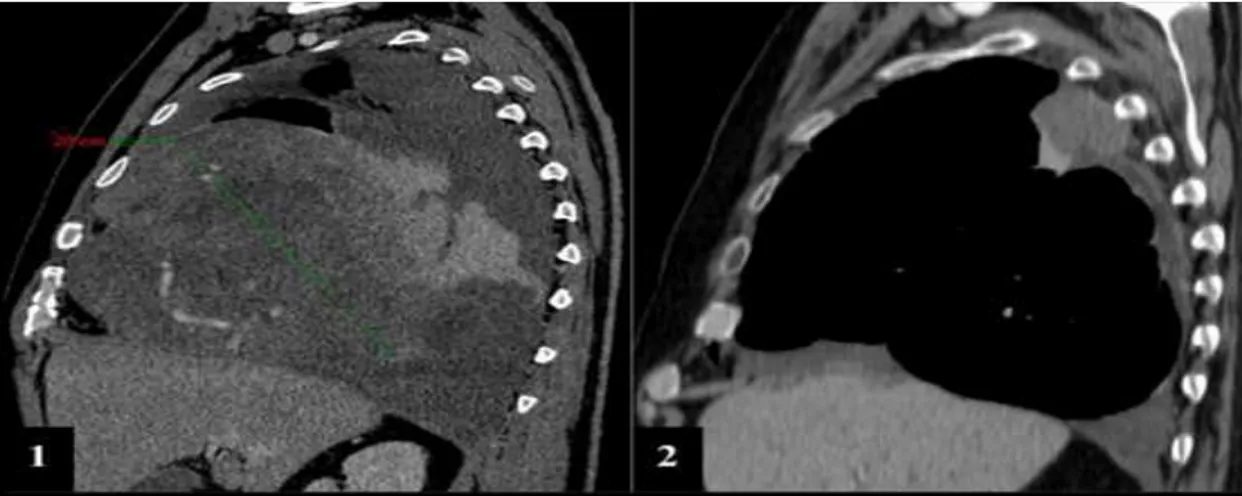
![Fig. I.5 – Radiographie thoracique de face et corrélation tomodensitométrique. Tumeur volumineuse ; opacité de l’hémithorax gauche [3]](https://thumb-eu.123doks.com/thumbv2/123doknet/13205036.392864/34.892.135.766.407.656/radiographie-thoracique-corrélation-tomodensitométrique-tumeur-volumineuse-opacité-hémithorax.webp)
![Fig. I.6 – Un exemple d’image TEP et les traceurs s’accumulent dans le cerveau, les tumeurs, le foie et la vessie [15]](https://thumb-eu.123doks.com/thumbv2/123doknet/13205036.392864/37.892.182.716.122.362/fig-exemple-image-traceurs-accumulent-cerveau-tumeurs-vessie.webp)
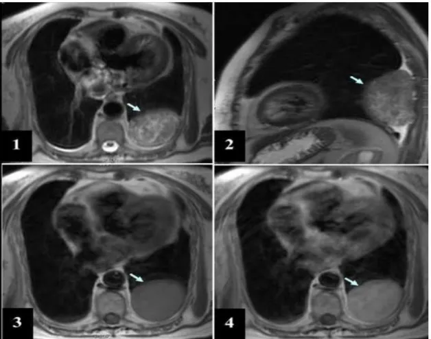
![Fig. I.8 – Tomographie par émission de positons au 18FDG. TFS bénigne (flèches) sans hypermétabolisme [9]](https://thumb-eu.123doks.com/thumbv2/123doknet/13205036.392864/39.892.163.795.377.622/fig-tomographie-émission-positons-fdg-bénigne-flèches-hypermétabolisme.webp)
![Fig. I.10 – Principe de la fusion TEP/TDM [3].](https://thumb-eu.123doks.com/thumbv2/123doknet/13205036.392864/42.892.149.785.227.525/fig-principe-la-fusion-tep-tdm.webp)
![Fig. I.11 – l’appareil IRM, (a) Scanner IRM de 1.5 T, (b) Coupe d'un scanner IRM [17]](https://thumb-eu.123doks.com/thumbv2/123doknet/13205036.392864/44.892.120.786.566.982/fig-appareil-irm-scanner-irm-coupe-scanner-irm.webp)
![Tab. I.1 – Ordre de grandeur des temps de relaxation à 1.5 Tesla [7].](https://thumb-eu.123doks.com/thumbv2/123doknet/13205036.392864/46.892.310.666.950.1060/tab-ordre-grandeur-temps-relaxation-tesla.webp)
Documents relatifs
L’analyse de la corrélation linéaire entre estimée et valeur vraie est utile pour une interprétation correcte du biais moyen et de sa variabilité. La caractérisation des
Figure 3: Single-tumor growth models’ analysis: parameter distributions Models for single tumor growth were independently fitted to the large and small growth curves from
In addition, approaches to estimate of model parameters from imaging data have been proposed [13, 20, 21], which are based on estimating the velocity of tumor growth and the
We establish uniform a priori estimates and existence theorems for symmetric hyperbolic-parabolic systems of partial differential equations with small second order terms and
We establish uniform a priori estimates and existence theorems for symmetric hyperbolic-parabolic systems of partial differential equations with small second order terms and
Levels of type VI collagen α1 and α3 chain fragments, derived from MMP proteolysis, appear higher in serum from cancer patients (breast, colon, gastric, ovarian, pancreas,
Tumor driven genes appearing in determined normal cells will be responsible for chromosomal instability with numerous chromosome breakages (fusions, deletions, etc)
(2015) developed a tumor trap for the metastasizing cells of breast and prostate cancers by imitating the red bone marrow microenvironment (Figure 1 Scheme II-3-b).. The strategy
