Modeling of the interaction of an x-ray free-electron laser with large finite samples
Texte intégral
Figure


Documents relatifs
We therefore conducted a prospective study to evaluate the prevalence of faecal carriage of acquired and intrinsic colis- tin-resistant Gram-negative rods among patients admitted to
Ce langage n’est pas hasardeux, il émane d’un milieu social très connu dans la réalité qui est la banlieue française appelée communément les cités beures. Ce langage
The cartridge used was const- ructed so that most of the solid material was outside both the cyclotron diameter of the electrons and the field of view of the collimator and thus
This paper studies by modelling, the phenomena resulting from the interaction photon X and material, by approximating a model which takes into account attenuation laws and the
can determine the laser characteristics needed as an efficient X-ray lithography source taking the resist. sensitivity as a parameter
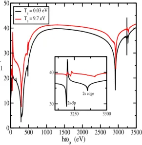
Relative transition energies and probabilities are calculated in a frozen-orbital approximation for the transition from a normal K state to singly ionized states of
L’archive ouverte pluridisciplinaire HAL, est destinée au dépôt et à la diffusion de documents scientifiques de niveau recherche, publiés ou non, émanant des
This is based on dark-field X-ray microscopy, where an X-ray objective lens is placed in the diffracted beam (Simons et al., 2015; Simons, Haugen et al., 2018), providing an
