HAL Id: hal-01663921
https://hal-amu.archives-ouvertes.fr/hal-01663921
Submitted on 14 Dec 2017
HAL is a multi-disciplinary open access
archive for the deposit and dissemination of
sci-entific research documents, whether they are
pub-lished or not. The documents may come from
teaching and research institutions in France or
abroad, or from public or private research centers.
L’archive ouverte pluridisciplinaire HAL, est
destinée au dépôt et à la diffusion de documents
scientifiques de niveau recherche, publiés ou non,
émanant des établissements d’enseignement et de
recherche français ou étrangers, des laboratoires
publics ou privés.
ABUNDANCE OF BRCA1 TRANSCRIPTS IN
HUMAN CANCER AND LYMPHOBLASTOID CELL
LINES CARRYING BRCA1 GERM-LINE
ALTERATIONS
Stéphane Ribieras, Frédérique Magdinier, Delphine Leclerc, Gilbert Lenoir,
Lucien Frappart, Robert Dante
To cite this version:
Stéphane Ribieras, Frédérique Magdinier, Delphine Leclerc, Gilbert Lenoir, Lucien Frappart, et al..
ABUNDANCE OF BRCA1 TRANSCRIPTS IN HUMAN CANCER AND LYMPHOBLASTOID
CELL LINES CARRYING BRCA1 GERM-LINE ALTERATIONS. International Journal of Cancer,
Wiley, 1997, 73 (5), pp.715-718. �10.1002/(SICI)1097-0215(19971127)73:53.0.CO;2-4�. �hal-01663921�
ABUNDANCE OF BRCA1 TRANSCRIPTS IN HUMAN CANCER AND
LYMPHOBLASTOID CELL LINES CARRYING BRCA1 GERM-LINE ALTERATIONS
Ste´phane RIBIERAS, Fre´de´rique MAGDINIER, Delphine LECLERC, Gilbert LENOIR, Lucien FRAPPARTand Robert DANTE*
Laboratoire de Ge´ne´tique, UMR 5641 CNRS, Lyon, France
A competitive polymerase chain reaction has been devel-oped for quantitation ofBRCA1mRN A. In human cancer cell lines, the amount ofBRCA1mRN A is relatively low, ranging from 6 to 38 copies per cell. T he decay rate of these transcripts in actinomycin-treated cells indicates that the half-life of these molecules is about 4 hr, suggesting that the low concentration ofBRCA1messagesisnot due to molecular unstability. In human lymphoblastoid cell lines derived from patients carrying germ-line alterations ofBRCA1,the amount ofBRCA1mRN A per cell is lowered only in cell lines exhibit-ing alterations leadexhibit-ing to specific loss of transcripts from the mutated allele. T hese data indicate that the amount of
BRCA1available in these cells can be related directly to the number of ‘‘active’’ allele.Int. J. Cancer73:715–718, 1997.
r
1997 Wiley-Liss, Inc.Breast cancer is the most common malignancy affecting women, and 5 to 10% of cases are estimated to be familial. Segregation analyses of families with multiple affected individuals led to the identification of the BRCA1 gene (Miki et al., 1994). Germline alterations (frameshift, nonsense, splice mutations) of this gene appear to account for about one half of inherited breast cancers (Friedman et al., 1994). Although mutations of BRCA1 were not detected in sporadic human breast cancers, and ovarian tumors exhibit BRCA1 mutations only exceptionally (Futreal et al., 1994; Mejraver et al., 1995), several experiments have correlated the level of BRCA1 expression and cell proliferation.
Analysis of the pattern of BRCA1 expression in mice indicated that BRCA1 is expressed in rapidly proliferating cell types undergo-ing differentiation, and suggested that this gene might be impli-cated in the processes of proliferation and differentiation in several tissues (Marquis et al., 1995). An essential role of BRCA1 in normal mammalian development has also been shown, since homozygous deletion results, in mice, in embryonic lethality (Gowen et al., 1996; Hakem et al., 1996). Modulations of BRCA1 expression are also observed in human tissues, a decreased BRCA1 mRNA level was observed during sporadic breast-cancer progres-sion (Thompson et al., 1995) and, in cell lines, steroid hormones may affect the level of this transcript by altering cellular prolifera-tion (Gudas et al., 1995).
The involvement of BRCA1 in cellular proliferation has been also addressed in human cell lines, using anti-sense oligonucleo-tides (Thompson et al., 1995) and in mouse cell lines, using anti-sense RNA (Rao et al., 1996). All these experiments indicate that BRCA1 negatively regulates cellular proliferation and may therefore act as a suppressor gene in human cancers. Taken together, these data strongly suggest that the level of BRCA1 mRNA may play an important role in human breast carcinogenesis. Hybridization methods have been widely used for the detection of BRCA1 transcripts in many cell types or human tumors. These studies have shown that the BRCA1-mRNA content in these samples can be modulated by several factors, including the presence of estradiol for estrogen-receptor-rich cell lines (Gudas et al., 1995) and the tumor types for sporadic human breast cancers (Thompson et al., 1995). However, all the methods used (Northern blots or RNase protection assays) give relative values, so that it is difficult to compare data between experiments. The competitive RT-PCR method appears to be an alternative to these more traditional methods, and has been successfully used to precisely quantitate low amount of RNA (Ramakrishnan et al., 1994; Laghami et al., 1997). This PCR method measures the absolute
amount of mRNA in an RNA sample; furthermore, this quantitation is not related to the amount of an another ‘‘control’’ mRNA, which may vary between samples or cellular types. Data obtained from different cell lines or samples can thus be compared directly with each other. In addition, for some experiments (stability, transcrip-tion rate) direct quantitatranscrip-tion of the amount of mRNA is needed.
We have therefore developed a competitive RT-PCR assay for the measurement of BRCA1 transcripts. BRCA1 mRNA molecules have been quantitated in several cell lines, and the effect of actinomycin D, an inhibitor of transcription, on the half-life of BRCA1 mRNA has been monitored in cell lines using this method.
MATERIAL AND METHODS RT PCR
A cDNA fragment of BRCA1 (from position 390 to position 647) was amplified by RT-PCR using C3 primers: 58 TGT GCT TTT CAG CTT GAC ACA GG 38 and 58 CGT CTT TTG AGG TTG TAT CCG CTG 38 (Friedman et al., 1994). After cloning in a pGEM-T vector (Promega, Lyon, France), the corresponding insert was cut with Van91 I at position 112 and a blunt-end oligonucleo-tide derived from pS2 sequence, nt2412 to nt 2365 (Jeltsch et al., 1983) was ligated at this site and cloned again in the same pGEM-T vector. Then the insert was characterized by sequencing. Since this vector contains a promoter site for T7 RNA polymerase the corresponding RNA was synthesized using the corresponding RNA polymerase according to the instructions of the manufacturer (Boehringer Mannheim, Meylan, France). After purification (DNaseI digestion and phenol-chloroform extraction), this competitor RNA was quantitated by spectrophotometry, and aliquots were diluted in presence of yeast tRNA as a carrier.
After extraction (Dante et al., 1994), RNA was quantitated by spectrophotometry and by electrophoresis on agarose gels of serial dilutions in comparison with known amounts of standard RNA (Boehringer Mannheim). Detection of transcripts were done as described (Dante et al., 1994) with some modifications. Briefly, reactions were performed with 0.3 or 0.6 µg of RNA in 100 µl containing 10 mM Tris-Hcl (pH 8.3), 3.0 mM MgCl2, 50 mM KCl, 0.1 mg/ml gelatin, 200 µM each of the 4 deoxyribonucleoside triphosphates. After initial denaturation at 92°C for 2 min, 6 units of M-Mulv reverse transcriptase (Boehringer Mannheim) were added to the reaction mixture and incubated for 35 min at 42°C. Reverse transcriptase was then inactivated by heating (94°C, 3 minutes) and after cooling to 0°C, PCR amplification of the cDNA product was accomplished by adding 0.6 units of Taq DNA polymerase (Boehringer Mannheim). The PCR amplification was accomplished using 35 cycles in a Eppendorf thermocycler in the following conditions: 1 min denaturation at 94°C, annealing at 55°C for 2 min and extension for 3 min at 72°C. In these conditions, heterodimers between PCR products derived from
Contract grant sponsors: Ligue Nationale pour la Recherche contre le Cancer; Association pour la Recherche contre le Cancer.
*Correspondence to: Laboratoire de Ge´ne´tique, UMR 5641 CNRS, Domaine Rockefeller, UCBL1, 8, avenue Rockefeller, 69373 Lyon cedex 08, France. Fax: 33 478 77 72 20. E-mail: dante@cismsun.univ-lyon1.fr
Received 25 April 1997; Revised 26 July 1997 Int. J. Cancer: 73, 715–718 (1997)
mRNA and the competitor were not detected. In addition, control experiments for each RNA sample were performed, omitting reverse transcriptase, to confirm that the signal was the result of RNA-specific and not DNA-specific amplification. Aliquots (17 µl) were then analyzed on 2% agarose gel containing 0.1 µg/ml of ethidium bromide. Photographs of the gels were scanned (UMAX) and the intensity of the 2 bands corresponding to PCR products were determined using image analyzer software (Wayne Rasband, NIH).
Cell lines
Lymphoblastoid cell lines were established from BRCA1-mutation carriers, and controls have been described (Serova et al., 1996). HeLa, Lovo clone C5, BT-20 and HBL-100 cell lines were grown as described (ATCC, Rockville, MD; Remy et al., 1993). MCF-7 cells were grown in Dulbecco’s modified Eagle’s medium (Gibco, Cergy-Pontoise, France) supplemented with SVF 10%, insulin 0.6 µg/ml and estradiol 1028M. Cells were then seeded in an estrogen-free medium (phenol-red-free medium supplemented with charcoal-treated serum); actinomycin D (43 1026M) was added 48 hr after seeding and for the experiments in presence of the hormone, estradiol (1028M) was added at same time. C5 cells were grown in standard medium and actinomycin D was added 48 hr after seeding.
RESULTS Validation of the RT-PCR method
To determine the amount of BRCA1 mRNA a competitive RT-PCR assay was developed, using a chimeric BRCA1 RNA as a competitor. In familial breast cancer, genetic alterations are more frequently localized in the 38 one third of the gene (Gayther et al., 1995), and only one case of alterations at exons 6 and 8 has been reported (Breast Cancer Information Core). Therefore, in order to amplify BRCA1 transcripts in a large panel of samples or cell lines, primers were chosen in these 2 different exons (exon 6 and exon 8). A 48-bp double-stranded oligonucleotide was inserted into a cloned
RT-PCR fragment of BRCA1 mRNA (see ‘‘Material and Meth-ods’’). After cloning, the corresponding chimeric RNA was synthe-sized using T7 RNA polymerase, and after quantitation it was used as a competitor in the RT-PCR experiments.
In order to evaluate the sensitivity of the RT-PCR, serial dilutions of the competitor were amplified; the amount of the corresponding PCR product was then determined by densitometry on ethidium-bromide-stained agarose gels and plotted against the initial concentration of synthetic RNA. Data obtained indicate that the signal was proportional to the log of the amount of synthetic RNA within a wide range of concentrations, from 33 103to 106 copies per assay (Fig. 1a,b). Moreover, the efficiency of PCR amplification for the cDNA corresponding to the BRCA1 mRNA fragment and to the synthetic competitor RNA was also compared using serial dilutions of plasmids containing these inserts. Ethidium-bromide staining of the agarose gels indicates that both fragments are amplified at the same rate (data not shown).
When 0.075 µg RNA extracted from HeLa cells is mixed with 0.23 µg of RNA from CHO cell line, the RT-PCR assay gives a value of 283 103copies. Since the primers used in this assay did not amplify the BRCA1 mRNA of CHO cells, this value is very close to the value obtained (1203 103, Table I) with 0.3 µg of RNA extracted from HeLa cells. Therefore, as expected from the titration curves obtained with the synthetic competitor (Fig. 1a,b), the quantitation of BRCA1 mRNA/µg of total RNA is not modified by the amount of total RNA analyzed.
Quantitation of BRCA1 mRNA in human cancer cell lines After these preliminary experiments, the amount of BRCA1 mRNA was determined in several cell lines from 0.3 µg of total RNA using the RT-PCR method, as described above. RT-PCR assays were performed from dilution series of the competitor added to a constant amount of sample RNA. The amount of PCR products corresponding to BRCA1 mRNA and to synthetic RNA was plotted against the initial number of synthetic RNA added to the test tubes (see Fig. 1c,d). The abscissa at the intersection of the curves
FIGURE1– RT-PCR assay of BRCA1 mRNA. (a) 15 µl of the RT-PCR reaction (total volume 100 µl) performed from serial dilutions of chimeric
BRCA1 RNA were analyzed on ethidium-bromide-stained 2% agarose gels; the 306-bp band corresponds to the expected size of the PCR product.
Initial concentrations of these standard molecules were: lane 1, 13 103; lane 2, 53 103; lane 3, 103 103; lane 4, 503 103; lane 5, 1003 103; lane 6, 5003 103; lane 7, 10003 103molecules. (b) Diagrammatic representation of the above data; the intensity of the bands corresponding to the PCR product were plotted against the initial number of standard molecules. (c) 15 µl of the RT-PCR reaction (total volume 100 µl) performed from 0.3 µg of total RNA of MCF-7 cells mixed with various amount of chimeric BRCA1 RNA as a competitor; 306-bp band, chimeric BRCA1 RNA and 258-bp band, wild-type BRCA1 RNA. Amount of standard molecules: lane 1, 1003 103; lane 2, 2503 103; lane 3, 5003 103. (d) Diagrammatic representation of the above data; the intensity of PCR products was plotted against the initial number of standard molecules; s, 258-bp band (WT
BRCA1 RNA); d, 306-bp band (chimeric competitor BRCA1 RNA).
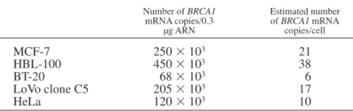
represents an estimation of the equivalence point between the initial amount of the competitor molecules and the number of copies of BRCA1 mRNA in 0.3 µg of sample RNA. Results (number of mRNA molecules per 0.3 µg of total RNA) obtained with RNA extracted from MCF-7, BT-20, HBL-100, LoVo clone C5 and Hela cell lines are shown in Table I. The abundance of BRCA1 mRNA appears relatively low, since, if we assume that 1 cell contains 25 pg of total RNA (Wilson et al., 1997), the number of BRCA1 copies per cell range from 6 copies (BT-20) to 38 copies (HBL100) (Table I). In addition, this low copy number does not seem to be dependent on cell type, since the breast-cancer cell lines exhibit approximately the same level of BRCA1 mRNA (Table I) than the cervix-cancer cell line (Hela) and the colon-cancer cell line (LoVo clone C5). Analysis of several human cancer cell lines shows that BRCA1 is expressed in all the cell lines studied at a relatively low rate (6 to 38 copies per cell). These low amounts of BRCA1 transcripts suggest a relatively low rate of transcription and/or very fast turnover of these molecules.
Turnover rates of BRCA1 mRNA
Several methods for half-live determination have been de-scribed, including pulse labelling, and short-term promoter activa-tion (Ross, 1995). In our case, none of these methods are easy to handle, since pulse-labelling methods are usually used for mRNA expressed at relatively high level, and little is known about transcription factors involved in BRCA1 regulation. Therefore, despite the inherent limitations of the use of chemical inhibitor of transcription, determination of the stability of BRCA1 mRNA was monitored in MCF-7 cells treated with the transcription inhibitor actinomycin D. Although this method does not give a very accurate measure of the half-lives of mRNA, this approach has been widely used to determine an order of magnitude (Ross, 1995).
To determine whether this low copy number is due to very fast turnover of BRCA1 mRNA, the stability of this transcript was investigated in 2 human cancer cell lines, MCF-7 cells (breast cancer) and LoVo clone C5 cells (colon cancer) during incubation in the presence of the transcription inhibitor actinomycin D. It had been shown, in estrogen-receptor-rich cells, that estradiol (E2) may modulate the half-life of some hormone-regulated transcripts in actinomycin-treated cells (Ross, 1995). Since MCF-7 cells are estrogen-responsive (Carr et al., 1995), we took this opportunity to investigate the effect of this hormone on the stability of BRCA1 mRNA. At time 0, MCF-7 cells were treated with actinomycin D, in the absence or the presence of estradiol. At several intervals, RNA samples were prepared and the amount of BRCA1 mRNA was determined, using the competitive RT-PCR method described above.
These transcripts appear to be relatively stable, since 4 hr after addition of actinomycin D, MCF-7 cells still contain about one half of the initial BRCA1 mRNA concentration (Fig. 2). The same decay rate was observed in the absence of estradiol (Fig. 2), suggesting that, in these conditions, the stability of BRCA1 mRNA is not hormone-dependent. In agreement with this observation, it is suggested that estradiol stimulation of BRCA1 transcription is not due to a direct effect of this hormone. In addition, in
actinomycin-treated colon-cancer cells (LoVo clone C5), the level of BRCA1 mRNA is similar to that observed in the breast-cancer cell line (Fig. 2), suggesting that the stability of BRCA1 mRNA is not cell-type-specific.
Taken together, these data suggest that the low amount of BRCA1 transcripts observed in the cell lines analyzed is not a consequence of fast turnover of these molecules. However, in situ hybridization methods have shown that BRCA1-linked breast tumors can be distinguished from sporadic breast tumors by their BRCA1 mRNA content (Kainu et al., 1996). We have, therefore, investigated BRCA1 expression in lymphoblastoid cell lines exhib-iting BRCA1 alterations and in matched controls.
The amount of BRCA1 mRNA is lowered only in lymphoblastoid cell lines carrying BRCA1 germ-line mutations leading to loss of transcripts
BRCA1 transcripts were assayed in 6 lymphoblastoid cell lines exhibiting different allele-specific BRCA1 alterations, and in 7 control lymphoblastoid cell lines (Table II). Alteration of BRCA1 transcripts leads to allele-specific polymorphisms, and cDNA analysis indicated that, in some of these cell lines (IARC 1506, 1514 and 1947), the mutated BRCA1 mRNA was undetectable (Serova et al., 1996; Puget et al., 1997). In these cell lines we found a reduced level of BRCA1 mRNA (1603 103 to 2303 103 copies/µg of RNA) when compared with lymphoblastoid cell lines expressing one normal allele and one mutated allele (4333 103to 5133 103copies/µg of RNA) or to cell lines exhibiting no BRCA1 alterations (4003 103to 7723 103copies/µg of RNA). These data strongly suggest that the amount of BRCA1 mRNA in lymphoblas-toid cell lines is directly related to the number of ‘‘active’’ alleles.
DISCUSSION
Values obtained with our RT-PCR assay are very similar to those obtained with a more classical method. Using a RNase protection assay, other authors (Wilson et al., 1997) have found in the HBL100 cell line 50 copies of BRCA1 mRNA/cell, while the RT-PCR assay gives 38 copies/cell. However, in the other cell lines analyzed the concentrations of BRCA1 transcripts are significantly lower than these values, since we found, for example, only 6 copies of BRCA1 message in the breast-cancer cell line BT-20. This low level of expression does not seem to be associated with instability of the mRNA. In cancer cell lines, BRCA1 transcripts remain stable several hours after inhibition of the transcription by actinomycin D, and in lymphoblastoid cell lines the level of transcripts was not modified by BRCA1 mutations. In addition, in lymphoblastoid cell lines carrying BRCA1 alterations leading to loss of transcripts, the level of the remaining allele is about one half of the level observed in cell lines expressing both alleles.
TABLE I– BRCA1 EXPRESSION IN HUMAN CANCER CELL LINES Number of BRCA1 mRNA copies/0.3 µg ARN Estimated number of BRCA1 mRNA copies/cell MCF-7 2503 103 21 HBL-100 4503 103 38 BT-20 683 103 6 LoVo clone C5 2053 103 17 HeLa 1203 103 10
The amount of BRCA1 mRNA was determined using a competitive RT-PCR method from 0.3 µg of total RNA. Data obtained for each point (2 to 4 independent assays) indicated that the variations were inferior to 15%. The number of copies/cell was estimated taking 25 pg as the amount of total RNA per cell.
FIGURE2– Stability of BRCA1 mRNA in human cancer cell lines. Actinomycin D was added to the culture medium 48 hr after seeding (see ‘‘Material and Methods’’), and BRCA1 mRNA was assayed at a number of intervals. h, MCF-7 cells in absence of estradiol; j, MCF-7 cells in presence of estradiol; d, LoVo clone C5 cells.
These data, therefore, show that low-level BRCA1 mRNA is not specifically associated with a cell type, but is observed in all cell lines analyzed. However, it has been reported, from in situ hybridization analysis, that BRCA1-linked breast-cancer tissues contain fewer BRCA1 transcripts than sporadic tumors (Kainu et al., 1996), and a decrease in BRCA1-mRNA level was observed during sporadic breast-cancer progression (Thompson et al., 1995). Taken together, these findings suggest that some specific events
involved in the regulation of BRCA1 expression are induced in human breast cancers. In line with this hypothesis, it has been reported that the relative proportion between an alternatively spliced message in which the majority of exon 11 has been deleted and the full-length BRCA1 mRNA is modified in some breast- and ovarian-cancer cell lines (Wilson et al., 1997).
The competitive RT-PCR method, developed for determining the level of BRCA1 mRNA, needs only very small amounts of cells or tissues. This quantitation, therefore, can be performed from fine-needle aspirates, since typically 1 µg or less of RNA can be extracted from such samples. In contrast to traditional protocols, this method does not require the use of radioisotopes or the development or purchase of specific antibodies. This method could be a very useful tool, since it has been suggested that BRCA1 expression might be linked to tumor progression. However, given the heterogeneity of cell types in fine-needle aspirates, it remains to established whether such assays parallel BRCA1 expression in normal or tumoral mammary glands.
In conclusion, our results indicate that this technique is feasible for evaluating BRCA1 transcript levels, and suggest that, in patients with germline alterations in BRCA1, the total level of BRCA1 transcripts is related to the stability of the transcript carrying the alteration.
ACKNOWLEDGEMENTS
S.R. is the recipient of a fellowship from the Ligue Nationale contre le Cancer, Comite´ de la Saoˆne et Loire, and F.M. is the recipient of a fellowship from the Ligue Nationale contre le Cancer, Comite´ de la Droˆme. The present work was supported by the Ligue Nationale pour la Recherche contre le Cancer and the Association pour la Recherche contre le Cancer.
REFERENCES BREASTCANCERINFORMATIONCORE ON THEINTERNET. (http://www.nchgr. nih.gov/Intramural_research/Lab_transfer/Bic/).
CARR, M., MAY, F.E.B., LENNARD, T.W.B. and WESTLEY, B.R., Determina-tion of estrogen responsiveness of breast cancer by competitive reverse-transcription-polymerase chain reaction. Brit. J. Cancer, 72, 1427–1434 (1995).
DANTE, R., RIBIERAS, S., BALDASSINI, S., MARTIN, V., BENZERARA, O., BOUTEILLE, C., BRE´MOND, A., FRAPPART, L., RIO, M.C. and LASNE, Y., Expression of an estrogen-induced breast-cancer-associated protein (pS2) in benign and malignant human ovarian cysts. Lab. Invest., 71, 188–192 (1994).
FRIEDMAN, L.S., OSTERMEYER, E.A., SZABO, C.I., DOWD, P., LYNCH, E.D., ROWELL, S.E. and KING, M.C., Confirmation of BRCA1 by analysis of germline mutations linked to breast and ovarian cancer in ten families.
Nature (Genet.), 8, 399–404 (1994).
FUTREAL, P.A. and 26OTHERS, BRCA1 mutations in primary breast and ovarian carcinomas. Science, 266, 120–122 (1994).
GAYTHER, S.A. and 15OTHERS, Germline mutations of the BRCA1 gene in breast- and ovarian-cancer families provide evidence for a genotype-phenotype correlation. Nature (Genet.), 11, 428–433 (1995).
GOWEN, L.C., JOHNSON, B.L., LATOUR, A.M., SULIK, K.S. and KOLLER, B.H.,
Brca1 deficiency results in early embryonic lethality characterized by
neuro-epithelial abnormalities. Nature (Genet.), 12, 191–194 (1996). GUDAS, J.M., NGUYEN, H., LI, T. and COWAN, K.H., Hormone-dependent regulation of BRCA1 in human breast-cancer cells. Cancer Res., 55, 4561–4565 (1995).
HAKEM, R. and 13OTHERS, The tumor-suppressor gene Brca1 is required for embryonic cellular proliferation in the mouse. Cell, 85, 1009–1023 (1996). JELTSCH, J.M., ROBERTS, M., SCHATZ, C., GARNIER, J.M., BROWN, A.M.C. and CHAMBON, P., Structure of the human estrogen-responsive gene pS2.
Nucleic Acid Res., 15, 1401–1414 (1983).
KAINU, T., KONONEN, J., JOHANSSON, O., OLSSON, H., BORG, A. and ISOLA, J., Detection of germline BRCA1 mutations in breast-cancer patients by quantitative messenger RNA in situ hybridization. Cancer Res., 56, 2912–2915 (1996).
LAGHAMI, K., BORENSZTEIN, P., AMBU¨HL, P., FROISSART, M., BICHARA, M., MOE, O.W., ALPERN, R.J. and PAILLARD, M., Chronic metabolic acidosis enhances NHE-3 protein abundance and transport activity in the rat thick limb by increasing NHE-3 mRNA. J. clin. Invest., 99, 24–30 (1997).
MARQUIS, S.T., RAJAN, J.V., WYNSHAW-BORIS, A., XU, J., YIN, G.Y., ABEL, K.J., WEBER, B.L. and CHODOSH, L.W., The developmental pattern of Brca1 expression implies a role in differentiation of the breast and other tissues.
Nature (Genet.), 11, 17–26 (1995).
MEJRAVER, S.D., PHAM, T.M., CADUFF, R.F., CHEN, M., POY, E.L., COONEY, K.A., WEBER, B., COLLINS, F.S., JOHNSTON, C. and FRANK, T.S., Somatic mutations in the BRCA1 gene in sporadic ovarian tumours. Nature (Genet.),
9,439–443 (1995).
MIKI, Y. and 44OTHERS, A strong candidate for the breast- and ovarian-susceptibility gene BRCA1. Science, 266, 66–71 (1994).
PUGET, N., TORCHARD, D., SEROVA, O.M., LYNCH, H.T., FEUNTEN, J., LENOIR, G.M. and MAZOYER, S.A., 1-kb alu-mediated germ-line deletion removing BRCA1 exon 17. Cancer Res., 57, 828–831 (1997).
RAMAKRISHNAN, R., FINK, D.J., JIANG, G., PRASHANT, D., GLORIOSO, J.C. and LEVINE, M., Competitive quantitative PCR analysis of herpes-simplex-type-1 DNA and latency-associated transcript RNA in latently infected cells of rat brain. J. Virol., 68, 1864–1873 (1994).
RAO, V.N., SHAO, N., AHMAD, M. and REDDY, S.P., Anti-sense RNA to the putative tumor-suppressor gene BRCA1 transforms mouse fibroblasts.
Oncogene, 12, 523–528 (1996).
REMY, L., JACQUIER, M.F., DAEMI, N., DORE´, J.F. and LISSITZKY, J.C., Comparative tumor morphogenesis of two human colon-adenocarcinoma cell clones xenografted in the immunosuppressed newborn rat.
Differentiation, 54, 191–200 (1993).
ROSS, J., mRNA stability in mammalian cells. Microbiol. Rev., 59, 423–450 (1995).
SEROVA, O., MONTAGNA, M., TORCHARD, D., NAROD, S.A., TONIN, P., SYLLA, B., LYNCH, H.T., FEUNTEN, J. and LENOIR, G.M., A high incidence of BRCA1 mutations in 20 breast-ovarian-cancer families. Amer. J. hum. Genet., 58, 42–51 (1996).
THOMPSON, M.E., JENSEN, R.A., OBERMILLER, P.O., PAGE, D.L. and HOLT, J.T., Decreased expression of BRCA1 accelerates growth and is often present during breast cancer progression. Nature (Genet.), 9, 444–450 (1995).
WILSON, C.A., PAYTON, M.A., ELLIOTT, G.S., BUAAS, F.W., CAJULIS, E.E., GROSSHANS, D., RAMOS, L., REESE, D.M., SLAMON, D.J. and CALZONE, F.J., Differential subcellular localization expression and biological toxicity of
BRCA1 and the splice variant BRCA1-D11b. Oncogene, 14, 1–14 (1997). TABLE II– BRCA1 EXPRESSION IN HUMAN LYMPHOBLASTOID CELL LINES
CARRYING BRCA1 MUTATIONS AND IN MATCHED CONTROLS
Cell lines BRCA1 mRNA copies/cell Mutation1 Loss of transcript1
IARC-1506 6 deletion exon 19 Yes
IARC-1514 5 deletion exon 19 Yes
IARC-1555 13 Cys-Gly (T-G) No
IARC-1162 11 Arg-Ter Not done
IARC-1947 4 deletion exon 17 Yes
IARC-1155 13 exon 5 missing in
transcript No
IARC-1468 13 Control cell line IARC-1235 10 Control cell line IARC-1475 13 Control cell line IARC-1625 10 Control cell line IARC-1220 10 Control cell line IARC-1220 15 Control cell line IARC-1102 20 Control cell line
1Mutation and transcript analyses are from Serova et al. (1996) and Puget et al. (1997). The amount of BRCA1 mRNA was determined as in Table I. Data obtained for each point (2 to 4 independent assays) indicated that variations were inferior to 15%.