HAL Id: hal-03025274
https://hal.archives-ouvertes.fr/hal-03025274
Submitted on 7 Dec 2020HAL is a multi-disciplinary open access archive for the deposit and dissemination of sci-entific research documents, whether they are pub-lished or not. The documents may come from teaching and research institutions in France or abroad, or from public or private research centers.
L’archive ouverte pluridisciplinaire HAL, est destinée au dépôt et à la diffusion de documents scientifiques de niveau recherche, publiés ou non, émanant des établissements d’enseignement et de recherche français ou étrangers, des laboratoires publics ou privés.
Quadruplex-interacting compounds for regulating the
translation of the Epstein–Barr virus nuclear antigen 1
(EBNA1) mRNA: A new strategy to prevent and treat
EBV-related cancers
Anton Granzhan, Rodrigo Prado Martins, Robin Fåhraeus, Marc Blondel,
Marie-paule Teulade-fichou
To cite this version:
Anton Granzhan, Rodrigo Prado Martins, Robin Fåhraeus, Marc Blondel, Marie-paule Teulade-fichou. Quadruplex-interacting compounds for regulating the translation of the Epstein–Barr virus nuclear antigen 1 (EBNA1) mRNA: A new strategy to prevent and treat EBV-related cancers. Stephen Neidle. Quadruplex Nucleic Acids As Targets For Medicinal Chemistry, 54, Elsevier, pp.243 - 286, 2020, Annual Reports in Medicinal Chemistry, 978-0-12-821017-8. �10.1016/bs.armc.2020.05.001�. �hal-03025274�
1
Quadruplex-interacting compounds for regulating the translation
of the Epstein–Barr virus Nuclear Antigen 1 (EBNA1) mRNA: A new
strategy to prevent and treat EBV-related cancers
Anton Granzhan,a,b Rodrigo Prado Martins,c ,# Robin Fåhraeus,c Marc Blondel,d,e
Marie-Paule Teulade-Fichoua, b
a CNRS UMR9187, Inserm U1196, Institut Curie, PSL Research University, F-91405 Orsay, France b CNRS UMR9187, Inserm U1196, Université Paris-Saclay, F-91405 Orsay, France
c Inserm UMR1131, Institut de Génétique Moléculaire (IGM), Université Paris 7, Hôpital St. Louis, F-75010 Paris, France
d Université de Brest, Inserm, EFS, UMR1078, GGB, F-29200 Brest, France
e CHRU Brest, service de génétique clinique et de biologie de la reproduction, F-29200 Brest, France # Current address: INRAE, Université de Tours, ISP, F-37380, Nouzilly, France
Abstract: The Epstein–Barr (EBV) virus is linked to at least 1% of cancers that include Burkitt’s and Hodgkin’s lymphomas, nasopharyngeal carcinoma, and 10% of gastric cancers. EBV is a latent virus that possesses a genome maintenance protein EBNA1, which is both essential for the virus and highly antigenic. Hence, EBV evolved a mechanism by which EBNA1 self-inhibits the translation of its own mRNA, thereby minimizing the production of EBNA1-derived antigenic peptides. Although not fully elucidated, this mechanism involves the Gly-Ala-rich (GAr) motif of EBNA1, encoded by a G-repeat-containing mRNA sequence able to form clusters of G-quadruplexes. This chapter summarizes recent significant advances in understanding of this phenomenon. Mechanistic investigations based on yeast chemical genetics, cellular assays and in vitro experiments have shown that the host cell factor nucleolin (NCL) is involved in this limitation of EBNA1 translation through binding to G-quadruplexes of EBNA1 mRNA. This interaction can be disrupted by the benchmark G4-ligand PhenDC3 acting as NCL competitor for binding to G4-RNA. Finally, exploration of the chemical space around PhenDC3 using combinatorial chemistry approach led to the generation of 20 compounds based on a bis(acylhydrazone) scaffold. Amongst these, two hits (PyDH2, PhenDH2) exhibit optimized properties with regard to the disruption of NCL/G4 interaction in cells, along with lower cytotoxicity. Consequently, treatment by PyDH2 or PhenDH2 increases EBNA1 production and stimulates the GAr-restricted antigenic response. Altogether, this innovative concept of antigenic stimulation sets the basis for further identification of lead candidates that may become promising candidate drugs for treating EBV-related cancers.
Keywords: Epstein–Barr virus (EBV), EBV-related cancers, G-quadruplex RNA, Nucleolin, Antigenic stimulation, G4-ligands
2
Contents
1 Epstein–Barr virus and cancers ... 2
2 Epstein–Barr virus Nuclear Antigen 1 (EBNA1): the Achilles’ heel of EBV ... 5
3 G-Quadruplexes in the GAr-encoding sequence of EBNA1 mRNA ... 7
4 Nucleolin: a new player in the hide-and-seek game ... 9
5 PhenDC3, a benchmark G4 ligand, disrupts NCL–EBNA1 mRNA interaction, enhances EBNA1 expression, and overcomes the immune evasion of EBV ... 14
6 Development of a novel series of G4-interacting ligands disrupting the NCL–G4 EBNA1 interaction ... 21
7 Conclusions ... 28
Acknowledgements ... 30
References ... 30
1 Epstein–Barr virus and cancers
The Epstein–Barr virus (EBV) belongs to the gamma-herpesvirus family (Gammaherpesvirinae) and is the first virus that has been isolated from a human cancer.1 It is one of the most ancient viruses, and
infects more than 95% of the human population worldwide. Human B-lymphocytes are the primary target of EBV infection, but other cell types (mainly, epithelial cells) can also be infected. Like other herpesviruses, EBV is a double-stranded DNA virus and, after infecting the cells, establishes a latent lifecycle with its genome existing as circular genetic elements (called episomes) inside the host cell’s nucleus. Viral episomes are produced in multiple copies that are closely associated to the host DNA whilst not being integrated, and thereby remains extrachromosomal.2 EBV genome is composed of
approximately 180 kb, which encode about 80–85 proteins. EBV infection persists as a lifelong, latent asymptomatic infection, indicating a fine balance between virus infection, the host cell and the host immune system.3 Periodically, the virus may become reactivated through mechanisms that are
currently unclear; the viral and immune factors that determine the outcome of infection remain poorly understood. Most infections take place at a young age and are asymptomatic, but from adolescence and onwards it may induce a number of immune disorders and diseases including infectious mononucleosis (the “kissing disease”), lymphomatoid granulomatosis, and multiple sclerosis. Moreover, under certain conditions that are still unknown, the virus may cause some human cancers, a reason for which the World Health Organization defined EBV as a class I carcinogen. Some cancers are 100% associated with EBV, such as the endemic forms of Burkitt’s lymphoma (BL) and the nasopharyngeal carcinoma (NPC).In other cases, such as Hodgkin’s lymphoma, the virus is detected in about 50% of tumor cells. Altogether, at least 1% of cancers worldwide are estimated to be linked to EBV.4 The incidence and characteristics of these EBV related cancers are detailed below (Table 1).
3
Table 1. EBV-associated cancers and their main characteristics: cell origin, geographic distribution, expression of
viral proteins during latency phases (adapted from Ref. 5).
Disease Characteristics
Burkitt's lymphoma (BL) • B-cell origin
• Endemic form in Africa is 100% EBV-associated • Only EBNA1 latent protein is expressed (latency I) Hodgkin's lymphoma (HL) • B-cell origin
• Common in western countries (USA and Europe) • 40% EBV-associated
• EBNA1, LMP1 and LMP2A/B latent proteins are expressed (latency II)
Lymphomas in immunosuppressed individuals (AIDS, post-transplant patients)
• B-cell or T-cell origin
• EBNA1, EBNA2, EBNA3A/B/C, EBNA-LP, LMP1 and LMP2A/B latent proteins are expressed (latency III) Nasopharyngeal carcinoma • Epithelial cell origin
• Common in south-east China and in North Africa • 100% EBV-associated
• EBNA1, LMP1 and LMP2A/B latent proteins are expressed (latency II)
T/NK cell lymphomas (nasal T/NK lymphoma, aggressive NK-cell leukemia, T cell lymphoproliferative disorder of childhood)
• T- or NK-cell origin
• Common in Asian and Latin American countries
• EBNA1, LMP1 and LMP2A/B latent proteins are expressed (latency II)
Gastric carcinoma • Epithelial cell origin • 10% EBV-associated
• EBNA1, LMP1 and LMP2A/B latent proteins are expressed (latency II)
• Burkitt’s lymphoma (BL) is the first cancer that has been linked to EBV infection. BL are classified into three groups, namely endemic (40–45%), sporadic (5–15%), and HIV-associated forms (40%). More than 90% of endemic BL are EBV-positive, and all are characterized by the chromosomal translocation of the c-Myc locus from its normal localization on chromosome 8 to one of the Immunoglobulin (Ig) heavy or light chain loci located on chromosomes 14, 2, or 22 respectively.
• Hodgkin’s Lymphoma (HL) is one of the most frequent lymphomas in the Western world, where it constitutes 20 to 50% of all lymphoma cases. HL can be divided into lymphocyte-predominant (LP-HL) and classical (cHL), including mixed cellularity, nodular sclerosis and lymphocyte-rich subtypes. About 40% of HL cases are EBV-infected. Clonal viral genomes can be found in all of the tumor cells, and the virus remains in malignant cells during the whole course of disease. Typically, most HL cells present a deregulation of B-cell specific genes and show a strong expression of the antiapoptotic factor NF-kB which contributes to the survival of cancer cells.
4
• T-cells and Natural Killer (NK)-cells lymphomas are rare, aggressive non-B-cell tumors that occur primarily in Asia and Latin America, but are comparatively uncommon in other areas. Activation of NF-kB is also typical of these cancers.
• Post-transplant lymphomas (PTL) present high risk for T-cell immunocompromised patients (mostly, after transplantation). Most post-transplant lymphomas have an early onset, typically within the first year of allografting, and almost all of these tumors are EBV-positive.
• Nasopharyngeal carcinoma (NPC) is a rare epithelial head and neck cancer with an incidence of about 1 per 100,000 per year in most parts of the world. However, NPC is very common in Southern China and in Tunisia where its prevalence is 25–30 cases per 100,000 people every year and represents one of the most predominant cancers among men (mostly of 40–60 years old). All NPC are associated with EBV. NPC is highly aggressive with an extensible local infiltration and early metastasis.
• Gastric carcinomas: Between 2 and 20% of gastric cancers are associated with EBV infection in epithelial cells, with a world average of 10%. It is most common in men and the predominant localization is the proximal stomach. The most common factor associated to gastric cancers is
Helicobacter pylori infection. However, H. pylori infection is not a risk factor for gastric
carcinoma associated to EBV, which suggests that both pathogens are involved in different carcinogenic pathways.
Treatment of EBV related cancers. A number of pharmacological and immunotherapeutic approaches are being developed to treat, or prevent, EBV-associated tumors. However, more than 50 years after the identification of EBV as an oncogenic virus, there is still no specific treatment against EBV-related cancers and others EBV-related diseases. As for cancers, the treatment relies on the use of radiation, classical polychemotherapy (anthracyclines, cylophosphamide, vincristine), and immunotherapy using monoclonal antibodies (Rituximab) either alone or in combination. Regarding EBV-related lymphoma, there are only few therapies specifically targeting the latent virus but, in most cases, the available treatments are the same than the ones used for EBV-negative lymphomas and are mainly based on adoptive immunotherapy (i.e., infusion of cytotoxic T-cells). This therapy has proven efficacy for treating post-transplant lymphomas (PTL) that arise in particular immunodepressed context. However, this is not applicable to EBV-related tumors in immunocompetent individuals who show more restricted viral gene expression and thus less immunogenicity.6
Of note, the expression profiles of EBV viral proteins are highly dependent on the type of EBV-cancer (Table 1), which may strongly modulate the effects of antiviral therapies based on anti-herpesvirus agents like Acyclovir or Ganciclovir. Indeed, these drugs are DNA polymerase inhibitors, but replication of latent EBV in proliferating B cells does not require the viral polymerase and therefore antiviral therapy is usually ineffective. In addition, Ganciclovir, a guanosine analogue inhibiting the viral replication, requires phosphorylation to become active, while viral kinases are typically not expressed in EBV-related tumors.
Finally, production of an effective vaccine for EBV have not yet been successful despite significant efforts towards production of recombinant EBV proteins in yeast.7,8 This is most certainly due to the
very complex lifecycle of the virus and to the deeply different immunogenic and viral expression patterns of the EBV-derived cancers. For all these reasons, there is a strong need to develop new therapies exploiting specific mechanisms of EBV biology, which should provide targeted strategies for EBV-related cancers. In particular, novel strategies based on molecular mechanisms that provide viral
5
antigenic peptides and on viral factors controlling the switch from latent to lytic phases currently represent new active research areas in the field.5
2 Epstein–Barr virus Nuclear Antigen 1 (EBNA1): the Achilles’ heel of EBV
The life cycle of EBV comprises two major phases: the latent phase and the lytic phase. During the latent phase, EBV has a strict gene expression pattern which includes six EBV nuclear antigens (EBNAs), two latent membrane proteins (LMPs), as well as EBV-encoded small RNAs (EBERs) and miRNAs (BART). Depending on the proteins expressed, EBV latent infection can be classified in three different categories (Latency I, II and III, Table 1). Despite their differences, all latency programs have in common the production of EBV Nuclear Antigen 1 (EBNA1), the genome maintenance protein (GMP) of EBV that is required for both replication and mitotic segregation of the episome during host cell division (hence its name). Therefore, the presence of EBNA1 protein in all EBV-carrying tumors constitutes a hallmark that distinguishes the virus-associated cancer cells from normal cells and thereby offers therapeutic opportunities for targeted interventions.
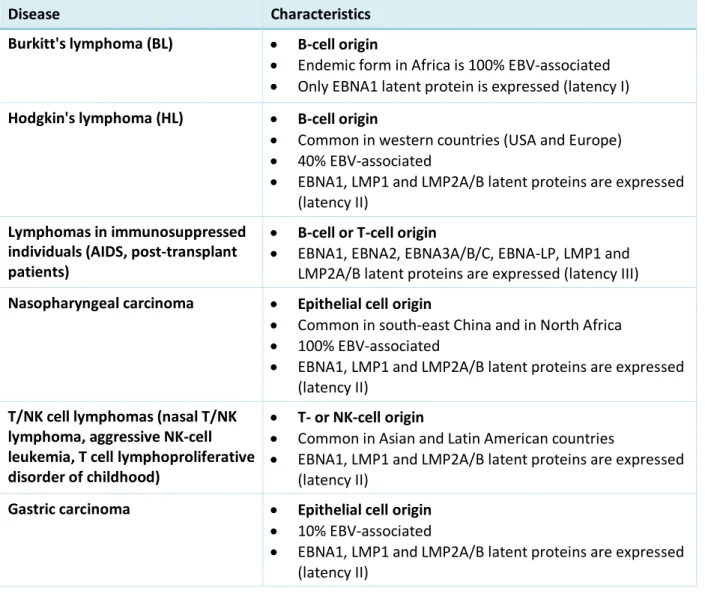
EBNA1 is a multifunctional protein that exerts essential functions in viral DNA replication and episome maintenance. Indeed, EBNA1 is required to ensure the extrachromosomal replication of viral episomes and mediates their segregation during cell division. EBNA1 also interacts with viral and cellular promoters contributing to the transcriptional regulation of both viral and cellular genes, directly or indirectly. EBNA1 is a dimeric protein containing three different functional domains: a central glycine-alanine (GAr) domain, two arginine-glycine (RG) domains (called linking regions, LR1 and LR2), and a C-terminal DNA-binding domain (DBD) involved inter alia in the association of the viral genome to the host cell chromatin (Figure 1).9,10 Interestingly, LR1 and LR2 domains of EBNA1 are known to interact
with G-quadruplex RNA structures (including those formed in its own mRNA, vide infra), and this interaction seems to play a critical role in recruiting the cellular origin recognition complex (ORC), ensuring the episome maintenance. Moreover, quadruplex-interacting drugs such as BRACO-19, TMPyP3 and TMPyP4 disrupt the interaction of EBNA1 with G4-RNA and interfere with recruitment of ORC, thereby inhibiting viral replication. Thus, treatment of EBV-infected Raji cells with BRACO-19 results in a moderate reduction of EBV copy number and selective death of EBV-positive cells, providing a first example of the use of a quadruplex-mediated strategy for the development of targeted therapy against EBV.10
6
Figure 1. A) Top, EBNA1 sequence showing the central Gly-Ala repeat (GAr) domain surrounded by the N and C
terminal domains; bottom, sequence of EBNA1 deleted for its GAr domain (EBNA1ΔGAr) used in the biological assays described below. B) Left, the G-rich motif represented 13 times in the GAr-encoding sequence of EBNA1 mRNA; right, the putative parallel two-quartet RNA G-quadruplex proposed as a model.
Immune response to non-self proteins: The detection of exogenous proteins originating from virus, bacteria or parasites by the host immune system is achieved through the production of derived antigenic peptides by the proteasome and/or by abortive translation. The antigenic peptides are shuttled to the endoplasmic reticulum (ER) where they are assembled into protein complexes called the Major Histocompatibility Complex (MHC). The loaded MHC complexes are then transported to the surface of the host cell where they display the antigenic peptides to activate recognition by T lymphocytes. Depending on the origin of the peptides (intra- vs. extracellular), the MHC are classified into class I and II, which are recognized by cytotoxic T-lymphocytes (CD8+ T) and T-helper lymphocytes
(CD4+ T) respectively through specific membrane receptors. This process of antigenic peptides
exposure, also called the antigen presentation pathway, allows the immune system to distinguish self from non-self, thereby ensuring the protection from viral, bacterial and parasite infections.11,12
EBNA1 and immune evasion: Like all the gamma-herpesviruses, EBV has evolved several strategies to evade the detection of its antigenic peptides and consequent elimination by the host immune system during both latent and lytic phases. Due to its crucial role in EBV genome replication and maintenance, and also to the fact that it is highly antigenic, EBNA1 plays a central role in the virus’ strategy to escape immune detection during the latent phase. Indeed, EBNA1 protein is expressed in all dividing EBV-infected cells and CD8+ T cells directed towards EBNA1 epitopes exist in all infected individuals. Despite
this, the immune system fails to detect and destroy EBNA1-expressing cells. To achieve this immune evasion, EBV has seemingly evolved a mechanism limiting EBNA1 production to the minimal level required for replication of the viral genome, but below the threshold for host immune detection. This
NL S DBD GAr RG-rich RG-rich N C NL S DBD RG-rich RG-rich N C EBNA1 EBNA1∆GAr 5′-GGGGCAGGAGCAGGAGGA-3′ 3′ 5′ A B Genome maintenance and replication Immune evasion G-quartet G-quadruplex structure
repeat motif (g4-EBNA1)
DNA binding and dimerization DNA binding and dimerization LR 1 LR 2 LR 1 LR 2 ♦ ♦♦♦♦♦ ♦♦♦♦♦♦♦ g4-EBNA1 (13×)
♦
7
is due to a tight regulation of the steady-state level of EBNA1 that minimizes the generation and, hence, the presentation of antigenic peptides. The immune evasion of EBNA1 depends on its central glycine-alanine repeat (GAr)13 and is based on the ability of EBNA1 to self-limit its own translation in cis (Figure
1, A).14–16 GAr consists of single alanine residues separated by 1–3 glycine residues, and is indeed a
potent inhibitor of its own mRNA translation.17The critical role of this domain was demonstrated
through infection by an EBV strain encoding a GAr-deleted version of EBNA1 (EBNA1ΔGAr), which induced high level of EBNA1 and efficient T cell response (Figure 1, A).13 In addition, the ability of GAr
to inhibit both its translation and antigenic presentation was shown to be tightly dependent on its length: longer GAr display a stronger inhibitory effect both on EBNA1 translation and antigen presentation.18 Another remarkable fact arguing for the strict involvement of the GAr domain is that
the fusion of GAr upstream to any open reading frame causes suppression of both protein synthesis and antigen presentation; this feature was subsequently used for the design of a test model involving the expression of chicken ovalbumin (OVA/Gar-OVA, see below). A number of mechanisms have been proposed to explain the ability of GAr to self-control EBNA1 synthesis (and thus of EBNA1-derived antigenic peptides), but two main hypotheses are currently admitted: the first one is based on the role of EBNA1 mRNA structure (e.g., G-quadruplexes) and is the topic of this book chapter, whereas the second one is based on the role of EBNA1 protein itself. Importantly, these two mechanisms are not mutually exclusive, and one may envision that both are required for efficient immune evasion of EBV.
3 G-Quadruplexes in the GAr-encoding sequence of EBNA1 mRNA
Supporting the role of EBNA1 mRNA structure, it was found that the structure of the GAr-encoding sequence of EBNA1 mRNA, rather than the peptide sequence itself, was involved in the self-control of EBNA1 synthesis.19,20 Indeed, EBNA1 mRNA, in particular the sequence encoding GAr, is G-rich and
form G-quadruplex (G4) structures.21 More recently, in 2014, Murat et al. identified a putative
quadruplex-forming sequence (PQS) motif comprised of 18 nucleotides (encoding for Gly-Ala-Gly-Ala-Gly-Gly) and repeated 13 times in the full length 300-nucleotide-long GAr-encoding sequence of EBNA1 mRNA.22 A panel of optical spectroscopy (TDS, CD and UV-melting) and NMR measurements indicated
that this short 18-nucleotide sequence forms a parallel G4 structure of high thermodynamic stability (Tm = 54–56 °C), likely corresponding to a two-quartet fold proposed as a model (Figure 1, B). Moreover,
the 300-nucleotide long fragment also showed a CD signature typical of parallel G4 with a clear K+
dependency, convincingly suggesting that GAr-encoding mRNA may fold into multiple G4 units in a pearl-on-a-necklace manner, as observed for other repeated sequences like telomeres and minisatellites.23–25It is straightforward to assume that G4 structures, like most secondary structures,
have a higher probability of occurrence in long tandem repeat sequences, which can give rise to G4 clusters. However, although the two-quartet quadruplex model is likely to be the predominant conformation in vitro, a strong G4-polymorphism can be expected in vivo considering the very high rich content of the GAr-encoding sequence of EBNA1 mRNA and the possible association of distal G-runs, which might be favored by formation of central long loops.26 Finally, although the thermodynamic
and kinetic parameters that regulate G4 formation in vitro are well characterized, the conditions of their formation and the factors influencing their lifetime in vivo are more difficult to determine since many cellular factors, in particular nucleic acid-interacting proteins, may have a strong impact in a way or another. Thus, the number of G4 structures able to form in long tandem repeats in vivo clearly remains an open question.
8
As stated above, a polymorphism exists with respect to the length of GAr domain, a fact which may reflect a way for EBV to adapt to its host. In addition, as previously mentioned, the translation inhibition of EBNA1 synthesis and presentation of the antigenic peptides are strongly dependent on GAr length, as shown both in mammalian cells and the budding yeast Saccharomyces cerevisiae.27
Hence, as the GAr length determines the number of PQS, it is tempting to correlate this structural feature with the biological response, which provides additional support to the G4-mediated regulation of EBNA1 expression.
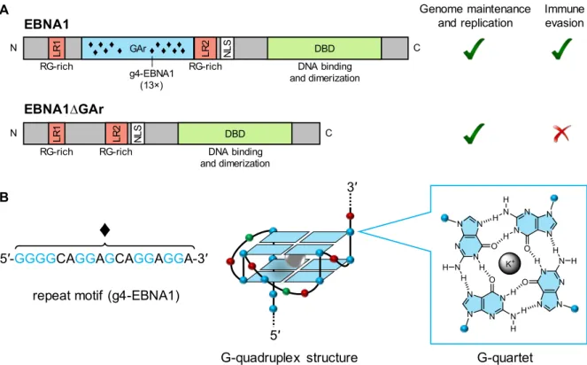
The study of Murat et al. reported the inhibitory role of G4 structures on the translation of EBNA1 mRNA in vitro, suggesting a ribosome stalling mechanism.22 The authors also showed data that suggest
that modulation of G4-RNA stability directly impacts EBNA1 levels, as antisense oligonucleotides that destabilize G4 (or prevent its formation) enhanced EBNA1 expression, whereas the G4-stabilizing compound Pyridostatin (PDS) decreases it (Figure 2). Following PDS treatment, inhibition of EBNA1 antigenic presentation was observed; at the same time, the authors did not exclude the involvement of other mechanisms due, in part, to the broad-spectrum activity profile of the drug.28 Also, it is
important to mention that the method employed in this study to evaluate the effect of G4 ligands on EBNA1 expression is an in vitro coupled transcription-translation assay, which does not allow to distinguish between transcriptional and translational effects. This point is particularly relevant given that G4 ligands are also known to affect transcription, as for example the transcription of the human immunodeficiency virus (HIV) genome.29 Nonetheless, these observations are consistent with the
repressor role of G4 observed in the translation of 5ʹ-UTR30and other inhibitory functions of mRNA
processing caused by steric blockade effect of G4.31,32
Figure 2. Schematic representation of a possible strategy of G4-mediated modulation of EBNA1 expression, as
proposed by Murat et al.22 ASO = antisense oligonucleotide.
… GAr domain ASO enhanced translation EBNA1 antigen presentation translation blockade Pyridostatin (PDS) no antigen presentation n
9
In parallel, other mechanisms related to the involvement of G4 structures on self-inhibition of EBNA1 translation were considered. As mentioned above, EBNA1 protein itself was demonstrated to bind to G-quadruplexes in its own mRNA transcript with moderate affinity, a fact that was proposed to explain its self-regulation in cis.10 More recently, the synthesis of EBNA1 and its antigenic presentation was
shown to be regulated by the host factor nucleolin (NCL).33,34 Altogether, these two studies set the
stage to explore more in depth the interplay between G-quadruplexes that form in EBNA1 mRNA, NCL and self-regulation of EBNA1 synthesis.
4 Nucleolin: a new player in the hide-and-seek game
The role of NCL in the regulation of EBNA1 synthesis was uncovered thanks to a yeast (Saccharomyces
cerevisiae)-based assay that recapitulates all the aspects of the GAr-based inhibition of translation
including the GAr-length dependency.35 This assay was used to perform a genetic that aimed at
identifying genes whose overexpression interfere (positively or negatively) with GAr-mediated translation inhibition.27,34 This way, the NSR1 gene which encodes the yeast homologue of human
nucleolin (Nsr1p) was identified as able, when overexpressed, to exacerbate the inhibitory effect of GAr on translation, whereas its elimination (by knock-out of the NSR1 gene which is not essential in yeast).33 Follow-up investigations in yeast and in EBV-infected human cells confirmed and extend this
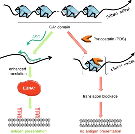
effect to human nucleolin (NCL) and have shown that nucleolin is a conserved host cell factor critically involved in the GAr-based regulation of EBNA1 expression, a mechanism at the basis of EBV immune evasion. In particular, unambiguous demonstration was provided by overexpressing or downregulating NCL in three EBV-infected B-cells (Mutu-1, B95-8 and Raji), which led, respectively, to a decrease or an increase in EBNA1 expression, and to an increase of GAr-limited antigen presentation in the case of downregulation of NCL (Figure 3A).33 The inhibitory effect of NCL on EBNA1 synthesis was also studied
in mammalian cells (HT1299 lung carcinoma cells) transfected with expression vectors encoding EBNA1 or its GAr-deleted version (EBNA1ΔGAr). As shown in Figure 3B (left), NCL downregulation induced EBNA1 repression only when the GAr domain was present, as no impact was observed on EBNA1ΔGAr protein. The same results were obtained with a second set of constructs encoding the chicken ovalbumin fused (or not) to a 235 amino acid-long GAr domain (235GAr-OVA vs. OVA), leading to the same conclusions (Figure 3B, right). These two settings represent benchmark assays giving crucial mechanistic information on the GAr-dependency of translation of EBNA1 or OVA, the latter further illustrating the generality of this mechanism of translation regulation. Importantly, the OVA/235 OVA assay also allows to determine the effect of GAr on antigen presentation on the host cell membrane using specific antibodies against SINFEKL (an OVA-derived antigenic peptide) associated to MHC class I receptors (Kb). Indeed, when fused to OVA, 235GAr lead to a strong inhibition of presentation of OVA-derived antigenic peptide.16,35 Using this assay, NCL downregulation was shown to induce a significant
GAr-dependent increase of antigen presentation (Figure 3C, left panel).33 Altogether, the test workflow
described on Figure 3 have proven particularly useful for evaluation of GAr-interacting drugs and was used throughout our follow-up studies (below).
10
Figure 3. A) Western blot analysis showing EBNA1 expression level as a function of NCL overexpression (HA-NCL)
(left) and downregulation (using siRNA, right) in EBV-infected Mutu-1 cells. B) Effect of NCL downregulation (using siRNA) on EBNA1 or EBNA1ΔGAr (left), or OVA or 235GAr-OVA (right) protein expression following transfection of HT1299 cells by the corresponding constructs. C) Effect of NCL downregulation (using siRNA) on
antigen presentation after transfection of HT1299 cells with OVA or 235GAr-OVA and murine MHC class I Kb
encoding plasmids. Cells were incubated with naive OVA CD8+ T cells whose activation is measured. Adapted
with permission form Ref. 33.
OVA/GAPDH A Mutu-1 cells 100 55.7 EBNA1 / Actin EBNA1 HA-NCL Actin EBNA1 NCL GAPDH 100 NCL / GAPDH 60 100 EBNA1 / GAPDH 150 235GAr-OVA OVA GAPDH EBNA1 EBNA1ΔGAr GAPDH NCL NCL/GAPDH NCL/GAPDH 100 60 100 60 100 181 100 96 + + – – – – + + si-control si-NCL NCL 100 50 100 45 100 153 100 90 + + – – – – + + si-control si-NCL EBNA1/GAPDH B HT1299 cells All MHC-I (Kb) SIINFKL-Kb complex C
11
NCL is a multifunctional DNA/RNA-binding protein widely conserved among eukaryotes and implicated in many cellular functions such as ribosome biogenesis, rRNA maturation, cell cycle control, apoptosis, transcription and translation regulation.36 As such, NCL binds a variety of nucleic acids thereby showing
a broad substrate promiscuity. NCL is one of the first G4-interacting protein identified.37 One of its best
documented mechanism of action on G4 is its role as repressor of transcription through stabilization of G4 in the c-Myc promoter.38,39 More recently, NCL was shown to recognize G4-DNA within the LTR
promoter of HIV, thereby silencing the provirus transcription.29 NCL is ubiquitous and is therefore likely
to interact at many G4 loci of DNA or RNA. The structural determinants of G4/NCL interactions are not really known, but recently three independent groups, including some of us, reported on the strong preference of NCL for binding G4-DNA harboring a long central loop.40–42 Whether this trend is a
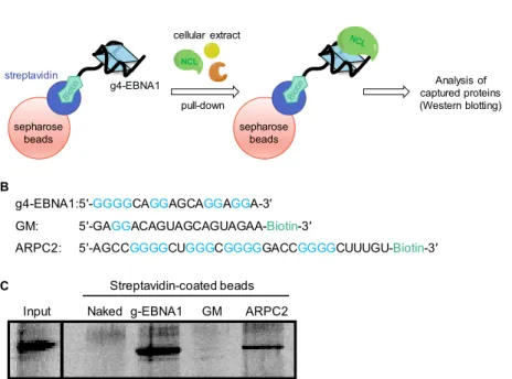
general rule that can be extended to recognition of G4-RNA remains to be explored. Therefore, after having uncovered the role of NCL in EBNA1 translation, our first line of investigation has been focused on finding whether NCL was directly interacting with the G4 formed in the GAr-encoding mRNA. This has been done by a classical affinity pull-down approach using the 18-nt oligonucleotide corresponding to the most probable G4-forming motif in the GAr-encoding sequence of EBNA1 mRNA (cf. Figure 1, A). This oligonucleotide sequence (g4-EBNA1), as well as a control mutated oligonucleotide containing adenine or uridine in place of guanine residues to prevent G4 folding (GM, negative control), and a 30-nt RNA sequence known to bind NCL (ARPC2, positive co30-ntrol) were 3ʹ-tagged by biotin, immobilized on streptavidin-covered sepharose beads, and incubated with lysates from HT1299 (lung cancer) cells, then thoroughly washed and eluted with increasing concentration of KCl (200–800 mM) (Figure 4, A– B). After elution, the pulled-down proteins were analyzed by western blotting against NCL. As shown from Figure 4, C, NCL was pulled down by g4-EBNA1 and RNA-G4 control (ARPC2), whilst no interaction was detected with the mutated sequence (GM). Importantly, the same results were obtained with recombinant NCL, showing that the interaction between NCL and G4 of the GAr-encoding sequence of EBNA1 mRNA is direct.33
Figure 4. A) Schematic representation of the pull-down assay using G4-RNA immobilized on streptavidin-covered
sepharose beads. B) RNA sequences used in the assay. C) Western blotting analysis of proteins after the pull-down.
Input Naked g-EBNA1 GM ARPC2 Streptavidin-coated beads g4-EBNA1:5′-GGGGCAGGAGCAGGAGGA-3′ GM: 5′-GAGGACAGUAGCAGUAGAA-Biotin-3′
ARPC2: 5′-AGCCGGGGCUGGGCGGGGGACCGGGGCUUUGU-Biotin-3′
A B C sepharose beads streptavidin g4-EBNA1 cellular extract pull-down Analysis of captured proteins (Western blotting) NCL sepharose beads
12
Affinity pull-down is a useful method for identification of proteins interacting with a given target (nucleic acid or protein), but should be considered only as a first indication. Therefore, other techniques are required to evaluate if the identified partners do indeed interact in a native environment. Thus, to verify whether the NCL–mEBNA1-G4 interaction was occurring in cellulo, a proximity ligation assay (PLA) was devised to detect this association in EBV-infected cells.
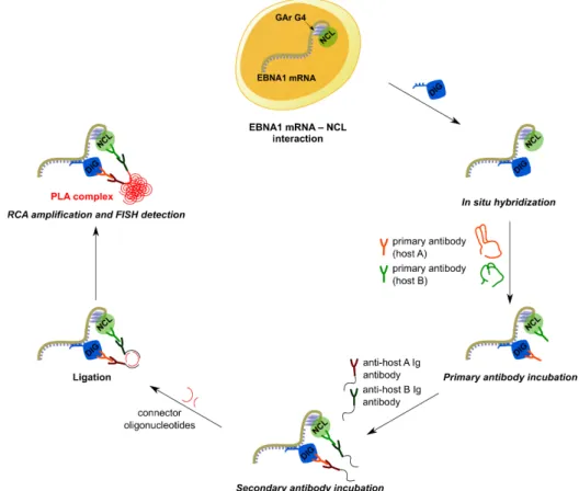
Briefly, PLA is a technique originally developed to detect proteins localized in close proximity to each-other (theoretically, at a maximum distance of 40 nm) and is based on the use of a pair of antibodies raised in two different species, each targeting one of the two proteins of interest. By using oligonucleotides labelled with haptens (digoxigenin or biotin), PLA has been adapted to the study of protein–DNA and protein–RNA interactions.43 To this end, the RNA target (herein, EBNA1 mRNA) was
tagged through in situ hybridization with a digoxigenin-labelled DNA probe detected by a mouse anti-digoxigenin antibody, whereas NCL was recognized by a rabbit anti-nucleolin antibody. If the two partners are located in close proximity (strongly suggesting that they are interacting), subsequent incubation with secondary antibodies directed against each primary species and covalently coupled to specific DNA primers will generate a circular DNA template. The latter is then amplified by rolling circle amplification (RCA) and detected by in situ hybridization with specific fluorescent probes. Thus, the close proximity of the two partners is visualized through formation of bright fluorescent foci (PLA dots) that appear at the place of the monitored interaction. The principle of the PLA assay is depicted in Figure 5.
Figure 5. Principle of the proximity ligation assay (PLA) applied to the study of EBNA1 mRNA–NCL interaction.
The DNA probe (5ʹ-CTTTCCAAACCACCCTCCTTTTTTGCGCCTGCCTCCATCAAAAA-digoxigenin-3ʹ) was chosen such that hybridization occurs outside the G4-forming region, to avoid eventual interference between G4 formation and RNA–DNA hybridization.
13
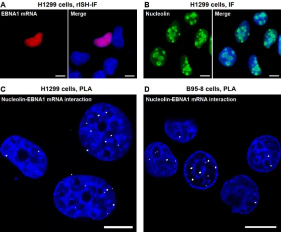
In a first step, to verify the specificity of the EBNA1–digoxigenin probe and the detection of the hybridization complex by anti-digoxigenin antibody, HT1299 (lung carcinoma) cells were transfected with an EBNA1 construct and analyzed by RNA in situ hybridization coupled to immunofluorescence (rISH-IF). Primary EBNA1-immunocomplexes were detected using an anti-mouse secondary antibody labelled with Alexa Fluor 568. Fluorescence imaging revealed the accumulation of EBNA1 mRNA in the nucleus (Figure 6 A, red staining). In parallel, immunostaining was performed to determine the appropriate conditions for detecting endogenous NCL using a rabbit anti-NCL antibody (Figure 6 B). Altogether, these assays demonstrated an accumulation of NCL and EBNA1 mRNA in the nucleus. Then the complete set of PLA reagents (i.e., EBNA1-rISH probe, primary antibodies, secondary antibodies, and RCA kit) was applied to the same EBNA1-transfected cell line (HT1299) and to an EBV-infected cell line (B95-8, EBV-producing marmoset lymphoblastoid cells). In both cell lines PLA complexes (depicted as white dots) were detected in or at close vicinity of the nuclear compartment (Figure 6 C, D). This confirms that NCL binds to EBNA1 mRNA, and that this interaction mostly takes place in the nucleus of EBV-infected as well as EBNA1-transfected cells.
Figure 6. A) RNA in situ hybridization (rISH) with the EBNA1 digoxigenin probe followed by mouse
anti-digoxigenin immunostaining and anti-mouse IgG labelled with Alexa Fluor 568. DAPI was used for nuclear counterstaining. B) Detection of NCL by rabbit anti-NCL antibodies. C–D) PLA in C) EBNA1-transfected HT1299 cells and D) EBV-infected B95-8 cells. PLA was performed using mouse anti-digoxigenin and rabbit anti-nucleolin, followed by anti-rabbit and anti-mouse Ig PLA probes applied together with fluorescent probes for RCA product detection. Scale bars represent 10 µm. Adapted with permission from Ref. 44.
The presence of PLA dots in the nucleus is consistent with the predominant nuclear localization of NCL and more specifically with the recent notion that early translation events coupled to transcription occur in the nucleus.45,46 In line, recent findings by some of us have shown that translation of EBNA1
14
mRNA as well as antigen presentation may be regulated by NCL in the nucleus.47 Altogether, these
results denote that EBV may exploit host cell mRNA maturation process(es) in order to hamper translation and thus immune response. Finally, and importantly, it was shown that the presence of GAr-encoding sequence, that is, the G4-forming motif of EBNA1 mRNA, was absolutely required for its interaction with NCL, as no PLA signals were detected when HT1299 cells were transfected with an EBNA1 construct lacking the GAr domain (EBNA1ΔGAr).33 In summary, the yeast assay combined with
PLA have been instrumental for unravelling the interaction between NCL and EBNA1 mRNA-G4 on one hand, and for visualizing and localizing this interaction in cells on the other hand. Thus, NCL is the first identified host cell factor involved in EBNA1 stealthiness and, as shown previously, NCL expression is directly linked to the regulation of EBNA1 level. For these reasons, the interaction of NCL with G4s in EBNA1 mRNA immediately appeared as a pertinent target for pharmacological intervention with the aim of modulating EBNA1 translation and, in fine, triggering immune response against EBV-infected cancer cells.
5 PhenDC3, a benchmark G4 ligand, disrupts NCL–EBNA1 mRNA interaction,
enhances EBNA1 expression, and overcomes the immune evasion of EBV
Following these results, the effect of G4-interacting compounds (G4-ligands) on the NCL–EBNA G4 interaction and immune evasion of EBV was evaluated. As a first step, the benchmark G4-ligand PhenDC3 was examined since this compound exhibits high affinity (with Kd in the nanomolar range)
and selectivity for most G4 structures and has been validated as one of the best probe for targeting G4 DNA or RNA in cells through numerous studies.25,48–52 A remarkable advantage of PhenDC3 is its high
permeation capacity with regard to live mammalian cell lines and yeast (S. cerevisiae). In parallel, we also evaluated Pyridostatin (PDS), another benchmark G4-ligand widely used for in-cell G4-studies, which was shown by Murat et al. to inhibit EBNA1 expression through stabilization of G4s in EBNA1 mRNA (cf. Figure 2). Both compounds strongly stabilize G4 and, as such, are expected to increase the lifetime of G4, thereby emphasizing their biological roles. The question whether small molecules can enhance or disrupt G4–protein interactions was much less investigated and is highly topical, raising questions about the precise mechanism of action of quadruplex-targeting drugs.42,53,54
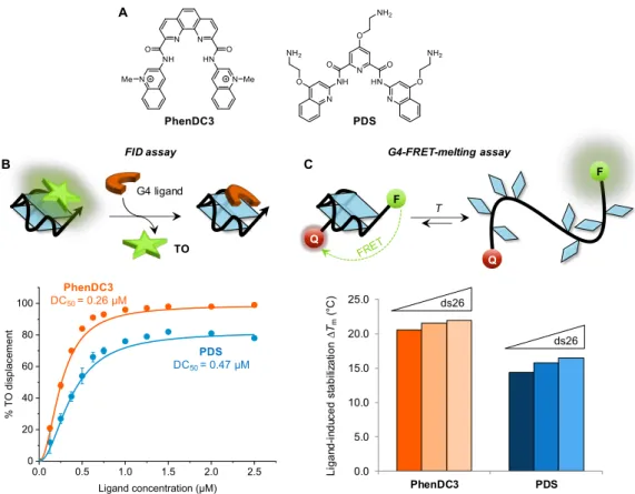
Prior to cell-based assays, the capacity of the two ligands to interact with the G4 structure of EBNA1 mRNA was evaluated in vitro, using classical biophysical assays (G4-FID and G4-FRET-melting) (Figure 7). The low DC50 value (i.e., high displacement activity) and high ∆Tm (i.e., thermal stabilization)
determined in both cases indicate that the two compounds bind to and strongly stabilize the g4-EBNA1 structure. Also, excellent selectivity for G4 vs. duplex DNA was evidenced by the absence of impact of the duplex competitor (ds26) on the variation in melting temperature induced by the ligands. Thus, both PhenDC3 and PDS can be considered as strong binders of g4-EBNA-G4 used as a model structure with a slight, but significant, advantage for PhenDC3 that displayed globally a higher performance.
15
Figure 7. A) Chemical structures of PhenDC3 and Pyridostatin (PDS) and evaluation of their G4 binding properties
to G4-RNA oligoribonucleotide (g4-EBNA1) in vitro by B) FID (fluorescent-indicator displacement) assay and C) FRET-melting assays. The principle of each assay is illustrated above the graphs. TO = Thiazole Ornage dye; DC50
= ligand concentration inducing 50% decrease in TO fluorescence; ∆Tm (°C) = increment of melting temperature
induced by the ligand alone or in presence of a 26-bp duplex competitor (ds26).
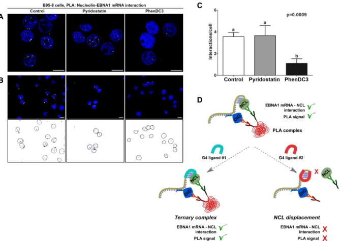
We then took advantage of the previously developed PLA assay to examine the direct in cellulo effect of both compounds on the NCL-GAr interaction. To this end, B95-8 cells were treated with PhenDC3 and PDS at sub-toxic concentration (< 1 µM) to avoid cell death, then fixed and PLA was performed. A qualitative look at the resulting confocal fluorescence images indicates clearly that PhenDC3 reduces significantly the PLA signal as compared to the untreated control, whereas PDS does not impact it (Figure 8, A). PLA is most often used as a qualitative method to visualize and localize the interaction between two biological partners in cells. However, in the case of drugs acting on a given interaction, quantitative evaluation is absolutely required to draw proper comparison of drug activity within a series. Thus, a protocol was further developed to achieve quantitative PLA. In detail, one hundred cells from control (DMSO) and drug-treated groups were imaged and the number of interactions (i.e. the number of PLA dots) per cell was counted using a customized image analysis protocol as depicted in Figure 8, B.44 The experiments were done in triplicate to obtain accurate quantification. As shown from
the resulting histograms (Figure 8, C), upon treatment with PhenDC3 the number of PLA dots per cell was reduced by a factor of 3.3 (from 3.6 ± 0.3 to 1.1 ± 0.4), whilst this number was not significantly modified in PDS-treated cells (3.7 ± 0.9).
FID assay PhenDC3 PDS A B G4 ligand TO 0.0 0.5 1.0 1.5 2.0 2.5 0 20 40 60 80 100 % TO disp lace ment Ligand concentration (µM) PhenDC3 DC50= 0.26 µM PDS DC50= 0.47 µM Q F T C 0.0 5.0 10.0 15.0 20.0 25.0 PhenDC3 PDS G4-FRET-melting assay Q F Li gan d -i nd uce d st a b ili za tio n ∆ Tm (° C) ds26 ds26
16
Figure 8. Evaluation of PhenDC3 and PDS in B95-8 cells using the quantitative PLA assay. A) Microscopy images
of cells treated with DMSO (control), PhenDC3 or PDS (5 µM). Nuclei are stained by DAPI (blue). White dots (PLA signals) indicate the interaction between NCL and EBNA1 mRNA. B) Quantification of PLA signals: 100 cells per condition were imaged and the number of dots per cell was estimated using a customized protocol in ImageJ. C) Graph showing number of PLA dots per cell as function of ligand. D) Schematic representation of the proposed mode of action of G4-ligands explaining the different effects on the PLA patterns. Left: Ternary interaction (G4-RNA/NCL/ligand) induces no modification of the PLA dots as compared to control; right: competitive binding of ligand displaced NCL and reduces the number of PLA dots. Adapted with permission from Ref. 44.
Thus, the quantitative PLA clearly indicates that PhenDC3 can disrupt the interaction between NCL and G4 that form in the GAr-encoding sequence of EBNA1 mRNA in a cellular context, whilst PDS has no effect. This discrepancy between the two G4 ligands can be explained in several ways. If we assume that NCL and drugs have common binding site(s) on GAr-G4, then competitive binding is possible and the result reflects the relative affinities of the endogenous (i.e., protein) and exogenous (i.e., small molecule) partners. In line, the higher affinity of PhenDC3 enables a competition with NCL for G4 binding and thus disrupts the interaction of NCL with GAr-mRNA. On the opposite PDS, that has a significantly lower G4 affinity cannot prevent NCL–mRNA association. Alternatively, it could be speculated that the drug (e.g., PhenDC3) induces a strong conformational change of the G4-mRNA target thereby hampering further recognition by NCL, while the non-active drug (PDS) does not modify the G4 structure. Finally, the drug may be bound to an allosteric site (G-quartet, groove, or loop) that does not disturb RNA recognition by the protein (Figure 8, D). This case has been reported for carboxyPDS and 7ODT (hexaoxazole analogue of telomestatin) that were shown to bind G4 without affecting their recognition by the antibody or proteins, namely through formation of ternary complexes.55,56 Finally, since the drugs are incubated in living cells, differences in cell uptake, or
A
C
D B
17
subcellular distribution, or metabolism cannot be excluded, and may contribute to various extents to the observed differences in activity.
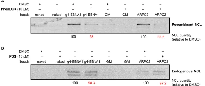
To further explore the comparison between PhenDC3 and PDS with regard to NCL/GAr-G4 interaction, we used the affinity pull-down assay described in Figure 4. Beads coated with g4-EBNA1 were incubated with endogenous or recombinant NCL in presence or in the absence of the drugs. In good agreement with the PLA results, inhibition of NCL binding to beads was achieved in presence of PhenDC3, whereas, in the same conditions, PDS had no effect (Figure 9). The same trend was also observed with the positive control G4-RNA (ARPC2) that also binds NCL.
Figure 9. Inhibition NCL–G4 binding by PhenDC3 and PDS, as determined by the affinity pull-down assay. for the
principle of the assay, cf. Figure 4, A. Adapted with permission from Ref. 33.
Interestingly, we recently investigated in vitro the ability of G4 drugs to disrupt NCL interaction with G4-DNA and found again that PhenDC3 was the more efficient competitor of NCL, as compared to PDS and other G4-binding compounds (RHPS4, 360A) frequently used as G4 probes.42 Therefore, our data
from PLA and pull-down assays are fully consistent with these recent findings and demonstrate that G4 ligands that exhibit comparable binding parameters for a given G4 structure in vitro may behave differently in cellulo, in part due to the differences in their capacity to compete with associated proteins.
In conclusion, it is worth noting that PLA coupled with quantitative imaging represents an attractive alternative to overcome the limitations of in vitro assays and to characterize the G4-RNA binding drugs targeting specific protein–RNA interactions in cells. In particular, this approach may represent a new and powerful tool for ranking and refining the selection of drug candidates during “hit-to-lead optimization” processes.
Biological effects: The effects of the two compounds on GAr-mediated protein expression inhibition and on antigenic stimulation have been evaluated using the set of biological assays described above. First, HT1299 cells transfected with plasmid expressing the ovalbumin fragments (OVA/235GAr-OVA) were treated, or not, with PhenDC3 or PDS (5 µM, 48 h) and OVA and 235GAr-OVA expression levels were evaluated by western blotting (Figure 10). This analysis revealed that PhenDC3 induced a
DMSO
PhenDC3 (10 μM)
naked naked g4-EBNA1 g4-EBNA1 + + – – – + – + Recombinant NCL 100 58 NCL quantity (relative to DMSO) GM GM + – – + ARPC2 ARPC2 + – – + 100 35.5 PDS (10 µM) Endogenous NCL DMSO
naked naked g4-EBNA1 g4-EBNA1 + + – – – + – + GM GM + – – + ARPC2 ARPC2 + – – +
100 98.3 100 97.2 NCL quantity(relative to DMSO)
beads beads
A
18
significant increase of the steady state level of 235-OVA while producing no notable variation of OVA expression (Figure 10 A). This result demonstrates that PhenDC3 is able to affect the protein expression exclusively when the G4-forming domain (235GAr) is present in the construct. As shown from control experiments, the corresponding mRNA levels were not modified, allowing to conclude that, at least in these experiments, PhenDC3 enhances the expression of the protein without significant effect on mRNA level. As both EBNA1 and OVA are very stable proteins, PhenDC3 most probably increases EBNA1 translation. In stark contrast, no clear effect on 235GAr-OVA or OVA was observed after treatment with PDS in the same conditions (Figure 10A). The effect of the two drugs on the expression of viral EBNA1 was also evaluated in two EBV-infected cancer cell lines, Mutu-1 (Burkitt’s lymphoma) and NCP-6661 (nasopharyngeal carcinoma). Western blotting analysis indicated that PhenDC3 treatment enhanced EBNA1 synthesis (Figure 10B). Again, PDS showed no activity even when applied at a higher concentration range as compared to PhenDC3 (up to 9 µM, Figure 10C). These data are fully consistent with the capacity of the compounds to disrupt (or not) the NCL-EBNA1 mRNA G4 interaction observed by PLA and in pull-down experiments and confirm that pharmacological modulation of NCL binding to G4 of EBNA1 mRNA is feasible, paving a way to counteract the inhibitory control of GAr on the EBNA1 production in EBV-infected cancer cells.
Figure 10. Protein expression after treatment of cells with PhenDC3 or PDS. A) HT1299 cell line transfected with
OVA or 235GAr-OVA constructs, western blotting (left) and mRNA quantification (right). B) Western blotting analysis after treatment of Mutu-1 and NCP6661 with PhenDC3 (1 µM). C) Western blotting analysis (left) and quantification of EBNA1 level (right) after treatment of Mutu-1 cells with PDS at concentrations of 3, 6 or 9 µM (left). 100 265 PhenDC3 GAPDH OVA 100 116 235GAr-OVA GAPDH
OVA/GAPDH 235GAr-OVA OVA
OV A mR N A Ac ti n mR N A PDS 235GAr-OVA OVA GAPDH GAPDH 100 104 100 105 OVA/GAPDH A 100 173.3 Mutu-1 EBNA1/Actin EBNA1 Actin NPC-6661 100 162.4 EBNA1 Actin EBNA1/Actin Mutu-1 + PDS EBNA1 GAPDH EB N A 1 e x pre ss ion le ve l PDS B C
19
Finally, in contrast to the study of Murat et al., in our hands PDS showed no significant effect on the translation of EBNA1 despite its high binding affinity for EBNA1-G4 as determined by FID and FRET-melting. Obviously unfavorable pharmacological features (poor uptake, undesired subcellular distribution) and eventual off-target binding affecting PDS capacity to reach G4 in EBNA1 mRNA could explain this discrepancy. Of note, differences in experimental conditions between the two studies should also be considered: indeed, PDS was initially evaluated using an in vitro transcription-translation coupled assay, thus it cannot be excluded that the impact on EBNA1 synthesis was, at least in part, due to effect of the compound on transcription rather than on translation.22
As a next step, PhenDC3 has been evaluated with respect to its ability to stimulate the presentation of antigenic peptide that, as shown before, is directly related to increase in EBNA1 level. This was done using the OT1 T-cell proliferation assay as described previously (Figure 11), again employing the HT1299 cells transfected with OVA or 235GAr-OVA constructs. As shown on Figure 11, PhenDC3 significantly increased (2-fold change) the proliferation of T cells added to the pool of cells expressing 235GAr-OVA, whereas it has no effect on OT1-T cells incubated with the OVA-expressing pool of cells. The stimulation of the immune response induced by PhenDC3 is fully consistent with the inhibitory role of NCL on antigen presentation and with the ability of PhenDC3 to interfere with the binding of NCL on the G4 motifs of EBNA1 mRNA. This data is in sharp contrast with the observations by Murat et al. who found that PDS further decreases GAr-restricted presentation of antigenic peptides.
Figure 11. FACS analysis of T-cell proliferation (T-cell assay). HT1299 cells were transfected with mouse Kb and A) 235GAr-OVA or B) OVA plasmids, and treated with PhenDC3 (5 µM) or DMSO (control). Cells were mixed with mouse naïve OVA257–264 (SINFEKL) specific CD8+ T-cells and stained with Celltrace Violet. Quantification of proliferating T lymphocytes by FACS following treatment are shown in the graphs on the right (ns = not significant). Reproduced with permission from Ref. 33.
A
20
Altogether, and despite these discrepancies, it can be concluded from these two studies that G4 motifs formed in the EBNA1 GAr domain are negative regulators of EBNA1 synthesis, acting either through NCL recruitment (our work) or as ribosome roadblocks (the work of Murat et al). Of note, these two possibilities are not mutually exclusive.
Taken together, our data demonstrate, firstly, that the role of NCL in GAr-based inhibition of translation involves its ability to bind to G4 in GAr-encoding sequence of EBNA1 mRNA. Secondly, this work provides a proof of concept that disrupting the NCL–G4 EBNA1 mRNA in cells by G4 ligands increases the GAr-restricted antigenic presentation of EBNA1, thus validating this class of compounds as potential agents to unveil EBV to the immune system and thereby for treating and/or preventing EBV-related cancers. However, in cellulo PLA imaging combined with in vitro biophysical data clearly indicate that the ability to disrupt in cellulo the NCL-G4 EBNA1 mRNA interaction is not a general feature shared by all G4 ligands. Further illustration of this specific point recently came from a novel series of G4 ligands (vide infra). Finally, from a practical viewpoint, this corpus of studies allowed to establish a pipeline of in vitro and in cellulo benchmark assays amenable to robust and drastic selection of drug candidates.
Following this work, we launched a program to identify other compounds with higher patentability and higher therapeutic index (or patentable properties). First, a selection of G4-selective and non-G4-selective DNA ligands (Figure 12) was screened by western blot to assess their effect on EBNA1 synthesis in human cells. The results confirmed PhenDC3 as a “hit” dramatically enhancing EBNA1 production, as well as tBisQ-Cu and a number of other G4-binding metal complexes.57 At the same
time, it was observed that other G4 binders belonging to different chemical classes, such as condensed polyaromatic compounds (i.e., MMQ1, MMQ12, TrisQ) and styryl dyes (PhenDV) either decreased, or had no effect on EBNA1 expression. Most interestingly, cationic bis(acylhydrazone) PyDH2 synthesized in our laboratory enhanced EBNA1 expression to a level comparable to the one induced by PhenDC3, as could be judged from this semi-quantitative screen. On these premises, a medicinal chemistry program was launched to devise a second generation of compounds derived from PhenDC3 and PyDH2 with optimized properties and evaluate a broader range of derivatives.
21
Figure 12. Small-molecule DNA and RNA binders tested as modulators of EBNA1 synthesis. Compounds that
increased the expression of EBNA1 are colored in green; those that decreased EBNA1 expression are colored red; those shown in black had no significant effect. Acri-2,7-Py is a non-G4-selective DNA binder.
6 Development of a novel series of G4-interacting ligands disrupting the NCL–G4
EBNA1 interaction
With the aim to explore novel pharmaceutical scaffolds as putative G4 ligands and disruptors of the NCL–EBNA1 G4-mRNA interactions, a series of cationic bis(acylhydrazones) (“HetDH”) was designed as shape analogues of PhenDC3. In addition, the N-acylhydrazone group has been selected as a privileged scaffold in drug design, due to a combination of hydrogen-bond acceptor and donor sites capable of interactions with a wide range of biomolecules. Despite the fact that acylhydrazone derivatives are rapidly metabolized, this motif is encountered in several approved drugs (e.g., nifuroxazide and dantrolene)58 and drug candidates.59–61 The facile synthetic availability of acylhydrazone derivatives,
which allows furnishing congeneric series of compounds with distinct physico-chemical properties and bioactivities, makes it a promising scaffold in the drug discovery field.62–64
The design of the HetDH series features a central heterocyclic core (Het), i.e., a residue of pyridine (Py), pyrimidine (Pym), 1,8-naphthyridine (Naph), or 1,10-phenanthroline (Phen), connected via two acyhydrazone linkages to lateral, N-substituted cationic heterocycles (Ar = pyridinium or quinolinium, R = Me, Et, or Bn). Altogether, we obtained 20 HetDH derivatives (PyDH1–5, PymDH1–5, NaphDH1–5, and PhenDH1–5, Table 1) in good yields by combinatorial synthesis starting from bis-acylhydrazides and cationic heteroaromatic aldehyde precursors.65
22
Table 2. Generic chemical structure and members of the HetDH family of bis(acylhydrazone) ligands.
Ar Het
(1) (2) (3) (4) (5)
(Py)
PyDH1 PyDH2 PyDH3 PyDH4 PyDH5
(Pym)
PymDH1 PymDH2 PymDH3 PymDH4 PymDH5
(Naph)
NaphDH1 NaphDH2 NaphDH3 NaphDH4 NaphDH5
(Phen)
PhenDH1 PhenDH2 PhenDH3 PhenDH4 PhenDH5
In silico evaluation: The drug-like properties of HetDH derivatives were initially evaluated through the
use of physicochemical descriptors, allowing the estimation of pharmacokinetics and drug-likeness of small molecules.66 The results of in silico assessment indicated satisfactory bioavailability for most
HetDH derivatives, as given by the combination of six physicochemical descriptors (lipophilicity, molecular weight, polarity, solubility, saturation and number of rotatable bonds). However, insufficient saturation (i.e., low fraction of sp3 carbons) could be identified as the limiting factor for
bioavailability of most compounds, as well as high molecular weight (M > 700 Da) in several cases (NaphDH3, NaphDH5, PhenDH3 and PhenDH5. With the exception of these four compounds, all HetDH derivatives satisfied the Lipinski’s rule, and most derivatives also satisfied the Muegge’s rule,67
indicating a high potential to serve as drugs. Finally, gastrointestinal absorption prediction was made using the BOILED-egg model (Figure 13, A).68 According to this model, most compounds, again with the
exception of NaphDH3, NaphDH5, and PhenDH3–5, could have a high rate of passive gastrointestinal absorption; however, none of compounds could cross the blood-brain barrier. In conclusion, the results of in silico evaluation indicate that HetDH scaffolds are compatible with their use as RNA-G4
23
targeting drugs, while heavy and aromatic substitutes (e.g., R = benzyl) should be avoided for the sake of bioavailability.
Figure 13. A) BOILED-egg evaluation68 of passive gastrointestinal absorption (HIA) and brain (BBB) penetration of HetDH derivatives. The white region indicates high probability of passive gastrointestinal absorption, while the yellow region indicates high probability of BBB penetration. B–C) In vitro assessment of binding of HetDH compounds and PhenDC3 (red) to g4-EBNA1: B) Thermal stabilization F-g4-EBNA1-T (0.2 µM) by tested compounds (1 µM), assessed by fluorescence melting experiments in the absence (dark blue bars) or in the presence of duplex DNA competitor ds26 (blue bars: 3 µM, pale blue bars: 10 µM). Conditions: 10 mM LiAsO2Me2, 10 mM KCl, 90 mM LiCl buffer, pH 7.3. C) Ligand-induced displacement of TO (0.5 µM) from g4-EBNA1 (0.25 µM). n.d. = no displacement (DC50 > 2.5 µM). Conditions: 10 mM LiAsO2Me2, 100 mM KCl, 1 mM EDTA, buffer, 1% v/v DMSO. Adapted with permission from Ref. 65.
Biophysical assessment: The capacity of HetDH derivatives to bind to g4-EBNA1 quadruplex was studied in vitro using the previously described biophysical methods, namely FRET-melting and G4-FID assay. Interestingly, these derivatives displayed great variability with respect to their capacity to bind to and stabilize the G4 target, revealing some structure–activity relationships. Specifically, most derivatives of the PyDH and NaphDH subfamilies, as well as PhenDH1, demonstrated significant stabilization of g4-EBNA1 (∆Tm = 10 to 20 °C), and other phenanthroline derivatives (PhenDH2–5)
demonstrated an even higher stabilization of the substrate (∆Tm = 20 to 30 °C), comparable to the
result obtained with PhenDC3 (∆Tm = 30.0 °C). In contrast, all derivatives of the PymDH family, as well
as compounds PyDH1 and NaphDH1, demonstrated low or very low stabilization of G4-RNA (∆Tm <
10 °C), illustrating the importance of the nature of heterocyclic residues on the G4-RNA binding properties of ligands. Finally, most derivatives that stabilized g4-EBNA1 also displayed significant level of selectivity with respect to ds-DNA, as their stabilizing effect was almost unaffected by the presence of ds-DNA competitor. The results of the FID assay (Figure 13, C) indicated high g4-EBNA1 affinity (DC50
< 0.5 µM) for five compounds (PyDH2, PyDH3, and PhenDH1–3), which was comparable to the result obtained for PhenDC3 (DC50 = 0.31 µM). In contrast, the derivatives PyDH1, PymDH1, PymDH4 and
PyDH5 were not able to displace the fluorescent probe (TO) from g4-EBNA1. Of note, none of tested ligands was able to induce displacement of TO from the double-stranded DNA substrate ds26 (DC50 >
2.5 µM in all cases). 0 50 100 150 200 -4 -2 0 2 4 6 8 log P (WLO GP)
Topological polar surface area (TPSA, Å3) BBB
HIA
PhenDH3, PhenDH5 NaphDH3, NaphDH5 PyDH3, PyDH5 PhenDH4
PymDH3, PymDH5 PhenDH2 NaphDH4 NaphDH2 PymDH4 PymDH2 PyDH1 NaphDH1 PymDH1 PyDH4 PhenDC3 PyDH2 0 5 10 15 20 25 30 Li gand-i nduc ed s tabi liz ati on ∆ Tm (°C
) PyDHn PymDHn NaphDHn PhenDHn
PyDH 1 PyDH 2 PyDH 3 PyDH 4 PyDH 5 Pym DH1 Pym DH2 Pym DH3 Pym DH4 Pym DH5 Naph DH1 Naph DH2 Naph DH3 Naph DH4 Naph DH5 Phen DH1 Phen DH2 Phen DH3 Phen DH4 Phen DH5 Phen DC3 1 2 3 4 1 / DC 50 (µM -1) n.d. n.d. n.d. A B C
24
The systematic comparison of 20 derivatives belonging to the same family allowed us to reveal some relationships between the structure of ligands and their binding to G-quadruplexes in vitro. Thus, the following trend can be deduced with respect to the impact of Het moiety on G4-binding properties in
vitro: PhenDH > NaphDH ≈ PyDH >> PymDH. This drastic difference between the subfamily of PymDH
derivatives, which did not, or very weakly, bind to G-quadruplexes, and most other derivatives could be rationalized considering the differences in compound geometry imposed by the central heteroaromatic residue, as evidenced by single-crystal structural studies. Indeed, PyDH and PhenDH derivatives adopt V- and U-shaped conformations, respectively, due to the coordination of water molecules between the two acylhydrazone groups and the central Het residue. Indeed, this conformation is beneficial for binding to G-quadruplex targets, as previously demonstrated in the case of PhenDC3 bound to a parallel-stranded G4-DNA substrate.69 In contrast, PymDH derivatives were
found to adopt a linear conformation, stabilized by intramolecular bonds between the amide NH groups and nitrogen atoms of the pyrimidine core. Hence, obviously, this conformation is deleterious for interaction with G-quadruplex targets.
Within each sub-family of ligands, derivatives with lateral pyridinium residues (i.e., PyDH1, PymDH1, NaphDH1, and PhenDH1) systematically demonstrated less efficient binding to G4 structures, in contrast to the derivatives bearing lateral quinolinium heterocycles. Consistently with literature data on related derivatives,70 this fact can be attributed to the limited π-stacking surface of pyridinium
derivatives. Conversely, side-chain substituents (R) seem to have little influence on G4-binding properties, even though their impact could not be comprehensively evaluated with our combinatorial matrix.
In summary, the N-acylhydrazone group appeared to be essentially as efficient as the carboxamide group in the design of cationic poly-heteroaromatic G4 ligands, as can be evidenced from comparison of the results obtained with PhenDC3 and its acylhydrazone analogue PhenDH2 (the former appears as a slightly more efficient binder in fluorescence melting assay, but not in the FID assay). Taken together, these data demonstrate that cationic bis(acylhydrazones) represent a promising scaffold for G4-DNA and G4-RNA binders, providing wide possibilities for further functionalization and modulation of physico-chemical properties.
Biological testing: The effect on EBNA1 expression of the novel derivatives was evaluated using the tests already described above. First, to determine whether this effect was, or not, dependent of the G4-forming GAr-encoding sequence of EBNA1 mRNA, the level of EBNA1 was assessed in H1299 cells transfected with EBNA1 or EBNA1∆GAr constructs and treated, or not, with all compounds at a 10 µM concentration. Interestingly, among the 20 HetDH derivatives, only PyDH2 and PhenDH2 induced GAr-dependent increase in EBNA1 expression (i.e., they increased EBNA1 level while having no significant effect on EBNA1ΔGAr), and three compounds (PyDH3, PymDH5, and PhenDH1) increased EBNA1 level in a GAr-independent manner (i.e., they increased both EBNA1 and EBNA1ΔGAr levels). As we were interested in compounds able to interfere with the GAr-dependent limitation of EBNA1 expression and antigen presentation (a mechanism at the basis of EBNA1 immune evasion), we focused only on the compounds that increase EBNA1 level while having no effect on EBNA1ΔGAr, namely PhenDH2 and PyDH2 (Figure 14).
25
Figure 14. A) Chemical structures of PyDH2 and PhenDH2. B) Expression of EBNA1 (top panel) or EBNA1∆GAr
(bottom panel) in transfected H1299 cells treated with DMSO (control), PyDH2, or PhenDH2 (10 µM each) 40 h post-transfection. Adapted with permission from Ref. 65.
Next, we assessed the effect of PyDH2 and PhenDH2 on the GAr-dependent suppression of antigen presentation. In this experiment, H1299 cells were transfected with murine MHC class I Kb and 235GAr-OVA or 235GAr-OVA plasmid constructs, treated for 48 h by the G4 ligands to be tested and then co-cultured for 24 h with naive OVA257–264 (SL8 peptide)-specific CD8+ T-cells from mice. The levels of antigen
presentation were determined by measuring the release of IL-2 (a potent growth factor stimulating T-cell proliferation) in the supernatant. IL-2 release increased following the treatment of 235GAr-OVA-expressing cells with compounds PyDH2 or PhenDH2, as compared to DMSO-treated cells (Figure 15). These data indicate that PyDH2 used at a concentration of 10 µM, as well as PhenDH2 used at a concentration of 5 or 10 µM significantly increased the presentation of antigenic peptides from GAr-OVA (more than a two-fold increase), while having no significant effect on cells expressing GAr-OVA. Thus, both compounds interfere with the immune evasion of EBNA1.
64 51 39 EBNA1 GAPDH EBNA1 / GAPDH 100 141 164 EBNA1∆GAr 51 39 GAPDH EBNA1 / GAPDH 100 115 109 PyDH2 PhenDH2 A B