HAL Id: hal-03065156
https://hal.archives-ouvertes.fr/hal-03065156
Submitted on 14 Dec 2020
HAL is a multi-disciplinary open access archive for the deposit and dissemination of sci-entific research documents, whether they are pub-lished or not. The documents may come from teaching and research institutions in France or abroad, or from public or private research centers.
L’archive ouverte pluridisciplinaire HAL, est destinée au dépôt et à la diffusion de documents scientifiques de niveau recherche, publiés ou non, émanant des établissements d’enseignement et de recherche français ou étrangers, des laboratoires publics ou privés.
Distributed under a Creative Commons Attribution| 4.0 International License
To cite this version:
Judith Miné-Hattab, Xavier Darzacq. Chromatin Dynamics upon DNA Damage. Chromatin, Inte-chOpen, 2020, �10.5772/intechopen.83559�. �hal-03065156�
Selection of our books indexed in the Book Citation Index in Web of Science™ Core Collection (BKCI)
Interested in publishing with us?
Contact [email protected]
Numbers displayed above are based on latest data collected. For more information visit www.intechopen.com Open access books available
Countries delivered to Contributors from top 500 universities International authors and editors
Our authors are among the
most cited scientists
Downloads
We are IntechOpen,
the world’s leading publisher of
Open Access books
Built by scientists, for scientists
12.2%
126,000
145M
TOP 1%
154
Chromatin Dynamics upon DNA Damage
Judith Miné-Hattab and Xavier Darzacq
Additional information is available at the end of the chapter© 2016 The Author(s). Licensee InTech. This chapter is distributed under the terms of the Creative Commons Attribution License (http://creativecommons.org/licenses/by/3.0), which permits unrestricted use, distribution, and reproduction in any medium, provided the original work is properly cited.
Judith Miné-Hattab and Xavier Darzacq
Additional information is available at the end of the chapterAbstract
The dynamics organization of the nuclear genome is essential for many biological pro-cesses and is often altered in cells from diseased tissue. In the presence of double-strand break (DSBs) in S. cerevisiae and some mammalian cell lines, DNA mobility is dramati-cally altered. These changes in DNA mobility act as a double-edged sword since they promote homologous pairing in diploid yeast for example, but in some cases, they lead to potentially mutagenic DNA repair event and are the source of chromosomal transloca-tions. In this chapter, we will present the state of the art in the field of chromosomes mobility in response to DNA damage. After introducing the importance of genome orga-nization and dynamics, we will present in a clear and accessible manner several methods used in the literature to measure and quantify chromatin mobility inside living cells. We will then give an overview of the important findings in the field, both in yeast and in mammalian cells.
Keywords: chromatin dynamics, DNA repair, homologous recombination,
global and local increased mobility, tracking
1. Introduction
1.1. Levels of chromatin organization
The eukaryotic genome is highly packaged into chromatin, a nucleoprotein complex com-posed of DNA wrapped into nucleosomes. Chromatin displays several levels of organization ranging from the 2-nanometers diameter of the DNA double helix to a few micrometers of chromosome territories in the nucleus.
© 2020 The Author(s). Licensee IntechOpen. This chapter is distributed under the terms of the Creative Commons Attribution License (http://creativecommons.org/licenses/by/3.0), which permits unrestricted use, distribution, and reproduction in any medium, provided the original work is properly cited.
The primary chromatin structure consists in nucleosomes distributed along a DNA fiber simi-lar to a “beads on a string” structure. For many years, it has been proposed that this structure spontaneously folds into a thicker 30-nm fiber observed in vitro [1, 2]. However, several studies failed to observe such a structure inside living cells, and this classical view has been revised [3, 4]. More recently, a study of chromatin structure at super resolution in mammalian cells proposed that nucleosomes associate along the chromatin fiber in heterogeneous groups of varying sized named “nucleosomes clutches” [5]. These clutches are spaced by nucleosomes-depleted regions, and the number of nucleosomes per clutch is very heterogeneous in a given nucleus, arguing against the existence of a well-ordered chromatin fiber. The size of these nucleosomes clutches is dependent on the differentiation state with differentiated cells con-taining on average larger and denser clutches than stem cells [5]. The existence of nucleosomes clutches remains to be confirmed, and future microscopy studies at super resolution will prob-ably clarify the precise chromatin structure at this scale.
The secondary level of chromatin organization consists of “chromatin loops” formed by long-distance interactions along the chromatin fiber [6]. These loops frequently link promoters and enhancers, correlate with gene activation, and show conservation across cell types and species [6]. The size of chromatin loops is around 100 kilobases long; however, their precise distribu-tion along the genome and their dynamics remain unknown. At a larger scale, these chroma-tin loops group together to form a larger level of chromachroma-tin organization named “Topological Associated Domains” or TADs. TADs are continuous regions of enriched contact frequency, corresponding to about 1 megabases of DNA, in which physical interactions occur relatively frequently [7–9]. It has been proposed that TADs are formed through a dynamics process of loop extrusion [10], and it has been proposed that cohesin and CTCF proteins are associated with the dynamics formation of TADs and loops [11]. Chromatin organization in TADs is not found in all organisms: for example, TADs appear to be absent in A. thaliana (genome size 135 Mb) [12, 13] but present in other plants [14]; in M. pneumonia, bacterial TAD-like domains of 15–33 kb (named chromosomal interaction domains, CIDs) have been described [15]. In
Saccharomyces cerevisiae yeast, the primary level of organization appears to be shorter than TADs, with domains of 1–5 genes forming compact gene crumples, or globules, rather than loops [16], although the conclusion that TADs and loops are absent in budding yeast remains contentious [17, 18].
The third level of chromatin organization corresponds to chromosomes compartments [19]. These compartments are constituted of several DNA megabases and regroup several TADs sharing similar characteristics such as chromatin compaction, genes density, etc. The chromo-some positioning inside the nucleus is not random and can be altered in cells from diseased tissue [20].
Within these complex levels of organization, chromatin is generally constrained in its motion, but large movements can occur in specific situations [21]. Indeed, chromatin movements up to 1 μm have frequently been reported both in yeast and in mammalian cells [22–24]. Chromatin mobility plays an essential role in many biological processes such as transcrip-tion, DNA repair [23, 25, 26], or differentiation [27]. Here, we will focus on the changes
in chromatin mobility in response to DNA damage in both mammalian and Saccharomyces
cerevisiae yeast cells.
1.2. Studying chromatin dynamics in the context of DNA damage
Our genome is constantly damaged by a variety of exogenous and endogenous agents. These DNA damages can result in missing or altered bases, bubbles due to deletion or insertion of a nucleotide, linked pyrimidines, single or double strand breaks, or cross-linked strands [28]. Each human cell undergoes tens of thousands lesions per day, among them, 50 endogenous double-strand breaks (DSBs) per cell cycle [29]. In contrast with their small numbers, DSBs are the most cytotoxic and genotoxic, since both strands of the DNA double helix are simultane-ously cut [30]. Failure to repair such lesions leads to genomic instability and/or cell death. In higher eukaryotes, mutations in DNA repair genes lead to diseases such as Werner, Bloom, and other cancer predisposition syndromes [31].
DNA repair is an essential process for genome integrity preservation. Chromatin dynamics have mainly been studied following DSBs, the most deleterious form of DNA damage. Eukaryotic organisms use two major mechanisms to repair DSBs: non-homologous end-joining (NHEJ) and homologous recombination (HR). NHEJ consists in directly ligating the two broken ends with no or minimal end processing [32]. NHEJ can occur throughout the cell cycle and is the preferred pathway in mammalian cells.
In contrast to NHEJ, HR occurs primarily in S/G2 phase cells and uses the undamaged homologous sister chromatid DNA sequence as a template for copying the missing infor-mation. The genetics and biochemistry of DSB repair by homologous recombination have been extensively investigated in vitro and in vivo [33–37]. In eukaryotes, HR is orchestrated by mega-Dalton multiprotein complexes of 500–2000 proteins that co-localize with the DSB [38]. These protein centers can be visualized in living cells as fluorescent foci using fluorescently tagged HR proteins [38, 39]. Among the proteins occupying these centers are the enzymes of the highly conserved Rad52 epistasis group, including Rad51, Rad52, and Rad54 [40, 41]. When a DSB forms and the HR recombination pathway is chosen, the 5′ ends of the DNA break undergo resection by nucleases to yield 3′ single-stranded DNA (ssDNA) tails on which the repair proteins polymerize [34, 42]. This repair protein-ssDNA complex, called a nucleoprotein filament, then searches for homologous sequences among neighboring double-stranded DNA (dsDNA) molecules. A common source for an intact duplex DNA donor is the undamaged sister chromatid; however, homologous sequences on either the homolog or on a different chromosome can be captured by the presynaptic nucleofilament to perform inter-homolog recombination or ectopic recombination, respec-tively. Once homology is found, the invading strand primes DNA synthesis on the homolo-gous template, ultimately restoring genetic information disrupted by the DSB. The search for a homologous dsDNA across the genome is a key step of HR; however, the pairing of homologous sequences remains the most enigmatic stage of HR with implications reaching beyond the range of DNA repair alone [43].
Considerable progress has been recently made to understand the molecular basis of repair pathway choice, pointing toward cell cycle stage and chromatin landscape as key determi-nants for the choice of the repair pathway. Although chromatin packing may protect the genome against DNA damage [44], multiple studies suggest that DNA repair processes are less efficient in densely packed heterochromatin [45], leading to an accumulation of mutations in these regions [46]. Recent findings also suggest that DSB occurring in tran-scriptionally active genes displays dedicated repair mechanisms. Indeed, in contrast to the rest of the euchromatic genome (intergenic and inactive genes), damaged active genes are preferentially repaired by HR in G2 cells, thanks to a chromatin modification (H3K36me3) dependent pathway [47], while in G1, they exhibit delayed repair and enhanced clustering [48]. Such results thus highlight the major impact of the chromatin landscape on the DNA repair processes.
Investigating the nature of DNA diffusion in the context of DNA repair is particularly relevant to understand how cells maintain genome integrity. When a DSB occurs, the two broken ends first need to stay in close proximity, both for NHEJ and HR. Following this first step, chroma-tin mobility probably differs depending on the repair pathway used. Since HR requires the search for a homologous sequence, many studies investigated chromatin mobility in response to DSB repaired by HR. In the following section, we will present several techniques that have been used in the literature to investigate chromatin mobility.
2. Methods to quantify chromatin dynamics
2.1. Microscopy techniques to visualize chromatin mobility
During the last 15 years, powerful microscopy techniques have allowed the visualization of chromatin mobility inside living cells. One method to image chromatin dynamics consists in uniformly labeling chromatin, using for example fluorescently tagged histones, or DNA intercalant. Local chromatin movements can then be investigated by FRAP (Fluorescence Recovery After Photo-bleaching) or image correlation methods for example [49]. However, this approach is limited in resolution. In the FRAP approach, the size of the laser spot used to photo-bleach or photo-convert the tagged chromatin or labeled DNA probably encompasses several megabases of DNA wrapped around thousands of nucleosomes. Another common labeling approach uses repeated bacterial sequences (lac or tet operators) integrated into the genome [50]. These lacO/tetO arrays are bound by LacI/TetR proteins, which are fused to fluorescent proteins (Figure 1A). These arrays are visible by microscopy as distinct spots that can be tracked through time to measure the chromatin dynamics. Importantly, the lacO/ LacI and tetO-TetR systems offer the possibility to fluorescently tag genomic loci at a defined genomic locus. Another tagging method, consisting of the ParB-INT DNA labeling system, has also been developed to fluorescently mark genomic loci [51]. To increase the resolu-tion and have access the posiresolu-tion and the dynamics of individual histones, a uniform chro-matin labeling can be performed using photo-activable fluorophors. Such approach allows
the visualization of chromatin organization at 20 nm resolution [52] or the measurement of histones dynamics [53, 54] (Figure 1B). However, in contract with the previous approaches (lacO, tetO arrays or ParB-INT DNA labeling system), a uniform chromatin labeling does not give access to the DNA sequence to which a specific histone is bound to. To combine the visualization of a single region of the genome at super resolution, a recent technique has been developed, named Oligopaint FISH [55]. Overall, these different approaches allow us to access different scales of chromatin organization and dynamics, from several megabases to a single nucleosome.
Figure 1. (A) Illustration of the lacO/LacI-GFP system to fluorescently mark a specific genomic locus. (B) Single-nucleosome image of a human HeLa cell nucleus expressing H2B-PA-mCherry. Each dot represents single Single-nucleosome [54]. (C) Representative trajectories of fluorescently labeled single nucleosome (50 ms per frame), with permission from [54].
2.2. Quantification of chromatin loci mobility
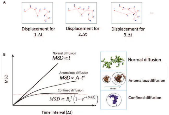
Several theoretical studies have shown that the mode of diffusion of a moving object drasti-cally changes the way it explores the available space. The time to reach a specific target can dramatically change depending on the way a particle samples its surrounding environment [56]. To quantify the mobility of a chromatin locus marked using a lacO/LacI system, the most common method consists in measuring its position (x, y, z) over time and calculating its mean square displacement (MSD) (Eq. (1)) [57]. The MSD curve represents the amount of space a locus has explored in the nucleus (Figure 2A).
MSD(n ⋅ Δt) = ____N − n1 ∑ i=0 N−1−n [ ( x i+n − x i ) 2 + ( y i+n − y i ) 2 + ( z i+n − z i ) 2] (1)
The shape of MSD curves then reveals the nature of DNA motion. Four main types of motion have been described in the literature (Figure 2B): confined motion, anomalous sub-diffusion, Brownian motion, and directive motion.
The simplest type of motion is Brownian diffusion: when a particle freely diffuses, its MSD curve is linear with time and its motion is called “Brownian.” However, in living cells, DNA motion is often slower than Brownian diffusion and is called “sub-diffusive” [58]. Several
Figure 2. (A) Illustration of trajectories: from the left to the right, points are represented spaced by 1.Δt, 2.Δt and 3.Δt and (B) mean square displacement for normal anomalous and confined diffusion, with a representation of the corresponding trajectories.
types of sub-diffusive motion have been observed. When a chromosomal locus is confined inside a sub-volume of the nucleus, the motion is called confined sub-diffusion and the MSD exhibits a plateau. Confined motion has been observed and quantified in living cells, when chromatin motion is examined during several minutes [23, 59, 60]. In that case, the MSD curves can be fitted by MSD ∝ L 2 (1 − e −4Dt/ L 2
) , where L is the plateau of the MSD curve (proportional
to the radius of confinement) and D is the diffusion coefficient.
When the force or structure that restricts the motion is not a simple confinement but is modu-lated in time and space with scaling properties, the motion is called anomalous sub-diffusion [58, 61]. In this case, sub-diffusive loci are constrained, but unlike confined loci, they can diffuse without boundary and thus reach further targets if given enough time. For
sub-diffu-sive motion, the MSD exhibits a power law (MSD ~ Atα), where α, the anomalous exponent,
is smaller than 1. The anomalous exponent α is linked to the degree of recurrence of DNA exploration, that is, the number of times a DNA locus reiteratively scans neighboring regions before reaching a distant position [62]. When α is small, the locus explores recurrently the same environment for a long time, while a large α indicates that the locus is able to explore new environments often. The anomalous diffusion coefficient A represents the amplitude of DNA motion; it is proportional to the diffusion coefficient only in the case of normal diffusion (when α = 1), which is rarely observed in biological systems [58]. Finally, a moving particle moves in a directive manner toward a target, and the motion is called directive.
The MSD is a standard statistical tool that describes a set of trajectories of similar objects. However, numerous artifacts perturb this statistic. The localization accuracy can have a strong impact on the MSD curve, even computed on simple Brownian motion [63]. Considering the movement of a single photon emitter, the localization accuracy can be divided into:
i. The error in the determination of the accurate particle position due to convolution with the point spread function (PSF) and the finite number of photons. This error is more important for short acquisition times since the number of photons collected is small. ii. The error due to the movement of the particle during the camera acquisition. This error is
more important with higher exposure times and is sometimes referred to as “motion blur.” For 2D Brownian motion with a diffusion coefficient D, Michalet computed the formula of the converged MSD including the corrections for localization accuracy (see Eq. (2)) [64]:
MSD(t) = 4Dt + σ 0 2 (1 + Dt E ___ s 02 ) − 4 __ 3 Dt E (2) where σ 0
2 is the localization accuracy of an immobile particle; s 0
2 is the variance of the PSF; t
E is
the exposure time of the camera.
The term 4Dt is the theoretical MSD for simple Brownian motion. The term σ
0 2 (1 + Dt E ___ s 02 ) accounts for
the motion blur of the particle along its path during acquisition. Since the localization accuracy
σ 02 is inversely proportional to the number of collected photons, we have σ
0 2 ∝ __1
t
E. The motion
blur term therefore converges to a fixed value as the exposure time increases. The term __34 Dt E accounts for the correlation between successive displacements due to the exposure overlap.
For anomalous motion with a diffusion coefficient D, the MSD formula including the correc-tions for localization accuracy is described in [65].
3. Increased mobility in response to DNA damage
3.1. Evidence of increased mobility in response to DSB
Most of the studies on chromatin mobility are based on the analysis of MSD curves calculated from the trajectories of fluorescently labeled chromatin loci. Using this approach, the diffusion coefficients reported in the literature varies from 5 × 10⁻⁵ to 10−3 μm2/s depending on the organ-isms, the loci studied, and the type of damage [66, 67]. In several studies, chromatin undergoes confined diffusion [22, 59, 68–71], while others have reported anomalous diffusion [69, 70, 72–75]. So far, no consensus has been reached to describe the nature of DNA motion prob-ably because these studies have been performed using different microscopy techniques and illumination settings. Indeed, multi time-scales observation of chromatin motion revealed that chromatin is driven by different types of diffusion at each time scale [76–78]. As a consequence, the type of diffusion depends on the time scale of observation. While comparing studies on chromatin dynamics, it is thus important to compare studies performed at similar time scales. Several studies have investigated DNA motion in the context of DNA damage. Since DSB is the most deleterious type of damage in the cell, chromatin dynamics in the context of DNA repair has been mainly investigated in response to DSBs. The changes in chromatin architecture and dynamics following DSB have been studied mostly in yeast, Drosophila and mammalian nuclei. In budding yeast, chromatin mobility has been investigated during the process of HR, when a Rad52 focus is already formed at the damaged locus [23, 25]. In diploid, where a homologous template is available, chromatin mobility is dramatically increased at the damaged site, allow-ing the damaged locus to explore a nuclear volume 10 times larger [23]. Increased mobility may facilitate homology search; however, haploid yeast cells, where no homologous template is present, also exhibit increased mobility in response to DSBs [25]. Since the two broken ends stay in contact during the process of HR repair [79], the current view is that the two broken ends explore the nuclear space together.
Importantly, only induced DSB associated to Rad52 foci display increased mobility. Indeed, dur-ing resection, the early stage of HR, a strong inhibition of chromatin mobility has been reported in yeast, highlighting the importance of the stage of DNA repair in chromatin mobility changes [51]. Finally, different types of damages have very different consequences on DNA mobility. For example, in yeast, spontaneous DSBs occurring during DNA replication exhibit decreased mobility [80]; DSBs induced by a protein-DNA adduct display no change in motion and campto-thecin (CPT)-induced Rad52 foci display no increased mobility [25].
In mammalian cells, while several studies have reported increased chromatin mobility upon DNA damage, others fail to observe significant changes. In HeLa cells, after α-particle-induced DSBs, γH2AX foci are more mobile [81, 82]. Similarly, uncapped telomeres exhibit
increased mobility in mouse cells, and this movement is dependent on the 53BP1 repair pro-tein [83]. Movement of heterochromatic DSBs toward euchromatin was observed in mouse embryo fibroblasts (MEFs), HeLa cells [84], and Drosophila cells [45]. It was proposed that re-localization of heterochromatic DSBs close to euchromatin regions prevents rearrange-ments between repetitive DNA sequences present in heterochromatin. Taken together, these studies suggest that chromosome mobility increases significantly in the presence of DSBs. By contrast, in other studies using MEFs [85], HeLa, or U2OS cells [86], DSBs generated by UV laser or γ-irradiation did not significantly alter chromosome mobility. Only energy-dependent local expansion of chromatin was observed around the initial damaged zone immediately after DNA damage [85]. These contradictory observations in mammalian cells probably result from variation between cell lines, the regions of chromatin damaged, and the type of damage induced. Recent studies suggest that DSBs induced in active genes, naturally enriched in the trimethyl form of histone H3 lysine 36 (H3K36me3), are repaired by HR [87]. These DSBs are susceptible to exhibit increased mobility, while DSBs repaired by Non-Homologous End Joining are rather immobile. Further systematic studies will be necessary to confirm these observations.
3.2. Local versus global increased mobility
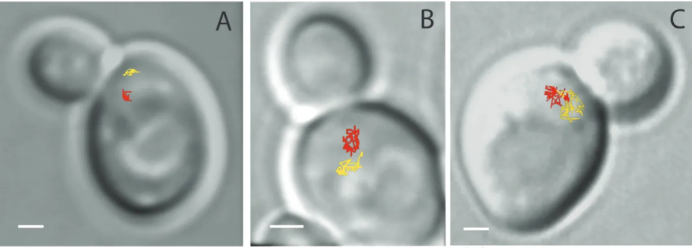
Changes in chromatin conformation have been extensively described around the site of dam-age but an important question is whether these changes in chromatin mobility also affect the rest of the genome. Interestingly, in budding yeast, increased chromatin mobility is not an intrinsic property of the damaged locus. Indeed, in diploid yeast, after induction of four random DSBs per nucleus by γ-irradiation, undamaged loci explore a 2.4 times larger nuclear volume than in the absence of irradiation [23]. Moreover, the global increased mobility is dose-dependent since upon induction of approximately 20 DSBs, the chromosomes explore almost the entire yeast nucleus [23]. Figure 3 illustrates the mobility observed for different levels of γ-irradiation in a diploid yeast cell. Global increased mobility is observed in haploid yeast, although it required higher doses of damages and it has been tested with a different type of DSBs (zeocin-induced DSBs) [88].
In mammalian cells, changes in mobility far from a damaged locus are not reported in the lit-erature. Since mammalian nuclei are much larger than the yeast nuclei, but chromatin motion exhibits very similar constrained (~0.5 μm of confined radius), it is likely that global mobility is specific to organisms with small nuclei and is therefore not present in mammalian cells. Importantly, most of these studies investigated chromatin mobility at one specific time scale. However, when studying the diffusion of a specific locus, the time scale at which data are collected reflects a specific spatial scale of the exploration studied. From nucleosomes to fiber, the different scales of chromatin organization might exhibit different diffusion behaviors. Using fast microscopy, a recent study investigated DNA mobility at several time scales, up to 1000 times faster than previously observed [65]. These experiments revealed that DNA motion following DNA damage is more complex than what had been previously described. Chromatin dynamics therefore appears to be scale-dependent: in response to DNA damage,
chromatin is more mobile at large time scales but, surprisingly, its mobility is reduced at short time scales, this effect being stronger at the damaged site. Such a pattern of dynamics is consistent with a global chromatin stiffness that has been proposed to arise in response to DNA damage [65, 89, 90]. These results underline the importance of performing multiscale tracking to fully understand the complex dynamics of chromatin at each scale.
3.3. DSBs clustering
In addition to the increased chromatin mobility, several studies indicate that multiple DSBs cluster together into a single “repair center.” Clustering of unrelated DSBs might be a con-sequence of increased mobility, although such clusters of DSBs might promote transloca-tions and their functransloca-tions is not clear. In yeast, several DNA lesions collapse together into the same repair focus, suggesting that these multiple DSBs are driven to a shared loca-tion, in so-called “repair centers” or “repair factories” [91]. In mammalian cells, clusters of radiation-induced foci have been observed in many independent studies [45, 81, 85, 86]. DSB clusters could be formed by collisions and fusion of several DSBs. One interesting hypothesis is that repair foci have a higher viscosity than the rest of the nucleus, due to their high concentration of repair proteins around the damaged site. It has been proposed that several repair foci act as dynamic liquid droplets that are able to fuse together when they collide [92].
3.4. Factors controlling local and global increased mobility
To investigate the mechanism of increased chromatin mobility in response to DSBs, sev-eral studies have tested chromatin mobility in mutant cells [23, 25, 65, 81, 83, 88, 90, 93–95].
Figure 3. Examples of the dynamics of URA3 loci (chromosome V) in budding yeast as a function of the number of double-strand breaks (DSBs) in the nucleus. The lines indicate a 2D projection of the trajectories of the two URA3 loci taken at between 10- and 30-s time intervals for approximately 15 min. (A) In the absence of DSBs, the two homologous loci are distant and explore only 3% of the nuclear volume. (B) In the presence of one to four DSBs induced on chromosomes III, the mobility of the URA3 loci increases and each locus can explore 11% of the nuclear volume. (C) After about 20 random γ-irradiation-induced DSBs per nucleus (200 Gy), URA3 loci explore almost the entire nuclear volume and their trajectories overlap. The scale is 1 μm. These three examples illustrate that more DSBs in the nucleus induce greater DNA mobility, thereby increasing the probability of collisions between loci (with permission from [66]).
These studies revealed that several genes involved in DSBs repair and chromatin remod-eling are involved in chromatin mobility changes. In diploid yeast, Rad51, the central protein of HR, is required for chromate in mobility [23], as well as for increased chro-matin rigidity [65]. Several checkpoint proteins acting earlier than Rad51 nucleoprotein filament formation are also essential for increasing the mobility of the damaged locus. For example, Rad9, a protein containing a BRCT domain roughly equivalent to human MDC1, BRCA1, and 53BP1, is also required for increased mobility at the damaged site in haploid yeast [25]. MEC1 and SML1, but not RAD53 and Tel1, are essential for increasing the mobility of the damaged locus [23, 25, 66]. Interestingly, the activation of the checkpoint protein Mec1 at a specific locus is sufficient to promote increased mobility, even in the absence of physical DSB [88]. Finally, in mouse cells, increased chromosome movements are associated with uncapped telomeres, and this movement is dependent on the 53BP1 repair protein [83].
Another study proposed that centromere and telomere release following DSBs are at the origin of chromatin changes in mobility. Strecker et al. found that a combined disruption of telomeres and centromeres can reproduce chromatin mobility observed after a DSB [96]; they identified the Mec1-dependent phosphorylation of Cep3, a kinetochore component, as an essential player in global increased chromatin mobility on DSBs.
More recently, it has been shown that molecular motors play an important role in DSBs mobil-ity [93–95]. For example, in fly in human genomes, heterochromatin constitutes about 30% of the genome [97], and “safe” repair of heterochromatic DSBs by homologous recombination relies on the relocalization of repair foci to the nuclear periphery. During this process, nuclear actin filaments form at repair sites to drive heterochromatin DSBs at the periphery and disas-semble after relocalization [93]. Actin filaments act in concert with Smc5/6, Arp2/3, Arp2/3 activators Scar and Wash, nuclear myosins Myo1A, Myo1B, and MyoV. Interestingly, in U2OS cells, ARP2/3-mediated actin polymerization enhances DSBs motion during homologous recombination, increasing the clustering of repair foci [94]. In budding yeast, DNA-damaged induced nuclear microtubule filaments (DIMs) form in response to endogenous or exogenous DNA damage [95]. These DIM filaments, formed at repair sites, reach the nuclear periphery to dive irreparable DSBs and disassemble after relocalization. Such DSBs motion is mediated by the Rad9 DNA damage response mediator and the Kar3 kinesin motor. Another model impli-cating microtubules has been proposed by Lawrimore et al. to explain the global increased mobility observed in yeast in response to DNA damage: in their model, microtubules would be responsible for a global chromatin shake-up that would be essential for global increase mobility on DSBs [98].
In these different examples, DSBs mobility is promoted by molecular motors, and DSBs exhibit a complex motion including mixture of directive and Brownian motions [93] or nonlinear directive motion [95]. Overall, these recent studies revealed the essential role of molecular motors in DSBs mobility to drive heterochromatic or irreparable DSBs to the nuclear periphery or in clustering of multiple DSBs. Importantly, these mechanisms are conserved through several organisms (human, Drosophila melanogaster, Xenopus laevis, and budding yeast).
3.5. Modifications of chromatin compaction in response to DSB and perspectives
In addition to chromatin mobility, many studies investigated the modulation of the chro-matin compaction state both at a specific damaged site and throughout the genome. Several studies showed a chromatin decondensation visible at the micrometer scale accessible by conventional light microscopy [85, 99]. In the recent studies, super resolution imaging of a
lacO array before and after damage allows the visualization of chromatin decompaction at the
damaged site in haploid yeast [90, 100]. In mammalian cells, most of the studies report chro-matin decondensation at the damaged site. However, it has been shown that following this initial fast decondensation, the damaged chromatin area slowly recondenses to reach higher compaction levels than before damage induction [101]. In addition, both chromatin expansion and compaction occur at the same time but in different regions of the chromatin near the DSBs [49]. Overall, chromatin changes in compaction are tuned in space and time upon DSBs, but the precise role of each step remains to be elucidated.
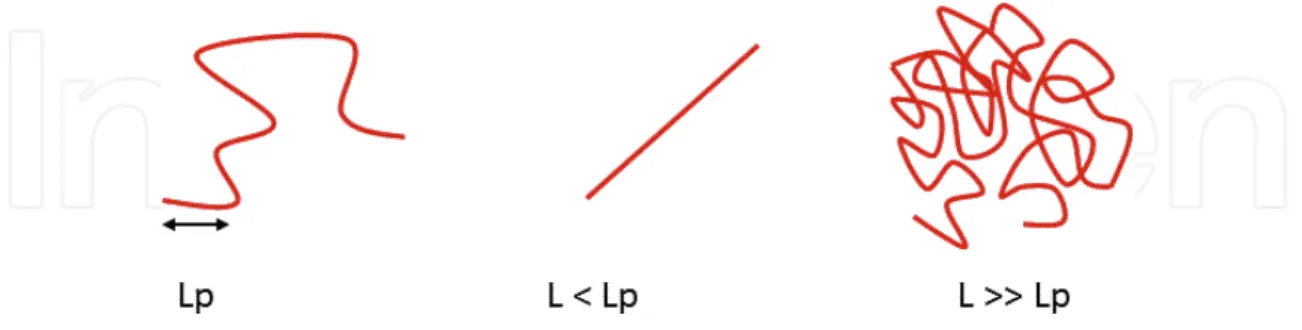
An interesting way to interpret chromatin changes in dynamics and compaction upon DSBs is to think in terms of mechanical properties of chromatin, such as chromatin stiffness (or per-sistence length). As illustrated in Figure 4, the perper-sistence length of a polymer is a mechanical property that quantifies its stiffness. The persistence length is the length over which correla-tions in the direction of the tangent are lost.
Changes in chromatin persistence length following DNA damage have been discussed, however, with contradictories interpretations. While some studies suggest that chromatin is more flexible following DSBs [100, 102, 103], other results indicate that chromatin is globally stiffer upon DSBs [65, 90]. One proposed explanation for chromatin stiffening upon DSBs could be the presence of negative charges due to H2A S129 phosphorylation [90]. Further studies will be required to solve to this open question and more generally to understand the physical mechanisms underlying the modifications of chromatin dynamics in response to DNA damage.
4. Conclusion
Thanks to recent advanced in fast and high-resolution microscopy, it became possible to quantify chromatin mobility with unprecedented precision and to understand how chromatin
Figure 4. Illustration of the persistence of a polymer, where Lp is the polymer persistence length and L is the polymer length.
explores the nuclear space. Following DSBs, chromatin mobility is dramatically altered in budding yeast both at the site of DNA damage and genome wide. In mammalian cells, DSBs mobility is strongly influenced by cell cycle stage, chromatin state, and repair pathway choice. For example, while DSBs induced in pericentric heterochromatin during the S and G2 phases of the cell cycle are more mobile and relocate to the nuclear periphery, DSBs generated during the G1 phase remain stable. Changes of chromatin mobility upon DNA damage is an intrigu-ing phenomenon, and over the last five years, several views to explain it have been proposed in the literature. They can be grouped into two classes: (1) increase in chromatin motion is due to intrinsic chromatin modifications which require chromatin remodelers, kinases, and repair proteins involved in the DNA response machinery; (2) increase in chromatin motion due to changes of external mechanical constraints that maintain chromatin and to the action of molecular motors. It is likely that both mechanisms act in concert to drive differently dam-aged chromatin depending on the type of damages, the chromatin state, or the cell cycle. Several questions remain open: how changes in chromatin dynamics alter its organization at the scale of TADs? Is there a global change in mechanical properties of the chromatin upon DSBs, such as an increase in chromatin rigidity? In the future, it will be an exciting challenge to investigate changes in chromatin organization and dynamics upon DNA damage com-bining different approaches from live cell microscopy, super resolution imaging, and Hi-C.
Acknowledgements
This work was funded by the ANR-12-PDOC-0035-01.
Author details
Judith Miné-Hattab1,2* and Xavier Darzacq3
*Address all correspondence to: [email protected]
1 Institut Curie, PSL Research University, CNRS, UMR3664, Paris, France 2 Institut Curie, Sorbonne Université, CNRS, UMR3664, Paris, France
3 Department of Molecular and Cell Biology, Li Ka Shing Center for Biomedical and Health Sciences, CIRM Center of Excellence, University of California, Berkeley, United States
References
[1] Robinson PJ et al. EM measurements define the dimensions of the "30-nm" chromatin fiber: Evidence for a compact, interdigitated structure. Proceedings of the National Academy of Sciences of the United States of America. 2006;103(17):6506-6511
[2] Schalch T et al. X-ray structure of a tetranucleosome and its implications for the chroma-tin fibre. Nature. 2005;436(7047):138-141
[3] Joti Y et al. Chromosomes without a 30-nm chromatin fiber. Nucleus. 2012;3(5):404-410 [4] Maeshima K, Hihara S, Eltsov M. Chromatin structure: Does the 30-nm fibre exist
in vivo? Current Opinion in Cell Biology. 2010;22(3):291-297
[5] Ricci MA et al. Chromatin fibers are formed by heterogeneous groups of nucleosomes
in vivo. Cell. 2015;160(6):1145-1158
[6] Rao SS et al. A 3D map of the human genome at kilobase resolution reveals principles of chromatin looping. Cell. 2014;159(7):1665-1680
[7] Nora EP et al. Spatial partitioning of the regulatory landscape of the X-inactivation cen-tre. Nature. 2012;485(7398):381-385
[8] Dixon JR et al. Topological domains in mammalian genomes identified by analysis of
chromatin interactions. Nature. 2012;485(7398):376-380
[9] Sexton T et al. Three-dimensional folding and functional organization principles of the Drosophila genome. Cell. 2012;148(3):458-472
[10] Fudenberg G et al. Formation of chromosomal domains by loop extrusion. Cell Reports. 2016;15(9):2038-2049
[11] Hansen AS et al. Recent evidence that TADs and chromatin loops are dynamic struc-tures. Nucleus. 2018;9(1):20-32
[12] Feng S et al. Genome-wide Hi-C analyses in wild-type and mutants reveal high-resolu-tion chromatin interachigh-resolu-tions in Arabidopsis. Molecular Cell. 2014;55(5):694-707
[13] Wang C et al. Genome-wide analysis of local chromatin packing in Arabidopsis thaliana. Genome Research. 2015;25(2):246-256
[14] Wang M et al. Evolutionary dynamics of 3D genome architecture following polyploidi-zation in cotton. Nature Plants. 2018;4(2):90-97
[15] Trussart M et al. Defined chromosome structure in the genome-reduced bacterium
Mycoplasma pneumoniae. Nature Communications. 2017;8:14665
[16] Hsieh TH et al. Mapping nucleosome resolution chromosome folding in yeast by micro-C. Cell. 2015;162(1):108-119
[17] Eser U et al. Form and function of topologically associating genomic domains in
bud-ding yeast. Proceebud-dings of the National Academy of Sciences of the United States of America. 2017;114(15):E3061-E3070
[18] Lazar-Stefanita L et al. Cohesins and condensins orchestrate the 4D dynamics of yeast chromosomes during the cell cycle. The EMBO Journal. 2017;36(18):2684-2697
[19] Lieberman-Aiden E et al. Comprehensive mapping of long-range interactions reveals folding principles of the human genome. Science. 2009;326(5950):289-293
[20] Misteli T. Higher-order genome organization in human disease. Cold Spring Harbor Perspectives in Biology. 2010;2(8):a000794
[21] Chubb JR, Bickmore WA. Considering nuclear compartmentalization in the light of nuclear dynamics. Cell. 2003;112(4):403-406
[22] Heun P et al. Chromosome dynamics in the yeast interphase nucleus. Science. 2001; 294(5549):2181-2186
[23] Miné-Hattab J, Rothstein R. Increased chromosome mobility facilitates homology search during recombination. Nature Cell Biology. 2012;14(5):510-517
[24] Levi V et al. Chromatin dynamics in interphase cells revealed by tracking in a two-photon excitation microscope. Biophysical Journal. 2005;89(6):4275-4285
[25] Dion V et al. Increased mobility of double-strand breaks requires Mec1, Rad9 and the homologous recombination machinery. Nature Cell Biology. 2012;14(5):502-509
[26] Roukos V et al. Spatial dynamics of chromosome translocations in living cells. Science. 2013;341(6146):660-664
[27] Meshorer E et al. Hyperdynamic plasticity of chromatin proteins in pluripotent embry-onic stem cells. Developmental Cell. 2006;10(1):105-116
[28] Lodish H, Berk A, Zipursky S. DNA damaged and repair and their role in carcinogen-esis. In: Molecular Cell Biology. 4th ed. New York: W. H. Freeman; 2000
[29] Vilenchik MM, Knudson AG. Endogenous DNA double-strand breaks: Production,
fidelity of repair, and induction of cancer. Proceedings of the National Academy of Sciences of the United States of America. 2003;100(22):12871-12876
[30] Wyman C, Kanaar R. DNA double-strand break repair: All's well that ends well. Annual Review of Genetics. 2006;40:363-383
[31] Rassool FV. DNA double strand breaks (DSB) and non-homologous end joining (NHEJ)
pathways in human leukemia. Cancer Letters. 2003;193(1):1-9
[32] Symington LS, Rothstein R, Lisby M. Mechanisms and regulation of mitotic recombina-tion in Saccharomyces cerevisiae. Genetics. 2014;198(3):795-835
[33] Lisby M et al. Choreography of the DNA damage response: Spatiotemporal relation-ships among checkpoint and repair proteins. Cell. 2004;118(6):699-713
[34] Finkelstein IJ, Greene EC. Single molecule studies of homologous recombination. Molecular BioSystems. 2008;4(11):1094-1104
[35] Sugawara N, Haber JE. Repair of DNA double strand breaks: In vivo biochemistry. Methods in Enzymology. 2006;408:416-429
[36] Sugawara N, Wang X, Haber JE. In vivo roles of Rad52, Rad54, and Rad55 proteins in Rad51-mediated recombination. Molecular Cell. 2003;12(1):209-219
[37] Lisby M, Rothstein R. Cell biology of mitotic recombination. Cold Spring Harbor Perspectives in Biology. 2015;7(3):a016535
[38] Lisby M, Rothstein R, Mortensen UH. Rad52 forms DNA repair and recombination
cen-ters during S phase. Proceedings of the National Academy of Sciences of the United States of America. 2001;98(15):8276-8282
[39] Nagy Z, Soutoglou E. DNA repair: Easy to visualize, difficult to elucidate. Trends in Cell Biology. 2009;19(11):617-629
[40] Krogh BO, Symington LS. Recombination proteins in yeast. Annual Review of Genetics.
2004;38:233-271
[41] Lisby M, Rothstein R. Choreography of recombination proteins during the DNA dam-age response. DNA Repair (Amst). 2009;8(9):1068-1076
[42] Miné J et al. Real-time measurements of the nucleation, growth and dissociation of single Rad51-DNA nucleoprotein filaments. Nucleic Acids Research. 2007;35(21):7171-7187
[43] Barzel A, Kupiec M. Finding a match: How do homologous sequences get together for
recombination? Nature Reviews. Genetics. 2008;9(1):27-37
[44] Takata H et al. Chromatin compaction protects genomic DNA from radiation damage. PLoS One. 2013;8(10):e75622
[45] Chiolo I et al. Double-strand breaks in heterochromatin move outside of a dynamic HP1a domain to complete recombinational repair. Cell. 2011;144(5):732-744
[46] Schuster-Bockler B, Lehner B. Chromatin organization is a major influence on regional mutation rates in human cancer cells. Nature. 2012;488(7412):504-507
[47] Aymard F et al. Transcriptionally active chromatin recruits homologous recombination at DNA double-strand breaks. Nature Structural & Molecular Biology. 2014;21(4):366-374 [48] Aymard F et al. Genome-wide mapping of long-range contacts unveils clustering of
DNA double-strand breaks at damaged active genes. Nature Structural & Molecular Biology. 2017;24(4):353-361
[49] Hinde E et al. Chromatin dynamics during DNA repair revealed by pair correlation analysis of molecular flow in the nucleus. Biophysical Journal. 2014;107(1):55-65
[50] Robinett CC et al. In vivo localization of DNA sequences and visualization of large-scale chromatin organization using lac operator/repressor recognition. The Journal of Cell Biology. 1996;135(6 Pt 2):1685-1700
[51] Saad H et al. DNA dynamics during early double-strand break processing revealed by non-intrusive imaging of living cells. PLoS Genetics. 2014;10(3):e1004187
[52] Recamier V et al. Single cell correlation fractal dimension of chromatin: A framework to interpret 3D single molecule super-resolution. Nucleus. 2014;5(1):75-84
[53] Nozaki T et al. Dynamic organization of chromatin domains revealed by super-resolu-tion live-cell imaging. Molecular Cell. 2017;67(2):282-293 e7
[54] Shinkai S et al. Dynamic nucleosome movement provides structural information of topological chromatin domains in living human cells. PLoS Computational Biology. 2016;12(10):e1005136
[55] Beliveau BJ et al. Single-molecule super-resolution imaging of chromosomes and in situ haplotype visualization using Oligopaint FISH probes. Nature Communications. 2015;6:7147
[56] Guerin T, Benichou O, Voituriez R. Non-Markovian polymer reaction kinetics. Nature
Chemistry. 2012;4(7):568-573
[57] Meister P et al. Visualizing yeast chromosomes and nuclear architecture. Methods in
Enzymology. 2010;470:535-567
[58] Barkai E, Garini Y, Metzler R. Strange kinetics of single molecules in living cells. Physics Today. 2012;65(8):29-35
[59] Marshall WF et al. Interphase chromosomes undergo constrained diffusional motion in
living cells. Current Biology. 1997;7(12):930-939
[60] English BP et al. Single-molecule investigations of the stringent response machinery in living bacterial cells. Proceedings of the National Academy of Sciences of the United States of America. 2011;108(31):E365-E373
[61] Metzler R et al. Anomalous diffusion models and their properties: Non-stationarity, non-ergodicity, and ageing at the centenary of single particle tracking. Physical Chemistry Chemical Physics. 2014;16(44):24128-24164
[62] Ben-Avraham D, Havlin S. Diffusion and Reactions in Fractals and Disordered Systems. Cambridge United Kingdom: Cambridge University Press; 2000
[63] Saxton MJ, Jacobson K. Single-particle tracking: Applications to membrane dynamics.
Annual Review of Biophysics and Biomolecular Structure. 1997;26:373-399
[64] Michalet X. Mean square displacement analysis of single-particle trajectories with local-ization error: Brownian motion in an isotropic medium. Physical Review. E, Statistical, Nonlinear, and Soft Matter Physics. 2010;82(4 Pt 1):041914
[65] Mine-Hattab J et al. Multi-scale tracking reveals scale-dependent chromatin dynamics after DNA damage. Molecular Biology of the Cell. 2017
[66] Miné-Hattab J, Rothstein R. DNA in motion during double-strand break repair. Trends
in Cell Biology. 2013;23(11):529-536
[67] Lebeaupin T et al. Chromatin dynamics at DNA breaks: What, how and why? AIMS Biophysics. 2015;2(4):458-475
[68] Masui O et al. Live-cell chromosome dynamics and outcome of X chromosome pairing events during ES cell differentiation. Cell. 2011;145(3):447-458
[69] Backlund MP, Joyner R, Moerner WE. Chromosomal locus tracking with proper account-ing of static and dynamic errors. Physical Review. E, Statistical, Nonlinear, and Soft Matter Physics. 2015;91(6-1). DOI: 062716
[70] Cabal GG et al. SAGA interacting factors confine sub-diffusion of transcribed genes to the nuclear envelope. Nature. 2006;441(7094):770-773
[71] Taddei A et al. Nuclear pore association confers optimal expression levels for an induc-ible yeast gene. Nature. 2006;441(7094):774-778
[72] Weber SC, Spakowitz AJ, Theriot JA. Bacterial chromosomal loci move subdiffusively
through a viscoelastic cytoplasm. Physical Review Letters. 2010;104(23):238102
[73] Hajjoul H et al. High-throughput chromatin motion tracking in living yeast reveals the flexibility of the fiber throughout the genome. Genome Research. 2013;23(11): 1829-1838
[74] Lucas JS et al. 3D trajectories adopted by coding and regulatory DNA elements: First-passage times for genomic interactions. Cell. 2014;158(2):339-352
[75] Burnecki K et al. Universal algorithm for identification of fractional Brownian motion. A case of telomere subdiffusion. Biophysical Journal. 2012;103(9):1839-1847
[76] Bronstein I et al. Transient anomalous diffusion of telomeres in the nucleus of
mam-malian cells. Physical Review Letters. 2009;103(1):018102
[77] Miné-Hattab J et al. Fast imaging of DNA motion reveals distinct sub-diffusion regimes at the site of DNA damage. BioRXiv. 2016
[78] Vazquez J, Belmont AS, Sedat JW. Multiple regimes of constrained chromosome motion
are regulated in the interphase Drosophila nucleus. Current Biology. 2001;11(16): 1227-1239
[79] Lisby M, Rothstein R. DNA repair: Keeping it together. Current Biology. 2004;14(23): R994-R996
[80] Dion V et al. Cohesin and the nucleolus constrain the mobility of spontaneous repair foci. EMBO Reports. 2013;14(11):984-991
[81] Aten JA et al. Dynamics of DNA double-strand breaks revealed by clustering of dam-aged chromosome domains. Science. 2004;303(5654):92-95
[82] Krawczyk PM et al. Chromatin mobility is increased at sites of DNA double-strand breaks. Journal of Cell Science. 2012;125(Pt 9):2127-2133
[83] Dimitrova N et al. 53BP1 promotes non-homologous end joining of telomeres by increas-ing chromatin mobility. Nature. 2008;456(7221):524-528
[84] Jakob B et al. DNA double-strand breaks in heterochromatin elicit fast repair protein recruitment, histone H2AX phosphorylation and relocation to euchromatin. Nucleic Acids Research. 2011;39(15):6489-6499
[85] Kruhlak MJ et al. Changes in chromatin structure and mobility in living cells at sites of DNA double-strand breaks. The Journal of Cell Biology. 2006;172(6):823-834
[86] Jakob B et al. Live cell microscopy analysis of radiation-induced DNA double-strand break motion. Proceedings of the National Academy of Sciences of the United States of America. 2009;106(9):3172-3177
[87] Clouaire T, Legube G. DNA double strand break repair pathway choice: A chromatin based decision? Nucleus. 2015;6(2):107-113
[88] Seeber A, Dion V, Gasser SM. Checkpoint kinases and the INO80 nucleosome
remodel-ing complex enhance global chromatin mobility in response to DNA damage. Genes & Development. 2013;27(18):1999-2008
[89] Faller R, Müller-Plathe F. Chain stiffness intensifies the reptation characteristics of poly-mer dynamics in the melt. Chemphyschem. 2008;2(3):180-184
[90] Herbert S et al. Chromatin stiffening underlies enhanced locus mobility after DNA dam-age in budding yeast. The EMBO Journal. 2017;36(17):2595-2608
[91] Lisby M, Mortensen UH, Rothstein R. Colocalization of multiple DNA double-strand
breaks at a single Rad52 repair centre. Nature Cell Biology. 2003;5(6):572-577
[92] Altmeyer M et al. Liquid demixing of intrinsically disordered proteins is seeded by poly(ADP-ribose). Nature Communications. 2015;6:8088
[93] Caridi CP et al. Nuclear F-actin and myosins drive relocalization of heterochromatic breaks. Nature. 2018;559(7712):54-60
[94] Schrank BR et al. Nuclear ARP2/3 drives DNA break clustering for homology-directed repair. Nature. 2018;559(7712):61-66
[95] Oshidari R et al. Nuclear microtubule filaments mediate non-linear directional motion of chromatin and promote DNA repair. Nature Communications. 2018;9(1):2567
[96] Strecker J et al. DNA damage signalling targets the kinetochore to promote chromatin mobility. Nature Cell Biology. 2016;18(3):281-290
[97] Ho JW et al. Comparative analysis of metazoan chromatin organization. Nature. 2014; 512(7515):449-452
[98] Lawrimore J et al. Microtubule dynamics drive enhanced chromatin motion and mobi-lize telomeres in response to DNA damage. Molecular Biology of the Cell. 2017;28(12): 1701-1711
[99] Ziv Y et al. Chromatin relaxation in response to DNA double-strand breaks is modu-lated by a novel ATM- and KAP-1 dependent pathway. Nature Cell Biology. 2006;8(8): 870-876
[100] Amitai A et al. Visualization of chromatin decompaction and break site extrusion as predicted by statistical polymer modeling of single-locus trajectories. Cell Reports. 2017;18(5):1200-1214
[101] Burgess RC et al. Activation of DNA damage response signaling by condensed chroma-tin. Cell Reports. 2014;9(5):1703-1717
[102] Seeber A, Dion V, Gasser SM. Remodelers move chromatin in response to DNA
dam-age. Cell Cycle. 2014;13(6):877-878
[103] Hauer MH et al. Histone degradation in response to DNA damage enhances chromatin dynamics and recombination rates. Nature Structural & Molecular Biology. 2017;24(2): 99-107