HAL Id: hal-01524114
https://hal.archives-ouvertes.fr/hal-01524114
Submitted on 17 May 2017HAL is a multi-disciplinary open access archive for the deposit and dissemination of sci-entific research documents, whether they are pub-lished or not. The documents may come from teaching and research institutions in France or abroad, or from public or private research centers.
L’archive ouverte pluridisciplinaire HAL, est destinée au dépôt et à la diffusion de documents scientifiques de niveau recherche, publiés ou non, émanant des établissements d’enseignement et de recherche français ou étrangers, des laboratoires publics ou privés.
Role of Alkali Metal Ions in G-Quadruplex Nucleic Acid
Structure and Stability
Eric Largy, Jean-Louis Mergny, Valérie Gabelica
To cite this version:
Eric Largy, Jean-Louis Mergny, Valérie Gabelica. Role of Alkali Metal Ions in G-Quadruplex Nucleic Acid Structure and Stability. The Alkali Metal Ions: Their Role for Life, Springer Verlag (Germany), pp.203-258, 2016, �10.1007/978-3-319-21756-7_7�. �hal-01524114�
This is the author post-print (accepted manuscript) version of a book chapter published in final form in: E. Largy, J.-L. Mergny, V. Gabelica, Met. Ions Life Sci. 16 (2016) 203–258. (http://dx.doi.org/10.1007/978-3-319-21756-7_7)
7
The Role of Alkali Metal Ions in G-Quadruplex Nucleic Acid Structure and
Stability
Eric Largy,1,2 Jean-Louis Mergny,1,2 and Valérie Gabelica1,2
1Université Bordeaux, IECB, ARNA Laboratory, F-33600 Pessac, France 2INSERM, U869, ARNA Laboratory, F-33000 Bordeaux, France
<eric.largy@inserm.fr> <jean-louis.mergny@inserm.fr>
<valerie.gabelica@inserm.fr> Contents
ABSTRACT
1 INTRODUCTION: G-QUADRUPLEX NUCLEIC ACIDS 1.1 Overview of Structure
1.2 Stabilizing Interactions
1.3 Alkali Metal Ion Coordination in G-Quartets 1.3.1 Cation Preference
1.3.2 Cation Binding Energetics 1.3.3 Cation Location
2 METHODS TO STUDY G-QUADRUPLEX NUCLEIC ACIDS 2.1 Folding Topology
2.1.1 X-Ray Crystallography
2.1.2 Nuclear Magnetic Resonance Spectroscopy 2.1.3 Molecular Modeling
2.1.4 Electronic Circular Dichroism Spectroscopy 2.1.5 UV Absorption Spectroscopy
2.1.6 Separative Techniques 2.1.7 Native Mass Spectrometry 2.1.8 Miscellaneous
2.2 Cation Coordination
2.2.1 X-Ray Crystallography
2.2.2 Nuclear Magnetic Resonance Spectroscopy 2.2.3 Native Mass Spectrometry
3 ROLE OF ALKALI METAL IONS IN G-QUADRUPLEX STABILITY 3.1 Case Study: dTG3-5T Tetramolecular G-Quadruplexes
3.2 General Trends 3.2.1 Libraries
3.2.2 Human Telomeric Sequences 3.2.3 Other Sequences and Overview
2 3.2.4 Summary
3.3 Kinetics of Strand Association, Dissociation, Folding and Unfolding 3.4 Cation Exchange Mechanisms
4 INFLUENCE OF ALKALI METAL IONS ON G-QUADRUPLEX STRUCTURES 4.1 Case Study: The Human Telomeric G-Quadruplex Sequence
4.1.1 The Intramolecular Folding of dAGGG(TTAGGG)3
4.1.2 Other Human Telomeric Sequences 4.2 Other Sequences
4.3 General Trends
5 CATION-DEPENDENT CONFORMATIONAL SWITCHING 6 CONCLUDING REMARKS AND FUTURE DIRECTIONS ABBREVIATIONS AND DEFINITIONS
ACKNOWLEDGMENTS REFERENCES
Abstract G-quadruplexes are guanine-rich nucleic acids that fold by forming successive quartets of guanines (the G-tetrads), stabilized by intra-quartet hydrogen bonds, inter-quartet stacking, and cation coordination. This specific although highly polymorphic type of
secondary structure deviates significantly from the classical B-DNA duplex. G-quadruplexes are detectable in human cells and are strongly suspected to be involved in a number of
biological processes at the DNA and RNA levels. The vast structural polymorphism exhibited by G-quadruplexes, together with their putative biological relevance, makes them attractive therapeutic targets compared to canonical duplex DNA. This chapter focuses on the essential and specific coordination of alkali metal cations by G-quadruplex nucleic acids, and most notably on studies highlighting cation-dependent dissimilarities in their stability, structure, formation, and interconversion. Section 1 surveys G-quadruplex structures and their
interactions with alkali metal ions while section 2 presents analytical methods used to study G-quadruplexes. The influence of alkali cations on the stability, structure, and kinetics of formation of G-quadruplex structures of quadruplexes will be discussed in sections 3 and 4. Section 5 focuses on the cation-induced interconversion of G-quadruplex structures. In sections 3 to 5, we will particularly emphasize the comparisons between cations, most often K+ and Na+ because of their prevalence in the literature and in cells.
Keywords DNA · folding · G-quadruplex · G-quartet · interconversion · metal ions · methods · RNA · stability · structure
3
1 Introduction: G-Quadruplex nucleic acids
G-quadruplexes (G4) encompass guanine-rich nucleic acids that fold by forming successive quartets of guanines (also called G-tetrads), stabilized by intra-quartet hydrogen bonds (Fig. 1A), inter-quartet stacking, and cation coordination (Fig. 1B). This specific although highly polymorphic type of secondary structure deviates significantly from the classical B-DNA duplex [1-3]. Such G-rich sequences are found in telomeres, and at a statistically remarkable frequency in other part of the genome, notably in promoters [4-6]. G-quadruplexes are detectable in human cells and are strongly suspected to be involved in a number of biological processes at the DNA and RNA levels [7-15]. The vast structural polymorphism exhibited by G-quadruplexes (see section 1.1), together with their putative biological relevance, makes them attractive therapeutic targets compared to canonical duplex DNA [16-20]. A very large – and exponentially increasing – number of studies have consequently been dedicated to G-quadruplexes involved in telomeric sequences, oncogenes, 5’-untranslated regions (5’-UTRs) to cite the most common [21]. Most studies so far involve potassium or sodium cations because of their prevalence in human cells, but a number of other monovalent and divalent cations promote quadruplex formation (vide infra). However, among alkali metals, only potassium, sodium and rubidium are truly effective at stabilizing G4s.
This chapter focuses on the essential and specific coordination of alkali metal cations by G-quadruplex nucleic acids, and most notably on studies highlighting cation-dependent dissimilarities. Section 1 surveys G-quadruplex structures and their interactions with alkali metal ions while section 2 presents analytical methods used to study G-quadruplexes. The influence of alkali cations on the stability, structure, and kinetics of formation of
G-quadruplex structures of G-quadruplexes will be discussed in sections 3 and 4. Section 5 focuses on the cation-induced interconversion of G-quadruplex structures. In sections 3 to 5, we will particularly emphasize the comparisons between cations, most often K+ and Na+ because of their prevalence in the literature and in cells.
1.1 Overview of Structure
The core structure of all quadruplexes is constituted by the stacking of at least two G-tetrads (or G-quartets), each formed by the quasi co-planar association of four guanines linked by a network of eight hydrogen bonds (Figure 1A,B). The guanines forming the tetrads
coordinate some mono- or divalent cations in their center (referred to as central stem), typically K+ and Na+, via their oxygen O6. The single-strand sections linking the guanines tracts are called loops, and can adopt a variety of geometries (Figure 1C): lateral (or
edgewise), diagonal, and double chain reversal (or propeller). Furthermore, the four guanine tracts can be oriented (regarding their 5’ to 3’ polarity) into four different topologies: parallel (the four strands share the same polarity), antiparallel (two strands in a way, the two other in the opposite way; these pairs may correspond to two adjacent or diagonally-opposed strands, resulting in very different geometries), or hybrid (three strands sharing the same polarity, and the last one the opposite) [22,23].
4 Figure 1 (A) Guanine tetrads contain four guanines linked by eight hydrogen bonds (donor and acceptor groups in blue and green, respectively). Guanine O6 selectively coordinate a metal cation (red). (B) Tetrads can stack to form G-quadruplexes. Cations of larger ionic radii are located between the tetrads (case of K+; blue), while smaller ones can also coordinate within the plane of the tetrads, or assume an intermediate position (case of Na+; green). (C) G-quadruplexes can fold into a variety of topology, that differ mainly by the relative orientation and number of strands (1 to 4), the number of tetrads (at least 2), and the geometry of the loops.
5 In antiparallel topologies, the strands sharing the same direction can be diagonally opposed or adjacent. This distinguishes ‘chair’-type G-quadruplexes (three lateral loops, e.g. the thrombin binding aptamer [24]) and ‘basket’-type G-quadruplexes (lateral-diagonal-lateral loops, e.g. the human telomeric sequence d[AG3(T2AG3)3] in Na+ conditions [25]; Figure 2, 143D) [25-32]. Intramolecular parallel structures (three double-chain reversal loops) are sometimes referred to as ‘propeller’ topologies (e.g., the crystal structure of d[AG3(T2AG3)3] in K+ conditions [26], Fig. 2, 1KF1).
Figure 2 Examples of human telomeric G-quadruplex structures deposited in the PDB that were solved by NMR or X-ray crystallography: 143D [25], 1KF1 [26], 2GKU [27], 2YH9 [28], 2JSM and 2JSL [29], 2JPZ [30], 2KF8 [31], and 2KKA [32]. Guanosines are depicted in brown, inosine in yellow, adenosines in blue, thymidines in green, the phosphate backbones as white ribbons, and K+ as purple spheres where available. All structures except 143D (Na+) were solved in K+ conditions.
6 The glycosidic bond angle of guanines involved in tetrads can adopt an anti or syn geometry depending on the relative strand orientation: parallel topologies contain almost exclusively anti guanines (for exceptions, see [33-35]), whereas a mixture of syn and anti is observed for antiparallel and hybrid structures [36]. Consequently, parallel G-quadruplexes generally contain anti/anti stacks (syn/syn is not favored; see section 1.2), antiparallel ones have anti/syn and syn/anti, and hybrid ones can have all of the above.
Furthermore, G-quadruplexes may be formed by the folding of a single strand or the association of two to four strands, and some sequences have a tendency to oligomerize [37-39]. One should therefore not confuse the term “quadruplex” or “G-quadruplex” (designating any structure containing stacked guanine taetrads) with the strand molecularity (for which we will adopt the nomenclature “intramolecular”, “bimolecular”, "trimolecular" [40], and
“tetramolecular”).
Finally, a number of uncommon features have been observed, such as strand bulges and snapbacks [41-43], base-pairing in loops [44], alternative tetrads [45], and other stacked planar entities (triads, pentads, hexads, heptads, and octads) [31,46-52]. The combination of different loop geometries, strand orientations, and molecularities, implies that G-quadruplexes display an important polymorphism. It has been reported that there is a theoretical number of 26 possible G-quadruplex topologies, not taking into account unexpected folds (e.g. isolated guanines involved in tetrads [53]), but only a few of them (six) have been observed in vitro [22,23], possibly meaning that some are energetically disfavored, thermodynamically or kinetically. A number of insightful review articles and books cover the G-quadruplex structures published so far [3,12,36,54-59].
1.2 Stabilizing Interactions
The factors contributing to the stabilization of G-quadruplexes and yielding a particular folding topology or mixture of topologies are: stacking interactions, hydrogen bonding, solvation, and cation binding. Cation effects on G-quadruplexes differ significantly from those on duplexes (see chapter 6). Cation coordination is indeed absolutely required to form G-quadruplexes, to stabilize the G-tetrad stacks. Moreover, sufficient ionic strength is
required to compensate electrostatic repulsion between the phosphate oxygens of four strands in G-quadruplexes, instead of two for duplexes (loops may be considered as unfolded single strands depending on the structure).
The total free energy can be decomposed in a number of free energy contributions (e.g. Coulombic forces, hydrogen bonding, hydration, van der Waals terms), which can themselves be decomposed in entropic and enthalpic contributions. Analysis of G-quadruplex melting and calorimetric data revealed that G-quadruplex formation is enthalpically driven [60,61]. This results from a more negative (favorable) enthalpy of tetrad formation, only partially
compensated by more negative (less favorable) entropies of tetrad formation. Below are presented some key elements to understand G-quadruplex stabilization, and cation
coordination per se is explored in the section 1.3. An excellent review discussing in details the stability of G-quadruplexes has been published by Lane et al. [62].
7 1.2.1. Stacking
Similarly to other nucleic acid secondary structures, G-quadruplex stabilization relies in part on stacking of aromatic bases, and more precisely of guanine from consecutive tetrads. Molecular mechanics simulations have predicted the relative stability order among guanine stacks to be (from 5’ to 3’): syn/anti > anti/anti > anti/syn > syn/syn [63]. This is not fully consistent with the structures solved so far as it does not explain the parallel orientation of the strands in tetramolecular assemblies, possibly because of force-field biases. Quantum
mechanical (QM) dispersion-corrected density functional theory (DFT-D3) calculations on stacking of two tetrads, containing one K+ cation gave a more consistent picture with the structures solved so far: anti/anti > syn/anti > anti/syn > syn/syn (E vs anti/anti = 1.2, 3.5 and 7.8 kcal.mol-1, respectively) [64]. It was also suggested in the same study that 5’-terminal H-bonds present a stabilizing effect on 5’-terminal syn guanines.
Ultimately, the topology of a G-quadruplex depends on its precise sequence, which can provide additional stabilizing interactions (see below), and on buffer conditions (cations, co-solvents). Because the stacking interactions likely account for a large part of the net energetic gain, increasing the number of quartets is energetically favorable. The number of consecutive tetrads is typically equal to the length of the guanine tracts, although exceptions have been observed. For instance, the 22 mer of the human telomeric sequence d[(G3T2A)3G3T) (PBD ID: 2KF8)contains four tracts of three guanines, but folds predominantly in a two-tetrad G-quadruplex (Figure 2) [31].
The formation of higher-order G-quadruplex structures (usually dimers) via the stacking of external tetrads of monomer units also provides additional stabilization. Almost all
published dimer structures exhibits a 5’-5’ interface [65], although 3’-3’ stacking remains possible [66,67]. QM and 100-ns MD simulations suggest that while 5’-5’ interface readily stack in a favorable manner (60—65° rotation), the 3’-3’ interface cannot reach the same type of geometry because of guanine-sugar clashes and therefore yields less favorable stacks (45° or 30° rotations, E vs 5’-5’ = 4 and 10 kcal.mol-1, respectively) [65].
Stacking of other nucleotides (non-tetrad bases) might also contribute to the overall stabilization of the quadruplex [31,68]. For instance, in the aforementioned two-tetrad G-quadruplex 2KF8, loop nucleotides provide G•G•G and A•G•G triads, which stack on both tetrads [31]. Alternative planar entities have been identified, such as G•C•G•C tetrads [45], as well as the larger pentads [46,47], hexads [48-50], heptads [51] and octads [52] that provide additional hydrogen bonds. Finally, formation of base pairs is also possible [69]. Searle et al. have described a bimolecular G-quadruplex whose two loops form mini-hairpin motifs [44]. The above mentioned 2KF8 structure involves mismatched T•T base pairs [31]. Base-pairing can mediate the formation of stacked G-quadruplex dimers, as observed by NMR for the c-kit2 sequence that exhibits an A•A base pair at the interface [70].
8 1.2.2. Hydrogen bonding
Hydrogen bonding is another stabilization element found in biomacromolecules, and in particular between bases in nucleic acids, typically interacting through two (A•T/U) or three (G•C) H-bonds. In such Watson-Crick base pairing, the stability of H-bonds is linked to donor-acceptor orbital interactions and polarization of the system by partially neutralizing the charges in the -electrons system. In G-quadruplex nucleic acids, there is a network of (C2)NH2:N7 and O6:N1H hydrogen bonds. As acceptor and donor groups are likely
hydrogen-bonding to water for unfolded/unassociated strands, there is only a small enthalpy change, but the release of water molecules to the bulk medium may induce an entropy gain [62].
A combination of high-resolution variable-temperature STM and DFT calculations suggested that the stabilization of a tetrad induced by the network of eight hydrogen bonds is higher than the sum of four individual G•G pairs [71]. This cooperativity was first ascribed to assistance, however, DFT-D calculations by Fonseca-Guerra et al. suggested that the cooperativity in guanine tetrads more likely arises from the charge separation that is
associated to charge transfer between pairs of guanine, i.e. donor-acceptor orbital interactions in the -electron system [72]. This results in an interruption of the -electron system, and an enhancement of both the positive charges on the H atoms of H-bond donor groups and negative charges of N and O atoms of acceptor groups. In the same study, it was evidenced that in aqueous solution, the hydrogen bond energy is significantly diminished as compared to the gas phase (-34 vs -80 kcal/mol). Compared to solvation, stacking of three tetrads (in water) has a weak favorable (-2 kcal/mol) effect on the cooperativity. However, introduction of two Na+ cations in this system entirely restores the stability observed in the gas phase, although it weakens the hydrogens bonds as compared to the system without cation.
1.3 Alkali Metal Ion Coordination in G-Quartets
1.3.1 Cation preference
Pinnavaia and co-workers’ pioneering work on 5’-GMP provided the first indication of the formation of anionic cavities in G-quadruplex-type structures that can selectively complex cations small enough to fit, but large enough to bridge the carbonyl oxygens [73], analogously to the binding of metal alkali to crown ethers [74]. This led the authors to propose the
complexation of Na+ in the plane of tetrads, while potassium would fit in the interplanar spacing and be classically octa-coordinated. Follow-up studies [75,76] expanded the scope to four-stranded poly(G) strands [77], helical poly(I) structures including the binding of
ammonium [78-80].
A seminal report by Blackburn et al. suggested the existence of “G•G base pairs” in the telomeric sequence of several organisms [81]. Following these findings, Williamson,
9 Raghuraman and Cech proposed a model for telomeric sequences where they formally
described the G-quadruplex structure for the first time [82]. The studies conducted on
Oxytricha and Tetrahymena telomeric sequences provided the first hints regarding the critical
importance of monovalent cations for G-quadruplex formation, stoichiometry, and stability. It was postulated that Li+ is too small to bind in the center of G-quadruplex tetrad, whereas Na+ fits perfectly, and larger ions such as K+ and Cs+ bind in the interplane cavity [82]. It is now widely accepted that cation coordination is essential for the stabilization of G-quadruplexes. By compiling a number of studies, one can estimate that G-quadruplex stabilization follows the general trend: Sr2+ > K+ > Ca2+ > NH4+, Na+, Rb+ > Mg2+ > Li+ ≥ Cs+ (alkali cations in
bold)[83-85].
Although the bulkiness of these cations is certainly critical for binding within a G4 cavity, and is historically presented as the main explanation for the cation-dependent stability differences (vide supra), other factors play an important role in the stabilization of the
complexes [86-89]. Cation binding to G-quadruplexes can be classified as non-specific (diffuse [90]), where the cations retaining their outer-sphere hydration bind the negatively charged phosphates, or specific (site-bound) by coordination to the guanine O6, where the hydration sphere has been lost and the binding follow the law of mass action (Figure 3). For instance, Gray and Chaires have determined that, at concentration of K+ above 2.5 mM, d[AG3(T2AG3)3] attracts up to 6—8 more cations than the predicted two specific binding sites [91]. The binding of these non-specific cations to the negatively charged phosphate backbone reduces the electrostatic repulsions and thus also promotes folding. Note that external
coordination to G-quadruplex is not necessarily diffuse: specific external coordination to loops has been suggested for Tb3+ on the basis of CD and luminescence data [92]. Notably, cations coordinated between the top G-quartet and the loop are often detected in the density maps provided by X-ray crystallography (see section 1.3.3). By comparing K+ binding to the 22-mer G-quadruplex d[AG3(T2AG3)3] with the binding to the unfolded d[T22], it was suggested that the G-quadruplex may specifically bind up to five cations on external sites [91]. However, the use of a control oligonucleotide with such a different sequence and different structure (random coil vs globular) may not mimic properly the diffuse binding of the G-quadruplex.
10 Figure 3 Multiple equilibria involved in cation coordination and G-quadruplex folding. Each cation can either bind to a preformed G-quadruplex (K1 then K2), or bind to the random coil that subsequently fold in a G-quadruplex (K4 then K3). The folded species on the right shows the differences between specific and unspecific binding as well as other stabilizing (stacking) and destabilizing (phosphate repulsions) factors.
1.3.2 Cation binding energetics
In specific coordination to the tetrads, the cations are involved in electrostatic and donor-acceptor orbital interactions with the lone pairs of guanine O6, yielding tight M+O
coordination bonds [72]. Stabilization is also provided by a screening of electronic repulsion of these O6 lone pairs [93]. In solution, potassium is typically hexa-hydrated, and hence the coordination per se by guanine O6 instead of water molecules shall not provide a large enthalpy change. Alkali cations specifically bind within the electronegative cavity formed by the O6 of tetrad guanines in distinct fashions.
As with crown ethers, sodium and potassium bind G-quadruplexes with different affinities, although other factors than the cation radii account for it. The difference in free energies is estimated to be around 2±0.5 kcal.mol-1 [88,94-97]. The thrombin binding aptamer sequence (TBA; d(G2T2G2TGTG2T2G2)), which contain only two tetrads binds a single cation, with a folding constant Kfold measured by monitoring the folding by spectroscopic means of 1.3 x 10⁷ and 5.5 x 10⁵ M-1 at 10 °C for K+ and Na+, respectively [98], or an
association constant Ka of 2 x 10⁵ M-1 for K+, determined by mass spectrometry [99]. Specific ion binding has been described thermodynamically by Lane et al. by multiple equilibrium involving the folded and unfolded G-quadruplex as shown in Figure 3 [62].
Cation coordination to G-quadruplexes is typically accompanied by G-quadruplex folding that brings further stabilization, which is not experimentally distinguishable except in molecular modeling studies (see section 2.1). Regarding the equilibrium constants defined in
11 Figure 3, in most cases the K1 value is large, and the apparent dissociation constant is K1K2 (exclusive binding mechanism) [95]. When the solution is devoid of alkali cation, the fraction of folded strand is presumably small. Cation binding to the unfolded strand is also small, and thus an important stabilization is attributed to specific cation binding (K2/K4 ratio) [62]. The analogy with alkali cations binding to crown ethers is noteworthy. In aqueous solutions, the crown ether 18-crown-6 binds K+ and Na+ with log K ≈ 2 and 0.7, respectively, and
complexation enthalpy rH° of -6 and -2 kcal.mol-1, at 37 °C, including the desolvation and structural change energies [74,100-102]. Comparatively, the net G at 37 °C of three-tetrad G-quadruplex formation has been estimated to 5—10 kcal.mol-1, which supports the idea that the energy of potassium binding and of subsequent conformation changes accounts for most of the stabilization [62,91]. For instance, the 22-mer of the human telomeric sequence
d[AG3(T2AG3)3] has an overall -2.4 kcal.mol-1 folding free energy, while in 5 mM potassium, K+ contributes to roughly -4.9 kcal.mol-1 [91]. This is also consistent with the unfolded state being largely populated in absence of cation (Figure 3, left). Consequently, binding of potassium or sodium is likely fast and cooperative, whereas the subsequent structural change is quasi irreversible with a net binding energy of around 5 kcal.mol-1. This is the case of the human telomeric sequence for instance [95].
The apparent binding of Na+ and K+ to human telomeric sequences is very cooperative and lies within 5—15 and 0.5—2 mM, respectively [94,95,103]. Comparatively, Na+ forms 1:1 complexes with isolated 5’-GMP with a Ka of 2.85 M-1 at 5 °C [104]. Feigon et al., and Leszczynski et al., have shown that the free energy of hydration of specific cations can explain the stability difference observed between Na+ and K+ solutions, the dehydration of the former inducing a greater energetic cost [88,105]. Indeed, unlike diffusely bound cations, tetrad-bound cations lose their whole hydration sphere [106]. The energetics of cation binding to G4s can be decomposed into a positive free energy of dehydration and a negative free energy of coordination per se. Na+ gives favorable coordination energy of coordination, as shown by NMR [88,107], but its binding is penalized by its stronger hydration as compared to K+. Meyer et al. have also observed by DFT calculations that solvation effects explain the favorable coordination to K+, and shown that the cation coordination contribution accounts for 50% of the total interaction energy of a two-tetrad construct [108]. Hydration energy of alkali cations is usually presented as being inversely proportional to their ionic radii. K+
systematically presents the best compromise, and stabilizes G4s more than Na+ and Rb+ (other alkali cations provide very weak to no stabilization). However, the extent to which it does so depends hugely on the studied sequence, and in particular, on the sequence’s ability to adopt different structures in presence of different cations (see section 3.2) [85,89,94,109-119].
1.3.3 Cation location
It is often stated that Na+ is small enough (0.95 Å) to fit in the plane of a quartet, while any cation larger than that, such as K+ (1.33 Å) or Rb+ (1.52 Å), are coordinated between two planes. In reality, there is a continuum of possible binding sites resulting from (i) the
12 abovementioned cation lone pair attraction, (ii) the presence of additional cation coordination sites (e.g. loops, dimer interface), and (iii) cation-cation repulsion. Indeed, when more than one cation is bound by the G4, the cations’ mutual repulsion also influences their precise locations and the fine structure of the G-quadruplex. Phillips et al. showed crystals of [d(TG4T)]4 coordinated to Na+ and Ca2+ that provides a nice illustration of this phenomenon for Na+ [120] (Figure 4A,B,C) [120,121]. The G-quadruplex is arranged in a head-to-head dimer with eight consecutive tetrads. Out of the seven bound sodium cations, only the two outer ones lie in the G-tetrad’s plane, in a four-coordinate fashion, completed by the binding of water molecules. The 5 other sodium cations are equally spaced between the two other ones, and adopt intermediate coordination positions in a continuum ending at the central cation that is sandwiched exactly halfway between two tetrads, with a bipyramidal geometry similar to the one usually adopted by K+. As a result, the average distance between Na+
cations (4.2 Å for outer cations, 3.6 Å for internal cations) is higher than the distance between G-tetrads. A slight distortion of the external quartets is also observed in this crystal, putatively allowing the inter-cation distance to increase. One should note however, that crystal packing forces might account for the position of the cations, which therefore could not reflect the reality of solution-based G-quadruplexes.
13 Figure 4 Crystal structure of [d(TG4T)]4 coordinating Na+(PDB ID 352D, [120]) (A, B, C), and d(G3CG4AG5A2G3A) coordinating K+ (PDB ID 4H29, [121] ) (D, E, F): tetrad geometry depicted as balls and sticks (A, D) and spheres (B, E), the head-to-head dimer coordinating seven Na+ adopting positions ranging from within the tetrads (external cations) to sandwiched midway between two quartets (central cation) (C), and the intertwined dimer of B-raf
coordinating six sandwiched K+ cations (F). Hydrogen bonds are shown in blue, Na+ and K+ cations and coordination in purple. Carbon atoms are in grey, nitrogen in blue, and oxygen in red except in panels C and F where guanosines are colored in brown and the phosphate backbone is a white ribbon. For the sake of clarity, dT, dA, and dC residues, Ca2+, H2O, and hydrogen atoms are omitted.
14 Creze et al. obtained a similar dimeric structure with Na+ being coordinated
increasingly more within the tetrad planes when going towards the extremities of the G-quadruplex (dNa+-Na+ = 3.4—4.7 Å), with water molecules capping at the external tetrads (dNa+-H2O= 2.4—2.5 Å) [122]. Crystallization was carried out from a sodium solution of quadruplex using lithium sulfate as a precipitating agent. A single Li+ ion was observed in a groove, and none of them in the central stem.
Incidentally, in the first reported crystal structure of a small molecule (daunomycin) bound to a G-quadruplex, the Na+ cations are all coordinated in the plane of the tetrads [123]. The tetramolecular [d(TG4T)]4 G-quadruplex is also observed as a dimer, with two
daunomycin molecules at the interface between the individual tetramers, which might also be linked to the difference of sodium positioning. In a more recent report on the high-resolution crystal of the dimeric [d(TG4T)]4/daunomycin complex employing syn glycosyl linkages, Na+ cations are not in the plane of the tetrads, but rather sandwiched in various fashions [124]. Notably, the Na+ closer to the 5’-interface lies almost midway between two tetrads, with a square prism geometry, due to the syn glycosyl orientation of the first guanine residue. Interestingly, Na+ also occupies a site at a daunomycin-daunomycin interface.
Conversely, in the crystal structure of the intertwined dimeric quadruplex formed by the B-raf sequence d(G3CG4AG5A2G3A) (PDB ID: 4H29), the six potassium cations are all observed in-between the tetrads, in a quasi linear arrangement, and are equidistant from each other (dK+-K+ = 3.44 ± 0.09 Å) (Figure 4D,E,F) [121].
The differences in location of sodium and potassium cations has been extensively investigated by NMR and X-Ray crystallography for the telomeric sequence from Oxytricha
Nova d(G4T4G4), forming an antiparallel bimolecular G-quadruplexes, where the thymines form two diagonal loops (Figure 5) [96,107,125-128]. The crystal structures 1JPQ and 1JRN exhibits five equidistant K+ cations, spaced by 3.4 Å on average, three being sandwiched between the tetrads and the two other coordinated between the external quartets and within the loops [128]. The K+-K+ distance is the same that has been measured for the human telomeric sequence [26]. Similar results were observed with Tl+ cations, which have a similar radius than K+ (5 bound cations with a 3.6 Å spacing; average RMSD = 0.26 Å) [129]. Conversely, only four Na+ cations are coordinated, the binding sites in the loops being empty (PDB ID 1JB7) [127]. Sodium cations are coordinated in the planes of the central tetrads while the external cations are bound slightly outer of the tetrads, towards the loops, and are coordinated by the thymines O2. This partial loop coordination leads to a distribution of Na+ location that differs from [d(TG4T)]4, but here too the distance between sodium cations is higher than the distance between tetrads.
15 Figure 5 Crystal structure of [d(T4G4T4)]2 coordinating K+ (PDB ID 1JPQ) or Na+(PDB ID 1JB7). Guanines are shown in brown, thymines in green, the cations as purple spheres, and the oxygens from water molecules completing the spheres of coordination as red balls.
Overall, K+ cations are specifically coordinated in between tetrads, in a nearly octahedral way, or alternatively within loops above tetrads, satisfying the usual
hexacoordinate stereochemistry of these cations. Positioning of bases outside of the plane of the tetrad may be necessary to comply with this geometry, to an extent balanced by stacking and hydrogen bonding energies. Smaller Na+ cations can be coordinated within the plane of tetrads, and can occupy a range of positions owing to lower steric constraints, hence reducing the electrostatic repulsions. K+ is bound with higher affinity than Na+ and Rb+, while Li+ and Cs+ are poorly coordinated by G-quadruplexes. Although the cationic radius certainly
accounts for these differences, the energy of hydration has been demonstrated to be the cause of the energetic preference for K+ vs Na+.
All these examples highlight that the stability of G-quadruplexes is massively driven by cation binding and the resulting structural (re-)organization, rather than by other weak
interactions.
2 Methods to study G-quadruplex nucleic acids
2.1 Folding
Topology
A number of high- and low-resolution analytical methods are used to determine the topology of G-quadruplexes [130], and some are presented hereafter. NMR spectroscopy, X-ray crystallography, and molecular modeling give access to atomic-scale structure information
16 [36,131-134], and have revealed over the last decades an impressive structural polymorphism among G-quadruplexes (Figure 2). A number of other spectroscopic and spectrometric methods (e.g. UV absorption spectroscopy, electronic circular dichroism spectroscopy, native mass spectrometry, electrophoresis, and chromatography) are employed to determine strand orientations and stoichiometry, and molecular sizes. As each of these techniques give different types of information, and furthermore presents certain drawbacks, it is advisable to combine a number of these techniques before drawing final conclusions.
2.1.1 X-Ray crystallography
X-Ray crystallography gives access to atomic-scale resolution structure of DNA, RNA, or LNA G-quadruplexes, including the cations (see section 2.2.1), water molecules, and binders [132,133]. A fair number of structures containing alkali cations (or Tl+ as a K+ surrogate; vide
infra) have been solved using this method [26,67,120-124,127-129,135-161], mainly by the
teams of Parkinson and Neidle, and the late prof. Sundaralingam. Screening of sequences, as well as base modification (e.g. heavy-atom addition) and loop/flanking sequence changes are typically performed in order to obtain a sequence that crystallizes and diffracts well. These aspects, as well as crystallization protocols are discussed in details in reference [133].
Almost all of the available crystal structures are tetrameric [120,122,123,135-145] or dimeric, via association of distinct strands or external stacking of monomer units, often templated by binders [26,67,121,124,127-129,145-160]. Moreover, stacked monomer units and intermolecular species [26,158,159] are systematically parallel-stranded within each subunit, except for the thrombin binding aptamer (TBA) bound to -thrombin [161]. This raises questions as to the possibility to crystallize structures such as intramolecular
antiparallel-stranded or hybrid structures (either because they do not crystallize or because the crystal packing forces induce conversion to parallel and multimeric folds), and this in turns casts some doubts about the complete relevance of this method for solution-based folding studies. Crystallography is nevertheless very powerful when it comes to characterize cation coordination (see section 2.2.1), and more generally ligand binding (reviewed in [162]). It also provides better defined structures than NMR in cases where the latter is limited by internal dynamics or the presence of mixtures of conformations, with or without inter-conversion between structures.
2.1.2 Nuclear magnetic resonance (NMR) spectroscopy
High-field NMR is broadly used to study G-quadruplex structures and their dynamics [36,131,163,164]. The full topology of many structures has now been solved, including the human telomeric DNA [25,27-32,45,165-172] and RNA sequences (TERRA) [50,173,174], telomeres from other species [46,68,83,96,125,126,175-177], human oncogenes [53,70,178-182], minisatellites [43,183], and aptamers against biological targets [24,41,184-186] or
17 viruses [15,69]. Besides, even when solving the full topology is not possible or required, several types of NMR experiments can provide information on particular aspects of the structure.
First, the formation of G-quadruplexes can be affirmed by the presence of guanine imino protons H1 in the 10—12 ppm range, typical of G-tetrads, whereas canonical Watson-Crick base-pairing shifts the protons to 13—14 ppm [134,187]. Moreover, compared to Watson-Crick protons, tetrad protons – particularly the ones from central tetrads – exchange more slowly with water, which results in sharp peaks [125,188].
Second, the number of imino proton peaks is linked to the number of tetrads (one peak per guanine, hence four peaks per tetrad, unless some protons are equivalent) [183], and a number of peaks higher than the number of guanines indicates a mixture of species. The kinetics of quadruplex formation are easily accessible given that the time-scale is compatible with NMR experiments [164].
To fully solve a structure however, a number of issues must be tackled: formation of multiple species by a single sequence, higher-order structures, ambiguous assignments [131]. Sequences folding in multiple conformers (e.g. [167,170]) can be characterized if the peaks are sufficiently resolved, or more frequently by screening for or favoring a given
conformation by sequence modification (flanking and loop bases, modified bases [170,172]), sample preparation, and buffer and cationic conditions (reviewed in [131]). Stoichiometry can be studied by titration [170,176], or diffusion ordered spectroscopy (DOSY) experiments [189,190], or alternatively by other methods described hereafter (ESI-MS, SE-HPLC, PAGE). Peak assignments was historically performed by through-space NOE experiments [125], possibly helped by base modifications (typically, guanine to inosine or bromoguanine, or thymine to uracil). Site specific 15N- and 13C-enrichement of defined residues allow the unambiguous elucidation of G-quadruplex structures routinely [191]. Fold determination can be typically performed by studying either the connectivity between guanine H1 and H8 from the following residue in NOE patterns, the J-couplings through tetrad hydrogen bonds [131]. Hydrogen bonds have been characterized by the H-bond scalar coupling [107].
2.1.3 Molecular modeling
Molecular modeling is a third way to get atomic-scale information on the structure of quadruplexes. Advantageously, it gives access to individual energetic terms, dynamics, intermediates, and does not suffer from mixtures of conformations because it studies one molecule at a time [64,65,192-200]. As a result, the structure, energetics and dynamics of rare conformations or of putative reaction intermediates, which would not be possible to isolate experimentally, can be explored. The quality of force fields is however an issue, notably the treatment of the loops, inter-cation repulsion, and the treatment of specific and non-specific electrostatic interactions [193-196,201,202], and combination with biophysical data can prove useful [203].
18 2.1.4 Electronic circular dichroism (CD) spectroscopy
The most common and straightforward low-resolution technique for the study of
G-quadruplex topology is circular dichroism (CD) [204-207]. CD is a spectroscopic method that measures the difference in absorption of left- and right- circularly polarized light by chiral compounds or by compounds in a chiral environment. G-quadruplex nucleic acids are typically characterized by their bands in the UV area (210—300 nm), arising from electronic transitions between stacked guanines. More precisely, -* transitions within guanine exciton couplets occur at 279 and 248 nm, giving rise to CD bands. The sign and intensity of these bands is dictated by the relative orientation of the stacked guanines (head-to-head:
anti/syn, syn/anti; head-to-tail: anti/anti, syn/syn).
As a result, the G-quadruplex topology can be inferred from the CD spectrum by analysis of the characteristic bands. Parallel topologies exhibit an intense positive bands centered at 260 nm and a negative bands at 240 nm, antiparallel topologies are characterized by a positive band at 290 nm, a (sometimes shallow) negative band at 260 nm, and a positive band at 245 nm, and hybrid-type structures have a positive band at 290 nm, and a shoulder at 260—270 nm. CD is relatively fast and easy to perform but has a low resolution as it only provides a global information on guanine stacks from the sample. It is not possible to easily distinguish mixtures of structures from pure species. For instance, mixtures of antiparallel and hybrid or parallel structures might lead to spectra that can be confused with a pure hybrid-type signature (see the controversy over the topology of d[AG3(T2AG3)3] in section 4.1.1).
Finally, other supramolecular features (additional base stacking, double helices, mismatches) can account for the overall spectrum and possibly lead to misinterpretations [205]. An example of additional guanine stacking modifying the CD signature is provided by the 2KF8 structure, described in section 1.2 (Figure 2) [31]. Although this G-quadruplex has a 2-tetrad antiparallel topology where the tetrad guanines are stacked in a syn/anti fashion, its CD signature exhibits a positive band at 260 nm that could be mistakenly interpreted has a hybrid-type topology, due to additional base stacking on both tetrads of triads composed of loop nucleotides, yielding anti/anti stacks [205]. Additionally, non-classical tetrads (such as G•C•G•C as in the human mutant telomeric GGGCTA motif) may also alter the CD spectra [45]. Similarly, the presence of two loops forming a mini-hairpin motifs in an antiparallel G-quadruplex leads to the presence of a wide peak centered at 280 nm, characteristic of a B-DNA duplex, masking the expected minimum at 260 nm [37,44].
2.1.5 UV absorption spectroscopy
The UV absorption spectrum slightly differ between single structures, and differences in topologies are usually appreciated by variations in absorbance spectra, monitored by difference spectra between folded and unfolded forms. For example, thermal difference spectra (TDS) fingerprints monitor the difference between unfolded spectrum (high
19 temperature) and the folded spectrum (low temperature) [205]. TDS fingerprints of
G-quadruplexes typically exhibit local maxima at 240, 255, and 275 nm, and a minimum at 295 nm [113]. As with CD, different guanine-guanine stacks may indeed modify the intensity and wavelength of absorption bands. Hence, parallel topologies appear to be discriminated from hybrid and antiparallel topologies thanks to their greater A240nm/A295nm ratio.
The use of TDS signatures is however not optimal because high melting temperatures leading to decreased TDS intensities, and temperature-dependent changes in molar absorption coefficient both contaminate the signature. Isothermal difference spectra (IDS), which are calculated by subtraction of the UV-spectra of a given sample acquired in absence or presence of cation, at constant temperature. IDS are not strictly identical to TDS because they do not suffer from the drawbacks mentioned above. It is possible to study kinetics of folding and interconversion by this method [208]. In both cases the main advantages are the high throughput and ease of use that compensate their low resolution. Coordination, can be indirectly monitored by either of these methods because it translates into quadruplex folding. Displacement of a cation by another can be observed by IDS, although the signature can differ significantly from case to case, and a significant change in guanine stacking is required for a signal to be observed [208].
UV-melting at 295 nm is a very common methodology to assess the stability of G-quadruplexes [209,210]. As mentioned above, the formation of G-G-quadruplexes lead to a change in absorbance at 295 nm that can be followed as a function of temperature: increasing temperatures induce an unfolding of the G-quadruplexes that translates into a hypochromism at 295 nm. Alternatively, oligonucleotides tagged with two fluorophores compatible with Förster resonance energy transfer can be used for melting experiments (FRET-melting) [94,98,211,212]. This is not the favored technique for structural studies since the fluorophores tend to form stacked exciton couplets that might affect both the structure and the stability of the G-quadruplex. Whatever the method used to detect the fraction folded, cation binding is related only indirectly to the fraction folded, monitored by the melting curve. Because the relationship is indirect, thermodynamics parameters extracted from such curves do not directly give access to the Kd of coordination.
2.1.6 Separative techniques
Native gel electrophoresis can be employed to determine strand stoichiometry, although it suffers from a number of drawbacks (charge screening effects, smearing, and
tedious/ambiguous quantification). It was recently combined to CD for the study of multimers [38]. Size-exclusion HPLC (SE-HPLC) was shown to be a valuable alternative to get insight into oligonucleotides secondary structures, and most notably the strand stoichiometry of quadruplexes [37,164,213-215]. The salient advantages of this technique is the possibility to work in a variety of conditions including cation nature and concentration, and various strand concentrations (µM—mM) that are not all accessible by NMR and X-Ray crystallography. Additionally, it is possible to work with mixture of structures, which are abundant with
G-20 quadruplex-forming sequences, whereas it is more difficult with techniques such as NMR or CD. Thermodynamics and kinetics of G-quadruplex folding can be studied with this method. It however does not provide information regarding the strand orientation or the number of quartets. Analytical ultracentrifugation (AUC) is a thermodynamically rigorous approach for the determination of absolute molecular weights and get insight into the hydrodynamic shapes of macromolecules in solution [216], and is thus broadly used for the determination of
topology and stoichiometry of quadruplexes [203,214,217-223].
2.1.7 Native mass spectrometry
Mass spectrometry (MS) in non-denaturing conditions (both in solution and in the mass spectrometer) is called native MS. Electrospray ionization (ESI) ensures non-denaturing ionization and vaporization. Native ESI-MS is another straightforward way to determine strand stoichiometry [39,224], as well as kinetics and thermodynamics of folding of quadruplexes [225], from pure species or from mixtures, without however getting direct information on the structures. Matrix-assisted laser desorption/ionization (MALDI) has also been used to determine the strand stoichiometry [173], but this ionization method has higher risk of denaturing the complexes than ESI [226,227].
Interestingly, the number of potassium cation bound by a given G-quadruplex can be determined by ESI-MS (see section 2.2.3 for more details), which can give insight into its folding topology [228,229]. Indeed, given the preferred location of specifically bound K+ cations, the number of tetrads of a G-quadruplex is usually equal to the number of coordinated K+ cations plus one. In order to get additional insights into structures of macromolecules, ion-mobility spectrometry (IMS) can be coupled in-line to ESI-MS [230]. ESI-IMS-MS is a technique that, in addition to mass-to-charge ratio, further separates ions on the basis of their mobility in a drift tube filled with gas (typically, nitrogen or helium), where a static electric field is applied [231]. The travel time is related to the collision cross section of the ions with the gas: compact structures collide less with gas and travel faster as a result. Gas phase structural determination by MS- and spectroscopy-based methods have been reviewed in a recent book [232].
2.1.8 Miscellaneous
Other methods have been employed to study the topology, stability, and formation of G-quadruplexes, as well as cation binding, albeit tangentially, including single molecule FRET [233], fluorescence melting [94,234-236], denaturing PAGE [237,238], 125I-radioprobing [239,240], electron paramagnetic resonance (EPR) [222], surface plasmon resonance [241], fluorescence spectroscopy [242,243], Raman spectroscopy [244,245], calorimetry (DSC, ITC) [61,84,112,246-248], temperature-jump relaxation experiments [62,170,236,249,250]. G-quadruplex characterization assays are reviewed in references [62,130,251,252].
21
2.2 Cation
Coordination
2.2.1 X-Ray crystallography
X-ray crystallography is an invaluable method for the study of cation binding in G4 nucleic acids because it directly locates cation binding sites through electron density. Crystal packing forces may however change the location of the cation as compared to solution-state structures [107,190]. Tl+ cations can be used as probes for potassium binding sites. Tl+ and K+ have close atomic radii (1.44 and 1.33 Å, respectively), dehydration energies and bond lengths, and can therefore lead to similar folds, yet Tl+ exhibits a higher X-ray scattering potential [142]. Crystals obtained in the presence of K+ cations can be converted to Tl+-crystals by soaking in thallium acetate [129].
2.2.2 NMR spectroscopy
NMR provides evidence not only on the location of cations within G-quadruplex structures but also on their dynamics [97,163]. First examples of alkali cation coordination by tetrads of 5’-GMP, detected by NMR involved 23Na+, 39K+ and 87Rb+ [75,253,254]. Notably, the
resonance of 23Na+ cations undergoes a significant line broadening and up-shifting upon binding [254]. The detection of these cations was extended to G-quadruplexes by Wu et al. [190,255]. The coordination and dynamics of sodium cation coordination can also be directly monitored by solution- and solid-phase 23Na NMR experiments [190,256,257]. In the same vein, 39K exhibits a characteristic signature on solid-state magic-angle spinning (MAS) NMR spectra upon coordination by G-quadruplexes [258].
Indirect observation of potassium binding has been performed based on proton NMR, chemical exchange and dynamic analysis on d(G3T4G3) and the Oxytricha telomeric sequence d(G4T4G4) bound to Na+ and K+ [88,96]. Another indirect mean of studying K+ coordination is the use of Tl+ cations as surrogate, in a strategy reminiscent of the one used in X-Ray crystallography studies (vide supra). 1H-205Tl scalar couplings provide important constraints for structure determination [129,259].
15NH4+ dynamicscan be monitored directly with heteronuclear correlations exchange spectroscopy (15N-1H NzExHSQC) [97], and serve as a reporter probe. Ammonium can indeed displace Na+, or be displaced by K+, allowing an indirect measurement of Na+ and K+
movements within G-quadruplexes [260]. Changes in topology by displacement of Na+ by K+ can also be used to study cation binding, albeit more indirectly [88]. Finally, Na+ cation dynamics have been studied by nuclear magnetic relaxation dispersion study [261].
22 2.2.3 Native mass spectrometry
Mass spectrometry seems to be an obvious choice for the study of cation binding to G-quadruplexes, since each coordinated cation increases the mass of the complex. A number of mass spectrometry studies were devoted to NH4+ binding determination [52,224,225,231,262-266], because ammonium acetate is a traditional volatile buffer used in biomolecule mass spectrometry. The study of alkali cations is however more tricky because (i) they tend to form clusters even at submillimolar concentrations, and (ii) they are not volatile like the classically employed ammonium acetate. The former issue leads to very noisy spectra from which no information can be inferred, while the latter means that diffuse (unspecific) coordination of alkali cation is at least partially preserved in the gas phase, which results in a higher number of bound cations than tetrad coordination sites.
These issues have recently been partially tackled by Gabelica et al. with the use of trimethyl ammonium acetate (TMAA) as a volatile co-solvent [228]. TMA is too bulky to be coordinated by the O6 of tetrad guanines and is therefore a cation of choice to fix the ionic strength and neutralize the backbone phosphates while allowing to study the specific binding of potassium (or other cations) to the tetrads. Mathematical subtraction of diffusively bound cation can be performed in a second step, by using a reference sequence that does not form a G-quadruplex [229], yielding cleaned mass spectra. Titration of G4-forming sequences with increasing potassium concentrations, and subsequent cleaning of the non-specific adducts, give access to the Kd of coordination of potassium for each tetrad binding site. This methodology is only applicable to G-quadruplexes stable at low (submillimolar) cation concentrations, which is usually not the case when working with sodium. Gross et al. have also developed a method that utilizes the gas-phase signal fractions from the bound and unbound species as input into a mathematical model that determines the binding constants [99]. This method was applied to the measure of the Kd of potassium (and strontium) to TBA.
Another bulky ammonium derivative, namely tetrabutylammonium phosphate, had already been used for the same purpose by Gray and Chaires in spectroscopic experiments involving the human telomeric sequence [91]. This allows the determination of apparent binding constants by CD. Another salient point of this study is the use of a potassium-binding benzofuran-isophthalate crown ether indicator (PBFI) to determine the concentration of free potassium by fluorescent titration, and hence deduce the concentration of coordinated cations and thermodynamic parameters [91,267].
3 Role of alkali metal ions in G-quadruplex stability
The stabilization of G-quadruplexes by cations has been studied for the past fifty years on increasingly complex systems. Decades after the first observation of guanosine gels [268], Gellert and co-workers have reported in 1962 the formation of gels by 5’-GMP in presence of sodium, at acidic pH, and postulated the structuration of guanines in tetrads (or quartets), stacked upon each other to form helices [269]. Similar structures were observed a decade later
23 using various guanosine derivatives, in presence of potassium [270]. However, the seminal paper regarding the necessary and selective complexation of cations by such structures, at neutral pH, dates from the late seventies [73]. Pinnavaia and co-workers have demonstrated by NMR experiments that K+, Na+, and Rb+ all lead to the formation of stacks of three tetrads of 5’-GMP, in a head-to-tail or alternating head to tail and head to head stacking, whereas Li+ and Cs+ were found to be ineffective. Additionally, it was evidenced that potassium induces a higher thermal stability than sodium. In the same manuscript is reported the first qualitative ranking of alkali cation-induced stability for 5’-GMP, K+ > Na+, Rb+ >> Li+, Cs+, which remarkably contrast with the ranking observed for duplex DNA where the binding constant decreases with increasing ionic radii. Research on that topic moved to biologically relevant sequences, notably the telomeres of various species, and model G-quadruplexes such as [d(TG4T)]4. Hereafter, we will review studies highlighting the influence of alkali cations on the stability of simple tetramolecular G-quadruplexes (section 3.1), and sets of telomeric or artificial sequences (section 3.2). The contribution of folding and unfolding rates in the cation-dependent stability of G-quadruplexes is discussed in the section 3.3.
3.1 Case
Study:
dTG
3-5T Tetramolecular G-Quadruplexes
Parallel-stranded tetramolecular G-quadruplexes formed by dTGnT (n = 3—5) sequences have
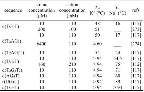
been used extensively as models. These short sequences form unambiguous G-quadruplex structures devoid of loops that makes them “minimal” G-quadruplexes ideal to conduct basic research. A typical measure of the stability of G-quadruplexes is the melting temperature (Tm), usually provided by UV-melting [209,210,271], CD-melting, and DSC [61,84,112,246] experiments. Mergny et al. have reported the melting temperatures obtained by UV-melting for a number of DNA and RNA tetramolecular G-quadruplexes [117]. Typically,
tetramolecular RNA G-quadruplexes are more stable than their DNA counterparts, thanks to higher association rates and lower dissociation rates. Noteworthy, RNA tetramolecular G-quadruplexes were shown as early as 1991 to be highly stable in K+, and more than in Na+ and Li+ [272]. For instance, the difference in Tm is large (35 °C) between [r(UG4U)]4 and
[d(TG4T)]4, in 110 mM Na+ solutions (Table 1) [112,117,273,274]. Finally, no dissociation is observed in most cases for G5 and longer tracts in Na+, and G4 and longer tracts in K+, further highlighting the cation effect on the stability of tetramolecular G-quadruplexes.
The short tetramolecular G-quadruplex [d(TG3T)]4 is characterized by an apparent melting temperature of 48 °C in presence of 110 mM KCl and 16 °C (Tm = 32 °C) in presence of 110 mM Na+ [117]. In the case of slow association/dissociation kinetics, the melting curve is not an equilibrium curve, and therefore depends on the temperature gradient. Melting of intermolecular quadruplexes is also affected by the strand concentration. A close result was obtained in 100 mM K+ by Balasubramanian et al. (51 °C; 200 µM strand
concentration) [273]. Almost identical stability differences are observed with the addition of flanking bases mimicking the human telomeric sequence, as in [d(T2AG3)]4 (50 vs. 17 °C; Tm = 33 °C) and [d(T2AG3T)]4 (55 vs. 24 °C, Tm = 31 °C) [117]. NMR experiments on a
24 high d(T2AG3) strand concentration (6.4 mM), in a 110 mM KCl buffer, NMR produced a Tm of around 60 °C [274].
The stability of the longer [d(TG4T)]4 has been studied by different groups
[89,112,117]. In presence of 110 mM KCl, the melting temperature is higher than 94 °C [89,117], indicating a very high thermal stability. An increase in Na+ concentration did not affect significantly the stability (Tm < 2 °C in the 50—400 mM Na+ range), but changed dramatically the association rate (see section 3.3) [117]. A large difference (Tm = 75 °C) can be noticed with another study by Petraccone et al. [112], performed in 210 mM NaCl but at a higher strand concentration (160 µM). Addition of thymines at both termini [d(T2G4T2)]4, substitution of the 5’-dT by a dA [d(AG4T)]4, or use of the RNA counterpart [r(UG4U)]4, all increase the thermal stability in 110 mM Na+ (Tm = 71, 60, and 89 °C, respectively) [117]. Further increase of the guanine tract leads to even more stable G-quadruplexes: the melting temperature is higher than 94 °C in both K+ and Na+ conditions for [d(TG5T)]4.
All these examples highlight the influence of the cation nature and concentration on the stability of G-quadruplexes, where little to no structural changes are expected, and thus the cation dehydration and binding per se accounts for most of the stabilization.
Table 1 Melting temperatures of tetramolecular G-quadruplexes. sequence strand concentration (µM) cation concentration (mM) Tm K+ (°C) Na+T (°C)m refs d(TG3T) 10 110 48 16 [117] 200 100 51 ___ [273] d(T2AG3) 10 110 50 17 [117] 6400 110 ≈ 60 ___ [274] d(T2AG3T) 10 110 55 24 [117] d(TG4T) 10 110 > 94 54.5 [117] 160 210 > 94 75 [112] d(T2G4T2) 10 110 > 94 71 [117] d(AG4T) 10 110 > 94 60 [117] r(UG4U) 10 110 > 94 89 [117] d(TG5T) 10 110 > 94 > 94 [117]
25
3.2 General
Trends
3.2.1 Libraries
The Mergny group contributed several systematic UV-melting based studies on a very large number of oligonucleotides, illustrating the difference of G-quadruplex stabilization by coordination of K+ or Na+. In a 1998 publication illustrating the use of UV-vis spectroscopy to follow the folding of G-quadruplexes, very large differences of melting temperatures in potassium- and sodium-rich conditions were observed for the 26- and 27-mer sequences d[T3A2G3(TGTG3)3] (63 and 37 °C, respectively) and d[G3(TGTGTG3)3] (62 and 37 °C, respectively) [111]. In a more recent study, the melting temperature of eighty different sequences containing four tracts of three guanines with loops of variable length (between 1 and 15 bases), following the template d(G3LaG3LbG3LcG3), where La, Lb, and Lc are thymines or TTA loops, were determined in presence of 100 mM K+ or Na+ [115]. Potassium stabilizes these sequences by on average 18.3 °C more than sodium, but the difference is highly variable since it can be as low as 1.2 °C, and as high as 39.2 °C. The Tm decreases when the loop length increases both in K+ and Na+, but to different extents – particularly for short loops – as seen from the large Tm variability. For loops of 7–15 nt, the difference within a given
sequence family tends to be relatively constant, from 1–2 °C (for La = Lc = TTA) to more than 30 °C (for La = Lc = T). More generally, there is a strong inverse correlation between total loop length and Tm for K+ (each added base leads to a 2 °C drop) but the trend is less clear in Na+. Also, the presence of adenines in the loops is favorable in presence of sodium, when the central loop contains at least two nucleotides.
Thirty-six sequences following the general formula d(G3T3G3HNHG3T3G3) were analyzed in similar conditions (N can be any base, H = C, T or A) [116]. The average difference of Tm is 12.7 °C in favor of potassium, but is also sequence dependent, ranging from 9.5 °C (for ACC and TGC central loops) to 16.2 °C (for AAT). In the same study, twenty-six additional sequences that vary in length, number of quartets and loop composition and positions were also investigated. The average of (measurable) potassium-sodium Tms is 14.4 °C, with a very large sequence dependence, ranging from 7.1 °C
(d(G3T2AG3CGCG3T2AG3)) to more than 40 °C (d(G3TG3ACTG3TG3)). Overall, K+ stabilizes particularly well G-quadruplexes containing YDH loops, and poorly ACH loops, while Na+ favors YDC loops and disfavor ACW loops (Y = C or T; H = A, C or T; W = A or T and D = A, G or T.).
Risitano and Fox have examined a randomized library of oligonucleotides based on the sequence d[T(G3H2)3G3], by FRET-melting, in presence of various concentrations of K+ and Na+ (5—100 mM) [94]. A clear increase in Tm is observed with the concentration, ranging from 27 to 85 °C for K+. For Na+, lower concentrations (5 and 10 mM) do not allow sufficient folding for the Tm to be measured, and Tms at higher concentrations (20—100 mM) range from 24 to 57 °C. The difference of stabilization, where measurable, decreases slightly with increasing cation concentration (29.4—27.6 °C).
26 Smargiasso et al. have investigated the melting temperature of libraries of sequences based on the sequence d(G3WiG3WjG3WkG3), where W is either a thymine or an adenine, and
the loop sizes (i, j, k) were systematically varied between 1 and 3, yielding a total of 2744 distinct sequences grouped as a function of their loop lengths [39]. The stability in all the groups is higher with 150 mM K+ than with 150 mM Na+, but again this is very sequence-dependent. The stability in K+ is inversely dependent on loop length: shorter loops promote parallel structures with average Tm above 80 °C, whereas longer loops (at least one loop of two and one loop of three nucleotides) promote hybrid structures with average Tm in the 65— 70 °C range. Conversely, longer loops yields more stable G-quadruplexes in Na+, and the loop length has less influence. Note however that some of these sequences are suspected of
forming multimeric G-quadruplexes, notably in K+ conditions [39].
3.2.2 Human telomeric sequences
The human telomeric sequence is certainly the most investigated intramolecular G-quadruplex-forming sequence. Telomeres consist of thousands of tandem repeats of the sequence d(T2AG3), with a 3’-end overhang of 100—200 nucleotides [275,276]. Telomere sequences are involved in the protection of the ends of chromosome and exhibit similar repeats of guanines in numerous species such as plants (d(T3AG3)), Oxytricha (d(T4G4)),
Tetrahymena (d(T2G4)), or Bombyx (d(T2AG2)).
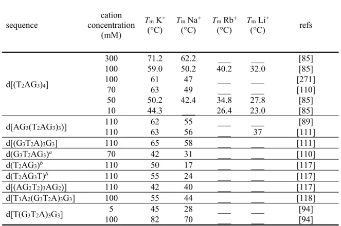
Włodarczyk et al. have measured the melting temperature of the 24-mer d[(T2AG3)4] sequence in presence of various concentration of alkali metals [85] (Table 2)
[85,89,94,110,111,117,118,271]. At ~ 100 mM in K+, Na+, Rb+, and Li+, the Tm is
respectively 59, 50, 40, and 32 °C, nicely illustrating the important role of alkali metal ions in G4 stability. This ranking is conserved at lower concentrations (~ 50 mM), ranging from 50 to 28 °C, and the G-quadruplex is still reasonably stable at 10 mM in K+ (Tm = 44.3 °C) and to a lower extent Rb+ (Tm = 26.4 °C). Higher Na+ and K+ concentrations bring further stabilization (62 and 71 °C, respectively, at ~ 300 mM), and partial folding in observed at room
temperature with 180—230 mM Cs+ (Tm = 27.4—27.7 °C). CD spectra were acquired at 2 °C and 20 °C with increasing concentrations of cations. Saturation was not observed for Li+ at 20 °C and Cs+ at both temperatures, consistent with their weak G-quadruplex stabilization properties. The presence of high Cs+ concentrations seemingly destabilizes the G-quadruplex. Conversely, only 4 and 30 mM were enough to reach saturation for K+ and Na+, respectively.
Sugimoto and co-workers have found similar results in 100 mM Na+ (~ 47 °C) and K+ (~ 61 °C) [271], and Balagurumoorthy and Brahmachari have observed melting temperatures of 63 and 49 °C in 70 mM KCl and NaCl, respectively [110]. This is somewhat higher than found by Włodarczyk et al., maybe because the values were extracted from 10-data point melting profiles. The 9-mer d(G3T2AG3), forming a bimolecular G-quadruplex, was also studied in the same conditions (Tm = 42 and 31 °C, respectively; 20.5 µM strand
concentration). The 22-mer d[AG3(T2AG3)3)] was found to be more stable in K+ than in Na+ by 7°C in two distinct publications (62/63 vs. 55/56 °C), and than in Li+ by 26 °C [89,111].
27 The difference of stability is identical for the shorter oligonucleotide 21-mer sequence
d[(G3T2A)3G3], with both the K+-form (Tm = 65 °C) and the Na+-form being slightly stabilized (Tm = 58 °C) as compared to the 22-mer counterpart [111]. The short 6- and 7-mers d(T2AG3) and d(T2AG3T) forming tetramolecular G-quadruplexes are unstable in presence of Na+ (17 and 24 °C), and much more stable in K+ (50 and 55 °C) [117].
Modified human telomeric sequences are also under scrutiny. An 18-mer sequence that contains repeats of only two guanines (d[(AG2T2)3AG2)]) is destabilized as compared to the unmodified 22-mer sequence counterpart, and has almost the same melting temperature in K+ and Na+ conditions (42 and 40 °C, respectively) [117]. The addition of extra nucleotides in 5’ to obtain the 26-mer d[T3A2(G3T2A)3G3] is also detrimental to the stability but yields a higher
Tm difference (55 vs. 44 °C) [118]. Risitano and Fox have examined the sequence
d[T(G3T2A)3G3], which contains an additional dT nucleotide in 5’ as compared to the non-modified 21-mer sequence, by FRET-melting in presence of various concentrations of K+ and Na+ (5—100 mM) [94]. A clear increase in Tm is observed with the concentration, ranging from 45 to 82 °C in K+ and 28 to 70 in Na+, with the difference of stabilization decreasing from 17 to 12 °C. The high melting temperatures could be attributed to the presence of fluorophores at both ends of the oligonucleotides that may stabilize the G-quadruplexes. Table 2 Melting temperature of human telomeric G-quadruplexes.
sequence concentration cation (mM) Tm K+ (°C) Tm Na+ (°C) Tm Rb+ (°C) Tm Li+ (°C) refs d[(T2AG3)4] 300 71.2 62.2 ___ ___ [85] 100 59.0 50.2 40.2 32.0 [85] 100 61 47 ___ ___ [271] 70 63 49 ___ ___ [110] 50 50.2 42.4 34.8 27.8 [85] 10 44.3 ___ 26.4 23.0 [85] d[AG3(T2AG3)3)] 110 110 62 63 55 56 ___ ___ ___ 37 [111] [89] d[(G3T2A)3G3] 110 65 58 ___ ___ [111] d(G3T2AG3)a 70 42 31 ___ ___ [110] d(T2AG3)b 110 50 17 ___ ___ [117] d(T2AG3T)b 110 55 24 ___ ___ [117] d[(AG2T2)3AG2)] 110 42 40 ___ ___ [117] d[T3A2(G3T2A)3G3] 100 55 44 ___ ___ [118] d[T(G3T2A)3G3] 100 5 45 82 28 70 ___ ___ ___ ___ [94] [94]
a bimolecular structure; 20.5 µM strand concentration b tetramolecular structure;10 µM strand concentration
![Figure 2 Examples of human telomeric G-quadruplex structures deposited in the PDB that were solved by NMR or X-ray crystallography: 143D [25], 1KF1 [26], 2GKU [27], 2YH9 [28], 2JSM and 2JSL [29], 2JPZ [30], 2KF8 [31], and 2KKA [32]](https://thumb-eu.123doks.com/thumbv2/123doknet/14216424.482782/6.918.148.773.302.899/figure-examples-telomeric-quadruplex-structures-deposited-solved-crystallography.webp)
![Figure 8 Crystal structure of TBA coordinating Na + (left; purple spheres: two positions; PDB ID: 4DIH) or K + (right; purple sphere: one position; PDB ID: 4DII), bound to -thrombin (not shown except for His71, in orange) [161]](https://thumb-eu.123doks.com/thumbv2/123doknet/14216424.482782/45.918.250.665.422.676/figure-crystal-structure-coordinating-spheres-positions-position-thrombin.webp)
