1
Cell-to-Cell Variability and Culture Conditions during Self-Renewal
Reversibly Affect Subsequent Differentiation of
Mouse Embryonic Stem Cells
byJit Hin Tan
Bachelor of Science in Chemical Engineering Cornell University, Ithaca, NY, 2003
Master of Science in Chemical Engineering Practice Massachusetts Institute of Technology, Cambridge, MA, 2004
Submitted to the Department of Chemical Engineering in Partial Fulfillment of the Requirements for the Degree of
Doctor of Philosophy in Chemical Engineering Practice at the
Massachusetts Institute of Technology June 2013
© 2013 Massachusetts Institute of Technology All Rights Reserved
Signature of Author……… Department of Chemical Engineering
September 6, 2011
Certified by………... Clark K. Colton Professor of Chemical Engineering Thesis Supervisor
Accepted
by………... William M. Deen Professor of Chemical Engineering Chairman, Committee for Graduate Students
2
3
Cell-to-Cell Variability and Culture Conditions during Self-Renewal Reversibly Affect Subsequent Differentiation of Mouse Embryonic Stem Cells
by Jit Hin Tan
Submitted to the Department of Chemical Engineering on September 6, 2011 in Partial Fulfillment of the Requirements for the Degree of
Doctor of Philosophy in Chemical Engineering Practice
Cell-to-cell variability in clonal populations is reflected in a distribution of mRNA and protein levels among individual cells, including those of key transcription factors governing embryonic stem cell (ESC) pluripotency and differentiation. This may be a source of heterogeneity resulting in mixtures of cell types in differentiated populations despite efforts to control the differentiation conditions and the use of a clonal starting population. In addition, this distribution may be affected by the cell microenvironment during self-renewal. Prior studies on self-renewal culture of ESC, however, focused on long term proliferation and pluripotency. The effects of culture conditions during self-renewal on the effectiveness of subsequent differentiation protocols remains
understudied.
Using a mouse ESC line harboring a GFP reporter, we examined cell-to-cell variability in clonal undifferentiated populations and how such variability affects subsequent differentiation. Subpopulations sorted according to their levels of Oct4-GFP expression displayed distinctly different expression levels of pluripotency and early differentiation markers and differentiated into cardiomyocytes at different efficiencies. However, when allowed to self-renew after sorting, the subpopulations regenerated the parental
distributions of Oct4-GFP and subsequent differentiation after regeneration did not show differences.
In addition to differences between cells in a clonal population, self-renewal conditions affecting Oct4 expression on the population-level were examined. Changes in culture conditions during self-renewal by low oxygen culture or small molecule dual inhibition (2i) of mitogen-activated protein kinase and glycogen synthase kinase reversibly affected levels of Oct4 expression in cells that were otherwise pluripotent. Effects of different self-renewal conditions immediately preceding differentiation are manifested by changes in subsequent differentiation to cardiomyocytes.
This study demonstrates that manipulation of self-renewal culture conditions can lead to changes in the outcomes of defined differentiation protocols, a novel dimension to explore for directed differentiation of pluripotent stem cells.
Thesis Supervisor: Clark K. Colton Title: Professor of Chemical Engineering
4
5
Acknowledgements
The single name on the front page belies the generous contributions of many, without whom this thesis would not have been possible. When I returned to MIT for the PhD program, a research area heavily underpinned by biology was not on my radar, due to a lack of prior training. In spite of this, I was welcomed with open arms into the research group by my advisor, Professor Clark Colton. As Clark mentioned during a tribute to his graduate advisor Ed Merrill, he “did not know that a graduate student was supposed to do the research that his advisor chooses for him”, and I am very grateful that Clark has allowed me the same ignorance during my thesis work to explore questions of my own choice. I must also thank the members of my thesis committee: Professor William Deen, for his invaluable advice in navigating the ins and outs of the doctoral process, and for reminding me of the value of my training in Chemical Engineering; Professor Rudolf Jaenisch, whom, despite his busy schedule, never refused a meeting, and is able, in a short discussion, to open up new horizons to explore; and Professor Narendra Maheshri, who has been generous in his suggestions on both experiments worth doing, and
perhaps more importantly, not worth doing.
To the members of the Colton Lab – Kevin Brower, Anna Coclite, Amanda DiIenno, Amy Johnson, Clarke Low, Michelle Miller, Jeff Millman: No one else understands the journey we all undertake as well as all of you do, and I am extremely grateful for your fellowship and for allowing me to commiserate during my time here. Also, I offer sincere thanks to Kellie Young, my UROP, who remained enthusiastic despite some of the mundane tasks I had assigned during her time with the lab. In particular, I am deeply indebted to Jeff, who had been instrumental in showing me the ropes and also holding long discussions on practically every aspect of my work throughout the entire time. Jeff inspires me with his insight and enthusiasm for research, and has taught by example hard work and perseverance in the face of fickle cell cultures and impenetrable
protocols. I leave the lab in Amanda’s very capable hands, and am confident that she will take the research ever further. Many thanks must also go to the underappreciated Val Grimm, our administrative assistant, who has skillfully helped me to wrangle schedules and untangle paperwork.
I would like to thank the many facilities and laboratories who have offered assistance, expertise and equipment that we were lacking and were essential to the completion of this work: Dr Malgorzata Borowiak (Melton Lab, Harvard University), for providing the Oct4-GFP mESC reporter line that made this work possible; Glenn Paradis, Michael Jennings, and Michele Griffin of the Koch Institute Flow Cytometry Core Facility, whose expertise and patience I have taken advantage of repeatedly; Han-Hwa Hung and Linda Bragman of the Centre of Biomedical Engineering’s qPCR Facility for allowing me to hog the equipment on many occasions, and T.L To, formerly of the Maheshri lab, for generously sharing equipment and expertise in the many common biological assays and protocols that I was regretfully short of.
As Michael Mosley opined, “Science is a human endeavor. It’s been shaped as much by what’s outside the laboratory as inside.” To ignore the people who have been a part of
6
my life outside of research would be to ignore those who have provided an equal, though indirect, contribution to this thesis. I have been extremely blessed to be part of two warm and welcoming groups outside of the laboratory, who have provided safe haven and sanity when I have needed it most, shared in the highs and provided
unquestioning support during the lows – my fellow Singaporeans, who I have subjected to many culinary experiments and hijacked conversations steered towards research; and the graduate community of Sidney-Pacific, who have given me free reign over the kitchen without the obligation of responsibility. When you are away from home, your friends become your family – as you all have. I will always be impressed and humbled by your great achievements and diverse abilities, honest humility and generous spirits. Sometimes things do not go according to plan, and the journey takes an unexpected detour. Looking back, I see a path that I may not have chosen at the start, but I am happy that I have taken, because of the people I would not have met, lessons I would not have learnt, and experiences I would have not have gained had I not done so. Half a world away, there are four people who continue to shower me with their
unconditional love and support - my parents, Michael and Doreen, and my siblings Jit Quan and Michelle, who, despite not fully understanding why this process has taken me away from home for so long, remain patient and understanding. Sometimes the most important things are the hardest to say and I find myself at a loss for words, except Thank you.
7
8
9
Table of Contents
Chapter 1 Introduction ... 15
1.1 Pluripotent stem cells ... 15
1.2 Self-renewal and propagation in the pluripotent state ... 16
1.3 Directed differentiation ... 18
1.4 Cell-to-cell variability in clonal undifferentiated PSC populations ... 20
1.5 Effects of oxygen on the undifferentiated phenotype ... 21
1.5.1 Derivation of new embryonic stem cell lines ... 22
1.5.2 Pluripotency ... 23
1.5.3 Proliferation ... 26
1.5.4 Clonal recovery ... 26
1.5.5 Reprogramming ... 27
1.5.6 Control of pO2cell ... 27
1.5.7 Effects on subsequent differentiation ... 29
1.6 Use of 2i in PSC derivation and propagation ... 30
1.7 Objectives ... 31
1.8 Overview ... 32
Chapter 2 Cell-to-cell variability within an undifferentiated clonal population affects subsequent differentiation of mouse embryonic stem cells ... 35
2.1 Abstract ... 35
2.2 Introduction ... 37
2.3 Methods ... 40
2.3.1 Self-renewing culture of mESC ... 40
2.3.2 Cell cycle analysis ... 40
2.3.3 Fluorescence-Activated Cell Sorting (FACS) of undifferentiated O4G cells ... 41
2.3.4 Flow cytometric analysis of Oct4-GFP expression ... 41
2.3.5 Differentiation of mESC to cardiomyocytes ... 42
2.3.6 Real-time polymerase chain reaction (PCR) ... 43
2.3.7 Cell enumeration ... 44
2.3.8 Flow cytometric analysis of MF20+ cells ... 44
2.3.9 Statistics ... 45
2.4 Results ... 46
2.4.1 Large cell-to-cell variability in Oct4-GFP expression in undifferentiated mESC which corresponds to Oct4 mRNA expression ... 46
10
2.4.3 Levels of Oct4-GFP mark undifferentiated subpopulations with different levels of
pluripotency and early differentiation markers ... 47
2.4.4 Populations differentiated from cells with higher initial Oct4-GFP levels had a greater fraction and number of cardiomyocytes ... 47
2.4.5 Sorted subpopulations recapitulate the parental Oct4-GFP distribution when allowed to propagate in self-renewing culture ... 49
2.4.6 Recapitulation of the parental Oct4-GFP distribution erases subpopulation differences in pluripotency and early differentiation markers ... 50
2.4.7 Recapitulation of the parental Oct4-GFP distribution removes the differences in subsequent differentiation to cardiomyocytes ... 50
2.5 Discussion... 52
2.6 Future Work ... 55
2.7 Tables and Figures ... 56
Chapter 3 Culture oxygen levels and use of small molecule inhibitors during self-renewal reversibly affect subsequent differentiation of mouse embryonic stem cells ... 68
3.1 Abstract ... 69
3.2 Introduction ... 71
3.3 Methods ... 73
3.3.1 Self-renewing culture of mESC ... 73
3.3.2 Flow cytometric analysis of Oct4-GFP expression ... 74
3.3.3 Differentiation of mESC to cardiomyocytes ... 74
3.3.4 Control of culture oxygen levels (pO2cell) ... 75
3.3.5 Real-time polymerase chain reaction (PCR) ... 75
3.3.6 Cell enumeration ... 76
3.3.7 Flow cytometric analysis of MF20+ cells ... 76
3.3.8 Estimation of spontaneously contracting areas ... 77
3.3.9 Statistics ... 77
3.4 Results ... 78
3.4.1 Self-renewal of mESC at low oxygen reduces expression of Oct4-GFP but does not increase rates of spontaneous differentiation ... 78
3.4.2 Reduction of Oct4-GFP expression by low oxygen culture is reversed by continued self-renewal in 20% oxygen ... 78
3.4.3 Low oxygen preconditioning decreases subsequent differentiation of mESC to cardiomyocytes ... 79
3.4.4 Total cell number and fraction of residual undifferentiated Oct4-GFP+ cells are not affected by preconditioning, but by oxygen conditions during differentiation ... 80
3.4.5 Reduction in efficiency of cardiomyogenesis due to low oxygen preconditioning is reversible by self-renewal in 20% oxygen before differentiation ... 81
11
3.4.6 2i use during self-renewal increases expression of naïve pluripotency markers, decreases early differentiation markers synergistically with high oxygen, and is reversible
by subsequent self-renewing culture without 2i ... 82
3.4.7 High oxygen and 2i use during preconditioning separately, synergistically, and reversibly increases subsequent differentiation to cardiomyocytes ... 84
3.4.8 Fraction of MF20+ cells in differentiated populations correlates positively with mean Oct-GFP levels of the starting populations ... 86
3.5 Discussion and future work ... 88
3.6 Figures ... 91
Chapter 4 Integrative Perspective Paper: Opportunities and Obstacles in the Commercialization of Pluripotent Stem Cell-Based Products and Services ... 114
4.1 Executive Summary ... 115
4.2 Introduction ... 116
4.3 Pluripotent Stem Cell Research and Diagnostic Consumables and Equipment ... 117
4.4 Pluripotent Stem Cell-Derived Tools for Disease Modelling, Drug Discovery, and Toxicology ... 120
4.5 Replacement Cell Therapies Derived from Pluripotent Stem Cells ... 124
4.6 Discussion... 130
12
13
List of Figures
Figure 2-1 Characterization of Oct4-GFP (O4G) mESC reporter line demonstrates large cell-to-cell variability in Oct4-GFP expression which corresponds to Oct4 mRNA levels ... 56 Figure 2-2 Oct4-GFP variability cannot be explained by differences in cell cycle stage ... 57 Figure 2-3 Real-time PCR analysis of sorted O4G mESC subpopulations reveal that levels of Oct4-GFP mark undifferentiated subpopulations with different levels of pluripotency and early differentiation markers ... 58 Figure 2-4 Populations differentiated from cells with higher initial Oct4-GFP levels had a greater fraction and number of cardimyocytes ... 60 Figure 2-5 Oct4-GFP fluorescence distributions of sorted subpopulations cultured in
self-renewing conditions for up to 30 days after initial sorting demonstrate recapitulation of the parental Oct4-GFP distribution over time. ... 62 Figure 2-6 Time course of mean Oct4-GFP fluorescence of the subpopulations relative to the All subpopulation... 63 Figure 2-7 Effects of regeneration of parental Oct4-GFP expression on gene expression of subpopulations... 64 Figure 2-8 Recapitulation of the parental Oct4-GFP distribution removes the differences in subsequent differentiation to cardiomyocytes ... 66 Figure 3-1 Effects of culture O2 in self-renewing culture on distributions of Oct4-GFP expression
in O4G mESC. ... 91 Figure 3-2 Mean Oct4-GFP fluorescence and fraction of Oct4-GFP+ cells from self-renewal at different O2. ... 92
Figure 3-3 Effects of O2 preconditioning on subsequent differentiation. ... 94
Figure 3-4 Effects of O2 preconditioning on total cell number and fraction of residual Oct4-GFP+
cells after differentiation. ... 96 Figure 3-5 Reversibility of O2 preconditioning. ... 97
Figure 3-6 Correlation between mean Oct4-GFP fluorescence of the starting undifferentiated populations and the fraction of MF20+ cells in the resulting differentiated populations. ... 99 Figure 3-7 Experimental design and nomenclature for preconditioning with O2 modulation and 2i
supplementation. ... 100 Figure 3-8 Effects of 2i supplementation and O2 during self-renewal on Oct4-GFP fluorescence
distributions of undifferentiated mESC. ... 101 Figure 3-9 Effects of 2i supplementation and culture O2 on mean Oct4-GFP fluorescence and
fraction of Oct4-GFP+ cells during self-renewal. ... 102 Figure 3-10 Effects of 2i supplementation and culture O2 on gene expression of mESC after 18
days in self-renewing culture. ... 104 Figure 3-11 Effects of 2i supplementation and culture O2 on gene expression of mESC after 24
days in self-renewing culture. ... 106 Figure 3-12 Effects of 2i supplementation and culture O2 on subsequent differentiation after 18
days of preconditioning. ... 108 Figure 3-13 Effects of 2i supplementation and culture O2 on subsequent differentiation after 24
14
Figure 3-14 Correlation between the fraction of MF20+ cells in the resulting differentiated populations for cells preconditioned for 18 (red triangles) and 24 days (blue circles) before differentiation and the mean Oct4-GFP fluorescence of the starting undifferentiated populations. Cells were differentiated at 20% O2. For cells preconditioned for 18d, <R2>= 0.63; for cells
preconditioned for 24d, <R2>= 0.93, both from a linear least squares regression. ... 111 Figure 3-15 Linear and semi-log correlations between mean Oct4-GFP fluorescence of starting undifferentiated populations and the fraction of MF20+ cells in the resulting differentiated
populations for all cells differentiated at 20% O2. ... 112
Figure 3-16 Model fit for correlation between mean Oct4-GFP fluorescence of starting undifferentiated populations and the fraction of MF20+ cells in the resulting differentiated
15
Chapter 1 Introduction
1.1 Pluripotent stem cells
First isolated from mouse [1], then more recently human blastocysts [2], pluripotent embryonic stem cells (ESC) have the ability to self-renew in the undifferentiated state indefinitely and differentiate into all cell types of the adult organism. They hold immense clinical and research potential for drug screening, disease modeling, as a model to study mammalian development, and to generate replacement cells for application in regenerative medicine [3–9]. Cardiomyocytes [10], bone marrow [11], dopamine
neurons [12], and β-cells [13] have been differentiated from ESC as potential therapies for cardiac infarctions, leukemia, Parkinson’s disease, and diabetes respectively. Other studies have also demonstrated the ability to generate self-organized tissues [14] and to perform gene and cell therapy using ESC [15].
The discovery of another source of pluripotent cells, induced pluripotent stem cells (iPSC) [16–18] gave further hope for personalized therapies by autologous
transplantation of cells and tissues differentiated from iPSC derived from the patients themselves [19], and for recapitulation of disease development by generating disease specific iPSC [20], in addition to all of the potential applications of ESC [21–25]. iPSC and ESC have shown similar potential to differentiate into cell types of interest and fulfill other criteria for pluripotency [26–28], however, some differences remain [29].
For pluripotent stem cells (PSC) of both types, many obstacles remain before their immense potential can be fulfilled. This includes removing the danger of tumorigenicity
16
of residual undifferentiated cells [30], immunogenicity of the transplanted cells [31], the development of methods for propagation in their undifferentiated state, and efficient protocols for directed differentiation in order to generate sufficient numbers and purities of the desired cell types [32].
1.2 Self-renewal and propagation in the pluripotent state
In order to generate sufficient numbers of cells, PSC can be propagated in their undifferentiated state until differentiation is initiated. For mouse ESC (mESC), this is usually accomplished by either co-culture on mouse embryonic fibroblasts (MEFs) or the addition of leukemia inhibitory factor (LIF) [33], a cytokine which acts by activating the JAK-STAT3 pathway [34]. Human ESC (hESC) can be kept in the pluripotent state by culture medium supplemented with basic Fibroblast Growth Factor (bFGF) [35] on a layer of various feeder cells [36] or an animal-derived protein matrix [37], although recently a fully defined self-renewal culture system has been discovered [38].
The main concern of undifferentiated PSC culture is to ensure the maintenance of the pluripotent state and prevent spontaneous differentiation while sustaining normal karyotype and cell viability. To do so, it must support the highly complex cellular machinery for maintaining pluripotency that is underpinned at its core by three pluripotency genes – Oct4, Sox2, and Nanog [39–43]. Disruption of these core transcription factors caused spontaneous differentiation of PSC and loss of the pluripotent state.
17
An ectopic increase in Oct4 expression in mESC directed spontaneous differentiation down the mesodermal lineage [44, 45] while a decrease in Oct4, or increase in Cdx2 expression relative to Oct4, directed differentiation to trophectoderm [45, 46]. Similar behavior occurred in hESC with an ectopic decrease in Oct4 expression resulting in spontaneous differentiation to ectoderm and mesoderm, while an ectopic increase caused differentiation to endoderm [47]. Both Oct4 and Sox2 determine germ layer fate decisions during early differentiation in opposite directions, with Oct4 promoting
differentiation down the mesodermal and endodermal lineages, while Sox2 promoted differentiation down the ectodermal lineage, and each suppressed the promoted lineage of the other [48]. Expression of Nanog prevented differentiation, and mESC can exhibit transient down-regulation of Nanog expression. This down-regulation has been
hypothesized to generate a necessary intermediate state between maintaining
pluripotency and differentiation [49, 50]. In hESC, Nanog affected cell fate choice, with an extended expression of Nanog during differentiation leading to increased
mesendodermal specification [51].
Recent studies demonstrated that multiple metastable pluripotent states for PSC exist as distinct subpopulations within a clonal population. In addition to Nanog, Rex1 [52] and Stella [53] are also heterogeneously expressed in mESC. These subpopulations were able to reversibly interconvert when the population was kept in conditions supporting self-renewal of the pluripotent state. Given that the culture conditions that maintain pluripotency can also affect the transition of mESC between the different pluripotent states, and that pluripotency factors can specify differentiation to different
18
cell fates [48, 54], it is thus important to assess the effects of the self-renewal conditions on PSC and on the efficiency of subsequent differentiation to different lineages. Two aspects in particular – culture oxygen levels, and the use of small molecule inhibitors supporting undifferentiated culture – are discussed in greater detail in sections 1.5 and 1.6.
1.3 Directed differentiation
The design of directed differentiation protocols for pluripotent stem cells has employed many different strategies to enrich the resulting differentiated population with the desired cell type [55, 56]. To direct differentiation to cardiomyocytes from both mouse and human PSC [57], for example, protocols have included the use of growth factors such as Activin or Nodal and BMP4 [58], the use of small molecules
5-aza-2’-deoxycytidine [59] or ascorbic acid [60], genetic manipulation to ectopically express a cytokine, TNF-α [61], co-culture with a visceral endoderm-like cell line (END-2) [62], or, most recently, synthetic mRNA transfection [63]. Combinations of techniques, such as the addition of stauprimide with Activin A for directed differentiation to definitive
endoderm [64], or the step-wise protocol of sorting Flk1+ cells differentiated from miPSC, then co-culturing the purified intermediate population on OP9 stroma cells to obtain cardiomyocytes [65], have also been documented.
The designs of directed differentiation protocols have grown in sophistication, inspired in part by insights from developmental biology. Strict temporal control of the cellular
19
growth factors (Activin A for example, can induce both endodermal and mesodermal specification depending on concentration), and optimizing levels of growth factors depending on the stage of differentiation have yielded success [13, 58, 66]. The focus, however, has been predominantly on the time period and process during which the cells are already differentiating, and much remains unknown about how the culture conditions before the initiation of differentiation affects the cells subsequently. This is in spite of the many studies (summarized above in section 1.2) showing that pluripotency factors, which are highly active when PSCs are in their undifferentiated state, can play roles in specifying cell fate during differentiation.
The focus of this thesis is on cell-to-cell variability (population heterogeneity), culture oxygen levels, and the use of small molecules during self-renewing culture on
subsequent differentiation. It examines the relationship between starting undifferentiated state of the ESC and the resulting differentiated population, an aspect of self-renewal that has not received much attention. Experiments on cell-to-cell variability examine differences between clonal undifferentiated cells within a population, while studies combining culture oxygen levels and the use of small molecules in self-renewal culture examine their effects on self-renewal and the efficiency of subsequent directed
differentiation of undifferentiated populations as a whole. More detailed treatments of these topics follow.
20
1.4 Cell-to-cell variability in clonal undifferentiated PSC populations
Cell-to-cell variability or cellular heterogeneity in clonal populations has received increasing attention with the development of methods to measure characteristics of single cells, rather than traditional methods yielding population-averaged results. Studies showing heterogeneity in single mammalian cells of a clonal population in mRNA [67, 68] and protein levels [69] that can have important biological functions suggest that traditional ensemble measurements insufficiently capture the
characteristics of single cells [70, 71].
Numerous studies have reported heterogeneity in clonal stem cell populations that correlate with differences in subsequent cell fate choice. Hematopoietic stem cells have transcriptome-wide differences in subpopulations sorted by expression of a surface antigen, Sca-1, despite being clones generated from a single cell [72], and levels of Sca-1 correlated to differentiation efficiencies to different cell lineages. Within clonal populations of mESC, there were heterogeneities in expression of Nanog [50], Rex1 [52], and Stella [53], all normally associated with pluripotency, and in an early
endodermal marker Hex [73]. Subpopulations exhibiting high or low levels of expression of these genes exhibited different differentiation proclivities when allowed to differentiate. Levels of these genes in mESC allowed to self-renew in the undifferentiated state,
however, are dynamic, and single cells can switch between high and low levels of expression. In both human ESC and iPSC, heterogeneities occurred in mRNA levels of multiple genes, although single hiPSC exhibited higher levels of heterogeneity than hESC [74]. The multitude of metastable pluripotent states has led to a proposal that
21
pluripotency is not a set of distinct states, but instead a continuum of states [75]. This heterogeneity, however, is viewed as undesirable in directed differentiation protocols because it can lead to heterogeneity in the differentiated population, which can be detrimental to the production of clinically useful cells and tissues [76].
Cell-to-cell variability in mRNA expression levels can occur due to gene expression noise, arising from transcription occurring in bursts, rather than at a continuous steady rate [68, 77, 78]. Together with a positive feedback loop, for example, a gene that activates its own transcription, such stochastic gene expression is able to generate stable distinct phenotypes [79]. Many theories have been put forward to describe the mechanisms behind such cell-to-cell variability, and it has been proposed that the large interconnected network of genes and epigenetic states in mammalian systems, together with transcriptional noise, create multiple stable gene expression profiles that clonal single cells may inhabit [80]. More importantly, as evidenced by effects on subsequent differentiation of cells in different “states”, such cell-to-cell variability can have important developmental functions.
1.5 Effects of oxygen on the undifferentiated phenotype
Adapted and updated from Millman, J.R., J.H. Tan, and C.K. Colton, The effects of low oxygen on self-renewal and differentiation of embryonic stem cells. Current Opinions in Organ Transplantation, 2009. 14(6): p. 694-700 with permission from Lippincott Williams & Wilkins
22
One aspect of the culture microenvironment that has mostly been ignored in
conventional cell culture is the oxygen partial pressure experienced by the cells. All cells of the developing embryo are exposed to oxygen levels in vivo far below that of
atmospheric levels, and they differentiate and undergo organogenesis in a low oxygen environment [81–87]. The local oxygen environment is potentially an important variable for manipulation by researchers hoping to recapitulate specific differentiation pathways in vitro.
Mammalian cellular responses to oxygen levels are predominantly mediated by Hypoxia Inducible Factors (HIFs), a family of heteromeric transcription factors. HIF-α subunits are constitutively expressed but are post-translationally regulated through rapid degradation in the presence of oxygen levels higher than 5% [88–90]. The HIF-1β subunit (also known as the aryl hydrocarbon receptor nuclear translocator or ARNT) is oxygen insensitive, but is only active as a transcription factor when dimerized with the appropriate stabilized HIF-α subunit under sufficient cellular hypoxia. The mammalian hypoxic response includes up-regulation of a large number of genes, including the vascular endothelial growth factor (VEGF), the adipose differentiation related protein (ADRP), and RTP801, which is known to be regulated in development, amongst others [91].
1.5.1 Derivation of new embryonic stem cell lines
Derivation of new ES cell lines, both mouse [92, 93] and human [38, 94], benefit from being performed in low oxygen levels. Studies examining the establishment of new ES
23
cell lines from blastocysts under 5% oxygen have had greater success than under the 20% more commonly used. The benefits of low oxygen conditions included an increase in the number of individual colonies exhibiting alkaline phosphatase activity [93], an increase in the number of proliferating cells [92], and removal of the need for undefined animal-derived products such as serum or matrices in the process [38], the last of which can be crucial in the applicability of these derived cells to clinical applications. Most recently, it has been found that maintenance of constant low oxygen during derivation of hESC enables the establishment of pre-X inactivated cells, which are similar to mESC, and exposure to 20% oxygen or oxidative stress irreversibly causes X inactivation [94].
1.5.2 Pluripotency
Low oxygen affected the pluripotent phenotype of mESCs when they were cultured with LIF [33]. Normally, mESC colonies have smooth edges, but this morphological
phenotype was lost with low oxygen exposure, instead causing the colonies to have fibroblast-like morphologies [95] or to form jagged edges [96], both signs of early differentiation despite the presence of LIF. These morphological changes were also associated with reduced levels of alkaline phosphatase activity and pluripotency gene (Oct4, Nanog, and Sox2) expression [95, 96]. One study [95] proposed that HIF-1α protein, the concentration of which increases at low oxygen, mediated suppression of the LIF-STAT3 pathway as evidenced by a reduction in the expression of the LIF receptor under hypoxia. Interestingly despite these observations, the cells maintained some potential to differentiate into cells of all three germ layers [96].
24
In contrast to mESCs, expression of Oct4, Nanog, and Sox2 in hESCs were apparently not affected by hypoxic culture (2% to 5% oxygen) [94, 97, 98]. In one study, however, Nanog and Notch1 mRNA expression increased in hESCs cultured over a four-week period at 5% oxygen compared to those cultured under 20% oxygen. This difference was not maintained after long term culture under the hypoxic condition, since there was no significant difference in Nanog and Notch1 mRNA levels between cells cultured for 18 months under either 5% or 20% oxygen. Consistent with earlier results, no
differences in Oct4 expression were observed under both oxygen conditions with either short or long term culture [99]. However, a recent report showed down-regulation of Oct4, Nanog and Sox2 in HIF-2α silenced hESC, together with reduced proliferation [100].
Hypoxic culture resulted in wide-spread transcriptome differences in hESCs when compared to culture under ambient oxygen conditions, including downstream targets of the core pluripotency network Oct4, Nanog, Sox2. Genes marking early differentiation to all cell lineages were upregulated at 20% oxygen [94, 98]. In addition, Lefty2, a member of the TGFβ family of proteins, which is hypothesized to play a role in preventing
differentiation, was down-regulated at 20% oxygen compared to culture at 4% [98]. This may lead to a greater proclivity for the hESC to differentiate. In studies on the
transcriptome-wide effects of hypoxic culture on hESCs kept under self-renewing conditions, culture at 20% O2 resulted in greater heterogeneity amongst different hESC lines than when cultured under hypoxic (2-4% oxygen) conditions [101]. This
25
heterogeneity may arise from subpopulations of cells already undergoing early differentiation when cultured at 20% oxygen.
While low oxygen appears not to affect pluripotency gene expression directly,
differentiated areas of each colony expressing SSEA-1, a marker of early differentiation for hESCs, were reduced when cultured under 3% and 1% oxygen compared to culture under 20% oxygen[102]. In a different study, however, culture at 2% oxygen did not affect expression of either SSEA-4 or SSEA-1 in hESCs when compared to ambient oxygen conditions [97].
HIF-1α has been investigated as an important gene mediating ESC hypoxic response. One study [103] suggests a possible link connecting HIF-1α action with Notch signaling. In mouse embryonic teratocarcinoma cells and neural stem cells, hypoxia allowed for the accumulation of HIF-1α, which stabilized the Notch intracellular domain, in turn promoting the undifferentiated phenotype. The action of Notch in promoting self-renewal appears to be conserved in hESCs in view of a recent demonstration that self-renewal is mediated by activation of the Notch pathway and that benefits of hypoxic culture were eliminated with inhibition of Notch signaling [99]. Interestingly, although another
hypoxia-inducible factor in the same family as HIF1α, HIF-2α has not been shown to be active in mESC [91], other studies have shown that it is the HIF-2α heterodimer and not HIF-1α that can induce transcription of Oct4 [104], providing a potential link between culture oxygen levels and pluripotency gene expression in ESC. Several studies
26
with chronic hypoxia or normoxia [94, 99]. However, it is the HIF-2α response that may influence pluripotency gene expression after long-term continuous culture under hypoxic conditions [100].
1.5.3 Proliferation
Many studies have found that low oxygen affects proliferation, but this effect varied between studies and cell lines. In one study [93], proliferation of mESCs increased under 5% compared to 20% oxygen, while in another [96], no difference was observed in one cell line and a small increase in another. Culture at 1% oxygen or lower, however, reduced the rate of proliferation compared to 20% [95, 96]. mESCs were able to adapt and continue to proliferate under anoxia, albeit with a longer doubling time compared to 20% oxygen, and had the ability to utilize anaerobic respiration exclusively for their energy requirements [96]. Similarly disparate results have been reported for hESCs: In one study, proliferation rates at 5% oxygen were reduced compared to 20% [99], in another, no differences in the rates were observed at 5% and 3% and only a slight reduction at 1% [102]. A conflicting recent report showed that low oxygen culture increased proliferation of hESC [100].
1.5.4 Clonal recovery
Low oxygen culture improved clonal recovery after passaging. mESCs passaged at 5% oxygen [34] and hESCs at 2% [101, 105] had improved clonal survival. Cytogenetic instability at high passages for mESCs was not reduced in culture at 5% oxygen [92], and further reductions in oxygen to even 0% did not affect the amount of oxidative DNA
27
damage incurred by the cells [96]. In contrast for hESCs, 2% oxygen reduced
chromosomal abnormalities after multiple passages [101], suggesting a further benefit of hypoxic culture.
1.5.5 Reprogramming
Moderate hypoxic conditions improved reprogramming of somatic cells to iPSC for both mouse and human lines. Performing reprogramming at 5% oxygen increased the number of iPSC colonies generated over both 20% and 1% oxygen conditions [106]. It is possible that this increase in reprogramming efficiency may be due to increased proliferation under 5% oxygen, as reprogramming is a stochastic process that can be accelerated by increasing rates of cell division [107].
1.5.6 Control of pO2cell
Most of the studies reviewed in this section implicitly make the assumption that the oxygen levels in the bulk gas phase (pO2gas) was equal to the actual levels of oxygen experienced by the cell (pO2cell). However, pO2cell can differ drastically from
pO2gas,depending on the cell density, medium height, cellular oxygen consumption rate, and oxygen permeability of the culture substrate [108]. Furthermore, step changes in pO2gas due to removal of culture dishes from the incubators may result in transient changes in pO2cell with long time scales for equilibration. These problems are
ameliorated by culturing the cells on the surface of a membrane with very high oxygen permeability, such as silicone rubber.
28
Differences in pO2cell between different experiments carried out at the same pO2gas provide a possible explanation for the inconsistencies in data reported. For instance, one study [99] reported reduced proliferation of hESCs cultured at 5% oxygen, and another [102] reported no reduction in proliferation at the same oxygen condition. In the first study, there were four weeks between passages (initial and final cell numbers not reported). In the second study, 6x10⁵ cells were plated per well of standard six-well tissue culture plates, and there were only two weeks between passages, with final cell numbers of 1.5-2.5x10⁶ cells per well. It is possible that the pO2cell of the first study was significantly lower than the pO2gas because of higher cell densities, in particular after extended culture without passage, while pO2cell in the latter study was closer to pO2gas. This would help explain the differences in results because the authors of the second study report a reduced proliferation at 1% gas phase oxygen level, which may be closer in cellular oxygen level to the 5% gas phase oxygen level of the first study that also demonstrated a reduced proliferation rate. Moreover, the first group used the Xvivo system (BioSpherix, Lacona, NY), which allowed for continuous culture low pO2gas without interruption for media changes and passaging. The second group used a conventional sealed chamber from which cells were periodically removed for media changes and passaging, allowing for periods where pO2gas is at ambient levels, disrupting the low pO2cell conditions. Without information regarding the experimental conditions, especially the media heights and cell densities, it is impossible to draw any conclusions.
29
The lack of recognition that pO2cell is often different from pO2gas, sometimes by a substantial amount, is exacerbated because values of cell density and media height necessary for retrospective estimation of pO2cell are often not reported. Knowledge of pO2cell is necessary for interpreting the results of studies and comparing them to data from other studies. Efforts to estimate or control pO2cell are essential for rational advancement of this field.
1.5.7 Effects on subsequent differentiation
Given that low oxygen partial pressure (pO2) is an important physiological cue during development [109], it is not surprising that low pO2 during differentiation markedly influences resulting cell fates [110–116]. One example is the increase in fraction and number of cardiomyocytes by low oxygen culture during the first 6 days of differentiation [117]. However, despite the many studies on the effects of oxygen levels employed during self-renewal culture, its effect on subsequent differentiation of PSC is largely unknown. A naïve hypothesis based on the previous two observations may be that since low oxygen is beneficial to cardiomyogenesis in early differentiation, low oxygen culture during self-renewal immediately before differentiation would have a synergistic positive effect as well. Given the effects of oxygen levels on self-renewal culture, it is important to study the effects of modulating oxygen on subsequent differentiation, since it is the differentiated cells that are of therapeutic importance.
30 1.6 Use of 2i in PSC derivation and propagation
Small molecules that are able to target specific signaling pathways or proteins have been invaluable in advancing stem cell culture. They allowed for defined conditions and media to be used in the generation, propagation, and differentiation of the desired cell types [118]. In particular, this thesis focuses on the use of a pair of small molecule inhibitors – PD0325901, a Mitogen-Activated Protein Kinase (MEK) inhibitor [119], and CHIR99021, a Glycogen Synthase Kinase 3β (GSK3β) inhibitor [120], together known as ‘2i’ [121].
PD0325901 acts as a MEK1/2 inhibitor that prevents the phosphorylation of ERK1/2, a kinase that can activate transcriptional activity associated with activation of the RTK (Receptor Tyrosine Kinase) signal transduction pathway associated with the Fibroblast Growth Factor (FGF). CHIR99021 acts as a GSK3β inhibitor that mimics the activation of the canonical Wnt pathway. Activation of the Wnt pathway or application of
CHIR99021 prevents the degradation of β-catenin and allows for its translocation to the nucleus, dimerization with LEF/TCF, and subsequent transcriptional activity [122].
2i was first identified as a replacement for LIF to allow for undifferentiated propagation of mESC [34]. It improves the efficiency of reprogramming of mouse iPSC to the naïve state when used together with LIF [121]. Along the same vein, 2i use in culture of mouse epiblast stem cells (mEpiSC), considered a “primed” state of pluripotency, can convert the cells to a “mESC-like” naïve state [123]. More recently, 2i, when used with ectopic gene expression, LIF, and Forskolin, is able to convert hESC to become a naïve
31
LIF-responsive state [124]. Furthermore, a cocktail of 2i with PD173074, a Fibroblast Growth Factor Receptor (FGFR) inhibitor, and Y27632, a Rho Kinase (ROCK) inhibitor, was able to maintain the undifferentiated state of hESC without feeder cells with single-cell passaging [125].
The increasing use of highly specific small molecules in defined stem cell culture, in particular, in propagating undifferentiated cells, raises the question about their downstream effects on subsequent differentiation of the cells. Studies have shown effects on expression of core pluripotency genes including Oct4, which affect cell fate during differentiation. It is thus not inconceivable that small molecule use during self-renewal would have effects on cell fate after subsequent differentiation.
1.7 Objectives
In order for PSC to fulfill their immense clinical and research potential, there must exist efficient methods for directing differentiation to the desired cell types. Two aspects that have been largely ignored have been differences within a clonal population that may exist due to cell-to-cell variation, and differences between clonal populations that have been subjected to different culture conditions that support undifferentiated PSC self-renewal but may manifest differences in subsequent differentiation. The central
hypothesis behind this thesis is that the starting “state” of a mESC affects the resulting cell fate after subsequent differentiation, despite using undifferentiated mESC from the same cell line that were also subjected to the same differentiation protocol. The
self-32
renewing culture conditions (including culture oxygen levels and the use of small
molecule inhibitors) immediately before the initiation of differentiation on the efficiency of established directed differentiation protocols in order to further improve them. This research would establish a novel dimension to explore in the design of future directed differentiation protocols.
1.8 Overview
Chapter 2: Cell-to-cell variability within an undifferentiated clonal population affects subsequent differentiation of mouse embryonic stem cells
This chapter describes the effects of differences within cells of an undifferentiated population of mESC, specifically Oct4 expression in single cells, on their efficiency of subsequent differentiation to cardiomyocytes. Using a mESC line harboring a GFP
reporter driven by the Oct4 distal enhancer, subpopulations of mESC with high, medium, or low Oct4-GFP levels were sorted by FACS and assessed for pluripotency and early differentiation markers. Sorted cells were differentiated immediately or allowed to self-renew before subsequent differentiation. (1) Differentiation immediately after sorting examined the effects of the initial levels of Oct4 expression on the differentiated cell fate; (2) self-renewal after sorting examined if the subpopulations are stably distinct or if the differences are transient; and (3) differentiation after the subpopulations have been allowed to self-renew examines if subpopulations with different differentiation
efficiencies as found in (1) could be stably isolated and propagated. Differentiated cells were assessed for fractions of residual undifferentiated Oct4-GFP+ cells,
33
immunostained with MF20 antibodies for presence of sacromeric myosin heavy chain, and assessed for cardiac gene expression by qRT-PCR.
Chapter 3: Culture oxygen levels and use of small molecule inhibitors during self-renewal reversibly affect subsequent differentiation of mouse embryonic stem cells
This chapter describes the effects of culture oxygen levels and use of a small molecule inhibitor cocktail (‘2i’) during self-renewal (termed ‘preconditioning’) on subsequent differentiation of mESC. mESC were kept in undifferentiated culture with strict control of cellular oxygen levels using silicone rubber membrane-based culture plates in
conditions of 20%, 5%, or 1% oxygen, with or without addition of 2i in LIF supplemented medium. Cells self-renewed under different conditions were assessed for levels of Oct4-GFP expression and gene expression of pluripotency and early lineage markers. Cells were subsequently differentiated and assessed for efficiency of differentiation to
cardiomyocytes by immunostaining for sacromeric myosin heavy chain protein, qRT-PCR assessment of cardiac gene expression, and semi-quantitative assessments of spontaneously contracting cells. In addition, the reversibility of different preconditioning effects was examined – for cells that received low oxygen preconditioning, further self-renewal at 20% oxygen before differentiation; and for cells that received 2i
supplementation during the initial 9 day preconditioning, further self-renewal without 2i supplementation before differentiation. In addition, the synergistic effects of combining preconditioning that produces the most efficient differentiation to cardiomyocytes with low oxygen during differentiation that has previously been shown to improve
34
differentiation were examined. Finally, a mathematical model describing the relationship between the fraction of cardiomyocytes in the differentiated populations with the initial mean Oct4-GFP levels was developed.
35
Chapter 2 Cell-to-cell variability within an undifferentiated clonal
population affects subsequent differentiation of mouse embryonic
stem cells
2.1 Abstract
Differentiation of embryonic stem cells (ESC) typically results in a mixture of cell types despite use of a clonal starting population of genetically identical undifferentiated cells and of efforts to control the culture microenvironment. One cause may be cell-to-cell variability in gene expression, which results in a distribution of mRNA and protein levels, including those of key transcription factors governing ESC pluripotency and
differentiation. Here we investigate how different levels of Oct4 expression resulting from cell-to-cell variation in a clonal undifferentiated population of mouse ESC (mESC) affect their proclivities to differentiate into cardiomyocytes, chosen here as a model system. Using a mESC line harboring a GFP reporter driven by the distal Oct4 enhancer, we found large cell-to-cell variability in Oct4-GFP expression in a clonal population of undifferentiated cells. These differences could not be attributed to cell cycle variation alone. Subpopulations with different levels of Oct4-GFP exhibited large differences in expression of pluripotency and early differentiation markers.
Subpopulations sorted through FACS with higher starting Oct4-GFP levels differentiated into populations with greater fractions and numbers of cardiomyocytes. However, if the sorted populations were allowed to self-renew, they recapitulated the parental
distribution of Oct4-GFP expression over a time period measured in days, and this erased the differences in expression of pluripotency and early differentiation markers
36
found immediately after sorting. Sorted populations that were allowed to recapitulate the parental Oct4-GFP distribution before subsequent differentiation did not exhibit
differences in the resulting differentiated populations. These results indicate that cell-to-cell variation within a clonal population during self-renewal prior to differentiation of mESC can affect the outcome of differentiation.
37 2.2 Introduction
Pluripotent stem cells (PSC), including embryonic stem cells (ESC) and induced pluripotent stem cells (iPSC), with their ability to differentiate into all cell types of the adult organism, have enormous potential as a source of replacement tissue for regenerative medicine [4, 24], for studying disease development [22], and for drug screening [126]. One of the main obstacles to their successful application, however, is the need to produce sufficient quantities and purities of viable terminally differentiated or progenitor cells of the desired type [5].
Differentiation of PSC in vitro typically results in a heterogeneous mixture of different cell types [49], despite a starting population of genetically-identical cells and efforts to control the culture microenvironment. Prior studies on directing differentiation of PSC have focused on the process of differentiation, with much effort going to optimizing cellular microenvironments during differentiation [127]. However, heterogeneity in the resulting differentiated cells may have been caused by cell-to-cell variability in the undifferentiated population, even before differentiation had been initiated. This cell-to-cell variability manifests as a distribution of mRNA and protein levels, including those of key transcription factors governing ESC pluripotency and differentiation.
Indeed, this has shown to be true in clonal hematopoietic stem cells which exhibit cell-to-cell transcriptome-wide differences and also correspondingly different proclivities to differentiate into different cell lineages [72]. Studies with ESC have shown that there is heterogeneous expression of Nanog within a clonal undifferentiated population, and its
38
transient down-regulation allows for cells to differentiate [49], [50]. More recently, heterogeneous expression of Rex1 and Stella have also been shown in mESC, and mESC when kept in the undifferentiated state, can interconvert between Rex1+ and Rex1-, and Stella+ and Stella- states. More significantly, when these subpopulations are differentiated, Oct4+/Rex1- mESC showed increased efficiency in differentiation to somatic cells of mesodermal and ectodermal lineages, while Oct4+/Rex1+ mESC differentiated more efficiently to primitive endoderm [52]. Similarly, Oct4+/Stella-/Pecam- mESC showed increased efficiency in neuronal and trophectodermal differentiation compared to Oct4+/Stella+/Pecam+ mESC [53].
Prior studies have noted the role of Oct4 in fate choice in differentiating ESC. Ectopic, quantitative increases or decreases in expression of Oct4 control lineage commitment [45, 46, 128]. In addition, the interplay of Oct4, Sox2 and Sox17 during early
differentiation affects hESC differentiation to cardiomyocytes [129]. Finally, upon initiation of differentiation, Oct4 and Sox2 play significant roles in germ layer determination [48].
Here, we investigate how different levels of Oct4 expression resulting from cell-to-cell variation at the initiation of differentiation of a clonal mouse ESC (mESC) population affect their proclivities to differentiate into cardiomyocytes, chosen here as a model system. We use a transgenic mESC reporter line harboring a GFP sequence driven by the Oct4 distal enhancer, active in the naïve pluripotent state [130, 131], to allow for non-invasive measurements of Oct4 expression levels. We also study the behavior of
39
subpopulations within a clonal undifferentiated population with different measured levels of Oct4-GFP expression when these subpopulations were allowed to self-renew in order to determine if cell-to-cell variations in Oct4-GFP are fixed or transient and if they retain their differences in affecting subsequent differentiation.
40 2.3 Methods
2.3.1 Self-renewing culture of mESC
A transgenic R1 mESC reporter line harboring a GFP sequence driven by the Oct4 distal promoter (O4G) [132], generously donated by Professor Douglas Melton (Harvard University, Cambridge, MA, USA), was used for the experiments. Undifferentiated O4G mESC were propagated as previously described [96] in Dulbecco’s modified Eagles medium optimized for ES cells (ES-DMEM; SCRR-2010; American Type Culture
Collection (ATCC), Manassas, VA, USA) supplemented with 10% (v/v) ES cell qualified fetal bovine serum (FBS; SCRR 30-2020; ATCC), 2 mM L-alanyl-L-glutamine (SCRR 30-2115; ATCC), 100 units/mL penicillin and 100 µg/mL streptomycin (30-002-CI;
Mediatech, Manassas, VA, USA), 1x MEM non-essential amino acids (30-2116; ATCC), 100 µM 2-mercaptoethanol (M7522; Sigma-Aldrich, St. Louis, MO, USA), and 103
units/mL Leukemia Inhibitory Factor (ESGRO mLIF; ESG1106; Millipore, Billerica, MA, USA) on standard cell culture polystyrene plates (353043; BD Biosciences, Bedford, MA, USA) or flasks (353014; BD Biosciences) that have been previously treated for 30 min with a sterile 0.1% (w/v) solution of Type A gelatin (G-2500; Sigma-Aldrich) in tissue culture water (25-055-CM; Mediatech). Cells were plated at a density of 12,000
cells/cm2. Medium was replaced daily, and cells were detached with 0.25% trypsin (30-2101, ATCC) every 3 days and replated at 12,000 cells/cm2.
2.3.2 Cell cycle analysis
Undifferentiated O4G mESC were detached and incubated as a single cell suspension at a concentration of 106 cells/mL in undifferentiated culture media supplemented with
41
fumitremorgin C to a final concentration of 10 µM (F9054; Sigma-Aldrich) for 30 min before live cell staining of cellular DNA content was performed using Vybrant DyeCycle Violet Stain (V35003; Molecular Probes, Eugene, OR, USA) according to the
manufacturer’s instructions. Samples were analyzed using a FACS LSR HTS-2
(Beckton Dickinson) using 405 nm and 488 nm excitation sources (Koch Institute (KI), MIT, Cambridge, MA, USA). All flow cytometry data was analyzed using FlowJo (Tree Star, Ashland, OR, USA). Fluorescence was taken to be proportional to DNA content in individual cells, and the population distribution of DNA content was fitted to a Watson cell cycle model [133] in FlowJo, allowing for separation of the cell population into subpopulations of cells in G1, S, or G2 phases.
2.3.3 Fluorescence-Activated Cell Sorting (FACS) of undifferentiated O4G cells Undifferentiated O4G mESC were sorted according to Oct4-GFP fluorescence in
individual cells using a FACS Aria (Beckton Dickinson) or MoFlo (Beckman Coulter) into the highest 15% (‘High’), middle 15% (‘Mid’), and lowest 15% (‘Low’). As a control, cells were also passed through the sorter and all Oct4-GFP-positive cells were collected (‘All’). R1 mESC without the reporter transgene were used as the negative gating control for Oct4-GFP expression.
2.3.4 Flow cytometric analysis of Oct4-GFP expression
Oct4-GFP fluorescence in individual cells was measured using a FACScan (Beckton Dickson). The fraction of Oct4-GFP positive cells was calculated as the number of cells above the negative gating threshold as defined above, divided by the number of cells in
42
the entire population at each specific measurement time and condition. For
undifferentiated cells, the mean GFP fluorescence was determined from all Oct4-GFP positive cells in the population.
2.3.5 Differentiation of mESC to cardiomyocytes
Cells were differentiated as previously described [108]. Briefly, embryoid bodies (EBs) were formed with 500 mESC per drop in 20µL of cell culture medium as used in self-renewing culture but without LIF and including 0.2 mM ascorbic acid (A4034; Sigma-Aldrich). After 2 days, 30 EBs were transferred per well into fibronectin-coated (F1141; Sigma-Aldrich) silicone rubber membrane-based plates [108] filled with 2mL of the EB medium. After an additional 3 days, medium was replaced with 2.5mL of serum-free medium, consisting of 50% (v/v) DMEM (90-133-PB; Mediatech) in tissue culture water and 50% (v/v) Ham’s F-12 (10-080-CV; Mediatech) supplemented with 0.75% (w/v) sodium bicarbonate (25-035-CI; Mediatech), 9mM glucose (G8769; Sigma-Aldrich), 5 µg/mL human insulin (I9278; Sigma-Aldrich), 100 units/mL penicillin, 100 µg/mL
streptomycin, and 0.2 mM ascorbic acid. Cells were differentiated at either 20% or 1% O2 throughout the entire protocol for 10 days with oxygen conditions maintained by flowing premixed gases containing 5% O2, and 20% or 1% O2 with a balance of N2 (certified medical gas from Airgas, Hingham, MA, USA), which corresponded under humidified conditions to 142 and 7 mmHg pO2gas inside chambers maintained at 37oC [108].
43
2.3.6 Real-time polymerase chain reaction (PCR)
Samples were collected as single cell suspensions and stored at 4oC in RNAprotect Cell Reagent (76526; Qiagen, Valencia, CA, USA) according to manufacturer’s instructions for no longer than 7 days before RNA isolation. Total RNA was isolated using the RNeasy Plus Mini Kit (74134; Qiagen), cDNA synthesized using the High Capacity cDNA Reverse Transcription Kit (4368814, Applied Biosystems, Foster City, CA, USA). Real-time PCR was performed on a Fast Real-Time PCR System (7900HT, Applied Biosystems), at the Center for Biomedical Engineering (MIT) using Power SYBR Green PCR Master Mix (4367659, Applied Biosystems) or Taqman Gene Expression Master Mix (43669016, Applied Biosystems) as appropriate. 18S ribosomal RNA (rRNA) was used as an endogenous control. Primer sequences used for Nanog, Oct4, Sox2, and Sox17 have been previously reported [96]. Additional primer sequences used (in order of forward and reverse primer, and 5’ to 3’) were cardiac α-Actin
GCTTCCGCTGTCCAGAGACT and TGCCAGCAGATTCCATACCA, cardiac Troponin T (cTnT) AGATGCTGAAGAAGGTCCAGTAGAG and CACCAAGTTGGGCATGAAGA, Cdx2 TCACCATCAGGAGGAAAAGTGA and GCTCTGCGGTTCTGAAACCA, and GFP CTGCTGCCCGACAACCA and TGTGATCGCGCTTCTCGTT. Commercially available primers for 18S rRNA (Hs99999901_s1, Applied Biosystems) and Nestin
(Mm03053244_s1, Applied Biosystems) were also used. A standard curve was
44 2.3.7 Cell enumeration
Enumeration of viable cells in undifferentiated culture was performed by staining with Guava Viacount (4000-0041; Millipore) and data acquired using a Guava PCA flow cytometer (Millipore) according to manufacturer’s instructions. For differentiated cells, nuclei enumeration was performed by exposing dispersed cells to a lysis solution of 1% (v/v) Triton X-100 and 0.1 M citric acid (0627; Mallinckrodt Specialty Chemicals, Paris, KY, USA), then vortexed to obtain nuclei samples. Samples were then stained with Guava Viacount and data acquired [96].
2.3.8 Flow cytometric analysis of MF20+ cells
The fraction of MF20+ cells in dispersed cells was acquired with flow cytometry as
previously described [96]. Briefly, samples were fixed by a 20 min incubation in 1% (w/v) paraformaldehyde (36606; Alfa Aesar, Ward Hill, MA, USA) in PBS. Fixed cells were permeabilized in 0.5% saponin (S-4521; Sigma-Aldrich) for 10 min, non-specific binding blocked by a 30 min incubation in 2% (v/v) FBS in PBS, and then incubated at room temperature with primary antibodies against sarcomeric myosin heavy chain (MF20; MF20 supernatant; Developmental Studies Hybridoma Bank, Iowa City, IA, USA) diluted 1:10 in 2% FBS solution for 1 hr. Samples were then incubated with goat anti-mouse phycoerythin-conjugated secondary antibodies (115-116-146; Jackson
ImmunoResearch; West Grove, PA, USA) diluted 1:250 in 2% FBS solution for 30 min at room temperature in the dark, washed 3x in 2% FBS solution, then data acquired using a FACScan.
45 2.3.9 Statistics
Statistical analysis was performed with unpaired two-tailed t-tests, with p<0.05
considered statistically significant. Error bars are ± 1 standard deviation calculated from biological replicates.
46 2.4 Results
2.4.1 Large cell-to-cell variability in Oct4-GFP expression in undifferentiated mESC which corresponds to Oct4 mRNA expression
Flow cytometric analysis of Oct4-GFP fluorescence in a clonal population of undifferentiated O4G mESC revealed a large range of Oct4-GFP fluorescence intensities in individual cells, much larger the broadening attributable to instrument errors or resolution limitations as shown by measurements with reference fluorescence beads (SPHERO Rainbow Calibration Particles; RCP-30-5; Spherotech, Lake Forest, IL) (Figure 2-1A). Using FACS, cells were sorted into high, med, and low subpopulations (Figure 2-1B). Oct4-GFP fluorescence intensity, and Oct4 and GFP mRNA levels of the subpopulations correlated positively (Figure 2-1C & D). A clonal population of O4G mESC expressed cell-to-cell variation in Oct4-GFP fluorescence intensities that spanned over two orders of magnitude, which was reflected by the 60x difference in Oct4 mRNA levels between the high and low subpopulations.
2.4.2 Distribution of Oct4-GFP is not entirely cell cycle dependent
We next tested the hypothesis that this variation is cell cycle dependent by analysis of DNA content and corresponding Oct4-GFP fluorescence intensity in individual cells (Figure 2-2). Fractions of cells in G1, S, and G2/M phases were similar to other published data for mESC [134]. The Oct4-GFP fluorescence profiles for cells in each cell cycle phase exhibited the same large range of intensities as the whole population with large overlap amongst all three phases. Although peak intensity for cells in G2 was 1.2x higher than cells in S phase, and both the aforementioned were higher than cells in
47
G1 (G2 by 1.5x, S by 1.2x), the differences in peak intensities were much smaller than the range of fluorescence intensities for cells in all three phases, which spanned more than 2 orders of magnitude. These differences between the subpopulations in different phases of the cell cycle were not significant at the 95% confidence interval.
2.4.3 Levels of Oct4-GFP mark undifferentiated subpopulations with different levels of pluripotency and early differentiation markers
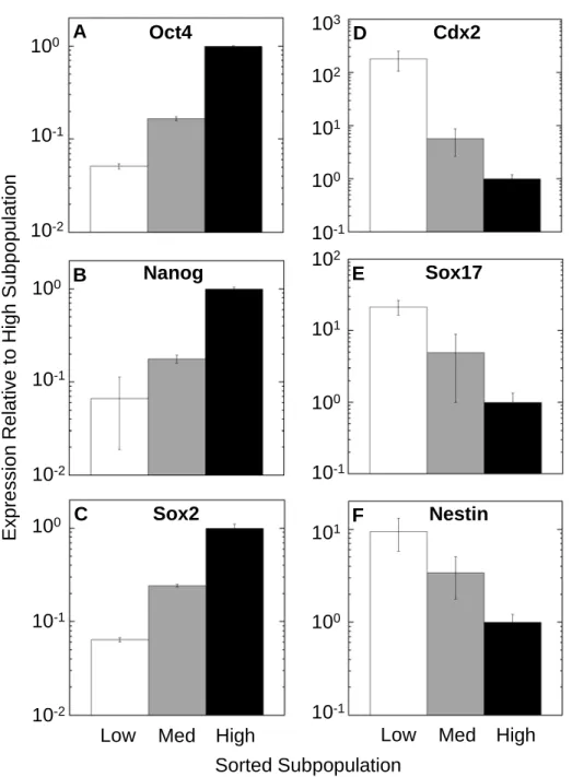
mRNA expression analysis by real-time PCR revealed cell-to-cell variability in
expression of pluripotency and early lineage markers within the clonal undifferentiated population. The sorted subpopulations showed that expression of pluripotency
associated genes Oct4, Nanog, and Sox2 [39–41] correlated positively with each other, with each gene showing a greater than an order of magnitude difference in expression between the high and low subpopulations (Figure 2-3A-C). Trophectodermal marker Cdx2 [135], early endodermal marker Sox17 [136] and neural stem cell marker Nestin [137] showed the reverse trend, with higher expression in the Low subpopulation. Expression of Cdx2 was 200x higher, Sox17 20x higher, and Nestin 10x higher in the Low subpopulation than in the High subpopulation (Figure 2-3D-F), despite all cells being Oct4-GFP+.
2.4.4 Populations differentiated from cells with higher initial Oct4-GFP levels had a greater fraction and number of cardiomyocytes
Prior studies have shown that ectopically increasing levels of Oct4 in ESC cause
48
in fate specification during early differentiation [48], [129]. We hypothesized that natural cell-to-cell variations in clonal mESC can affect the proclivity of individual cells in the population to differentiate into cells of different lineages, specifically that cells with higher Oct4-GFP at the initiation of differentiation would differentiate into
cardiomyocytes with greater efficiency. To test this, we sorted the cells as described above and immediately initiated differentiation by EB formation. In addition, as a control, a 4th population of cells that had simply been passed through the FACS instrument and all Oct4-GFP+ cells collected (‘All’) was differentiated in the same manner as well to
control for the effects of FACS. Each subpopulation was differentiated separately at 20% and 1% O2 throughout the entire differentiation protocol (Figure 2-4).
Despite the clonal nature of the starting cells, the subpopulations differentiated into cells positively stained for MF20 (anti-sacromeric myosin heavy chain, a cardiac specific antibody [112], [138], [139]) with different efficiencies. Cells differentiated at 20% O2 from the high subpopulation were 8.6% MF20+ compared to 4.2% from the low
subpopulation, and when differentiated at 1% O2, the high subpopulation produced 21.7% MF20+ compared to 14.1% MF20+ from the low subpopulation (Figure 2-4A). Following the trend, the med and all subpopulations both produced fractions in between the high and low subpopulations in both cases. These results also replicated earlier studies that low oxygen during differentiation increased cardiomyocyte formation [117], [140].
The positive correlation between initial Oct4-GFP fluorescence and differentiated MF20+ fractions was retained within each oxygen level by the total number of MF20+
49
cells produced (Figure 2-4B), which was calculated by multiplying the fraction of MF20+ cells by the total number of cells in each differentiated subpopulation (Figure 2-4C). There were no statistically significant differences in the total cell numbers in the populations that were differentiated at the same O2, although differentiation at 1% O2 produced less than 50% of the total cell number than differentiation at 20% O2. In addition, the fraction of residual Oct4-GFP+ cells (Figure 2-4D) and average fluorescence of these cells (data not shown) in each subpopulation were also only dependent on the differentiation O2 (with low O2 decreasing residual fraction of undifferentiated cells as previously shown [139, 140]). Finally, the trend of increasing MF20+ cells with increasing initial Oct4-GFP fluorescence were also corroborated by real-time PCR analysis (Figure 2-4E&F) of expression of cardiac genes Cardiac α-Actin [142] and cTnT [143].
2.4.5 Sorted subpopulations recapitulate the parental Oct4-GFP distribution when allowed to propagate in self-renewing culture
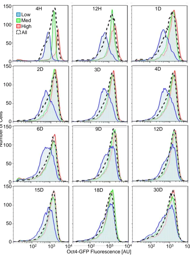
To investigate the stability of the cell-to-cell variation in Oct4-GFP expression intensities, we cultured the sorted subpopulations in self-renewing conditions and measured Oct4-GFP fluorescence for up to 30 days (Figure 2-5 & 6). Within 12 hr of sorting, the cells repopulated the entire range of fluorescence intensities of the parental population, and the high and med subpopulations had similar peak intensities 3 days after the sorting. However, the low subpopulation took much longer to reconstitute the parental
50
days after sorting with a broader distribution and proportionally more cells at lower intensities.
2.4.6 Recapitulation of the parental Oct4-GFP distribution erases subpopulation differences in pluripotency and early differentiation markers
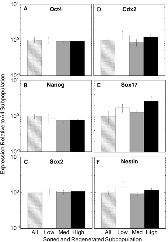
We then hypothesized that the regeneration of parental Oct4-GFP levels would also remove differences in pluripotency and early differentiation markers observed in the subpopulation immediately after sorting. RNA was isolated from cells allowed to self-renew for 30 days after sorting and levels of pluripotency and early differentiation genes were analyzed with real time PCR (Figure 2-7). The large differences in expression of all genes observed immediately after sorting (Figure 2-3) no longer existed in the subpopulations.
2.4.7 Recapitulation of the parental Oct4-GFP distribution removes the differences in subsequent differentiation to cardiomyocytes
Finally, we tested the efficiency of differentiation to cardiomyocytes of the regenerated subpopulations by differentiation at 1% O2 (Figure 2-8) There were no significant differences in the differentiated cells of the subpopulations in terms of fraction and number of MF20+ cells, and in the expression of Cardiac α-Actin and cTnT.
In addition, the fraction of MF20+ cells (averaging 23.7%) differentiated from cells that had undergone the 30d regeneration after sorting (Figure 2-8A) was higher than those differentiated immediately after FACS (Figure 2-4A). The fraction of MF20+ cells was
51
higher than in the cells differentiated from cells simply passed through FACS without sorting (all population) (14.1%) and comparable to the high subpopulation (21.7%). We postulate that this difference could be due to the harsh treatment of cells during FACS and the extended period that the cells were kept outside the incubators at the initiation of differentiation.

![Figure 2-2 Oct4-GFP variability cannot be explained by differences in cell cycle stage (A) Cell cycle analysis of undifferentiated and unsorted O4G mESC by fitting to a Watson cell cycle model [133] to identify cells in different cell cycle phases](https://thumb-eu.123doks.com/thumbv2/123doknet/14212426.482184/57.918.247.620.147.822/variability-explained-differences-analysis-undifferentiated-unsorted-identify-different.webp)