HAL Id: inserm-00156552
https://www.hal.inserm.fr/inserm-00156552
Submitted on 22 Jun 2007HAL is a multi-disciplinary open access archive for the deposit and dissemination of sci-entific research documents, whether they are pub-lished or not. The documents may come from teaching and research institutions in France or abroad, or from public or private research centers.
L’archive ouverte pluridisciplinaire HAL, est destinée au dépôt et à la diffusion de documents scientifiques de niveau recherche, publiés ou non, émanant des établissements d’enseignement et de recherche français ou étrangers, des laboratoires publics ou privés.
Reporting of radiographic methods in randomised
controlled trials assessing structural outcomes in
rheumatoid arthritis.
Gabriel Baron, Isabelle Boutron, Bruno Giraudeau, Philippe Ravaud
To cite this version:
Gabriel Baron, Isabelle Boutron, Bruno Giraudeau, Philippe Ravaud. Reporting of radiographic methods in randomised controlled trials assessing structural outcomes in rheumatoid arthritis.. Annals of the Rheumatic Diseases, BMJ Publishing Group, 2007, 66 (5), pp.651-7. �10.1136/ard.2006.063164�. �inserm-00156552�
Title: Reporting of radiographic methods in randomized controlled trials
assessing structural outcomes in rheumatoid arthritis
Authors: Gabriel Baron1, Isabelle Boutron1, Bruno Giraudeau2, Philippe Ravaud1
1
Gabriel Baron, MSc, Isabelle Boutron, MD, Philippe Ravaud, MD, PhD: INSERM U738, Paris, France ; Université Paris 7 Denis Diderot, UFR de Médecine, Paris, France ; AP-HP, Hôpital Bichat, Département d’Epidémiologie, Biostatistique et Recherche Clinique, Paris , France ;
2
Bruno Giraudeau, PhD: INSERM CIC 202, Tours, France.
Address correspondence and reprint requests to
Gabriel Baron, Département d'Epidémiologie Biostatistique et Recherche Clinique, INSERM
U738, Groupe Hospitalier Bichat-Claude Bernard, 46 rue Henri Huchard, 75018 Paris,
France.
Tel: +33 01 40 25 62 57
Fax: +33 01 40 25 67 73
E-mail: [email protected]
The Corresponding Author has the right to grant on behalf of all authors and does grant on
behalf of all authors, an exclusive licence (or non exclusive for government employees) on a
worldwide basis to the BMJ Publishing Group Ltd to permit this article (if accepted) to be
published in ARD and any other BMJPGL products and sublicences such use and exploit all
ABSTRACT
Objective. Because an increasing number of clinical trials evaluating disease modifying
anti-rheumatic drugs in rheumatoid arthritis (RA) emphasize radiographic outcomes as a primary
outcome, using a reproducible radiographic measure should be placed at a premium. We
aimed to evaluate the reporting of radiographic methods in randomized trials assessing
radiographic outcomes in RA.
Methods. We searched MEDLINE for randomized controlled trials assessing radiographic
outcomes published between January 1994 and December 2005 in general medical and
specialty journals with a high impact factor. One reader extracted data (radiographic
acquisition, assessment and reproducibility) using a standardized form.
Results. A total of 46 reports were included in the analysis. The mean (SD) methodological
quality scores on the Jadad scale (range 0-5) and the Delphi list (0-9) were 2.9 (1.2) and 6.4
(1.3), respectively. Use of a standardized procedure for the acquisition of the radiographs was
reported in 2 articles (4.3%). Two reports (4.3%) indicated that the quality of the radiographs
was evaluated. In 65.2% of the reports, 2 or more radiographic scores were used. Reporting of
radiographic assessment was well detailed for number of readers (91.3%), information on
readers (71.7%), blinding (91.4%) and how films were viewed (74.0%). The reproducibility
of the reading was reported in 39.1% of the articles.
Conclusion. Our results highlight that the reporting of results of randomized controlled trials
of radiographic outcomes in RA shows great variability in radiographic scores used and that
reporting of radiographic methods could be improved upon, especially the acquisition
procedure and the reproducibility of the reading.
Key words: rheumatoid arthritis, randomized controlled trials, reproducibility, radiographic
Rheumatoid arthritis (RA) is the most common chronic inflammatory joint disease
andis responsible for symptomatic manifestations (e.g., functional status, pain) and structural
damage (i.e., damage of the articular cartilage and bone) (1). The use of disease-modifying
anti-rheumatic drugs has increased for RA (2). Assessing such treatments requires the
measurement of structural outcomes in randomized controlled trials to demonstrate a
reduction or a retardation of disease progression.
Radiography provides an objective measure of the extent of anatomical joint damage.
It can be used to assess the severity of the structural destruction, to follow the course of the
disease and to establish effects of treatment (3). This highly accepted technique is widely
available and provides a permanent record of the structural image, allowing for comparison
over time and rereading if necessary (4, 5). The assessment of radiographic outcomes for
evaluating drug efficacy was recommended for the management of RA in controlled trials (6,
7) and the radiographic outcome is often used as a primary endpoint for assessing structural
severity (8).
The reproducibility (i.e., the extent to which repeated measurements on the same
subject yield the same results) is one of the prerequisites for a primary outcome (9-11). With
radiography, measurements can be biased and their precision compromised by 2
well-identified sources of variability -- image acquisition and assessment -- which can be a serious
limitation in its use. The image variability due to differences in acquisition processes is a
major concern. Because many parameters can affect the appearance of the radiographs (i.e.,
positioning for radiographs, film exposure and resolution, reproducibility), standardization of
the acquisition procedure is needed (12, 13). Similarly, radiographic assessment could be
influenced by many known parameters (e.g., the scoring method, the number of readers, films
Evaluating the reproducibility of an outcome measure theoretically supposes a detailed
reporting of the methods used to measure it. The CONSORT (Consolidated Standards of
Reporting Trials) statement has recommended the reporting of methods used “to enhance the quality of measurements”, especially when considering the primary outcome (15), including
how the outcome was measured and what steps were used to increase the reliability.
The purpose of this study was to evaluate the reporting of radiographic methods in
randomized controlled trials assessing radiographic outcomes in RA. We investigated
particularly radiographic acquisition, radiographic assessment and how the reproducibility
MATERIALS AND METHODS
Search strategy for identification of studies
The search method has been detailed elsewhere (16). Briefly, we searched MEDLINE and the
Cochrane Central Register of Controlled Trials for randomized controlled trials assessing
radiographic outcomes in RA published between January 1994 and December 2005 in general
medical and specialty journals with a high impact factor (Appendix A). We chose these
journals because a high impact factor is a good predictor of high methodological quality of
journal articles (17) and because our goal was not to be exhaustive but, rather, to raise
awareness of the reporting of radiographic assessment and acquisition. Potentially relevant
articles were selected on the basis of the title, abstract and keywords by one reader (GB) using
the following criteria: study population of adults aged 18 years or older, randomized
controlled trial, and presence of at least 1 radiographic outcome evaluated by scheduled
radiography. When duplicate publications of a trial existed, only the main publication was
included (i.e., when a report of the same trial appeared twice or more, the report of the
principal analysis planned by the protocol was selected). Reports of extended follow-up trials,
analyses of multiple trials and subgroup analyses were excluded. When the abstract indicated
that the article might be relevant, the entire paper was obtained.
Data extraction
One author (GB) extracted all the data using a standardized form. From each article, data were
(I) Characteristics of the included articles. Data were searched for number of centers, sample
size, and methodological quality scores (Jadad scale (18) and the Delphi list (19)). The
reviewer also determined whether the radiographic outcome was clearly defined as the
primary endpoint.
(II) Radiographic acquisition. The reviewer extracted data on regions assessed (e.g., hand,
feet), number of radiographic sessions, time between baseline session and the last session, and
time between 2 radiographic sessions. The reviewer checked whether methods used to take
the radiographs were detailed: radiographic view (e.g., dorsovolar, anteroposterior), use of a
standardized procedure to improve positioning, description of the image quality (film
exposure and use of resolution films), and use of digitized image or not.
(III) Radiographic assessment. The reviewer collected data on which score was used for
primary and secondary radiographic analysis and determined whether information was included on the reader’s experience (e.g., it was explicitly stated that radiographs were read
by a well-known expert), background (e.g., radiologist) and identity (e.g., initials reported);
and whether there were multiple observers (i.e., methods of consensus to obtain a single score
described when appropriate). The reviewer also checked whether the assessment of the
radiographs was blinded (i.e., to treatment assignment or patient identity and clinical data).
He also determined how films were read (paired reading with chronological or random order).
(IV) Reproducibility. The reviewer recorded whether intra- and inter-reader reproducibility
was evaluated and how.
Analysis
Categorical variables were described with frequencies and percentages and quantitative
also performed. The first describes characteristics of the radiographic assessment in reports
published before 2000 versus reports published in 2000 and later and the second compares
reports with radiographic outcome defined as a primary endpoint and reports with a
nonradiographic primary outcome. All data analyses involved use of SAS for Windows,
RESULTS
Characteristics of the included articles
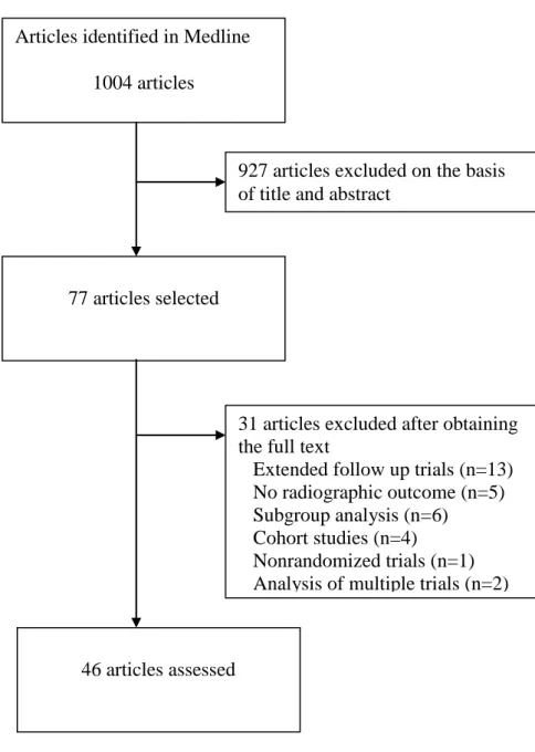
The search strategy generated 1004 citations. Forty-six studies were relevant according to the
title, abstract, and complete retrieval of the article (figure 1). Detailed references of the
selected articles are listed in appendix B.
Of the reports, 40 (87.0%) concerned multicenter studies. The median sample size of
the studies was 183 [20, 1446] patients. The mean (SD) methodological quality scores were
2.8 (1.0) for the Jadad scale and 6.5 (1.3) for the Delphi list. The radiographic outcome was
clearly defined as the primary endpoint in 26 reports (56.5%).
Radiographic acquisition
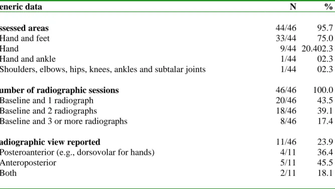
Both joints of hand and feet were the most frequently assessed areas (75.0%) (table 1). In
many reports (n=20, 43.5%), 2 radiographs (included baseline radiographs) were obtained
(table 1). The median time between the baseline session and the last session was 12 months
[5.5, 48 months] and the median time between 2 sessions was 53 weeks [24, 104 weeks].
The radiographic view (e.g., dorsovolar) was described in 11 reports (23.9%). Use of a
standardized radiographic procedure for optimal positioning was reported in 2 articles (4.3%).
The X-ray films were described as digitized in 3 reports (6.5%). The assessment of
radiographic quality was reported in only 2 articles (4.3%) and details on film resolution (e.g.,
single emulsion film) were provided in 2 (4.3%) reports.
Radiographic score
In 65.2% of the reports, 2 or more radiographic scores were used (table 2). Among scores
combining erosions and joint space narrowing, the Sharp score and the Larsen score
(including their modified versions) were the most represented methods (47.8% and 34.7%,
respectively).
Readers
Information on reader(s) (i.e., readers’ experience in evaluating radiographs, readers’ background or identity) were described in 33 articles (71.7%). Readers’ experience,
background and identity were reported in 17 (37.0%), 25 (54.3%), and 23 (50.0%) reports,
respectively (table 3).
In 4 reports (8.7%), the number of readers was impossible to determine. Radiographs
were read by at least 2 readers in 30 reports (65.2%) and by a single reader in 12 (26.1%).
Of the reports describing at least 2 readers, 22 described readers reading all the
radiographs (61.1%) and 1 all the readers did not read all the radiographs (3.3%), and in 7 the
reading was unclear (23.3%). When all the radiographs were read (n=22), a consensus method
to produce a single radiographic score was described in 8 reports (consensus with the same
readers for all disagreements [n=5], consensus for only important disagreements [n=2], or use
of another method of measurement [n=1]); in 10 reports, the radiographic score used came
from the mean score of the readers and from the lower score of the readers in 2 reports (in the
Blinding
Masked assessment of radiographs was described in 43 (93.5%) reports (Table 3). Blinding
was described to concern treatment assignment or patient identity (n=37, 86.0%) and clinical
data (n=11, 23.9%); 4 articles reported that the assessor was blinded, with no other details
given.
Reading session
In 12 articles (26.1%), no details were provided about the reading of the radiographs. When
reading was described (73.9%), paired reading in random order and paired reading in
chronological order were used in 18 (52.9%) and 15 (44.1%) articles, respectively. One article
(2.9%) used both random and chronological paired reading.
Reproducibility of the reading
Reproducibility of the reading was described in 18 articles (39.1%). Intrareader
reproducibility was reported in 9 (19.6) articles and inter-reader reproducibility in 13 (28.3) (4
articles [8.7%] reported both intra- and inter-reader reproducibility). Five articles (10.8%)
reported 2 reproducibility measures. Use of the intraclass correlation coefficient (n=8) and
coefficient of correlation (n=7) and reporting the smallest detectable difference (n=6) were the
most common methods used to assess reproducibility. Other methods were use of median
with minimum and maximum (n=1).
When comparing reports with radiographic outcome defined as a primary endpoint versus
other reports, we only found a major difference in the reporting of reproducibility (53.8%
versus 20.0%) (table 3).
No difference was found in the reporting of radiographic assessment between reports
DISCUSSION
Because an increasing number of clinical trials in RA emphasize radiographic
outcome as a primary outcome, investigating the reporting of the radiographic methods is
important. Our results show a great variability of the radiographic scores used and that
radiographic assessment was better described than radiographic acquisition. Measures of
reproducibility were reported in almost 40% of the assessed reports.
Reducing measurement error is an important objective, so use of a reproducible
radiographic measure should be placed at a premium. Reporting reproducibility measures and
describing methods of measurement are an essential step to confirm the validity of a measure
(15). Controlling the reproducibility of a radiographic measurement concerns 2 steps:
acquisition and assessment.
Although variability of acquisition can lead to measurement error (12, 13), only a few
articles described radiographic acquisition in detail. The standardization of the acquisition is
facilitated by centralized acquisition and entails training in radiographic acquisition and
similar conditions of assessment (e.g., positioning for radiography (20)). Even if
centralization of the acquisition is not always possible (e.g., because of multicenter studies or
financial reasons), the training of radiologists could be centralized to decrease the variability.
Radiographic assessment also depends on the technical quality of the radiographs, such as
film exposure or resolution (21). So, evaluation of radiographic quality could be a control
element that allows for limiting the number of nonassessable radiographs.
When designing a trial, the choice of a radiographic score for assessing structural
destruction can directly influence reproducibility (21, 22). When focusing on radiographic
because the radiographic score use has evolved over the years (21). However, in the last few
years, the modified Sharp and Larsen scores seem to have been preferentially used (23).
When considering other radiographic assessment characteristics, we noted that
blindness was well respected and described in our study. Similarly, recommendations on
reporting the number of observers and describing how to obtain a single measurement are
followed (e.g., mean radiographic score from 2 assessors to decrease measurement error (24)).
Specific training, as a calibration exercise, could be requested to come to an agreement when
there are more than 1 reader. Some other parameters could be better described. For instance,
experienced readers have been shown to have better agreement (22). A number of reports did
not state in what order radiographs were assessed, even though this parameter can influence
measurements (23, 25).
Almost 40% of the articles described the reproducibility of reading, even though
reporting agreement between observers is essential to assessing the quality of observations
(14, 26). If reproducibility was low or not assessed, then the use of radiographic outcomes as
a primary endpoint may be questionable. Most of the methods reported, such use of as
intraclass correlation coefficient or smallest detectable difference, were adapted for evaluating
reproducibility. However, the use of the correlation coefficient as a measure of agreement was
found in a surprising number of articles. Correlation coefficients measure the strength of an
association, not the concordance, and thus should be avoided in this indication (27).
Van der Heijde et al. give recommendations for improving the reporting of
radiographic methods in studies of radiographic outcomes (14). Because the optimal
interpretation of a radiograph also depends on conditions of acquisition, we suggest that the
radiographic acquisition should be more detailed or referenced in reports. Such a description
educated, whether the radiographs were digitized, whether the quality of films was assessed,
which view was chosen and what kind of film was used.
Our study is limited in that our search was restricted to articles published in
high-impact-factor journals. However, articles in low-high-impact-factor journals may have had the
same or lower methodological quality. Second, we pooled phase II and III studies with those
of a more epidemiological nature, even if they were also randomized controlled trials. The
degree of conformance with regulatory requirements and, consequently, required details and
rigor are probably more important for reporting phase II and phase III trials. Third, the
radiographic outcome was not always defined as a primary endpoint in our selected studies.
We could have presumed a more detailed description of the conditions of measurement if we
considered only reports in which the radiographic outcome was the primary endpoint, but
results were quite similar. We also decided to include all the studies in all the fields evaluated
because the primary outcome has been shown to differ between protocols and reports of
studies. In fact, Chan et al. demonstrated that primary outcome differed between protocol and
publication in 40% to 62% of trials (28, 29). Finally, some discrepancies may exist between
the real methods used and methods reported. Some deficiencies may appear simply because of
poor reporting, which does not necessarily mean that the methods were not applied (30, 31).
In fact, some details may not have been reported because the authors regarded them to be
standard and not necessary to mention (e.g., radiographic view or details about film
resolution). Similarly, authors are often forced by referees or editors to abbreviate the report,
and so important information such as details on acquisition technique are removed from
manuscripts. However, online appendices, supplemental information, or longer versions of a
paper could be provided. For example, the U.S. Food and Drug Administration provided in its
Website most of the required details of the radiographic methods used in reporting results of a
In conclusion, our results have highlighted that the reporting of results of randomized
controlled trials of radiographic outcomes measured by scheduled radiography in RA showed
a great variability of radiographic scores used and that reporting of radiographic methods
could be improved upon, especially the acquisition procedure and the reproducibility.
Investigators are encouraged to follow guidelines on reporting radiographic data in
REFERENCES
1. Symmons DP. Disease assessment indices: activity, damage and severity. Baillieres Clin Rheumatol 1995;9:267-85.
2. Lee DM, Weinblatt ME. Rheumatoid arthritis. Lancet 2001;358:903-11.
3. Soubrier M, Dougados M. How to assess early rheumatoid arthritis in daily clinical practice. Best Pract Res Clin Rheumatol 2005;19:73-89.
4. van der Heijde DM. Plain X-rays in rheumatoid arthritis: overview of scoring methods, their reliability and applicability. Baillieres Clin Rheumatol 1996;10:435-53.
5. Pincus T, Sokka T. Quantitative measures for assessing rheumatoid arthritis in clinical trials and clinical care. Best Pract Res Clin Rheumatol 2003;17:753-81.
6. Felson DT, Anderson JJ, Boers M, Bombardier C, Chernoff M, Fried B, et al. The American College of Rheumatology preliminary core set of disease activity measures for rheumatoid arthritis clinical trials. The Committee on Outcome Measures in Rheumatoid Arthritis Clinical Trials. Arthritis Rheum 1993;36:729-40.
7. Recommendations for the registration of drugs used in the treatment of rheumatoid arthritis. Group for the Respect of Ethics and Excellence in Science (GREES): rheumatoid arthritis section. Br J Rheumatol 1998;37:211-5.
8. Boers M, van der Heijde DM. Prevention or retardation of joint damage in rheumatoid arthritis: issues of definition, evaluation and interpretation of plain radiographs. Drugs
2002;62:1717-24.
9. Fleiss JL. Reliability of measurement. In: The design and analysis of clinical experiments. New York: Wiley; 1986. p. 1-32.
10. Giraudeau B, Ravaud P, Chastang C. Importance of reproducibility in responsiveness issues. Biom J 1998;40:685-701.
11. Vangeneugden T, Laenen A, Geys H, Renard D, Molenberghs G. Applying concepts of generalizability theory on clinical trial data to investigate sources of variation and their impact on reliability. Biometrics 2005;61:295-304.
12. Brower AC. Use of the radiograph to measure the course of rheumatoid arthritis. The gold standard versus fool's gold. Arthritis Rheum 1990;33:316-24.
13. Weissman BN. Imaging techniques in rheumatoid arthritis. J Rheumatol Suppl 1994;42:14-9.
14. van der Heijde D, Simon L, Smolen J, Strand V, Sharp J, Boers M, et al. How to report radiographic data in randomized clinical trials in rheumatoid arthritis: guidelines from a roundtable discussion. Arthritis Rheum 2002;47:215-8.
15. Altman DG, Schulz KF, Moher D, Egger M, Davidoff F, Elbourne D, et al. The revised CONSORT statement for reporting randomized trials: explanation and elaboration. Ann Intern Med 2001;134:663-94.
16. Baron G, Boutron I, Giraudeau B, Ravaud P. Violation of the intent-to-treat principle and rate of missing data in superiority trials assessing structural outcomes in rheumatic diseases. Arthritis Rheum 2005;52:1858-1865.
17. Lee KP, Schotland M, Bacchetti P, Bero LA. Association of journal quality indicators with methodological quality of clinical research articles. Jama 2002;287:2805-8.
18. Jadad AR, Moore RA, Carroll D, Jenkinson C, Reynolds DJ, Gavaghan DJ, et al. Assessing the quality of reports of randomized clinical trials: is blinding necessary? Control Clin Trials 1996;17:1-12.
19. Verhagen AP, de Vet HC, de Bie RA, Kessels AG, Boers M, Bouter LM, et al. The Delphi list: a criteria list for quality assessment of randomized clinical trials for conducting systematic reviews developed by Delphi consensus. J Clin Epidemiol 1998;51:1235-41. 20. van der Heijde DM. Radiographic imaging: the 'gold standard' for assessment of disease progression in rheumatoid arthritis. Rheumatology (Oxford) 2000;39 Suppl 1:9-16. 21. Boini S, Guillemin F. Radiographic scoring methods as outcome measures in rheumatoid arthritis: properties and advantages. Ann Rheum Dis 2001;60:817-27. 22. Sharp JT, Wolfe F, Lassere M, Boers M, Van Der Heijde D, Larsen A, et al. Variability of precision in scoring radiographic abnormalities in rheumatoid arthritis by experienced readers. J Rheumatol 2004;31:1062-72.
23. Ory PA. Interpreting radiographic data in rheumatoid arthritis. Ann Rheum Dis 2003;62:597-604.
24. Van Der Heijde D. Structural damage in rheumatoid arthritis as visualized through radiographs. Arthritis Res 2002;4 Suppl 2:S29-33.
25. van Der Heijde D, Boonen A, Boers M, Kostense P, van Der Linden S. Reading radiographs in chronological order, in pairs or as single films has important implications for the discriminative power of rheumatoid arthritis clinical trials. Rheumatology (Oxford) 1999;38:1213-20.
27. Lin LI. A concordance correlation coefficient to evaluate reproducibility. Biometrics 1989;45:255-68.
28. Chan AW, Hrobjartsson A, Haahr MT, Gotzsche PC, Altman DG. Empirical evidence for selective reporting of outcomes in randomized trials: comparison of protocols to published articles. Jama 2004;291:2457-65.
29. Chan AW, Krleza-Jeric K, Schmid I, Altman DG. Outcome reporting bias in
randomized trials funded by the Canadian Institutes of Health Research. Cmaj 2004;171:735-40.
30. Soares HP, Daniels S, Kumar A, Clarke M, Scott C, Swann S, et al. Bad reporting does not mean bad methods for randomised trials: observational study of randomised controlled trials performed by the Radiation Therapy Oncology Group. Bmj 2004;328:22-4. 31. Hill CL, LaValley MP, Felson DT. Discrepancy between published report and actual conduct of randomized clinical trials. J Clin Epidemiol 2002;55:783-6.
32. Sharp JT, Lidsky MD, Collins LC, Moreland J. Methods of scoring the progression of radiologic changes in rheumatoid arthritis. Correlation of radiologic, clinical and laboratory abnormalities. Arthritis Rheum 1971;14:706-20.
33. Sharp JT, Young DY, Bluhm GB, Brook A, Brower AC, Corbett M, et al. How many joints in the hands and wrists should be included in a score of radiologic abnormalities used to assess rheumatoid arthritis? Arthritis Rheum 1985;28:1326-35.
34. van der Heijde D, Dankert T, Nieman F, Rau R, Boers M. Reliability and sensitivity to change of a simplification of the Sharp/van der Heijde radiological assessment in rheumatoid arthritis. Rheumatology (Oxford) 1999;38:941-7.
35. van der Heijde DM, van Riel PL, Nuver-Zwart IH, Gribnau FW, vad de Putte LB. Effects of hydroxychloroquine and sulphasalazine on progression of joint damage in rheumatoid arthritis. Lancet 1989;1:1036-8.
36. Larsen A, Dale K, Eek M. Radiographic evaluation of rheumatoid arthritis and related conditions by standard reference films. Acta Radiol Diagn (Stockh) 1977;18:481-91.
37. Larsen A, Thoen J. Hand radiography of 200 patients with rheumatoid arthritis repeated after an interval of one year. Scand J Rheumatol 1987;16:395-401.
38. Larsen A. How to apply Larsen score in evaluating radiographs of rheumatoid arthritis in long-term studies. J Rheumatol 1995;22:1974-5.
39. Rau R, Wassenberg S, Herborn G, Stucki G, Gebler A. A new method of scoring radiographic change in rheumatoid arthritis. J Rheumatol 1998;25:2094-107.
40. Genant HK, Jiang Y, Peterfy C, Lu Y, Redei J, Countryman PJ. Assessment of rheumatoid arthritis using a modified scoring method on digitized and original radiographs. Arthritis Rheum 1998;41:1583-90.
Figure 1. Process of screening articles on randomized controlled trials of radiographic
outcomes in rheumatoid arthritis, for inclusion in the present analysis.
Articles identified in Medline
1004 articles
77 articles selected
927 articles excluded on the basis of title and abstract
31 articles excluded after obtaining the full text
Extended follow up trials (n=13) No radiographic outcome (n=5) Subgroup analysis (n=6)
Cohort studies (n=4)
Nonrandomized trials (n=1) Analysis of multiple trials (n=2)
Table 1. Generic data on radiographs described in reports of randomized controlled trials of
radiographic outcomes
Generic data N %
Assessed areas 44/46 95.7
Hand and feet 33/44 75.0
Hand 9/44 20.402.3
Hand and ankle 1/44 02.3
Shoulders, elbows, hips, knees, ankles and subtalar joints 1/44 02.3
Number of radiographic sessions 46/46 100.0
Baseline and 1 radiograph 20/46 43.5 Baseline and 2 radiographs 18/46 39.1 Baseline and 3 or more radiographs 8/46 17.4
Radiographic view reported 11/46 23.9
Posteroanterior (e.g., dorsovolar for hands) 4/11 36.4
Anteroposterior 5/11 45.5
Table 2. Radiographic outcomes in randomized controlled trials in rheumatoid arthritis
Radiographic outcomes N=46 %
Sharp score (original (32), modified by Sharp (33) or by van der Heijde’s (34, 35))
22 47.8
Erosion score and joint space narrowing score reported with composite score 16 34.8 Larsen score (original (36, 37) or modified version (38)) 18 39.0 Number of eroded joints counts 9 19.6 Erosion score and joint space narrowing score reported without composite
score
5 10.9
Number of erosions 3 06.5
Ratingen score (39) 2 04.3
Cumulative number of joints free of erosions at baseline in which at least one erosion developed during follow-up
2 04.3
Genant scoring method (40) 1 02.2
Table 3. Reporting of radiographic assessment in reports of randomized controlled trials with
radiographic outcomes in rheumatoid arthritis*
Reporting of All N=46 Reports with radiographic outcome as primary endpoint N=26 Reports with radiographic outcome not as primary endpoint N=20 Reports published before 2000 N=17 Reports published in 2000 and later N=29 Reader’s information 71.7 73.1 70.0 70.6 72.4 Experience 36.9 34.6 40.0 35.3 64.7 Background 54.4 57.7 50.0 52.9 55.2 Identity 50.0 50.0 50.0 52.9 48.3 Number of readers 91.3 96.2 85.0 94.1 89.7 1 26.1 19.2 35.0 29.4 24.2 >1 65.2 76.9 50.0 64.7 65.5 Not reported 08.7 03.9 15.0 05.9 10.3 Blinding 93.5 96.2 90.0 94.1 93.1 To treatment or patient identity 86.0 92.0 77.8 87.5 85.2 To clinical data 23.9 26.9 20.0 17.6 27.6
How films were
viewed 73.9 80.7 65.0 70.6 75.9
Reproducibility 39.1 53.8 20.0 35.3 41.4
Appendix A. Journals included in the search strategy for randomized controlled trials with
radiographic outcomes in rheumatoid arthritis
10 general and internal medicine journals (New England Journal of Medicine, Journal of the
American Medical Association, The Lancet, Annals of Internal Medicine, Annual Review of Medicine, Archives of Internal Medicine, British Medical Journal, American Journal of Medicine, Medicine, and Proceedings of the Association of American Physicians);
6 rheumatologic journals (Arthritis and Rheumatism, Seminars in Arthritis and Rheumatism,
Annals of Rheumatic Diseases, Rheumatology [Oxford, England], Journal of Rheumatology,
and Arthritis Care and Resarch);
6 orthopedic journals (Osteoarthritis and Cartilage/OARS, Arthroscopy, Journal of
Orthopeadic Research: Official Publication of the Orthopaedic Research Society, Journal of Bone and Joint Surgery, American Volume, Spine, and Gait & Posture);
6 rehabilitation journals (Archives of Physical Medicine and Rehabilitation, Supportive Care
in Cancer: Official Journal of the Multinational Association of Supportive Care in Cancer, Journal of Electromyography and Kinesiology: Official Journal of the International Society of Electrophysiological Kinesiology, Physical Therapy, Journal of Rehabilitation Research and Development, and Scandinavian Journal of Rehabilitation Medicine).
Appendix B. Detailed references of selected articles
1. Bathon JM, Martin RW, Fleischmann RM, Tesser JR, Schiff MH, Keystone EC, et al. A comparison of etanercept and methotrexate in patients with early rheumatoid arthritis. N Engl J Med 2000;343:1586-93.
2. Bluhm GB, Sharp JT, Tilley BC, Alarcon GS, Cooper SM, Pillemer SR, et al. Radiographic results from the Minocycline in Rheumatoid Arthritis (MIRA) Trial. J Rheumatol 1997;24:1295-302.
3. Boers M, Verhoeven AC, Markusse HM, van de Laar MA, Westhovens R, van Denderen JC, et al. Randomised comparison of combined step-down prednisolone, methotrexate and sulphasalazine with sulphasalazine alone in early rheumatoid arthritis. Lancet 1997;350:309-18.
4. Bresnihan B, Alvaro-Gracia JM, Cobby M, Doherty M, Domljan Z, Emery P, et al. Treatment of rheumatoid arthritis with recombinant human interleukin-1 receptor antagonist. Arthritis Rheum 1998;41:2196-204.
5. Capell HA, Madhok R, Hunter JA, Porter D, Morrison E, Larkin J, et al. Lack of radiological and clinical benefit over two years of low dose prednisolone for rheumatoid arthritis: results of a randomised controlled trial. Ann Rheum Dis 2004;63:797-803.
6. Choy EH, Kingsley GH, Khoshaba B, Pipitone N, Scott DL. A two year randomised controlled trial of intramuscular depot steroids in patients with established rheumatoid arthritis who have shown an incomplete response to disease modifying antirheumatic drugs. Ann Rheum Dis 2005;64:1288-93.
7. de Jong Z, Munneke M, Zwinderman AH, Kroon HM, Jansen A, Ronday KH, et al. Is a long-term high-intensity exercise program effective and safe in patients with rheumatoid arthritis? Results of a randomized controlled trial. Arthritis Rheum 2003;48:2415-24. 8. D'Elia HF, Larsen A, Mattsson LA, Waltbrand E, Kvist G, Mellstrom D, et al.
Influence of hormone replacement therapy on disease progression and bone mineral density in rheumatoid arthritis. J Rheumatol 2003;30:1456-63.
9. Dougados M, Combe B, Cantagrel A, Goupille P, Olive P, Schattenkirchner M, et al. Combination therapy in early rheumatoid arthritis: a randomised, controlled, double blind 52 week clinical trial of sulphasalazine and methotrexate compared with the single components. Ann Rheum Dis 1999;58:220-5.
10. Eggelmeijer F, Papapoulos SE, van Paassen HC, Dijkmans BA, Valkema R, Westedt ML, et al. Increased bone mass with pamidronate treatment in rheumatoid arthritis. Results of a three-year randomized, double-blind trial. Arthritis Rheum 1996;39:396-402.
11. Emery P, Breedveld FC, Lemmel EM, Kaltwasser JP, Dawes PT, Gomor B, et al. A comparison of the efficacy and safety of leflunomide and methotrexate for the treatment of rheumatoid arthritis. Rheumatology (Oxford) 2000;39:655-65.
12. Forre O. Radiologic evidence of disease modification in rheumatoid arthritis patients treated with cyclosporine. Results of a 48-week multicenter study comparing low-dose cyclosporine with placebo. Norwegian Arthritis Study Group. Arthritis Rheum 1994;37:1506-12.
13. Gerards AH, Landewe RB, Prins AP, Bruijn GA, Goei The HS, Laan RF, et al. Cyclosporin A monotherapy versus cyclosporin A and methotrexate combination therapy in patients with early rheumatoid arthritis: a double blind randomised placebo controlled trial. Ann Rheum Dis 2003;62:291-6.
14. Goekoop-Ruiterman YP, de Vries-Bouwstra JK, Allaart CF, van Zeben D, Kerstens PJ, Hazes JM, et al. Clinical and radiographic outcomes of four different treatment strategies in patients with early rheumatoid arthritis (the BeSt study): a randomized, controlled trial. Arthritis Rheum 2005;52:3381-90.
15. Hafstrom I, Ringertz B, Spangberg A, von Zweigbergk L, Brannemark S, Nylander I, et al. A vegan diet free of gluten improves the signs and symptoms of rheumatoid arthritis: the effects on arthritis correlate with a reduction in antibodies to food antigens. Rheumatology (Oxford) 2001;40:1175-9.
16. Hakkinen A, Sokka T, Kotaniemi A, Hannonen P. A randomized two-year study of the effects of dynamic strength training on muscle strength, disease activity, functional capacity, and bone mineral density in early rheumatoid arthritis. Arthritis Rheum 2001;44:515-22. 17. Hansen M, Podenphant J, Florescu A, Stoltenberg M, Borch A, Kluger E, et al. A randomised trial of differentiated prednisolone treatment in active rheumatoid arthritis. Clinical benefits and skeletal side effects. Ann Rheum Dis 1999;58:713-8.
18. Hasegawa J, Nagashima M, Yamamoto M, Nishijima T, Katsumata S, Yoshino S. Bone resorption and inflammatory inhibition efficacy of intermittent cyclical etidronate therapy in rheumatoid arthritis. J Rheumatol 2003;30:474-9.
19. Helliwell PS, O'Hara M, Holdsworth J, Hesselden A, King T, Evans P. A 12-month randomized controlled trial of patient education on radiographic changes and quality of life in
20. Jiang Y, Genant HK, Watt I, Cobby M, Bresnihan B, Aitchison R, et al. A multicenter, double-blind, dose-ranging, randomized, placebo-controlled study of recombinant human interleukin-1 receptor antagonist in patients with rheumatoid arthritis: radiologic progression and correlation of Genant and Larsen scores. Arthritis Rheum 2000;43:1001-9.
21. Keystone EC, Kavanaugh AF, Sharp JT, Tannenbaum H, Hua Y, Teoh LS, et al. Radiographic, clinical, and functional outcomes of treatment with adalimumab (a human anti-tumor necrosis factor monoclonal antibody) in patients with active rheumatoid arthritis
receiving concomitant methotrexate therapy: a randomized, placebo-controlled, 52-week trial. Arthritis Rheum 2004;50:1400-11.
22. Kirwan JR. The effect of glucocorticoids on joint destruction in rheumatoid arthritis. The Arthritis and Rheumatism Council Low-Dose Glucocorticoid Study Group. N Engl J Med 1995;333:142-6.
23. Klareskog L, van der Heijde D, de Jager JP, Gough A, Kalden J, Malaise M, et al. Therapeutic effect of the combination of etanercept and methotrexate compared with each treatment alone in patients with rheumatoid arthritis: double-blind randomised controlled trial. Lancet 2004;363:675-81.
24. Lipsky PE, van der Heijde DM, St Clair EW, Furst DE, Breedveld FC, Kalden JR, et al. Infliximab and methotrexate in the treatment of rheumatoid arthritis. Anti-Tumor Necrosis Factor Trial in Rheumatoid Arthritis with Concomitant Therapy Study Group. N Engl J Med 2000;343:1594-602.
25. Marchesoni A, Battafarano N, Arreghini M, Panni B, Gallazzi M, Tosi S.
Radiographic progression in early rheumatoid arthritis: a 12-month randomized controlled study comparing the combination of cyclosporin and methotrexate with methotrexate alone. Rheumatology (Oxford) 2003;42:1545-9.
26. Marchesoni A, Battafarano N, Arreghini M, Pellerito R, Cagnoli M, Prudente P, et al. Step-down approach using either cyclosporin A or methotrexate as maintenance therapy in early rheumatoid arthritis. Arthritis Rheum 2002;47:59-66.
27. Mottonen T, Hannonen P, Leirisalo-Repo M, Nissila M, Kautiainen H, Korpela M, et al. Comparison of combination therapy with single-drug therapy in early rheumatoid arthritis: a randomised trial. FIN-RACo trial group. Lancet 1999;353:1568-73.
28. Neustadt DH. Double blind evaluation of the long-term effects of etodolac versus ibuprofen in patients with rheumatoid arthritis. J Rheumatol Suppl 1997;47:17-22.
29. Pasero G, Priolo F, Marubini E, Fantini F, Ferraccioli G, Magaro M, et al. Slow progression of joint damage in early rheumatoid arthritis treated with cyclosporin A. Arthritis Rheum 1996;39:1006-15.
30. Proudman SM, Conaghan PG, Richardson C, Griffiths B, Green MJ, McGonagle D, et al. Treatment of poor-prognosis early rheumatoid arthritis. A randomized study of treatment with methotrexate, cyclosporin A, and intraarticular corticosteroids compared with
sulfasalazine alone. Arthritis Rheum 2000;43:1809-19.
31. Quinn MA, Conaghan PG, O'Connor PJ, Karim Z, Greenstein A, Brown A, et al. Very early treatment with infliximab in addition to methotrexate in early, poor-prognosis
rheumatoid arthritis reduces magnetic resonance imaging evidence of synovitis and damage, with sustained benefit after infliximab withdrawal: results from a twelve-month randomized, double-blind, placebo-controlled trial. Arthritis Rheum 2005;52:27-35.
32. Rau R, Herborn G, Menninger H, Sangha O. Radiographic outcome after three years of patients with early erosive rheumatoid arthritis treated with intramuscular methotrexate or parenteral gold. Extension of a one-year double-blind study in 174 patients. Rheumatology (Oxford) 2002;41:196-204.
33. Smolen JS, Kalden JR, Scott DL, Rozman B, Kvien TK, Larsen A, et al. Efficacy and safety of leflunomide compared with placebo and sulphasalazine in active rheumatoid
arthritis: a double-blind, randomised, multicentre trial. European Leflunomide Study Group. Lancet 1999;353:259-66.
34. St Clair EW, van der Heijde DM, Smolen JS, Maini RN, Bathon JM, Emery P, et al. Combination of infliximab and methotrexate therapy for early rheumatoid arthritis: a
randomized, controlled trial. Arthritis Rheum 2004;50:3432-43.
35. Strand V, Cohen S, Schiff M, Weaver A, Fleischmann R, Cannon G, et al. Treatment of active rheumatoid arthritis with leflunomide compared with placebo and methotrexate. Leflunomide Rheumatoid Arthritis Investigators Group. Arch Intern Med 1999;159:2542-50. 36. Svensson B, Boonen A, Albertsson K, van der Heijde D, Keller C, Hafstrom I. Low-dose prednisolone in addition to the initial disease-modifying antirheumatic drug in patients with early active rheumatoid arthritis reduces joint destruction and increases the remission rate: a two-year randomized trial. Arthritis Rheum 2005;52:3360-70.
37. Taylor PC, Steuer A, Gruber J, Cosgrove DO, Blomley MJ, Marsters PA, et al. Comparison of ultrasonographic assessment of synovitis and joint vascularity with
38. Valleala H, Laasonen L, Koivula MK, Mandelin J, Friman C, Risteli J, et al. Two year randomized controlled trial of etidronate in rheumatoid arthritis: changes in serum
aminoterminal telopeptides correlate with radiographic progression of disease. J Rheumatol 2003;30:468-73.
39. van der Heide A, Jacobs JW, Bijlsma JW, Heurkens AH, van Booma-Frankfort C, van der Veen MJ, et al. The effectiveness of early treatment with "second-line" antirheumatic drugs. A randomized, controlled trial. Ann Intern Med 1996;124:699-707.
40. van der Laan W, Molenaar E, Ronday K, Verheijen J, Breedveld F, Greenwald R, et al. Lack of effect of doxycycline on disease activity and joint damage in patients with
rheumatoid arthritis. A double blind, placebo controlled trial. J Rheumatol 2001;28:1967-74. 41. van Everdingen AA, Jacobs JW, Siewertsz Van Reesema DR, Bijlsma JW. Low-dose prednisone therapy for patients with early active rheumatoid arthritis: clinical efficacy, disease-modifying properties, and side effects: a randomized, double-blind, placebo-controlled clinical trial. Ann Intern Med 2002;136:1-12.
42. van Holten J, Pavelka K, Vencovsky J, Stahl H, Rozman B, Genovese M, et al. A multicentre, randomised, double blind, placebo controlled phase II study of subcutaneous interferon beta-1a in the treatment of patients with active rheumatoid arthritis. Ann Rheum Dis 2005;64:64-9.
43. van Jaarsveld CH, Jacobs JW, van der Veen MJ, Blaauw AA, Kruize AA, Hofman DM, et al. Aggressive treatment in early rheumatoid arthritis: a randomised controlled trial. On behalf of the Rheumatic Research Foundation Utrecht, The Netherlands. Ann Rheum Dis 2000;59:468-77.
44. van Schaardenburg D, Valkema R, Dijkmans BA, Papapoulos S, Zwinderman AH, Han KH, et al. Prednisone treatment of elderly-onset rheumatoid arthritis. Disease activity and bone mass in comparison with chloroquine treatment. Arthritis Rheum 1995;38:334-42. 45. Wassenberg S, Rau R, Steinfeld P, Zeidler H. Very low-dose prednisolone in early rheumatoid arthritis retards radiographic progression over two years: a multicenter, double-blind, placebo-controlled trial. Arthritis Rheum 2005;52:3371-80.
46. Willkens RF, Sharp JT, Stablein D, Marks C, Wortmann R. Comparison of
azathioprine, methotrexate, and the combination of the two in the treatment of rheumatoid arthritis. A forty-eight-week controlled clinical trial with radiologic outcome assessment. Arthritis Rheum 1995;38:1799-806.