The MIT Faculty has made this article openly available. Please share
how this access benefits you. Your story matters.
Citation Pluskal, Tomáš et al. "The biosynthetic origin of psychoactive
kavalactones in kava." Nature plants 5 (2019): 867-878.
As Published 10.1038/s41477-019-0474-0
Publisher Springer Science and Business Media LLC
Version Original manuscript
Citable link https://hdl.handle.net/1721.1/124692
Terms of Use Creative Commons Attribution-Noncommercial-Share Alike
The biosynthetic origin of psychoactive kavalactones in kava
Tomáš Pluskal1, Michael P. Torrens-Spence1, Timothy R. Fallon 1,2, Andrea De Abreu1,2,
Cindy H. Shi1,2, and Jing-Ke Weng1,2,*
1Whitehead Institute for Biomedical Research, 455 Main Street, Cambridge, MA 02142 USA. 2Department of Biology, Massachusetts Institute of Technology, Cambridge, MA 02139 USA.
Abstract
For millennia, humans have used plants for medicinal purposes. However, our limited understanding of plant biochemistry hinders the translation of such ancient wisdom into modern pharmaceuticals1. Kava (Piper methysticum) is a medicinal plant native to the Polynesian islands
with anxiolytic and analgesic properties supported by over 3,000 years of traditional use as well as numerous recent clinical trials2–5. The main psychoactive principles of kava, kavalactones, are
a unique class of polyketide natural products known to interact with central nervous system through mechanisms distinct from those of the prescription psychiatric drugs benzodiazepines and opioids6,7. Here we report de novo elucidation of the biosynthetic pathway of kavalactones,
consisting of seven specialized metabolic enzymes. Based on phylogenetic and crystallographic analyses, we highlight the emergence of two paralogous styrylpyrone synthases, both of which have neofunctionalized from an ancestral chalcone synthase to catalyze the formation of the kavalactone scaffold. Structurally diverse kavalactones are then biosynthesized by subsequent regio- and stereo-specific tailoring enzymes. We demonstrate the feasibility of engineering heterologous production of kavalactones and their derivatives in bacterial, yeast, and plant hosts, thus opening an avenue towards the development of new psychiatric therapeutics for anxiety disorders, which affect over 260 million people globally8.
Main text
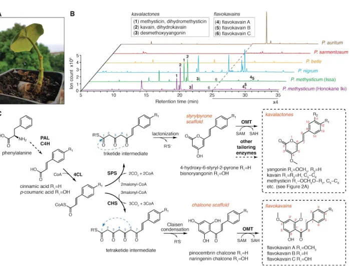
To investigate kavalactone biosynthesis, we first surveyed the occurrence of kavalactones in kava (Figure 1A) and four related Piper species by liquid-chromatography mass-spectrometry (LC-MS). While kavalactones are among the most abundant metabolites found in kava, they are absent in the other examined species (Figure 1B), suggesting their underlying biosynthetic machinery likely emerged after the diversification of the Piper genus. We also detected flavokavain A, B, and C in kava, which are specialized chalconoids with reported anti-cancer properties9,10 (Figure 1B).
Considering the structural relationship between kavalactones and flavokavains, which feature the styrylpyrone and chalcone backbones, respectively, we hypothesized that kavalactone biosynthesis likely involves styrylpyrone synthase (SPS), a polyketide synthase related to
chalcone synthase (CHS), which catalyzes the first committed enzymatic step in flavonoid biosynthesis and is ubiquitously present in all land plants11 (Figure 1C). Like CHS, SPS would
accept a 4-coumarate-CoA ligase (4CL)-generated hydroxycinnamoyl-CoA thioester (e.g., p-coumaroyl-CoA) as a starter substrate, and catalyzes iterative decarboxylative condensations of two-carbon ketone units derived from its co-substrate, malonyl-CoA. While CHS produces a tetraketide intermediate that further cyclizes via Claisen condensation to yield the chalcone backbone, SPS would yield a triketide intermediate that lactonizes to form the styrylpyrone backbone (Figure 1C). Additional tailoring enzymes, such as O-methyltransferases (OMTs) and oxidoreductases, are necessary to further decorate the initial scaffolds to give rise to the full repertoire of kavalactones and flavokavains present in kava plants (Figure 2).
Kava is a decaploid (2n = 10x = 130 chromosomes)12, rendering biosynthetic inquiry via
conventional genetic approaches infeasible. We therefore turned to a multi-omics-guided candidate gene approach13–15. We first identified three CHS-like genes in a kava transcriptome
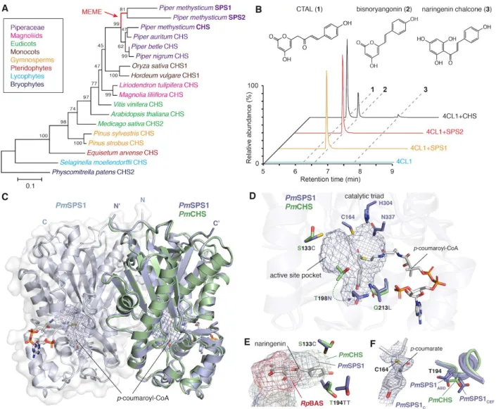
assembled de novo from leaf and root tissues (Supplementary Table 1). Phylogenetic analysis of these genes in the context of other land plant CHSs suggests that two of the three genes were likely derived from recent gene duplication events specific to kava (Figure 3A). In vitro enzyme assays using heterologously expressed and purified enzymes established that these two CHS-like
genes indeed encode functional SPSs. Both enzymes exclusively produce the triketide lactone bisnoryangonin from p-coumaroyl-CoA (Figure 3B), and are therefore named PmSPS1 and PmSPS2 hereafter. The assay also established that the third CHS-like gene encodes a bona fide CHS, which produces the expected tetraketide-derived naringenin chalcone, and is referred to as PmCHS hereafter (Figure 3B). It is noted that PmCHS also produces p-coumaroyltriacetic acid lactone (CTAL) and bisnoryangonin in addition to naringenin chalcone in vitro, although similar derailment products have been reported for a number of previously characterized CHSs16.
To assess the in vivo function of the three newly identified kava PKSs, we expressed each of them as stable transgenes in the Arabidopsis thalianaCHS-null mutant tt4-2 background17.
While PmCHS restored flavonoid biosynthesis in tt4-2, neither PmSPS1 nor PmSPS2 did, consistent with their respective in vitro biochemical activities (Figure 4). However, we could detect neither bisnoryangonin nor other styrylpyrones in transgenic Arabidopsis expressing PmSPS1 or PmSPS2. Similarly, Agrobacterium infiltration-mediated transient expression of PmSPS1 or PmSPS2 in the leaves of Nicotiana benthamiana18 also failed to yield any
styrylpyrone compounds. Upon careful examination, we instead identified a series of
benzalacetone-type metabolites that accumulated in transgenic Arabidopsis or Nicotiana plants expressing PmSPS1 (Supplementary Note 2). We conclude that styrylpyrones generated by heterologously expressed PmSPS1 are rapidly turned over by unknown enzymes present in Arabidopsis or Nicotiana to yield benzalacetones as breakdown products.
To probe the mechanistic basis for SPS neofunctionalization from the ancestral CHS, we solved the apo structure of PmCHS and the holo structure of PmSPS1 in complex with
p-coumaroyl-CoA by X-ray crystallography (Table 1). Both proteins are homodimers and share the canonical αβαβα thiolase fold typical for plant type III PKSs11 (Figure 3C). An evolutionary
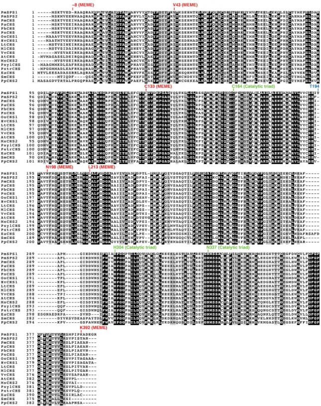
analysis using the Mixed Effects Model of Evolution (MEME)19 on the ancestral branch of kava
SPSs (Figure 3A) detected six amino acid residues under episodic diversifying selection (p<0.05) (Figure 5). Three of these substitutions, S133C, T198N, and Q213L, are mapped to the enzyme active site (Figure 3D). Notably, the T198N substitution was previously reported as one of the three mutations sufficient to convert CHS into a 2-pyrone synthase (2PS)20. We also observed an
S133C substitution, causing a reduction of the PmSPS1 active site volume compared to PmCHS in the direction along the polyketide chain elongation (Figure 3E). In comparison, the previously reported Rheum palmatum benzalacetone synthase (RpBAS) , which catalyzes only a single polyketide elongation21, features a further shortened active site compared to PmSPS1
(Figure 3E). These observations therefore support the notion that subtle changes of active site volume and shape of type III PKSs dictate the iterative cycles of polyketide elongation and alternative cyclization mechanisms22. In addition, we observed two different conformations of
T194 among the six protomers present in the asymmetric unit, suggesting the dynamic nature of the underlying loop and its potential role in the catalytic cycle of PmSPS1 (Figure 3F). One of the protomers in our PmSPS1-p-coumaroyl-CoA complex structure also captures a
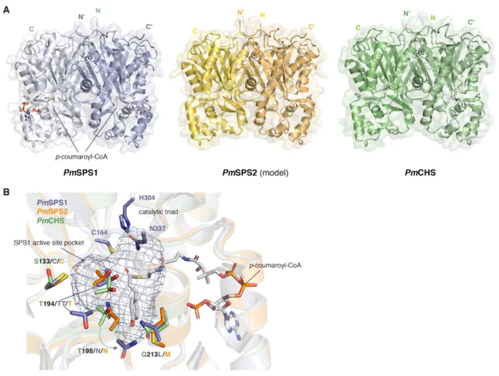
p-coumaroyl-monoketide intermediate covalently bound to the catalytic cysteine (Figure 3F), providing a rare glimpse of the active site configuration after the starter substrate loading step of the PKS catalytic cycle22. Homology modeling and sequence analysis of PmSPS2 suggest that its
active site contains a similar set of amino acid substitutions as observed in PmSPS1, except for the T194 insertion (Figure 5 and 6).
Structures of naturally occuring kavalactones implicate rich O-methylation reactions during their biosynthesis (Figure 2A). We hypothesized that kavalactone biosynthetic OMTs could have been recruited from a conserved OMT involved in plant phenylpropanoid
metabolism, e.g. the caffeic acid O-methyltransferase (COMT) in lignin biosynthesis23.
Combining phylogenomics and expression analysis, we identified six putative OMT candidate genes from kava transcriptomes (Supplementary Table 1). Since kavalactones contain four possible O-methylation sites (−OH groups attached to the C4, C10, C11, or C12 positions), we established a coupled enzymatic system using a 4CL enzyme cloned from kava (designated Pm4CL1) and PmSPS1 to produce five styrylpyrones featuring different aromatic ring
modification patterns derived from five hydroxycinnamic acids, namely cinnamic, p-coumaric, caffeic, ferulic, and umbellic acid (Supplementary Note 3). Subsequent OMT activity assays using recombinant kava OMTs revealed that two of the six kava OMT candidates showed activity against various styrylpyrones (Figure 7A), and were thus named kava
the most highly expressed enzymes in kava (Supplementary Table 1), and indeed is capable of methylating the −OH groups at the C4, C11, or C12 positions of the styrylpyrone backbone. On the other hand, PmKOMT2 activity is specific to the −OH group attached at the C10 position
(Figure 7A). Transient co-expression of PmKOMT1 together with PmSPS1 or PmSPS2 in Nicotiana resulted in the production of yangonin, a kavalactone with two methoxy groups at the C4 and C12 positions (Figure 7B), supporting its enzymatic activities observed in vitro. This result also suggests that O-methylation protects styrylpyrones produced by kava SPSs from being catabolized in Nicotiana.
The styrylpyrone backbone produced by SPSs contains two olefins (C5=C6 and C7=C8), which could be reduced to alkane at either position (e.g., kavain with C5–C6 or
7,8-dihydroyangonin with C7–C8) or at both positions (e.g., 7,8-dihydrokavain) (Figure 2A). Importantly, the reduction of the 5,6-olefin occurs in a stereospecific manner, suggesting that this reaction is likely enzymatic. Using similar approaches as described above, we identified and characterized twelve kava reductases that are homologous to various known reductases in plant specialized metabolism (Supplementary Table 1). By transgenic co-expression of each of these reductase candidates together with PmSPS1 and PmKOMT1 in Nicotiana, we identified a single reductase candidate that produced metabolites with masses corresponding to reduced
styrylpyrones (Supplementary Note 4). We thus named this enzyme kavalactone reductase 1 (PmKLR1). A combined enzyme assay using purified Pm4CL1, PmSPS1, PmKOMT1, and PmKLR1 enzymes starting from cinnamic acid successfully produced kavain in vitro
(Figure 8A). Furthermore, we determined using chiral chromatography that PmKLR1 produces only (R)-(+)-kavain, the same stereoisomer as present in kava root extract (Figure 8B). We further established that PmKLR1 can only accept unmethylated styrylpyrones, and is therefore competing with PmKOMT1 for the same substrates (Figure 9). This result provides an
explanation to the fact that only some of the major kavalactones contain the reduced C5−C6 bond (Figure 2A).
We hypothesized that the 7,8-saturated kavalactones could be derived from dihydro-hydroxycinnamoyl-CoA precursors already containing a reduced α−β bond. For example, two type III PKSs in the genera Pinus and Malus could accept
dihydro-p-coumaroyl-CoA as a starter substrate to produce 5-hydroxylunularic acid and phloretin, respectively24,25. To test this, we performed coupled Pm4CL1−PmSPS1 in vitro
enzyme assays using phloretic acid or phenylpropanoic acid as starter substrates, both of which contain a reduced α−β bond. These assays resulted in the production of the corresponding 7,8-saturated kavalactones, respectively, suggesting that Pm4CL1 and PmSPS1 can utilize substrates derived from dihydro-hydroxycinnamic acids with a reduced α−β bond, which are common phenylpropanoid metabolites in plants (Figure 10).
Methysticin-like kavalactones, which carry a methylenedioxy bridge at the C11−C12 position (Figure 2A), exhibit strong modulatory effects on human liver cytochromes P450, and thus contribute to the complex pharmacology of kava26. The CYP719 family of plant P450
enzymes is known to catalyze the formation of the methylenedioxy bridge moiety from vicinal methoxyl and hydroxyl groups, e.g. in the biosynthesis of berberine and etoposide27,28. We
identified a single gene of the CYP719 family in kava, PmCYP719A26 (Figure 8C), and hypothesized that it may catalyze the formation of methylenedioxy bridge using ferulic acid-derived styrylpyrone precursors carrying vicinal 11-methoxyl and 12-hydroxyl groups (Figure 8D). Indeed, co-expression of PmSPS1, PmKOMT1, and PmCYP719A26 in Nicotiana resulted in the transgenic production of a compound with molecular mass and retention time identical to that of methysticin (Figure 8E). Fragmentation pattern comparison with a
methysticin standard further identified this compound as 5,6-dehydro-7,8-dihydromethysticin (Supplementary Note 5). We therefore designated PmCYP719A26 as methysticin synthase 1 (PmMTS1).
Through OMT activity screen, we found that PmKOMT1 and PmKOMT2 also exhibit O-methylation activities against naringenin chalcone produced by CHS (Supplementary Note 6). Using enzyme assays and heterologous expression in Nicotiana, we confirmed that PmKOMT1 methylates the −OH groups at the C4 and C4’ positions on the chalcone scaffold, while
PmKOMT2 methylates the −OH group at the C2’ position (Figure 11). We thus established that the three known flavokavains (Figure 2B) are produced by the combinatorial activities of PmCHS, PmKOMT1, and PmKOMT2.
As a proof of concept, we examined the feasibility of kavalactone production in microbial hosts through the means of metabolic engineering. Using an operon-like construct co-expressing Pm4CL1 with PmSPS1 or PmSPS2 under the constitutive pGAP promoter, we could reconstitute the biosynthesis of the kavalactone precursor, bisnoryangonin, from supplemented p-coumaric acid in E.coli under shake flask condition (Figure 12A). Similarly, plasmid-based expression of the same combination of enzymes under the constitutive pTEF promoter in the yeast
Saccharomyces cerevisiae also resulted in bisnoryangonin production from supplemented p-coumaric acid under shake flask condition (Figure 12B).
The kava extracts display complex pharmacology in animals, which include modulation of GABAA and cannabinoid (CB1) receptors, blockade of voltage-gated sodium ion channels, reduced release and reuptake of several neurotransmitters, and interactions with monoamine oxidase B and liver cytochromes P4504,5,26. The capability to produce structurally diverse
kavalactones and flavokavains using their biosynthetic enzymes described herein will help delineate the action mechanism and structure-function relationship of kavalactones. Furthermore, combinatorial biosynthesis using additional tailoring enzymes from other sources will further enable diversification of kavalactones for novel biomedical applications. In light of several major challenges facing modern society, such as anxiety, opioid crisis, and the scarcity of effective treatments for psychiatric disorders, kavalactones and their derivatives present a promising class of psychoactive molecules to potentially answer these unmet needs.
Materials and Methods
Chemicals and reagents. Kavalactone standards (yangonin, methysticin, desmethoxyyangonin, 7,8-dihydrokavain, and 7,8-dihydromethysticin) were obtained from AvaChem Scientific. p-Coumaroyl-CoA was obtained from MicroCombiChem. Naringenin was obtained from AK Scientific. (±)-Kavain, malonyl-CoA, trans-cinnamic acid-d6, and other cofactors and reagents were obtained from Sigma-Aldrich.
RNA extraction and cDNA template preparation. Total RNA was extracted separately from kava root and leaf tissue using the RNeasy Plant Mini Kit (QIAGEN). First-strand cDNAs were synthesized by RT–PCR from the total RNA samples as templates using the SuperScript III First-Strand Synthesis System with the oligo(dT)20 primer (Thermo Fisher Scientific). Transcriptome sequencing and assembly. The transcriptome library preparation and
sequencing were performed at the Beijing Genomics Institute using the standard BGISEQ-500 RNA sample preparation protocol. The libraries were sequenced as 50×50 bp (PE-50) reads on the BGISEQ-500 platform. Sequence reads (FASTQ files) from leaf and root samples were merged into a single set of reads, trimmed for sequencing adaptors using Trimmomatic29, and
assembled into a de novo transcriptome using Trinity30, resulting in 285,753 transcript sequences.
Within those, we identified 302,366 putative open reading frames using Transdecoder31. We
evaluated the transcriptome as 78.6% complete by the metric of Benchmarking Universal Single-Copy Orthologs (BUSCO)32. Gene expression statistics were determined using RSEM33.
Transcripts and predicted protein sequences were annotated with transcript-per-million (TPM) values and closest BLAST hits from the UniProtKB/Swiss-Prot database using in-house scripts. Transcriptome mining was performed on a local BLAST server34. In addition, two kava root
RNAseq datasets previously published by the PhytoMetaSyn Project35 (NCBI SRA accessions
SRX202785 and SRX202184) were assembled and used as complementary sources of candidate enzyme sequences. Existing RNAseq datasets of Piper auritum, Piper betle, and Piper nigrum (NCBI SRA accessions ERX2099199, SRX691517, and SRX890122) were also assembled into de novo transcriptomes to obtain their corresponding CHS sequences.
Sequence alignment and phylogenetic analyses. Sequence alignments were performed using the MUSCLE36 algorithm in MEGA737. Evolutionary histories were inferred by using the
Maximum Likelihood method based on the JTT matrix-based model38. Bootstrap values were
Evolutionary analysis. The Mixed Effects Model of Evolution (MEME) analysis19 was
performed using the Hypothesis Testing using Phylogenies (HyPhy) package39, starting from
codon-aligned CHS nucleotide sequences and the corresponding phylogenetic tree. The p-value threshold was set to 0.05.
Cloning of candidate genes from cDNA. Phusion High-Fidelity DNA Polymerase (Thermo Fisher Scientific) was used for PCR amplifications from kava cDNA. Gibson assembly was used to clone the amplified genes into target vectors40. Restriction enzymes and Gibson assembly
reagents were purchased from New England Biolabs. Oligonucleotide primers were purchased from Integrated DNA Technologies. All primers used for cloning are listed in Supplementary Table 3.
Protein expression and purification. Candidate genes were cloned into pHis8-4, a bacterial expression vector containing an N-terminal 8×His tag followed by a tobacco etch virus (TEV) cleavage site for recombinant protein production in E. coli. Proteins were expressed in the BL21(DE3) E. coli strain cultivated in terrific broth (TB) and induced with 0.1 mM isopropyl β-D-1-thiogalactopyranoside (IPTG) overnight at 18 °C. For the COMTL enzyme screen, crude protein extracts were prepared from E. coli cultures using Bacterial Protein Extraction Reagent (B-PER, Thermo Fisher Scientific). For protein purification, E. coli cells were harvested by centrifugation, resuspended in 150 mL lysis buffer (50 mM Tris pH 8.0, 500 mM NaCl, 30 mM imidazole, 5 mM DTT), and lysed with five passes through an M-110L microfluidizer
(Microfluidics). The resulting crude protein lysate was clarified by centrifugation (19,000 g, 1 h) prior to QIAGEN nickel–nitrilotriacetic acid (Ni–NTA) gravity flow chromatographic
purification. After loading the clarified lysate, the Ni–NTA resin was washed with 20 column volumes of lysis buffer and eluted with 1 column volume of elution buffer (50 mM Tris pH 8.0, 500 mM NaCl, 300 mM imidazole, 5 mM DTT). 1 mg of His-tagged TEV protease41 was added
to the eluted protein, followed by dialysis at 4 °C for 16 h in dialysis buffer (50 mM Tris pH 8.0, 500 mM NaCl, 5 mM DTT). After dialysis, protein solution was passed through Ni–NTA resin to remove uncleaved protein and His-tagged TEV. The recombinant proteins were further
purified by gel filtration on a fast protein liquid chromatography (FPLC) system (GE Healthcare Life Sciences). The principal peaks were collected, verified by SDS–PAGE, and dialyzed into a storage buffer (12.5 mM Tris pH 8.0, 50 mM NaCl, 5 mM DTT). Finally, proteins were
concentrated to >10 mg/mL using Amicon Ultra-15 Centrifugal Filters (Millipore). The PmSPS1, PmSPS2, PmCHS, and PmKLR1 proteins purified as homodimers, while Pm4CL1, PmKOMT1 and PmKOMT2 purified as monomers.
In vitro enzyme assays. Enzyme assays were performed in 50 mM potassium phosphate buffer,
pH 7.6 containing 5 mM MgCl2, 3 mM adenosine triphosphate (ATP), 1 mM coenzyme A, 3 mM malonyl-CoA, and 0.25 mM initial substrate (typically, cinnamic acid or p-coumaric acid). Recombinant enzymes were added to final concentration of 10 µg/mL. For OMT assays,
additional 3 mM S-adenosyl-methionine was added. For reductase assays, additional 5 mM reduced nicotinamide adenine dinucleotide phosphate (NADPH) was added. Reactions were incubated at 30 °C until completion (typically overnight), followed by the addition of methanol to 50% final concentration. Samples were centrifuged (13,000 g, 20 min) and supernatants were collected for LC-MS analyses.
Transgenic Arabidopsis. Kava PKS genes were cloned into the pEarleyGate 100 vector42 and
transformed into Agrobacterium tumefaciens strain GV3130. The Arabidopsis thaliana tt4-2 mutant17 was then transformed using the Agrobacterium-mediated floral dip method43.
Transformants were selected by spraying with Finale (contains 11.33% glufosinate ammonium; Bayer) diluted 1:500 in water. Selection was repeated in each subsequent generation and
experiments were performed in the T2 or T3 generations.
qRT-PCR analysis. qRT-PCR reactions were performed on a QuantStudio 6 Flex system (Thermo Fisher Scientific) using SYBR Green Master Mix (Thermo Fisher Scientific) and primers listed in Supplementary Table 3. Gene expression Ct values were normalized using the reference gene At1g1332044.
Plant metabolite extraction. Approximately 100 mg of plant leaf tissue was dissected,
transferred into grinding tubes containing ~15 zirconia/silica disruption beads (2 mm diameter; Research Products International), and snap-frozen in liquid nitrogen. The frozen samples were homogenized twice on a TissueLyser II (QIAGEN). Metabolites were extracted using 5–10 volumes (w/v) of 50% methanol at 55 °C for 1 h. Extracts were centrifuged twice (13,000 g, 20 min) and supernatants were collected for LC-MS analysis.
LC-MS analysis. LC was conducted on a Dionex UltiMate 3000 UHPLC system (Thermo Fisher Scientific), using water with 0.1% formic acid as solvent A and acetonitrile with 0.1% formic acid as solvent B. Reverse phase separation of analytes was performed on a Kinetex C18 column, 150 × 3 mm, 2.6 μm particle size (Phenomenex). Chiral chromatography was performed on a Lux i-Amylose-1 column, 250 × 4.6 mm, 5 µm particle size (Phenomenex). The column oven was held at 30 °C. Most injections were eluted with a gradient of 5–60% B for 9 min, 95% B for 3 min, and 5% B for 3 min, with a flow rate of 0.7 mL/min. The initial analysis of Piper species leaf extracts was performed on a gradient of 5–80% B for 40 min, 95% B for 4 min, and 5% B for 5 min, with a flow rate of 0.8 mL/min. Chiral chromatography was performed on a gradient of 5–95% B for 30 min, 95% B for 5 min, and 5% B for 10 min, with a flow rate of 1 mL/min. MS analyses for the OMT enzyme screen and for chiral chromatography were performed on a TSQ Quantum Access Max mass spectrometer (Thermo Fisher Scientific) operated in positive ionization mode with full scan range of 100–400 m/z. Other MS analyses and plant metabolic profiling were performed on a high-resolution Q-Exactive benchtop Orbitrap mass spectrometer (Thermo Fisher Scientific) operated in positive ionization mode with full scan range of 100–1000 m/z and top 5 data-dependent MS/MS scans. Raw LC-MS data were
analyzed using XCalibur (Thermo Fisher Scientific), MZmine 245, and MetaboAnalyst46. The
SIRIUS tool was used to interpret MS/MS spectra47,48. Identified metabolites are listed in
Supplementary Table 2.
X-ray crystallography. The purified SPS1 protein (17.1 mg/mL) was incubated with 4 mM p-coumaroyl-CoA for 1 h prior to setting crystal trays. Crystals of PmSPS1 were obtained after 4
days at 21 °C in hanging drops containing 0.8 μL of protein solution and 0.8 μL of reservoir solution (50 mM HEPES pH 7.5, 10% w/v PEG 8000, 4% v/v ethylene glycol). Crystals were frozen in reservoir solution with ethylene glycol concentration increased to 15% (v/v). SPS1 X-ray diffraction data were collected on the 24-ID-C beam line equipped with a Pilatus-6MF pixel array detector (Advanced Photon Source, Argonne National Laboratory). Crystals of the purified PmCHS protein (17.6 mg/mL) were obtained from a screening plate after 4 days at 4°C in sitting drops containing 150 nL protein solution and 150 nL of reservoir solution (100 mM HEPES pH 7.0, 10% w/v PEG 6000). CHS X-ray diffraction data were collected on the 24-ID-E beam line equipped with an Eiger-16M pixel array detector (Advanced Photon Source, Argonne National Laboratory). Diffraction intensities were indexed and integrated with iMosflm49 and scaled with SCALA50. Initial phases were determined by molecular replacement
using Phaser51 in PHENIX52, using search models generated from protein sequences on the
Phyre2 server53. Subsequent structural building and refinements were conducted in PHENIX.
Coot was used for graphical map inspection and manual tuning of atomic models54. Root mean
square deviation (RMSD) of PmSPS1 and PmCHS structures was calculated using the Structure comparison function in PHENIX. Crystallography statistics are listed in Table 1.
Protein structure modeling and rendering. The PmSPS2 structure model was generated on the Phyre2 server using the PmSPS1 X-ray structure as a modeling template53. The active site
pocket in the PmSPS1 structure was determined using KVFinder55. Molecular graphics were
rendered with PyMOL (Schrödinger).
Transient expression in Nicotiana benthamiana. Target kava genes were cloned into the pEAQ-HT vector56,57 and transformed into the ElectroMAX Agrobacterium tumefaciens strain
LBA4404 (Invitrogen). Bacteria were cultivated at 30 °C to OD600 of 1.5 in 50 mL of YM medium (0.4 g/L yeast extract, 10 g/l mannitol, 0.1 g/L NaCl, 0.2 g/L MgSO4·7H2O, 0.5 g/L K2HPO4·3H2 O), washed with 0.5×PBS buffer (68 mM NaCl, 1.4 mM KCl, 5 mM Na2HPO4, 0.9 mM KH2PO4), and resuspended in 0.5×PBS buffer to OD600 of 0.8. For co-expressing multiple genes, individual A. tumefaciens cultures containing the transgene constructs were
grown, pelleted, and washed separately, and finally mixed to reach the final OD600 of 0.8 for each culture. 1 mL of the final culture was used to infiltrate the underside of 5-6 weeks old
N. benthamiana leaves. Leaves were harvested 5 days post infiltration for metabolite extraction. Expression in Escherichia coli.Pm4CL1, PmSPS1, and PmSPS2 were cloned into a
custom-made, operon-like expression vector pJKW1565. The genes were preceded by a constitutively expressed pGAP promoter of the E. coli glyceraldehyde-3-phosphate
dehydrogenase (GAPDH)58, and each gene was preceded by a 23-bp ribosome binding site
(RBS) sequence59. tSPY was used as a terminator60. The constructed plasmids (pJKW1574
containing the Pm4CL1 + PmSPS1 operon and pJKW1658 containing the Pm4CL1 + PmSPS2 operon) were transformed into the BW27784 E. coli strain61 and cultivated at 30 °C overnight in
TB medium supplied with 1 mM p-coumaric acid, followed by metabolite extraction from the culture fluid. An aliquot of the bacterial culture was mixed with an equal volume of methanol (50% final methanol concentration), centrifuged twice (13,000 g, 20 min) and supernatants were collected for LC-MS analysis. Since bisnoryangonin standard was not available, its
concentrations were estimated using naringenin as the calibration-curve standard.
Expression in Saccharomyces cerevisiae. The p42xTEF 2µ plasmids with various auxotrophic growth markers for constitutive expression in yeast were used62. The Pm4CL1 gene was cloned
into p425TEF (plasmid pJKW1413), while PmSPS1 and PmSPS2 were cloned into p426TEF (plasmids pJKW1538 and pJKW1547, respectively). The constructed plasmids were transformed into the BY4743 yeast strain63. Yeast cells were cultivated at 30 °C for 2 days in Yeast Nitrogen
Base (YNB) + histidine + dextrose medium supplied with 2 mM p-coumaric acid, followed by metabolite extraction from the culture fluid. An aliquot of the yeast culture was mixed with an equal volume of methanol (50% final methanol concentration), centrifuged twice (13,000 g, 20 min) and supernatants were collected for LC-MS analysis.
References
1. Li, F.-S. & Weng, J.-K. Demystifying traditional herbal medicine with modern approach. Nature Plants3, 17109 (2017).
2. Lebot, V. & Lèvesque, J. The origin and distribution of kava (Piper methysticum Forst. f., Piperaceae): a phytochemical approach. Allertonia5, 223–281 (1989).
3. Singh, Y. N. Kava: an overview. J. Ethnopharmacol.37, 13–45 (1992).
4. Cairney, S., Maruff, P. & Clough, A. R. The neurobehavioural effects of kava. Aust. N. Z. J. Psychiatry36, 657–662 (2002).
5. Sarris, J., LaPorte, E. & Schweitzer, I. Kava: a comprehensive review of efficacy, safety, and psychopharmacology. Aust. N. Z. J. Psychiatry45, 27–35 (2011).
6. Chua, H. C. et al. Kavain, the Major Constituent of the Anxiolytic Kava Extract, Potentiates GABAA Receptors: Functional Characteristics and Molecular Mechanism. PLOS ONE11, e0157700 (2016).
7. Jamieson, D. D. & Duffield, P. H. The antinociceptive actions of kava components in mice. Clin. Exp. Pharmacol. Physiol.17, 495–507 (1990).
8. Depression and other common mental disorders: global health estimates. (Geneva: World Health Organization, 2017).
9. Zi, X. & Simoneau, A. R. Flavokawain A, a novel chalcone from kava extract, induces apoptosis in bladder cancer cells by involvement of Bax protein-dependent and mitochondria-dependent apoptotic pathway and suppresses tumor growth in mice. Cancer Res.65, 3479–3486 (2005).
10. Zhou, P. et al. Flavokawain B, the hepatotoxic constituent from kava root, induces GSH-sensitive oxidative stress through modulation of IKK/NF-kappaB and MAPK signaling pathways. FASEB J. 24, 4722–4732 (2010).
11. Abe, I. & Morita, H. Structure and function of the chalcone synthase superfamily of plant type III polyketide synthases. Nat. Prod. Rep.27, 809–838 (2010).
12. Lebot, V., Aradhya, M. K. & Manshardt, R. M. Geographic survey of genetic variation in kava (Piper methysticum Forst. f. and P. wichmannii C. DC.). Pacific Science45, 169–185 (1991). 13. Torrens-Spence, M. P., Fallon, T. R. & Weng, J. K. A Workflow for Studying Specialized
Metabolism in Nonmodel Eukaryotic Organisms. in Methods in Enzymology 69–97 (2016).
14. Owen, C., Patron, N. J., Huang, A. & Osbourn, A. Harnessing plant metabolic diversity. Curr. Opin. Chem. Biol.40, 24–30 (2017).
15. Tatsis, E. C. & O’Connor, S. E. New developments in engineering plant metabolic pathways. Curr. Opin. Biotechnol.42, 126–132 (2016).
16. Akiyama, T., Shibuya, M., Liu, H. M. & Ebizuka, Y. p-Coumaroyltriacetic acid synthase, a new homologue of chalcone synthase, from Hydrangea macrophylla var. thunbergii. FEBS J.263, 834–839 (1999).
17. Burbulis, I. E., Iacobucci, M. & Shirley, B. W. A null mutation in the first enzyme of flavonoid biosynthesis does not affect male fertility in Arabidopsis. Plant Cell8, 1013–1025 (1996).
18. Sainsbury, F. & Lomonossoff, G. P. Transient expressions of synthetic biology in plants. Curr. Opin. Plant Biol.19, 1–7 (2014).
19. Murrell, B. et al. Detecting individual sites subject to episodic diversifying selection. PLoS Genet.8, e1002764 (2012).
20. Jez, J. M. et al. Structural control of polyketide formation in plant-specific polyketide synthases. Chem. Biol.7, 919–930 (2000).
21. Morita, H. et al. A structure-based mechanism for benzalacetone synthase from Rheum palmatum. Proc. Natl. Acad. Sci. U. S. A.107, 669–673 (2010).
22. Austin, M. B. & Noel, J. P. The chalcone synthase superfamily of type III polyketide synthases. Nat. Prod. Rep.20, 79–110 (2003).
23. Moinuddin, S. G. A. et al. Insights into lignin primary structure and deconstruction from Arabidopsis thaliana COMT (caffeic acid O-methyl transferase) mutant Atomt1. Org. Biomol. Chem.8,
3928–3946 (2010).
24. Eckermann, C. et al. Stilbenecarboxylate biosynthesis: a new function in the family of chalcone synthase-related proteins. Phytochemistry62, 271–286 (2003).
25. Gosch, C., Halbwirth, H. & Stich, K. Phloridzin: biosynthesis, distribution and physiological relevance in plants. Phytochemistry71, 838–843 (2010).
26. Li, Y. et al. Methysticin and 7,8-dihydromethysticin are two major kavalactones in kava extract to induce CYP1A1. Toxicol. Sci.124, 388–399 (2011).
27. Ikezawa, N. et al. Molecular cloning and characterization of CYP719, a methylenedioxy
bridge-forming enzyme that belongs to a novel P450 family, from cultured Coptis japonica cells. J. Biol. Chem.278, 38557–38565 (2003).
28. Lau, W. & Sattely, E. S. Six enzymes from mayapple that complete the biosynthetic pathway to the etoposide aglycone. Science349, 1224–1228 (2015).
29. Bolger, A. M., Lohse, M. & Usadel, B. Trimmomatic: a flexible trimmer for Illumina sequence data. Bioinformatics30, 2114–2120 (2014).
30. Grabherr, M. G. et al. Full-length transcriptome assembly from RNA-Seq data without a reference genome. Nat. Biotechnol.29, 644–652 (2011).
31. Haas, B. J. et al. De novo transcript sequence reconstruction from RNA-seq using the Trinity platform for reference generation and analysis. Nat. Protoc.8, 1494–1512 (2013).
32. Simão, F. A., Waterhouse, R. M., Ioannidis, P., Kriventseva, E. V. & Zdobnov, E. M. BUSCO: assessing genome assembly and annotation completeness with single-copy orthologs. Bioinformatics 31, 3210–3212 (2015).
33. Li, B. & Dewey, C. N. RSEM: accurate transcript quantification from RNA-Seq data with or without a reference genome. BMC Bioinformatics12, 323 (2011).
34. Priyam, A. et al. Sequenceserver: a modern graphical user interface for custom BLAST databases. bioRxiv 033142 (2015). doi:10.1101/033142
35. Xiao, M. et al. Transcriptome analysis based on next-generation sequencing of non-model plants producing specialized metabolites of biotechnological interest. J. Biotechnol.166, 122–134 (2013). 36. Edgar, R. C. MUSCLE: multiple sequence alignment with high accuracy and high throughput.
Nucleic Acids Res.32, 1792–1797 (2004).
37. Kumar, S., Stecher, G. & Tamura, K. MEGA7: Molecular Evolutionary Genetics Analysis Version 7.0 for Bigger Datasets. Mol. Biol. Evol.33, 1870–1874 (2016).
38. Jones, D. T., Taylor, W. R. & Thornton, J. M. The rapid generation of mutation data matrices from protein sequences. Comput. Appl. Biosci.8, 275–282 (1992).
39. Pond, S. L. K., Frost, S. D. W. & Muse, S. V. HyPhy: hypothesis testing using phylogenies. Bioinformatics21, 676–679 (2005).
40. Gibson, D. G. et al. Enzymatic assembly of DNA molecules up to several hundred kilobases. Nat. Methods6, 343–345 (2009).
41. Tropea, J. E., Cherry, S. & Waugh, D. S. Expression and purification of soluble His(6)-tagged TEV protease. Methods Mol. Biol.498, 297–307 (2009).
42. Earley, K. W. et al. Gateway-compatible vectors for plant functional genomics and proteomics. Plant J.45, 616–629 (2006).
43. Clough, S. J. & Bent, A. F. Floral dip: a simplified method forAgrobacterium-mediated transformation ofArabidopsis thaliana: Floral dip transformation of Arabidopsis. Plant J.16, 735–743 (1998).
44. Czechowski, T., Stitt, M., Altmann, T., Udvardi, M. K. & Scheible, W.-R. Genome-wide identification and testing of superior reference genes for transcript normalization in Arabidopsis. Plant Physiol.139, 5–17 (2005).
45. Pluskal, T., Castillo, S., Villar-Briones, A. & Oresic, M. MZmine 2: modular framework for processing, visualizing, and analyzing mass spectrometry-based molecular profile data. BMC Bioinformatics11, 395 (2010).
46. Xia, J., Sinelnikov, I. V., Han, B. & Wishart, D. S. MetaboAnalyst 3.0—making metabolomics more meaningful. Nucleic Acids Res.43, W251–W257 (2015).
47. Böcker, S., Letzel, M. C., Lipták, Z. & Pervukhin, A. SIRIUS: decomposing isotope patterns for metabolite identification. Bioinformatics25, 218–224 (2009).
48. Dührkop, K., Shen, H., Meusel, M., Rousu, J. & Böcker, S. Searching molecular structure databases with tandem mass spectra using CSI:FingerID. Proc. Natl. Acad. Sci. U. S. A.112, 12580–12585 (2015).
49. Battye, T. G. G., Kontogiannis, L., Johnson, O., Powell, H. R. & Leslie, A. G. W. \it iMOSFLM: a new graphical interface for diffraction-image processing with \it MOSFLM. Acta Crystallogr. D Biol. Crystallogr.67, 271–281 (2011).
50. Evans, P. Scaling and assessment of data quality. Acta Crystallogr. D Biol. Crystallogr.62, 72–82 (2006).
51. McCoy, A. J. Solving structures of protein complexes by molecular replacement with Phaser. Acta Crystallogr. D Biol. Crystallogr.63, 32–41 (2007).
52. Adams, P. D. et al. PHENIX: a comprehensive Python-based system for macromolecular structure solution. Acta Crystallogr. D Biol. Crystallogr.66, 213–221 (2010).
53. Kelley, L. A., Mezulis, S., Yates, C. M., Wass, M. N. & Sternberg, M. J. E. The Phyre2 web portal for protein modeling, prediction and analysis. Nat. Protoc.10, 845–858 (2015).
54. Emsley, P. & Cowtan, K. Coot: model-building tools for molecular graphics. Acta Crystallogr. D Biol. Crystallogr.60, 2126–2132 (2004).
55. Oliveira, S. H. P. et al. KVFinder: steered identification of protein cavities as a PyMOL plugin. BMC Bioinformatics15, 197 (2014).
56. Sainsbury, F., Thuenemann, E. C. & Lomonossoff, G. P. pEAQ: versatile expression vectors for easy and quick transient expression of heterologous proteins in plants. Plant Biotechnol. J.7, 682–693 (2009).
57. Peyret, H. & Lomonossoff, G. P. The pEAQ vector series: the easy and quick way to produce recombinant proteins in plants. Plant Mol. Biol.83, 51–58 (2013).
58. Charpentier, B., Bardey, V., Robas, N. & Branlant, C. The EIIGlc protein is involved in
glucose-mediated activation of Escherichia coli gapA and gapB-pgk transcription. J. Bacteriol.180, 6476–6483 (1998).
59. Olins, P. O. & Rangwala, S. H. A novel sequence element derived from bacteriophage T7 mRNA acts as an enhancer of translation of the lacZ gene in Escherichia coli. J. Biol. Chem.264,
16973–16976 (1989).
60. Chen, Y.-J. et al. Characterization of 582 natural and synthetic terminators and quantification of their design constraints. Nat. Methods10, 659–664 (2013).
61. Khlebnikov, A., Datsenko, K. A., Skaug, T., Wanner, B. L. & Keasling, J. D. Homogeneous
expression of the P(BAD) promoter in Escherichia coli by constitutive expression of the low-affinity high-capacity AraE transporter. Microbiology147, 3241–3247 (2001).
62. Mumberg, D., Müller, R. & Funk, M. Yeast vectors for the controlled expression of heterologous proteins in different genetic backgrounds. Gene156, 119–122 (1995).
63. Brachmann, C. B. et al. Designer deletion strains derived from Saccharomyces cerevisiae S288C: a useful set of strains and plasmids for PCR-mediated gene disruption and other applications. Yeast14, 115–132 (1998).
64. Ferrer, J. L., Jez, J. M., Bowman, M. E., Dixon, R. A. & Noel, J. P. Structure of chalcone synthase and the molecular basis of plant polyketide biosynthesis. Nat. Struct. Biol.6, 775–784 (1999). 65. Veit, M. & Pauli, G. F. Major flavonoids from Arabidopsis thaliana leaves. J. Nat. Prod.62,
1301–1303 (1999).
Acknowledgments
This work was supported by grants from the Edward N. and Della L. Thome Memorial Foundation, the Family Larsson-Rosenquist Foundation, and the National Science Foundation (CHE-1709616). T.P. is a Simons Foundation Fellow of the Helen Hay Whitney Foundation. J.K.W is supported by the Beckman Young Investigator Program, Pew Scholars Program in the Biomedical Sciences (grant number 27345), and the Searle Scholars Program (grant number 15-SSP-162). This work is based on research conducted at the Northeastern Collaborative Access Team (NE-CAT) beamlines, which are funded by the National Institute of General Medical Sciences from the National Institutes of Health (P41 GM103403). The Pilatus 6M detector on NE-CAT 24-ID-C beam line is funded by a NIH-ORIP HEI grant (S10 RR029205). This research used resources of the Advanced Photon Source, a U.S. Department of Energy
(DOE) Office of Science User Facility operated for the DOE Office of Science by Argonne National Laboratory under Contract No. DE-AC02-06CH11357. RNAseq service was provided free of charge by the Beijing Genome Institute in exchange for an evaluation of their
BGISEQ-500 sequencing platform. We thank Bryan Marotta for an introduction to kava, Chi Nguyen and Fadel A. Samatey for advice regarding crystallography, Gerald Fink for providing yeast strains and expression vectors, and Weng lab members for constructive comments. Kava plant photo courtesy of Randy Travis.
Author Contributions
T.P. and J.K.W. designed experiments. T.P. performed experiments, while M.P.T.S. assisted with cloning and crystallography and T.R.F. assisted with transcriptome assembly and LC-MS analyses. A.D.A. cloned genes and purified proteins. C.H.S. constructed expression vectors. T.P. analysed data. T.P. and J.K.W. wrote the paper.
Competing Interests
T.P. and J.K.W. have filed a patent application on metabolic engineering of kavalactones and flavokavains using the enzymes discovered in this study. J.K.W. is a co-founder, a member of the Scientific Advisory Board, and a shareholder of DoubleRainbow Biosciences, which develops biotechnologies related to natural products.
Figures
Figure 1. Chemotypes of select Piper species and the bifurcation of kavalactone and flavokavain biosynthesis from common hydroxycinnamoyl-CoA precursors in kava. A. A young root cutting of the Hawaiian kava cultivar Honokane Iki. B. LC-MS base peak plots of leaf tissue extracts from two different cultivars of kava (Pipermethysticum) and four other Piper species showing that kavalactones and flavokavains are major secondary metabolites in kava absent in the other examined Piper species. The intensity in the 25-35 min region was increased 4-fold to help visualize the flavokavain peaks. The flavokavain C (6) peak is present but below the plot baseline. C. Hypothesized mechanisms for
kavalactone and flavokavain biosynthesis. The polyketide cyclization mechanisms are denoted by blue arrows. PAL, phenylalanine ammonia-lyase; C4H, trans-cinnamate 4-monooxygenase; 4CL,
4-coumarate-CoA ligase; SPS, styrylpyrone synthase; CHS, chalcone synthase; OMT, O-methyltransferase; CoA, coenzyme A; SAM, S-adenosyl-L-methionine; SAH, S-adenosyl-L-homocysteine.
Figure 2. The chemical diversity of kavalactones and flavokavains. A. Chemical structures, formulas, and molecular masses of twenty known kavalactones. The six major kavalactones, which constitute over 96% kavalactone content in the plant rhizome, are highlighted within a dashed rectangle. B. Chemical structures, formulas, and molecular masses of three known flavokavains.
Figure 3. Mechanistic basis for the neofunctionalization of SPSs from ancestral CHS in kava. A. Maximum-likelihood phylogenetic analysis the three CHS-like sequences (PmSPS1, PmSPS2, and PmCHS) from kava with other CHS otholog sequences from select land plant lineages. Bootstrap values (based on 1000 replicates) are indicated at the tree nodes. The branch undergone episodic positive
selection as detected by the Mixed Effects Model of Evolution analysis19 is highlighted in red and denoted
with MEME. B. Combined LC-MS extracted ion chromatograms (XICs) of 273.076 m/z (naringenin chalcone and CTAL) and 231.065 m/z (bisnoryangonin) showing the in vitro activities of PmSPS1, PmSPS2, and PmCHS from a 4CL-coupled enzyme assay using p-coumaric acid as the starter substrate. C. An overlay of the PmCHS apo structure and the PmSPS1-p-coumaroyl-CoA holo structure
(RMSD=1.019 Å). Both proteins form homodimers (only a single protomer of PmCHS is shown). Active-site pocket is indicated with a mesh. D. Structural features of the PmSPS1 active site. The three active-site amino acid substitutions between PmSPS1 and PmCHS are highlighted. E. The C133 and
T194 residues of PmSPS1 dictate the size and shape of the PmSPS1 active site pocket in comparison to PmCHS and Rheum palmatum BAS (RpBAS). The naringenin coordinates are from the Medicago sativa CHS structure (PDB 1CGK64) based on structural alignment. F. p-Coumaroyl-monoketide intermediate
covalently bound to the catalytic cysteine as captured in chain C of the PmSPS1-p-coumaroyl-CoA structure. The |2Fo – Fc| electron density map is contoured at 1.2 σ. T194 residue of a nearby loop
captured in closed (chains A, B, and D) and open (chains C, E, and F) conformations, in comparison with the corresponding residue in the PmCHS structure.
Figure 4. Stable transgenic expression of PmSPS1, PmSPS2, and PmCHS in Arabidopsis tt4-2 background. A. Expression levels of PmSPS1, PmSPS2, and PmCHS relative to a reference gene, At1g13320, in three different Arabidopsis lines measured by qRT-PCR in the T2 generation. B. Transgenic expression of PmCHS, but not PmSPS1 and PmSPS2 rescues the anthocyanin phenotype observed at the stem base in the Arabidopsis tt4-2 mutant background. C. Transgenic expression of PmCHS partially rescues the transparent testa phenotype of the seed coat in the Arabidopsis tt4-2 mutant background. The difference in coloring intensity in WT and tt4-2-35S:PmCHS seed coat an be attributed to the strength of the 35S promoter in seeds. D. LC-MS quantification of two major flavonoids65 in the
Figure 5. Multiple sequence alignment of CHS homologs as in Figure 3A. The residues undergone episodic positive selection were detected based on the Mixed Effects Model of Evolution19 along the
ancestral branch of kava SPSs, and are annotated with MEME. Residue numbering is according to PmSPS1 sequence.
Figure 6. Structural comparison of PmSPS1, PmSPS2 and PmCHS. A. The overall fold of experimentally determined PmSPS1-p-coumaroyl-CoA and PmCHS structures in comparison with the computationally modeled PmSPS2 structure. B. PmSPS1 and PmSPS2 contain a similar set of amino acid substitutions in the active site relative to PmCHS. Although both PmSPS1 and PmSPS2 contain the T198N substitution, the actual position of this residue is different due to the T194 insertion in PmSPS1.
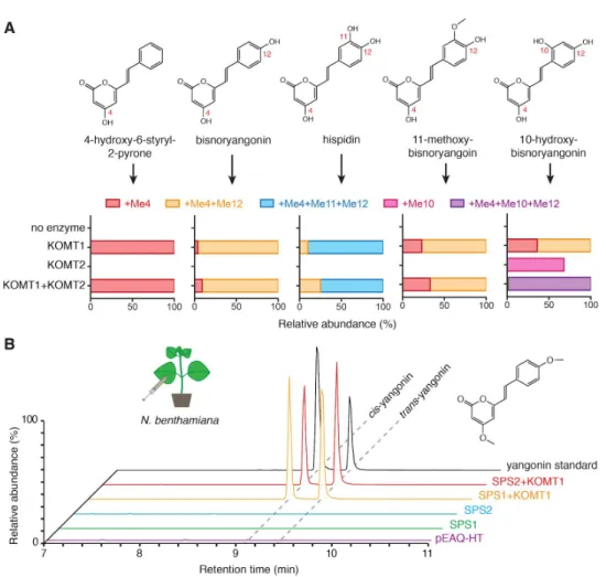
Figure 7. Functional characterization of kava OMTs. A. In vitro enzyme activities of PmKOMT1 and PmKOMT2 against five different styrylpyrone substrates. The bar charts indicate proportions of
differentially methylated products relative to the unmethylated precursor (e.g., +Me4+Me12 indicates methyl group addition to both C4 and C12 sites). Detailed results are shown in Supplementary Note 3. B. LC-MS XICs of 259.096 m/z (yangonin) in leaf extracts from Nicotiana transiently expressing indicated enzymes. Yangonin readily isomerizes between cis and trans configurations in solution.
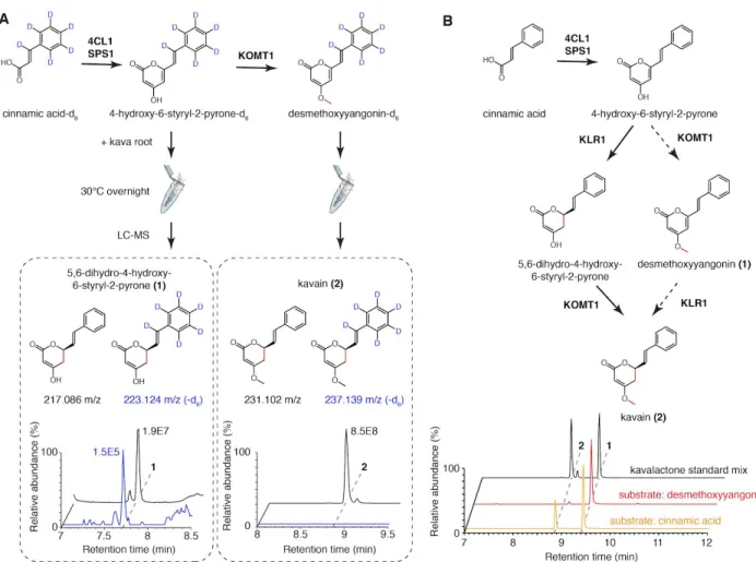
Figure 8. Functional characterization of kavalactone tailoring enzymes KLR1 and MTS1.
A. Combined LC-MS XICs of 215.070 m/z (1), 217.086 m/z (2), 229.086 m/z (3), and 231.102 m/z (4) from in vitro enzyme assays using the indicated combinations of enzymes and cinnamic acid as a starter substrate to show the specific activity of PmKLR1. B. Chiral chromatography-MS traces of 231 m/z (kavain) demonstrating stereo-selectivity of PmKLR1. C. Maximum likelihood phylogenetic analysis of PmCYP719A26 together with several characterized members of the CYP719 family P450s. Bootstrap values (based on 1000 replicates) are indicated at the tree nodes. The scale measures evolutionary
distances in substitutions per amino acid. D. Proposed reaction catalyzed by PmCYP719A26 (PmMTS1). E. LC-MS XICs of 275.091 m/z (methysticin) in Nicotiana leaves infiltrated with the indicated
combinations of enzymes to show the specific activity of PmMTS1. The two structural isomers shown were distinguished using MS/MS analysis (Supplementary Note 5).
Figure 9. Resolving the order of PmKOMT1 and PmKLR1 reactions in vivo and in vitro. A. Isotope tracing using labeled styrylpyrone or desmethoxyyangonin precursors in live kava root suggests that only the unmethylated precursor serve as a substrate for the reduction of C5=C6 olefin. B. Combined LC-MS XICs of 229.086 m/z (1) and 231.102 m/z (2) from in vitro enzyme assays using cinnamic acid and desmethoxyyangonin as starter substrates to show that PmKLR1 precedes PmKOMT1. The reaction from cinnamic acid successfully produced kavain, while the same assay starting from desmethoxyyangonin failed to show any activity. The determined order of the pathway is shown with solid arrows, while the rejected order is shown with dashed arrows.
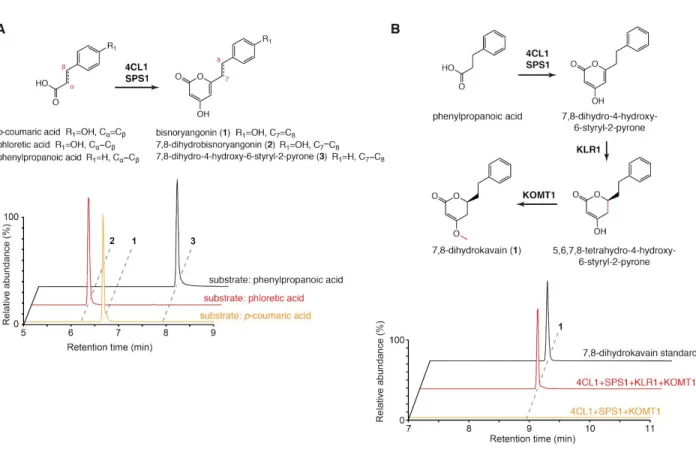
Figure 10. The biosynthesis of 7,8-reduced kavalactones. A. Combined LC-MS XICs of 231 m/z (1), 233 m/z (2), and 217 m/z (3) in coupled (Pm4CL1 + PmSPS1) in vitro enzyme assays to show that kavalactone biosynthesis is operational starting with dihydro-hydroxycinnamic acids. The use of α,β-reduced substrates (phloretic acid and phenylpropanoic acid) resulted in the production of corresponding 7,8-reduced styrylpyrones. B. LC-MS XICs of 233.117 m/z (7,8-dihydrokavain) in combinatorial in vitro enzyme assays using the indicated enzymes and phenylpropanoic acid as a starter substrate. The addition of PmKLR1 to the enzyme mix resulted in the production of 7,8-dihydrokavain, thus further supporting the conclusion that PmKLR1 acts as a NADPH-dependent kavalactone 5,6-olefin reductase.
Figure 11. The biosynthesis of flavokavains using PmCHS, PmKOMT1, and PmKOMT2.
A. Combined LC-MS XICs of 273.076 m/z (1), 287.091 m/z (2, 4), 301.107 m/z (3, 6), and 315.123 m/z (5) from in vitro enzyme assays using the indicated combinations of enzymes and p-coumaric acid as the starter substrate. MS/MS analysis interpretation is shown on the right to support the identification of individual products. B. Combined LC-MS XICs of 273.076 m/z (1), 287.091 m/z (2, 4), 301.107 m/z (3, 6) in leaf extracts from Nicotiana transiently expressing indicated enzymes. Expression in Nicotiana resulted in equivalent compounds to those produced by in vitro enzyme assays.
Figure 12. Heterologous production of the kavalactone precursor, bisnoryangonin, in E. coli and yeast. A. LC-MS XIC of 231.065 m/z (bisnoryangonin) from the media supernatants of E. coli cultures incubated overnight at 30°C with 1 mM p-coumaric acid. Cells were expressing the indicated enzymes from a single plasmid under the control of a constitutive pGAP promoter. Final bisnoryangonin yields are indicated next to each peak. B. LC-MS XIC of 231.065 m/z (bisnoryangonin) from the media
supernatants of S. cerevisiae cultures incubated for 2 days at 30°C with 2 mM p-coumaric acid. Cells were expressing the indicated enzymes from two plasmids under the control of a constitutive pTEF promoter.
Table 1. Crystallography data collection and refinement statistics. Statistics for the highest-resolution shell are shown in parentheses.
PmSPS1 PmCHS Wavelength 0.9793 Å 0.9793 Å Resolution range 82.2 - 2.7 (2.797 - 2.7) 62.7 - 2.91 (3.014 - 2.91) Space group P 31 P 21 21 21 Unit cell 164.402 164.402 87.1221 90 90 120 50.9096 120.57 125.408 90 90 90 Total reflections 3027278 (290476) 1083656 (107234) Unique reflections 72283 (7187) 17602 (1726) Multiplicity 41.9 (40.2) 61.6 (62.1) Completeness (%) 99.59 (99.31) 99.95 (100.00) Mean I/sigma(I) 6.90 (1.30) 12.54 (2.85) Wilson B-factor 44.73 R-merge 1.406 (4.383) 0.926 (2.025) R-meas 1.424 (4.445) 0.9341 (2.043) R-pim 0.2252 (0.7278) 0.1204 (0.2639) CC1/2 0.775 (0.157) 0.949 (0.614) CC* 0.935 (0.521) 0.987 (0.872)
Reflections used in refinement 72027 (7187) 17602 (1726)
Reflections used for R-free 1987 (199) 1771 (179)
R-work 0.2111 (0.2718) 0.2615 (0.3382)
R-free 0.2606 (0.3123) 0.3156 (0.3688)
CC(work) 0.668 (0.224) 0.921 (0.694)
CC(free) 0.403 (0.278) 0.881 (0.694)
Number of non-hydrogen atoms 18141 5880
macromolecules 17603 5846 solvent 135 34 Protein residues 403 762 RMS(bonds) 2293 0.002 RMS(angles) 0.004 0.53 Ramachandran favored (%) 0.62 94.20 Ramachandran allowed (%) 92.63 5.80 Ramachandran outliers (%) 6.45 0.00 Rotamer outliers (%) 0.92 1.59 Clashscore 0.05 24.22 Average B-factor 14.56 47.80 macromolecules 38.58 47.89 solvent 38.46 32.71 Number of TLS groups 8