ASICs Mediate Food Responses in an Enteric
Serotonergic Neuron that Controls Foraging Behaviors
The MIT Faculty has made this article openly available.
Please share
how this access benefits you. Your story matters.
Citation
Rhoades, Jeffrey L. et al. “ASICs Mediate Food Responses in an
Enteric Serotonergic Neuron that Controls Foraging Behaviors.” Cell
176 (2019): 85-97.e14 © 2019 The Author(s)
As Published
https://dx.doi.org/10.1016/J.CELL.2018.11.023
Publisher
Elsevier BV
Version
Author's final manuscript
Citable link
https://hdl.handle.net/1721.1/125439
Terms of Use
Creative Commons Attribution-NonCommercial-NoDerivs License
ASICs Mediate Food Responses in An Enteric Serotonergic
Neuron that Controls Foraging Behaviors
Jeffrey L. Rhoades1, Jessica C. Nelson2, Ijeoma Nwabudike1, Stephanie K. Yu1, Ian G. McLachlan1, Gurrein K. Madan1, Eden Abebe1, Joshua R. Powers1, Daniel A.
Colón-Ramos2, and Steven W. Flavell1,3,*
1Picower Institute for Learning and Memory, Department of Brain and Cognitive Sciences, Massachusetts Institute of Technology, Cambridge, MA 02139, USA.
2Department of Neuroscience and Department of Cell Biology, Program in Cellular Neuroscience, Neurodegeneration, and Repair, Yale University School of Medicine, New Haven, CT 06536, USA.
3Lead Contact
Summary
Animals must respond to the ingestion of food by generating adaptive behaviors, but the role of gut-brain signaling in behavioral regulation is poorly understood. Here, we identify conserved ion channels in an enteric serotonergic neuron that mediate its responses to food ingestion, and decipher how these responses drive changes in foraging behavior. We show that the C. elegans serotonergic neuron NSM acts as an enteric sensory neuron that acutely detects food ingestion. We identify the novel and conserved acid-sensing ion channels (ASICs) DEL-7 and DEL-3 as NSM-enriched channels required for feeding-dependent NSM activity, which in turn drives slow locomotion while animals feed. Point mutations that alter the DEL-7 channel change NSM dynamics and associated behavioral dynamics of the organism. This study provides causal links between food ingestion, molecular and physiological properties of an enteric serotonergic neuron, and adaptive feeding behaviors, yielding a new view of how enteric neurons control behavior.
Graphical Abstract
*Correspondence: flavell@mit.edu.
Author Contributions
J.L.R., J.C.N., I.N., S.K.Y., I.G.M., G.K.M., D.A.C-R., and S.W.F designed experiments. J.L.R., J.C.N., I.N., S.K.Y., I.G.M., G.K.M., E.A., J.R.P., and S.W.F. conducted experiments, analyzed data, and interpreted data. J.L.R. and S.W.F. wrote the paper.
Publisher's Disclaimer: This is a PDF file of an unedited manuscript that has been accepted for publication. As a service to our
customers we are providing this early version of the manuscript. The manuscript will undergo copyediting, typesetting, and review of the resulting proof before it is published in its final citable form. Please note that during the production process errors may be discovered which could affect the content, and all legal disclaimers that apply to the journal pertain.
HHS Public Access
Author manuscript
Cell
. Author manuscript; available in PMC 2020 January 10.Published in final edited form as:
Cell. 2019 January 10; 176(1-2): 85–97.e14. doi:10.1016/j.cell.2018.11.023.
A
uthor Man
uscr
ipt
A
uthor Man
uscr
ipt
A
uthor Man
uscr
ipt
A
uthor Man
uscr
ipt
Introduction
All animals must respond to the ingestion of food by generating adaptive behaviors. Enteroendocrine cells and neurons of the enteric nervous system line the gastrointestinal (GI) tract and play crucial roles in mediating responses to food ingestion. These cells have sensory roles in detecting the contents of the alimentary canal (Bellono et al., 2017) and regulate local processes like GI motility and secretion (Mawe and Hoffman, 2013). They can also signal to the CNS via action on vagal sensory neurons that innervate the gut. It is widely thought that gut-brain signaling might be important for the regulation of internal states like mood and appetite, but we currently have a poor understanding of how enteric neurons detect feeding cues and generate neural activity patterns that influence CNS circuits and complex behaviors.
Enteroendocrine cells and enteric neurons signal in large part through hormones and neuromodulators: they produce >90% of the serotonin (5-HT) found in the body, as well as other biogenic amines and neuropeptides. The role of 5-HT has received particular interest, due to its importance for human health. It is well known that 5-HT has local effects in the gut and that vagal afferent neurons express 5-HT receptors that should allow for signal propagation to the CNS (Mawe and Hoffman, 2013). However, it is not known how patterned 5-HT release in the gut influences behavior. Even within the CNS, the function of 5-HT is poorly understood. There are many molecularly distinct subtypes of 5-HTergic neurons in the Dorsal Raphe Nucleus (Okaty et al., 2015). Neural recordings from this heterogeneous population have revealed diverse activity profiles, reflecting rewarding stimuli like food (Li et al., 2016), and complex cognitive processes (Cohen et al., 2015). Optogenetic stimulation of 5-HTergic neurons in rodents can elicit many behavioral effects, including slow locomotion, waiting/perseverance, and changes in reward learning (Correia et al., 2017; Liu et al., 2014). Overall, the causal links between the molecular profiles of 5-HTergic neuron subtypes, their activity patterns, and the behaviors induced by these activity patterns remain enigmatic for this important modulatory system, both in the CNS and in gut-brain signaling.
In the well-defined nervous system of C. elegans, neuromodulators like 5-HT are known to regulate behaviors associated with feeding. Acute exposure to food alters animals’
A
uthor Man
uscr
ipt
A
uthor Man
uscr
ipt
A
uthor Man
uscr
ipt
A
uthor Man
uscr
ipt
locomotion, egg-laying, and pharyngeal pumping, and these effects require monoamines like 5-HT and dopamine, as well as neuropeptides (Li and Kim, 2008; Sawin et al., 2000; Waggoner et al., 1998). For example, the egg-laying events that C. elegans display while feeding require 5-HTergic HSN neurons (Waggoner et al., 1998). We previously
characterized how C. elegans locomotion is altered by the presence of food. While exploring food patches, C. elegans switch between two persistent behavioral states: dwelling states where they restrict their movement to a small area, and roaming states where they explore a large area (Ben Arous et al., 2009; Flavell et al., 2013; Fujiwara et al., 2002). Each state can last minutes, and the proportion of time spent in each state is influenced by food density, so that animals dwell more in favorable food environments. We previously found that
neuromodulators stabilize these states: stable dwelling states require 5-HT, and stable roaming states require the neuropeptide pigment dispersing factor (Flavell et al., 2013). This role of neuromodulation in driving persistent behavioral states has been observed for other C. elegans behaviors (Choi et al., 2013) and in other animals (Saper et al., 2010).
We previously identified the 5-HTergic neuron NSM as a key regulator of feeding-related dwelling states (Flavell et al., 2013). Indeed, this neuron is required for feeding-dependent switches to slow locomotion in several contexts (Iwanir et al., 2016; Sawin et al., 2000). NSM extends a synapse-less neurite into the alimentary canal (Axäng et al., 2008), suggesting a potential role in food sensation, but specific hypotheses and molecular mechanisms for NSM function remain unexplored.
Here we show that NSM acts as an enteric sensory neuron that acutely detects food ingestion through a mechanism that requires DEL-3 and DEL-7, two previously uncharacterized ion channels in the evolutionarily conserved ASIC family. These channels localize to NSM sensory endings in the alimentary canal and are required for feeding-dependent NSM activity, which in turn drives slow locomotion while animals feed. Point mutations that alter the DEL-7 channel alter NSM’s dynamical response to food ingestion and associated behavioral dynamics. This study identifies a molecular mechanism for bacterial detection by enteric neurons and illustrates how activity in enteric neurons can control behavioral responses to food ingestion.
Results
NSM is an Enteric Serotonergic Neuron that Senses Acute Food Ingestion
We and others have shown that NSM activation drives slow locomotion via its release of 5-HT (Flavell et al., 2013; Iwanir et al., 2016; Sawin et al., 2000). However, the mechanisms that drive endogenous NSM activity were unclear. The unique anatomy of NSM suggests that it might be involved in gut-brain signaling. NSM has one minor neurite (Fig. 1A, arrowheads) that, based on electron microscopy (EM) studies, lacks synapses and extends into the alimentary canal (Albertson and Thomson, 1976; Axäng et al., 2008). This neurite is rich in F-actin (Fig. S1A) and might act as a sensory dendrite. NSM also has two major neurites (Fig. 1A) with 5-HT release sites apposed to pseudocoelemic space that is in contact with the main neuropil of the C. elegans nervous system, the nerve ring (Axäng et al., 2008; Nelson and Colón-Ramos, 2013). Given its anatomy and position, we hypothesized that
A
uthor Man
uscr
ipt
A
uthor Man
uscr
ipt
A
uthor Man
uscr
ipt
A
uthor Man
uscr
ipt
NSM might act as an enteric sensory neuron that directly detects food ingestion and alters behavior via 5-HTergic signaling to downstream circuits.
When C. elegans are exposed to food (E. coli strain OP50), NSM displays calcium peaks that correlate with transitions to slow locomotion (Flavell et al., 2013). To test whether NSM calcium transients induced by food availability require synaptic input onto NSM, we examined NSM activity in unc-13(s69) mutants that display a dramatic deficit in synaptic transmission. NSM makes no electrical synapses in the C. elegans connectome. Through in vivo calcium imaging of NSM, we observed robust spontaneous NSM activity in feeding unc-13 animals, but this activity was absent when animals were removed from bacterial food (Figure 1B–C; additional controls in Figure S1B–C). unc-31 mutants that are defective in neuropeptide release also display robust NSM calcium transients in feeding animals, but no activity in the absence of food (Figure 1C). Finally, mutations in aex-2 or aex-6, which disrupt intestine-to-neuron neuropeptide signaling, also had no effect on NSM activity (Figure S1D). These data suggest that even with attenuated synaptic and neuropeptidergic inputs, NSM still responds to food availability by generating calcium transients.
Food is a complex stimulus, providing olfactory and mechanosensory cues, proprioceptive cues associated with eating, and post-ingestive cues. We next examined which aspects of food availability are required for NSM activation. First, we noted that NSM was inactive in unc-31 mutants removed from food (Figure 1C), even though these mutants exhibit robust pharyngeal pumping in the absence of food (Avery et al., 1993). Thus, the motor act of pharyngeal pumping is insufficient to drive NSM activity in the absence of food. We then treated the bacterial food with aztreonam, which prevents bacterial division and results in E. coli that are too large to consume (Ben Arous et al., 2009). NSM activity was absent when animals were exposed to aztreonam-treated OP50 (Figure 1C), suggesting that successful ingestion of bacteria is necessary for NSM to display feeding-dependent activity.
To test which features of the ingested bacterial food are critical for NSM activation, we heat-killed bacteria (at 95°C) and examined NSM responses when animals ingested the crude extract or the supernatant after pelleting high density components, like cell membranes. We found that NSM was activated by heat-killed bacteria, but not by the isolated supernatant (Figure 1C). To test for pure mechanical responses, we fed animals bacteria-sized nanobeads (at low pH; see below) and found that this failed to activate NSM (Figure 1C). These data suggest that a heat-insensitive component of bacterial membranes is necessary to drive NSM activation, but that pure mechanical cues do not drive NSM activation.
To examine the latency of NSM’s response to food ingestion, we devised an optogenetic strategy to control food intake. We expressed the red-shifted opsin Chrimson (Klapoetke et al., 2014) throughout the pharyngeal muscle and found that exposure to red light contracts the pharyngeal muscles of these transgenic animals, halting the rhythmic pumping required for food ingestion (Figure 1D). This effect occurs immediately upon red light exposure, can be sustained for hours, and is rapidly reversible (<5 sec) when red light is turned off. Using this tool, we examined NSM calcium transients in feeding animals while red light was turned on and off to control food ingestion. NSM activity was abolished within 30 sec after pumping stopped and resumed within 30 sec after pumping resumed (Figure 1D–E). These
A
uthor Man
uscr
ipt
A
uthor Man
uscr
ipt
A
uthor Man
uscr
ipt
A
uthor Man
uscr
ipt
data indicate that NSM can detect food within seconds of ingestion, suggesting a fairly direct mode of food sensing. Taken together, these data indicate that NSM acutely responds to the ingestion of bacterial food in a manner that does not require synaptic or
neuropeptidergic inputs.
The ASICs DEL-7 and DEL-3 are Enriched in NSM Neurons
To clarify how NSM is activated by acute food ingestion, we sought to identify receptors and channels in NSM that are required for its feeding-dependent activity. We performed
molecular profiling of NSM neurons from feeding, adult animals using Tagged Ribosome Affinity Purification (TRAP) (Heiman et al., 2008; Sanz et al., 2009). We expressed an HA-tagged rpl-22 cDNA in NSM, purified the HA-HA-tagged mRNA-ribosome complexes from adult animals, and subjected the isolated RNA to mRNA-seq, along with whole animal RNA for normalization (Figure 2A–B).
We performed three independent biological replicates of NSM TRAP and found a high level of reproducibility among replicates (Figure 2C and S2A). Genes known to have enriched expression in NSM, like those involved in 5-HT signaling (tph-1, cat-1, mod-5) and NSM development (ceh-2, unc-86), were among the top NSM-enriched genes in our genome-wide dataset (Table S1; Figure 2D). We were able to validate the mRNA-seq results using gene-specific qRT-PCR, both for positive controls and for newly discovered NSM-enriched genes (Figure S2B). These data suggest that single neuron TRAP reliably isolates NSM-enriched mRNAs.
We inspected all of the genes that were enriched in NSM >4-fold in all three replicates (Table S1). Out of 94 total genes, 16 encoded putative receptors or channels (Figure 2D). To determine which of these genes are required for feeding-dependent NSM activity and resulting locomotion changes, we performed a behavioral screen of mutant animals lacking these genes using an NSM-dependent exploration assay that measures the extent to which animals explore a bacterial food lawn (Flavell et al., 2013). We tested mutants for nine of the 16 genes and observed significantly increased exploration in mutants lacking del-7, which is predicted to encode a DEG/ENaC superfamily ion channel (Figure 2E). Mutants lacking del-3, which is a del-7 paralog also identified through NSM TRAP, displayed a modest increase in exploration (Figure 2E). Loss of both del-3 and del-7 resulted in a strong increase in exploration (Figure 2E). Importantly, del-3;del-7 mutants had normal food ingestion rates (Figure S3A; see also Figure S3B). These data indicate that del-3 and del-7 are NSM-enriched genes that suppress exploration, a phenotype that is consistent with the possibility that these genes promote NSM activation.
The genes del-3 and del-7 are predicted to encode DEG/ENaC superfamily sodium channels. del-7 encodes a DEG/ENaC subunit most closely related to ppk13 in Drosophila and ASIC1 in mammals (Figure 3A–C). del-3 encodes a related channel subunit (Figure 3D–F). ASICs form homomeric and heteromeric trimers that are permeable to sodium and occasionally calcium ions (Wemmie et al., 2013). In mammals, ASICs are expressed in the CNS and in peripheral sensory neurons; they are known to be involved in pain and GI function (Holzer, 2015; Wemmie et al., 2013). Many distinct activation mechanisms have been described for
A
uthor Man
uscr
ipt
A
uthor Man
uscr
ipt
A
uthor Man
uscr
ipt
A
uthor Man
uscr
ipt
ASICs, including low pH, small molecules, peptides, and mechanical stimuli. DEL-7 and DEL-3, like most other C. elegans ASICs, have not been characterized before.
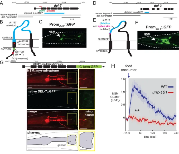
To determine the del-3 and del-7 expression patterns, we fused the promoter sequences for each of these genes to green fluorescent protein (GFP) and generated transgenic animals. We observed strong del-7::GFP expression exclusively in NSM neurons (Figure 3C). For del-3::GFP, we observed expression in NSM and ~8 other pairs of head neurons that we have not yet identified (Figure 3F). Although we cannot rule out additional sites of expression conferred by distal regulatory elements, these results are consistent with our TRAP data and confirm that del-7 and del-3 encode NSM-enriched ion channels.
To examine the subcellular localization of these channels in NSM, we used CRISPR/Cas9 to modify the endogenous del-7 gene, adding an in-frame flexible linker followed by GFP to the DEL-7 C-terminus (Fig. 3G). This strain displays normal exploratory behavior,
suggesting that addition of GFP does not disrupt del-7 function (Fig. S3C). The DEL-7::GFP channels localized exclusively to the posterior end of the NSM minor neurite, with faint or undetectable signal in the soma and major neurites of NSM (Fig. 3G; additional examples in Fig. S3D). The posterior end of the NSM minor neurite is located in the alimentary canal in close proximity to the grinder of the pharynx that mechanically lyses bacteria, suggesting that these channels are well-positioned to detect components of lysed bacteria.
Given the exclusive localization of DEL-7 channels in the NSM minor neurite, we examined whether the minor neurite might be involved in mediating feeding-induced NSM activation. We examined mutants lacking the unc-101 gene, which is required for formation of the NSM minor neurite, but not the other NSM neurites (Axäng et al., 2008). We measured NSM calcium levels in starved unc-101 mutants as they encountered a food patch (Figure 3H). In contrast to wild-type (WT) animals that display robust NSM calcium peaks upon food encounter (Iwanir et al., 2016), food-deprived unc-101 animals displayed no NSM response when they encountered food (Figure 3H), despite having robust pharyngeal pumping rates (Figure S3E). These data are consistent with the possibility that NSM’s minor neurite detects bacterial food cues in the alimentary canal. Taken together, these data suggest that DEL-7 and DEL-3 are NSM-enriched ASICs that are well-positioned to participate in the detection of bacterial food cues.
del-3 and del-7 Function in NSM to Promote Slow Locomotion in Response to Food
Ingestion
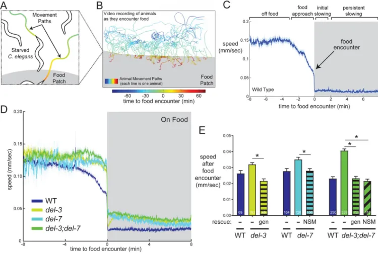
NSM plays an important role in driving abrupt transitions to slow locomotion when animals encounter food patches (Iwanir et al., 2016; Sawin et al., 2000). In WT animals, this
transition to slow locomotion occurs immediately upon food encounter and persists for many minutes (Fig. 4C), though some animals resume high speed locomotion 1–2 minutes after food encounter (Figure S4A). We characterized the functions of DEL-3 and DEL-7 during this behavior to specifically examine how these channels contribute to an NSM-dependent behavior that is acutely triggered by food (Figure 4A–C). Relevant to these ASICs, pH was not detectably altered on or near the bacterial lawn (Figure S4B).
A
uthor Man
uscr
ipt
A
uthor Man
uscr
ipt
A
uthor Man
uscr
ipt
A
uthor Man
uscr
ipt
It was not known whether food encounter-induced slowing in C. elegans is driven by food ingestion, changes in sensory cues like odors/gases, or both. Thus, we first examined the importance of food ingestion for food patch-induced slowing by optogenetically inhibiting food intake while animals encountered food. This caused a strong, albeit partial, attenuation of the behavioral slowing response (Figure S4C), indicating that both food ingestion and other food sensory cues contribute to this behavioral effect. A previous study showed that inhibiting NSM synaptic vesicle release also causes a partial attenuation of the behavioral slowing response to food (Iwanir et al., 2016). To confirm these results in a temporally-precise manner, we silenced NSM during food encounters using a chemogenetic approach (Pokala et al., 2014) and also observed a strong, though partial, attenuation of behavioral slowing (Fig. S4D). Thus, food ingestion and NSM neurons play important roles in driving behavioral slowing in response to food, but they do not explain the entire behavioral response.
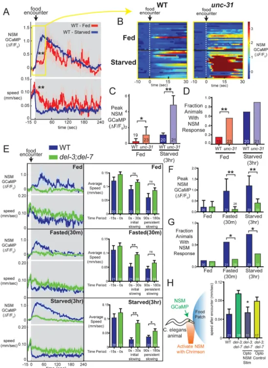
To assess the roles of del-3 and del-7, we examined the behavioral phenotypes of each single mutant and del-3;del-7 double mutants. We found that each single mutant had elevated speed at all time points after the food encounter, as did the del-3;del-7 double mutants, whose phenotype was slightly more severe than each single mutant (p<0.01, ANOVA and
Bonferroni-corrected t-test, del-3;del-7 vs. single mutants; Fig. 4D; S4E–F). Thus, del-3 and del-7 are critical for the initial slowing response to food encounter and for persistent slowing minutes later. To verify these effects were specifically due to the loss of the del-3 and del-7 genes, we performed genetic rescues. Restoring expression of each gene in the respective single mutants or both genes in the double mutant fully rescued the behavioral defects (Fig. 4E). A similar phenotype was also observed for a second, independent loss-of-function allele of del-7 (Fig. S4G). These data indicate that del-3 and del-7 play important roles in driving feeding-induced behavioral slowing.
To determine if del-3 and del-7 function specifically in NSM to promote behavioral slowing, we expressed both cDNAs under an NSM-specific promoter in double mutant animals. This fully rescued the post-food-encounter speed defects in these mutant animals (Figure 4E). We note that the del-3;del-7 mutants also display elevated speed immediately before
encountering food, though not when they are far away from a food patch (Figures 4D and S4F). Pre-food-encounter speed changes likely reflect sensory detection of nearby food cues (Iwanir et al., 2016). The pre-food-encounter speed defect was fully rescued by transgenic expression from full genomic fragments encompassing both genes, but not by NSM-specific expression of these genes (Figure S4H; similar analysis of del-7 single mutant in Figure S4I). This indicates that del-3 and del-7 function in other cells to promote slow locomotion as animals approach a food patch, but they are specifically required in NSM for post-food encounter slowing. These channels represent the first identified molecular mechanism linking food ingestion to 5-HT-dependent slowing.
NSM Calcium Responses Integrate Acute Food Ingestion with Satiety Levels
We next sought to determine how del-3 and del-7 contribute to feeding-dependent NSM activity. Previous work has suggested that NSM has a stronger impact on locomotion in starved animals (Sawin et al., 2000). Thus, we first examined how satiety alters NSM’s
A
uthor Man
uscr
ipt
A
uthor Man
uscr
ipt
A
uthor Man
uscr
ipt
A
uthor Man
uscr
ipt
response to food ingestion and associated behavioral slowing (Figure 5A–D). Then, we used this information to examine how del-7 and del-3 contribute to NSM activity across satiety states (see below).
We examined NSM activity and behavior as WT animals encountered food patches, comparing well-fed animals (“fed”) to those that had been deprived of food for 30 minutes (“fasted”) or 3 hours (“starved”). Fasted and starved animals displayed reliable NSM calcium peaks that began within seconds of encountering a food patch and lasted
approximately 30 sec to 3 min (Figure 5A). NSM activity did not detectably change before animals encountered the food, even as they came into close proximity to the food patch (Figure S5A). Consistent with previous studies, NSM calcium peaks triggered by food encounters were correlated with an abrupt, persistent transition to slow locomotion (Figure 5A, lower panel). Across animals, NSM activity was strongly associated with reduced speed immediately after food encounter (Figures 5A and S5B), though the magnitude of NSM activation was not linearly correlated with the magnitude of speed reduction (Figure S5B). Instead, NSM activation appears to trigger an all-or-none transition to slow locomotion, consistent with previous studies (Flavell et al., 2013). This interpretation is also consistent with the observation that optogenetically activating NSM with a range of different light intensities in the presence of food produces the same magnitude of initial slowing when the lights are turned on (Figure S5C).
In contrast to fasted and starved animals, well-fed animals almost never displayed NSM calcium peaks timed to the food patch encounter (Figure 5A–B). Instead, well-fed animals displayed stochastic, irregular NSM calcium peaks at later time points, as previously described (Flavell et al., 2013). These animals also failed to slow abruptly in response to the food encounter, gradually slowing instead (Figure 5A). These results indicate that NSM’s response to food ingestion is modulated by satiety: NSM is immediately activated by food ingestion in starved animals but is inhibited in well-fed animals until its activity is stochastically de-repressed at later time points during feeding.
Satiety states can be communicated through circuit-level mechanisms involving neuropeptide signaling. To examine whether circuit-level mechanisms suppress NSM’s sensory response to food in well-fed animals, we measured NSM activity during food patch encounters in well-fed and starved unc-31 mutants, which are defective in neuropeptide release. There was a robust de-repression of NSM activity in this mutant background in both well-fed and starved animals (Fig. 5B–D). Notably, NSM activation in well-fed unc-31 animals was as robust as NSM activation in starved WT animals (Fig. 5B–D). These experiments suggest that neuropeptide signaling inhibits NSM activity and that this inhibition is a major component of NSM suppression in well-fed animals.
DEL-3 and DEL-7 Are Required for NSM Calcium Responses to Food, which Drive Slow Locomotion
To examine how DEL-3 and DEL-7 contribute to feeding-dependent NSM activity, we recorded NSM calcium levels in well-fed, fasted, and starved del-3;del-7 double mutants during food patch encounters. Compared to WT animals, NSM calcium events were abolished in fasted double mutants and greatly attenuated in starved double mutants (Figure
A
uthor Man
uscr
ipt
A
uthor Man
uscr
ipt
A
uthor Man
uscr
ipt
A
uthor Man
uscr
ipt
5E–G). In addition, we observed no stochastic, irregular NSM activity in well-fed double mutants (Figure 5E–G). Consistent with the above behavioral analyses (Figure 4E), slowing was defective in fasted and starved conditions immediately after food encounter and minutes later, compared to WT (Figure 5E; Figure S5D). Similar defects were also observed in del-3 and del-7 single mutants (Figures S5E and S5F). These data indicate that del-3 and del-7 are critical for food-induced NSM activity and behavioral slowing.
We asked whether these defects reflect a specific function for DEL-7 and DEL-3 in food detection. To test whether del-3 and del-7 play general roles in controlling NSM excitability or calcium influx, we photo-activated NSM with Chrimson in non-feeding animals while recording NSM GCaMP (Fig. S5G). We found that activating Chrimson with increasing red light intensities led to stronger NSM GCaMP responses, which decayed after light cessation (Figure S5G). WT and del-3;del-7 double mutant animals had similar NSM GCaMP responses at all light levels examined (Figure S5G), suggesting these channels do not generically control NSM excitability or calcium influx.
To test whether other DEG/ENaC channels could substitute for del-3 and del-7 in mediating NSM food responses, we expressed the known mechanosensor deg-1 (also in the DEG/ ENaC superfamily) in NSM neurons of del-3;del-7 double mutants, but found this did not restore NSM calcium responses upon food encounter (Figure S5H). Thus, the roles of DEL-3 and DEL-7 in mediating NSM food responses cannot be flexibly substituted by other DEG/ENaC channels.
We hypothesized that the loss of NSM activity in del-3;del-7 mutants likely causes the deficit in food-induced slowing in this mutant background. To directly test this, we restored WT NSM calcium peaks to del-3;del-7 mutants at the moment of food encounter and examined the impact on behavior. We again co-expressed Chrimson and GCaMP in NSM in del-3;del-7 double mutants and then examined the behavior of animals that had been fasted for 30 minutes. At the moment these animals encountered the food patch, we activated Chrimson with red light (Figure 5H). We shaped the intensity of the red light so that animals received a temporal pattern of red light whose profile matched the kinetics of a WT NSM calcium response to food. Simultaneous NSM calcium imaging confirmed that this approach induced NSM calcium events with similar amplitudes and kinetics to those seen in fasted WT animals (Figure S5I). Furthermore, these animals displayed a slowing response to the food encounter similar to WT animals (Figure 5H). These results indicate that the novel ASICs del-3 and del-7 are required for food-induced NSM activity, which in turn drives behavioral slowing.
A Gain-of-function Point Mutation in del-7 Alters NSM Calcium Dynamics and Behavioral Dynamics
The above experiments indicate that del-3 and del-7 are critical for food-induced NSM activity and behavioral slowing. However, these experiments did not reveal whether these ASICs play an active role in shaping NSM’s dynamical response to food ingestion. We reasoned that additional gain-of-function mutations in DEL-3 or DEL-7 that alter the properties of these channels might provide such insights. To better understand how NSM serves its unique role in gut-brain signaling, we performed unbiased forward genetic screens
A
uthor Man
uscr
ipt
A
uthor Man
uscr
ipt
A
uthor Man
uscr
ipt
A
uthor Man
uscr
ipt
for mutants with defective 5-HTergic vesicle localization in NSM. Remarkably, from this orthogonal approach we also recovered an allele of del-7. The ola110 allele that we recovered contains a point mutation that causes a methionine-to-threonine substitution in amino acid 76 of DEL-7, which is in a juxtamembrane region in the N-terminal intracellular domain. This region has been implicated previously in channel gating and pore properties in related DEG/ENaCs (Figure 3B) (Tavernarakis et al., 2001). In contrast to WT animals that display punctate clusters of 5-HT vesicles in NSM, del-7(ola110) animals displayed diffusely distributed 5-HT vesicles (Fig. S6A). Based on complementation tests and comparisons to del-7 null animals (Fig. S6A), we determined that the ola110 mutation acts as a genetic gain-of-function with semi-dominant inheritance and is henceforth referred to as del-7(ola110gf). These data indicate that alterations in the DEL-7 channel can influence 5-HT release sites in NSM, and that the del-7(ola110gf) allele acts as a genetic gain-of-function.
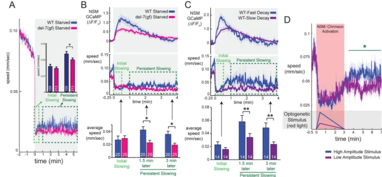
We next examined the effect of this M76T mutation on animal behavior. Starved
del-7(ola110gf) mutants displayed normal slowing immediately after food encounter, but their slow locomotion was unusually sustained so that del-7(ola110gf) animals had reduced speed relative to WT for many minutes after encountering food (Figure 6A). By comparison, del-7 null animals had increased speed at all of these time points (Figure 4D). As a genetic control, we induced a frameshift mutation in del-7(ola110gf) animals that results in a premature stop codon upstream of M76T (Figure S6B). These intragenic revertant
del-7(ola110flv3) animals had a behavioral phenotype matching del-7 null mutants (Figure S6C), indicating that background mutations in the del-7(ola110gf) strain are unlikely to explain its defects. These data suggest that del-7(ola110gf) animals display unusually persistent behavioral slowing in response to food encounter.
We next examined how food-induced NSM calcium dynamics were altered in
del-7(ola110gf) mutants. del-7(ola110gf) animals displayed an NSM calcium peak of lower amplitude and slower decay rate than that of WT animals (Figure 6B; compare to del-7(lf) in S6D). Thus, the exaggerated behavioral slowing in response to food encounter in
del-7(ola110gf) is accompanied by a low-amplitude, slow-decaying NSM calcium response upon food encounter, suggesting a potentially complex relationship between NSM calcium events and persistent behavioral slowing.
NSM Dynamics Shape Behavioral Dynamics
Prompted by our observations in the del-7(ola110gf) mutants, we sought to determine whether different patterns of NSM activity drive different levels of persistent slowing. To examine whether there was a similar relationship between NSM activity and persistent slowing in WT animals as was observed in del-7(ola110gf) animals, we segmented the WT animals that we had recorded into two groups based on their diverse NSM calcium
responses: a low-amplitude/slow-decay group and a high-amplitude/fast-decay group. Like del-7(ola110gf) mutants, WT animals displaying lower amplitude, slower decaying NSM calcium events displayed lower speed many minutes after food encounter compared with those displaying higher amplitude, faster decaying peaks (Figure 6C; all data points in
A
uthor Man
uscr
ipt
A
uthor Man
uscr
ipt
A
uthor Man
uscr
ipt
A
uthor Man
uscr
ipt
Figure S6E). Thus, in WT animals NSM calcium dynamics are associated with behavioral dynamics.
To further examine this issue, we optogenetically activated NSM using a variety of
stimulation patterns while recording behavior. To avoid interference from endogenous NSM activation, we performed these experiments on del-3;del-7 mutants. We activated NSM with uniform rise times (24sec to peak, based on endogenous NSM peaks), but varied the decay rate and peak intensity of stimulation. Slower-decaying patterns of NSM stimulation led to more prolonged reductions in locomotion (Figure S6F–G). Consistent with our calcium imaging results, we also found that animals receiving low-amplitude optogenetic stimulation displayed a less pronounced rebound in locomotion following light offset compared to animals receiving high-intensity stimulation (Figure 6D). This was most apparent in the time period immediately after optogenetic stimulation, as both stimuli had similar acute effects on locomotion during the lights-on time period (see also Figure S6F–K). Taken together, these data are consistent with the possibility that robust phasic activation of this 5-HTergic neuron drives a transient suppression of locomotion followed by a post-inhibitory rebound, whereas low/moderate increases in NSM activity are more effective at driving persistent slowing.
Discussion
All animals must respond to the ingestion of food by generating adaptive behavioral responses, but the role of gut-brain signaling in behavioral regulation remains poorly understood. Here, we show that the 5-HTergic neuron NSM acts as an enteric sensory neuron that acutely detects food ingestion through a mechanism that requires the newly characterized ASICs, DEL-7 and DEL-3. These channels localize to NSM sensory dendrites in the alimentary canal and are specifically required for responses to food ingestion. Point mutations that alter the DEL-7 channel change NSM dynamics and associated behavioral dynamics. These experiments identify a molecular mechanism for bacterial detection by enteric neurons and illustrate how activity in enteric neurons can drive adaptive behavioral responses to food ingestion.
ASICs Mediate Neuronal Responses to Bacteria and Control Feeding State
Prompted by the finding that NSM acts as an enteric sensory neuron that detects ingested bacteria, we examined the molecular mechanisms that allow this neuron to detect bacterial food cues. Through molecular profiling of NSM, we identified two novel ASICs, DEL-3 and DEL-7, that are required for feeding-dependent NSM activity. We also performed a forward genetic screen that recovered a gain-of-function mutation in del-7 that enhances the behavioral response to food ingestion. The identification of these ASICs using two orthogonal, unbiased approaches underscores the importance of these channels in NSM. This class of ion channels is highly conserved and mammalian orthologs of del-3 and del-7 are expressed in the CNS and in peripheral sensory neurons, such as taste receptor neurons, enteric neurons in the GI system, and dorsal root ganglia nociceptive neurons (Holzer, 2015; Shimada et al., 2006; Wemmie et al., 2013). Given that del-3 and del-7 are required in NSM for neural responses to bacteria, we speculate that mammalian orthologs of these channels
A
uthor Man
uscr
ipt
A
uthor Man
uscr
ipt
A
uthor Man
uscr
ipt
A
uthor Man
uscr
ipt
might mediate responses to bacteria in peripheral sensory neurons, which are also exposed to microbial populations in the mouth, gut, and during bacterial infections.
Our work has not yet fully resolved whether these channels are directly involved in sensory transduction or amplify another primary sensory response in NSM. However, we found that the DEL-7 channels localize to the posterior end of NSM’s minor neurite, a dendritic ending that is devoid of synapses, but protrudes into the alimentary canal and is located in close proximity to the site of bacterial lysis in the pharynx. Consistent with a possible sensory function for DEL-7/3 channels in the minor neurite, we found that NSM is acutely and directly activated by bacterial food ingestion and that mutants lacking either the DEL-7/3 channels or the minor neurite had abolished food-induced NSM activity. ASICs can be activated by many agents, including mechanical stimuli and small molecules. For example, DEG/ENaCs are known to mediate touch sensitivity in C. elegans sensory neurons (Chalfie, 2009). However, we found that pure mechanical stimuli do not activate NSM. In addition, ASICs were initially identified based on their response to low extracellular pH (Wemmie et al., 2013). However, pharyngeal pH does not change during food ingestion, so this might not be the signal driving NSM activity (Chauhan et al., 2013). Our current results are most consistent with an activation mechanism that involves a heat-insensitive bacterial product, though further studies will be necessary to fully answer this question.
NSM’s response to food ingestion is modulated by the animal’s satiety state, with well-fed animals showing suppressed and delayed NSM responses to food ingestion, compared to starved animals. We found that mutants defective in neuropeptide release had de-repressed NSM activity. It is possible that this reflects modulation of DEL-3 or DEL-7. ASICs are known to be modulated by neuropeptides, protein kinases, and other intracellular signaling pathways (Wemmie et al., 2013). Indeed, our NSM TRAP studies revealed new NSM-enriched neuropeptide receptors that could merit further investigation across satiety states (Table S1).
Enteric Sensory Neurons Control Behavioral Responses to Food Ingestion
How does patterned activity in the enteric nervous system drive changes in animal behavior? Here, we show that the enteric 5-HTergic neuron NSM integrates its acute detection of food with chronic satiety levels. Depending on the animal’s internal state, NSM generates calcium peaks of varying amplitudes and kinetics when it encounters food. NSM calcium peaks uniformly drive acute behavioral slowing, which allows animals to slow down and feed. Our data also suggest the specific pattern of NSM activity shapes long-term locomotion dynamics. High-amplitude, phasic NSM responses drive transient slowing, followed by a rebound speed burst when NSM activity decays. Moderate-amplitude NSM calcium peaks drive both transient and sustained behavioral slowing. However, we note that the relationship between NSM activity and locomotion is complex and that other neurons are likely also involved. The mechanism underlying the rebound speed bursts after phasic NSM activation is still unclear, though it could involve post-inhibitory rebound in downstream neurons, 5-HT receptor desensitization, circuit-level effects, or even co-transmission. Neuromodulation and post-inhibitory rebound are key features of circuits that generate rhythmic oscillations (Marder et al., 2014). Further studies of this circuit may clarify
A
uthor Man
uscr
ipt
A
uthor Man
uscr
ipt
A
uthor Man
uscr
ipt
A
uthor Man
uscr
ipt
whether similar mechanisms control behavioral state switching. Notably, optogenetic stimulation of 5-HTergic neurons in rodents drives acute behavioral slowing, but repeated stimulation causes increased locomotion afterwards (Correia et al., 2017). Thus, 5-HTergic neurons might have a conserved role in driving opposing short- and long-term behaviors. A previous study found that harmful environmental cues inhibit NSM (Li et al., 2012), which, when combined with our findings, suggests that NSM integrates appetitive (food) and aversive (harmful) cues. The drive to exploit a food patch is normally balanced by a drive to explore for other food sources. However, indicators of risk or danger in the environment cause animals to favor exploitation over risky exploration. NSM might integrate these disparate cues and generate precisely tuned neural responses that dictate how long to exploit a newly encountered food patch.
Conserved Role for 5-HT in Mediating Gut-To-Brain Signaling
NSM’s role as an enteric sensory neuron that signals through HT is reminiscent of 5-HTergic enteroendocrine cells in the mammalian GI epithelium. These cells can detect feeding cues in the gut and release 5-HT that has local and long-range effects (Bellono et al., 2017). 5-HTergic neurons in the mammalian CNS have also been shown to have phasic responses to food ingestion, which can be modulated by internal state (Li et al., 2016). In the compressed nervous system of C. elegans, NSM acts as an enteric sensory neuron, an integrator of internal state, and a site of 5-HT release that targets downstream neurons to drive adaptive behavioral responses to food ingestion.
STAR METHODS
CONTACT FOR REAGENT AND RESOURCE SHARING
Further information and requests for resources and reagents should be directed to and will be fulfilled by Lead Contact, Steven W. Flavell (flavell@mit.edu).
EXPERIMENTAL MODEL AND SUBJECT DETAILS
C. elegans strains—All wild-type, mutant and transgenic strains used in this study are listed in the Key Resources Table above.
Growth Conditions & Handling—Nematode culture was conducted using standard methods (Brenner, 1974). Populations were maintained on NGM agar plates supplemented with E. coli OP50 bacteria. Wild-type was C. elegans Bristol strain N2. For genetic crosses, all genotypes were confirmed using PCR. Transgenic animals were generated by injecting DNA clones plus fluorescent co-injection marker into gonads of young adult
hermaphrodites. We typically analyzed 2–3 independent transgenic lines per injection. One day old isogenic hermaphrodites were used for all assays. All assays were conducted at room temperature (~22°C) and replicated on two or more days.
METHOD DETAILS
Forward Genetic Screen and Characterization of del-7(ola110gf)—The ola110 mutant was recovered in a forward genetic screen for mutants with altered NSM 5-HT
A
uthor Man
uscr
ipt
A
uthor Man
uscr
ipt
A
uthor Man
uscr
ipt
A
uthor Man
uscr
ipt
release sites (Nelson and Colón-Ramos, 2013). These mutants displayed a diffuse pattern of the CAT-1::GFP vesicle marker, although active zone markers were normally localized in NSM. We initially mapped ola110 to the right arm of Chromosome IV through chromosome mapping. Subsequent interval mapping placed the ola110 lesion within a 0.55 centiMorgan interval (between SNPs haw57407 and haw153809). Genomic DNA was then collected from ola110 mutants and sent for whole genome sequencing. Within the region of interest, only 2 non-synonymous mutations were identified. These lesions, subsequently confirmed by Sanger sequencing, included a missense lesion in the gene K07H8.5, and a missense lesion in the predicted degenerin/epithelial Sodium channel (DEG/ENaC), del-7 (M76T). The allele is a presumed gain-of-function because none of the ola110 mutant phenotypes are observed in the del-7 null animals, but the phenotype can be reverted to a null phenotype via introduction of a frameshift mutation that induces a premature stop codon upstream of the M76T mutation in del-7. This mutation was introduced via Cas9/CRISPR using the Co-CRISPR method and is a 17-bp insertion that causes a premature stop codon. The sequence of the modified allele proximal to this mutation is
gcttccgtaaacacaacacaTGACACAATATGACACAatatgaggaagctttgaaaa (insertion in capital letters; premature stop codon underlined).
Tagged Ribosome Affinity Purification (TRAP) Analysis—Raw RNA-seq data and processed data analysis files for TRAP experiments are freely available in NCBI GEO Datasets (Accession number GSE110334).
We adapted the TRAP method for use in single C. elegans neurons, guided by previous protocols used in mammalian tissues (Heiman et al., 2008; Sanz et al., 2009). A Ribotag plasmid was constructed by fusing the C. elegans rpl-22 (C27A2.2a) cDNA with three tandem HA tags (rpl-22–3xHA). Plasmid design was based on the mammalian Ribotag (Sanz et al., 2009). A transgenic strain was generated that expressed both the Ribotag and GFP specifically in NSM. For each TRAP experiment, animals were synchronized by bleaching and grown on 15cm, OP50-seeded NGM plates with enriched peptone (20g/L). After three days, one-day old adult animals were prepared for lysis by collecting animals from the plates in liquid NGM supplemented with cycloheximide (CHX; 0.8 mg/ml). Animals were washed once in NGM+CHX, and then washed once in Minimal
Homogenization Buffer (10mM HEPES, pH 7.4, 150mM KCl, 5mM MgCl2, 0.8 mg/ml CHX). Finally, the worm pellet was resuspended in Complete Homogenization Buffer (10mM HEPES, pH 7.4, 150mM KCl, 5mM MgCl2, 0.5mM DTT, Complete EDTA-free protease inhibitor, 0.4U/uL RNasin, 10mM ribonucleoside vanadyl complex, 0.8 mg/ml CHX). The resuspended animals were flash frozen in liquid nitrogen and stored at −80C. The concentration of animals was estimated by counting the number of animals in 10uL of frozen solution. Then, frozen worm pellets were ground to a fine powder with an RNase-free pestle while keeping samples frozen. For immunoprecipitations, the samples were thawed, diluted in Complete Homogenization Buffer (as above, except with CHX at 0.1 mg/ml) to a final concentration of 8 animals per uL of extract. Based on the total volume of extract used in each TRAP experiment, we estimate that each pulldown was from ~150,000 animals. NP-40 (1% final) and DHPC (30mM final) detergents were added to the diluted samples. After 10min of 4°C incubation, samples were cleared twice by spinning at 15,000rcf for
A
uthor Man
uscr
ipt
A
uthor Man
uscr
ipt
A
uthor Man
uscr
ipt
A
uthor Man
uscr
ipt
12min at 4°C. Samples were then pre-cleared by incubating 150uL Protein G Dynabeads (Life Technologies) with samples for 30’ and then discarding the beads. After pre-clearing, 3.5 uL of anti-HA antibody (clone HA-7, Sigma) was incubated with sample for 30min, after which 150uL of Protein G Dynabeads were added and the sample was incubated for an additional 30min. The beads were then washed four times in Wash Buffer (10mM HEPES, pH 7.4, 350mM KCl, 5 mM MgCl2, 1% NP-40, 0.5mM DTT, 0.04U/uL RNasin, 0.1mg/mL CHX). Finally, RNA was eluted from beads by incubating with 100uL of Lysis Buffer from the Absolutely RNA Nanoprep kit (Stratagene). RNA was then purified, following the kit protocol. For whole-animal input material, RNA was also purified from 100uL of the pre-cleared lysate.
Whole-animal and NSM-Ribotag RNA samples were subject to mRNA-seq. We performed three independent biological replicates, as described in the main text. Due to low RNA amounts, samples were first amplified with Clontech SMARTer Ultra Low RNA kit and then prepared for sequencing with Illumina Nextera XT. We obtained 13–93M mapped reads per sample and used CuffLinks to identify genes with increased abundance in NSM-Ribotag samples. For the data shown in Figure 2 and Table S1, we required that each “NSM-enriched” gene (1) was 4-fold enriched in the NSM-Ribotag sample (vs. whole-animal) in all three biological replicates, (2) was <4-fold enriched in a mRNA-seq Ribotag pulldown from a different neuron, and (3) had FPKM values >0.25 in all three biological replicates. Reverse Transcription-Quantitative PCR—A subset of genes was tested to validate whether or not they were enriched in NSM Ribotag samples. Each gene’s expression was measured using reverse transcription-quantitative PCR (RT-qPCR). cDNA samples were prepared using Nugen Ovation RNA amplification system v2. Expression of specific mRNAs were measured in NSM-Ribotag and Whole-animal RNA samples by qPCR. In these experiments, we used Power SYBR Master Mix and a QuantStudio Realtime PCR system (Life Technologies). For each primer set, we performed standard curves to ensure that amplification was exponential, and a no-reverse-transcriptase control to ensure that there was no genomic DNA contamination. Primer sequences are available upon request.
Exploration Behavior Assays—Behavioral assays for exploration were conducted as previously described (Flavell et al., 2013). L4 animals were picked to 35mm plates seeded with OP50. After 16 hours, plates were superimposed on a grid and the number of squares intersected by the animal’s movement path were counted. This assay essentially measures the level of exploration of each animal, which correlates with the level of roaming across many genotypes (Flavell et al., 2013). Data were always normalized to N2 controls measured in parallel.
Pharyngeal Pumping Assays—Pharyngeal pumping was either counted manually (for myo-2::Chrimson line) or by video recording and subsequent manual counting (for WT, del-7;del-3 and unc-101). Videos were recorded at 20Hz for 30sec on a tracking microscope stage that followed each freely-moving animal for 30sec on or off food, as indicated in figures. Pharyngeal pumps were counted over this timeframe on videos played back in slow motion.
A
uthor Man
uscr
ipt
A
uthor Man
uscr
ipt
A
uthor Man
uscr
ipt
A
uthor Man
uscr
ipt
Food Patch Encounter Behavioral Assays—In an assay adapted from Iwanir et al (2016), wild-type, transgenic, and mutant one-day old adult worms were allowed to re-encounter lawns of OP50 after various levels of starvation. 16 hours prior to assays, 114×73mm NGM plates were seeded with OP50 to create a lawn with a straight edge that covered approximately 2/3 of the plate from edge to edge. For pH measurements, plates were made similarly, but Phenol Red (Sigma) was added at 18mg/L. Prior to assays, one-day old adult worms were washed off standard growth plates with 1 mL of M9 buffer and transferred to 1.7mL Eppendorf tubes, where they were rinsed once with 1mL of M9 before being transferred either to unseeded NGM plates for starvation, or directly to assay plates. For HisCl1 experiments, histamine dihydrochloride (Sigma) was added at 10mM to both the pre-starvation plates and to assay plates. Animals starved before assays were left on
unseeded plates for indicated durations of time before transfer to assay plates in M9. Animals were recorded at 3 fps using Streampix 7.0, a JAI SP-20000M-USB3 CMOS camera (41mm, 5120×3840, Mono) and a Nikon Micro-NIKKOR 55mm f/2.8 lens. Backlighting was achieved using a white panel LED (Metaphase Technologies Inc. White Metastandard 10” X 25”, 24VDC). Assay plates were placed on glass 3” above LEDs to avoid heat transfer to plates. Videos were processed using custom Matlab scripts, which included a step to manually confirm the exact frame of lawn encounter for each animal. For experiments with myo-2::Chrimson inhibition of pumping (Figure S4C), animals were grown on 50uM all-trans-retinal overnight, and red light was applied constantly during recordings using a 625nm Mightex BioLED light source.
We have plotted the absolute speed of animals throughout all datasets in an effort to present the maximum amount of information with regards to animal behavior, but also present speed data normalized to pre-food-encounter baselines in Fig. S4 and S5.
Throughout the study, genetic mutants are compared to complete datasets of wild-type N2 animals.
In vivo Calcium Imaging—In vivo calcium imaging of NSM was conducted in wild-type, transgenic, and mutant backgrounds. For calcium imaging in unc-13 and unc-31 mutant backgrounds, experiments were conducted with a 5x/0.25NA Zeiss objective and a Hamamatsu Orca Flash 4.0 sCMOS camera, or a 4x/0.2NA Nikon objective and an Andor Zyla 4.2 Plus sCMOS camera. Blue light application to animals was 10ms for each exposure at a rate of 10fps, as previously described (Flavell et al., 2013). In these experiments, one-day old adult animals were picked to flat NGM pads that either had (i) no bacteria, (ii) OP50, (iii) Aztreonam-treated OP50 (Ben Arous et al., 2009), (iv) heat-killed bacterial extract (95°C for 10min, with vortexing), (v) heat-killed bacterial extract supernatant (95°C for 10min; then 2 consecutive 15min spins at 17,000xg), or (vi) bacterial-sized beads (Fluorspheres with bead sizes 0.02um-1um in equal parts, Invitrogen) dialyzed into pH5 solution, with 5mM 5-HT to promote bead ingestion. The NGM pads were enclosed using a rubber gasket and cover glass. Animals were imaged for 30 minutes, beginning 15 minutes after they were transferred. Pixel intensity values for NSM and adjacent background were extracted from each video frame using custom ImageJ macros, as described previously
A
uthor Man
uscr
ipt
A
uthor Man
uscr
ipt
A
uthor Man
uscr
ipt
A
uthor Man
uscr
ipt
(Flavell et al., 2013). Gaps in recordings were due to animals adopting a body posture that obscured the head. For these recordings where animals were chronically on or off food, background-subtracted NSM GCaMP fluorescence values were normalized to baseline periods of NSM inactivity for each trace. For experiments with combined myo-2::Chrimson inhibition of pumping, animals were grown on 1–3uM all-trans-retinal overnight, and blue light levels were reduced to 1–5% output on the SOLA-LE solid-state LED light source. Red light was applied at defined times using a 617nm red LED light (Mightex). Under these recording conditions, blue light did not visibly alter pharyngeal pumping, but red light exposure caused a full cessation of pumping.
The above procedure was adapted to allow for imaging of one-day old adult animals during lawn encounters. Experiments were conducted with a 4x/0.2NA Nikon objective and an Andor Zyla 4.2 Plus sCMOS camera. Blue light application to animals was 100% output from a X-Cite 120LED system for 10ms of each exposure at a recording rate of 10fps, as above. Fed animals, as well as animals fasted for 30 minutes or starved for 3 hours were assayed on flat NGM pads enclosed by a rubber gasket and coverslip as described above. Pads were seeded with 20x concentrated OP50 liquid culture, and rectangular corrals laser cut from Apollo Laser Printer Transparency Film were placed on the slides to keep animals within the field of view while recording their food encounter. For experiments with combined Chrimson activation of NSM, animals were grown on 1–3uM all-trans-retinal overnight, and blue light levels were reduced to two 2-ms pulses 6ms apart at 20% intensity. For Optogenetic Rescue experiments, recordings were monitored so that red light
application (617nm Mightex LED) was timed with each animal’s food encounter. For activation of NSM in non-feeding animals, red light was applied for 30sec at intensities indicated in figure to animals immobilized in tetramisole. Under these recording conditions, blue light did not visibly alter NSM activation on or off food.
Optogenetic Stimulation of NSM During Behavioral Recordings—For optogenetic stimulation of NSM during behavioral recordings, L4 animals were picked onto 50uM all-trans-retinal (ATR) OP50 plates 16–24 hours before experiments. Where indicated, control animals were cultivated on plates without ATR.
15cm NGM plates used for ‘on food’ experiments were seeded evenly with 600ul of OP50 and let dry overnight. Immediately prior to experiments, a thin filter paper ring 1–1.5 cm wide, with a 15cm outer diameter, was dipped in 0.02M copper chloride solution and placed on either unseeded 15cm NGM plates (‘off food’) or seeded NGM plates (‘on food’). C. elegans’ aversion to heavy metals makes these copper rings useful in preventing animals from leaving the recording arena.
For behavioral recordings, one-day old adult animals were picked directly from ATR or control plates to the copper ringed ‘on food’ plates 45 minutes before experiments. In ‘off food’ experiments, animals were washed twice with liquid NGM solution to remove all bacteria, then placed on copper ringed NGM plates 15 minutes before experiments.
A
uthor Man
uscr
ipt
A
uthor Man
uscr
ipt
A
uthor Man
uscr
ipt
A
uthor Man
uscr
ipt
Animals were illuminated with 625nm red light from a Mightex BioLED while being recorded via the Streampix setup described above. Illumination patterns were delivered to the BioLED Light Source Control Module via custom Matlab scripts.
QUANTIFICATION AND STATISTICAL ANALYSES
The statistical tests performed in this study are indicated in the figure legends and Results section.
DATA AND SOFTWARE AVAILABILITY
The NSM TRAP mRNA-Seq datasets, including raw and processed data files, have been deposited in NCBI GEO Datasets under accession number GSE110334.
Supplementary Material
Refer to Web version on PubMed Central for supplementary material.
Acknowledgments
We thank Cori Bargmann for support and advice during early phases of this project, Josh Greene for early help with exploration assays, Lucelenie Rodriguez-Laureano for technical help, and David Hall for helpful discussions about NSM morphology. We thank Paul Greer, Cori Bargmann, and members of the Flavell Lab for helpful comments on the manuscript. We thank Olivia Foster Rhoades for her graphical contributions. We also thank the Research Center for Minority Institutions program, the Instituto de Neurobiología de la Universidad de Puerto Rico and the Marine Biological Laboratories for providing a meeting and brainstorming platform. We thank the Caenorhabditis Genetics Center (supported by P40 OD010440) for strains. S.W.F. acknowledges funding from the JPB Foundation, PIIF, PNDRF, the NARSAD Young Investigator Award Program, and NIH (R01NS104892). HHMI also supported early phases of this project (to C.B.). D.A.C.-R., and J.N. were supported by NIH (R01NS076558), NSF (IOS 1353845) and the Howard Hughes Medical Institute Faculty Scholars program.
References
Albertson DG, and Thomson JN (1976). The pharynx of Caenorhabditis elegans. Philos. Trans. R. Soc. Lond. B. Biol. Sci 275, 299–325. [PubMed: 8805]
Avery L, Bargmann CI, and Horvitz HR (1993). The Caenorhabditis elegans unc-31 gene affects multiple nervous system-controlled functions. Genetics 134, 455–464. [PubMed: 8325482] Axäng C, Rauthan M, Hall DH, and Pilon M (2008). Developmental genetics of the C. elegans
pharyngeal neurons NSML and NSMR. BMC Dev. Biol 8, 38. [PubMed: 18400083]
Bellono NW, Bayrer JR, Leitch DB, Castro J, Zhang C, O’Donnell TA, Brierley SM, Ingraham HA, and Julius D (2017). Enterochromaffin Cells Are Gut Chemosensors that Couple to Sensory Neural Pathways. Cell 170, 185–198.e16. [PubMed: 28648659]
Ben Arous J, Laffont S, and Chatenay D (2009). Molecular and sensory basis of a food related two-state behavior in C. elegans. PloS One 4, e7584. [PubMed: 19851507]
Brenner S (1974). The genetics of Caenorhabditis elegans. Genetics 77, 71–94. [PubMed: 4366476] Chalfie M (2009). Neurosensory mechanotransduction. Nat. Rev. Mol. Cell Biol 10, 44–52. [PubMed:
19197331]
Chauhan VM, Orsi G, Brown A, Pritchard DI, and Aylott JW (2013). Mapping the pharyngeal and intestinal pH of Caenorhabditis elegans and real-time luminal pH oscillations using extended dynamic range pH-sensitive nanosensors. ACS Nano 7, 5577–5587. [PubMed: 23668893]
Choi S, Chatzigeorgiou M, Taylor KP, Schafer WR, and Kaplan JM (2013). Analysis of NPR-1 reveals a circuit mechanism for behavioral quiescence in C. elegans. Neuron 78, 869–880. [PubMed: 23764289]
A
uthor Man
uscr
ipt
A
uthor Man
uscr
ipt
A
uthor Man
uscr
ipt
A
uthor Man
uscr
ipt
Cohen JY, Amoroso MW, and Uchida N (2015). Serotonergic neurons signal reward and punishment on multiple timescales. eLife 4.
Correia PA, Lottem E, Banerjee D, Machado AS, Carey MR, and Mainen ZF (2017). Transient inhibition and long-term facilitation of locomotion by phasic optogenetic activation of serotonin neurons. eLife 6.
Flavell SW, Pokala N, Macosko EZ, Albrecht DR, Larsch J, and Bargmann CI (2013). Serotonin and the neuropeptide PDF initiate and extend opposing behavioral states in C. elegans. Cell 154, 1023– 1035. [PubMed: 23972393]
Fujiwara M, Sengupta P, and McIntire SL (2002). Regulation of body size and behavioral state of C. elegans by sensory perception and the EGL-4 cGMP-dependent protein kinase. Neuron 36, 1091– 1102. [PubMed: 12495624]
Heiman M, Schaefer A, Gong S, Peterson JD, Day M, Ramsey KE, Suárez-Fariñas M, Schwarz C, Stephan DA, Surmeier DJ, et al. (2008). A translational profiling approach for the molecular characterization of CNS cell types. Cell 135, 738–748. [PubMed: 19013281]
Holzer P (2015). Acid-sensing ion channels in gastrointestinal function. Neuropharmacology 94, 72– 79. [PubMed: 25582294]
Iwanir S, Brown AS, Nagy S, Najjar D, Kazakov A, Lee KS, Zaslaver A, Levine E, and Biron D (2016). Serotonin promotes exploitation in complex environments by accelerating decision-making. BMC Biol. 14, 9. [PubMed: 26847342]
Klapoetke NC, Murata Y, Kim SS, Pulver SR, Birdsey-Benson A, Cho YK, Morimoto TK, Chuong AS, Carpenter EJ, Tian Z, et al. (2014). Independent optical excitation of distinct neural populations. Nat. Methods 11, 338–346. [PubMed: 24509633]
Li C, and Kim K (2008). Neuropeptides. WormBook Online Rev. C Elegans Biol. 1–36.
Li Y, Zhong W, Wang D, Feng Q, Liu Z, Zhou J, Jia C, Hu F, Zeng J, Guo Q, et al. (2016). Serotonin neurons in the dorsal raphe nucleus encode reward signals. Nat. Commun 7, 10503. [PubMed: 26818705]
Li Z, Li Y, Yi Y, Huang W, Yang S, Niu W, Zhang L, Xu Z, Qu A, Wu Z, et al. (2012). Dissecting a central flip-flop circuit that integrates contradictory sensory cues in C. elegans feeding regulation. Nat. Commun 3, 776. [PubMed: 22491324]
Liu Z, Zhou J, Li Y, Hu F, Lu Y, Ma M, Feng Q, Zhang J-E, Wang D, Zeng J, et al. (2014). Dorsal raphe neurons signal reward through 5-HT and glutamate. Neuron 81, 1360–1374. [PubMed: 24656254]
Marder E, O’Leary T, and Shruti S (2014). Neuromodulation of circuits with variable parameters: single neurons and small circuits reveal principles of state-dependent and robust neuromodulation. Annu. Rev. Neurosci 37, 329–346. [PubMed: 25032499]
Mawe GM, and Hoffman JM (2013). Serotonin signalling in the gut--functions, dysfunctions and therapeutic targets. Nat. Rev. Gastroenterol. Hepatol 10, 473–486. [PubMed: 23797870]
Nelson JC, and Colón-Ramos DA (2013). Serotonergic neurosecretory synapse targeting is controlled by netrin-releasing guidepost neurons in Caenorhabditis elegans. J. Neurosci. Off. J. Soc. Neurosci 33, 1366–1376.
Okaty BW, Freret ME, Rood BD, Brust RD, Hennessy ML, deBairos D, Kim JC, Cook MN, and Dymecki SM (2015). Multi-Scale Molecular Deconstruction of the Serotonin Neuron System. Neuron 88, 774–791. [PubMed: 26549332]
Pokala N, Liu Q, Gordus A, and Bargmann CI (2014). Inducible and titratable silencing of
Caenorhabditis elegans neurons in vivo with histamine-gated chloride channels. Proc. Natl. Acad. Sci. U. S. A 111, 2770–2775. [PubMed: 24550306]
Sanz E, Yang L, Su T, Morris DR, McKnight GS, and Amieux PS (2009). Cell-type-specific isolation of ribosome-associated mRNA from complex tissues. Proc. Natl. Acad. Sci. U. S. A 106, 13939– 13944. [PubMed: 19666516]
Saper CB, Fuller PM, Pedersen NP, Lu J, and Scammell TE (2010). Sleep state switching. Neuron 68, 1023–1042. [PubMed: 21172606]
Sawin ER, Ranganathan R, and Horvitz HR (2000). C. elegans locomotory rate is modulated by the environment through a dopaminergic pathway and by experience through a serotonergic pathway. Neuron 26, 619–631. [PubMed: 10896158]
A
uthor Man
uscr
ipt
A
uthor Man
uscr
ipt
A
uthor Man
uscr
ipt
A
uthor Man
uscr
ipt
Shimada S, Ueda T, Ishida Y, Yamamoto T, and Ugawa S (2006). Acid-sensing ion channels in taste buds. Arch. Histol. Cytol 69, 227–231. [PubMed: 17287577]
Tavernarakis N, Everett JK, Kyrpides NC, and Driscoll M (2001). Structural and functional features of the intracellular amino terminus of DEG/ENaC ion channels. Curr. Biol. CB 11, R205–208. [PubMed: 11301263]
Waggoner LE, Zhou GT, Schafer RW, and Schafer WR (1998). Control of alternative behavioral states by serotonin in Caenorhabditis elegans. Neuron 21, 203–214. [PubMed: 9697864]
Wemmie JA, Taugher RJ, and Kreple CJ (2013). Acid-sensing ion channels in pain and disease. Nat. Rev. Neurosci 14, 461–471. [PubMed: 23783197]
A
uthor Man
uscr
ipt
A
uthor Man
uscr
ipt
A
uthor Man
uscr
ipt
A
uthor Man
uscr
ipt
Highlights
• NSM is an enteric serotonergic neuron that is activated by food ingestion • Two ASIC channels, DEL-7 and DEL-3, mediate feeding-induced NSM
activation
• Activation of NSM neurons drives slow locomotion while animals feed • Changes in NSM response dynamics alter foraging behavior dynamics A sensory mechanism for food sensing and feeding-associated behaviors is mediated by two ASIC ion channels
A
uthor Man
uscr
ipt
A
uthor Man
uscr
ipt
A
uthor Man
uscr
ipt
A
uthor Man
uscr
ipt
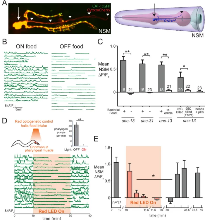
Figure 1. The serotonergic neuron NSM detects acute food ingestion.
(A) Left: confocal image of NSM neurons with soma, axons (yellow) and minor neurite (red) of NSM labeled via CAT-1::GFP (5-HT vesicle marker) and cytoplasmic-mCherry. Synaptic vesicles labeled via CAT-1::GFP are not found in the minor neurite. Right: cartoon depicting location of NSM in pharynx, reprinted from WormAtlas (wormatlas.org) with permission. Arrows mark NSM soma; arrowheads mark minor neurite. (B) NSM GCaMP traces from unc-13(s69) animals in the presence or absence of bacterial food. Each line is a recording from one animal. Gaps are due to the unc-13 animals adopting body postures that
A
uthor Man
uscr
ipt
A
uthor Man
uscr
ipt
A
uthor Man
uscr
ipt
A
uthor Man
uscr
ipt
obscured NSM GCaMP. (C) Average NSM GCaMP signals across animals for indicated conditions. “Inedible” is animals exposed to Aztreonam-treated E. coli. “s’ntnt” refers to bacterial supernatant. **p<0.001, t-test; *p<0.05, t-test. (D) Above: optogenetic strategy to control food intake with myo-2::Chrimson, and pharyngeal pumping in these animals in the absence or presence of red light (**p<0.001, paired t-test; n=6 animals). Below: NSM calcium traces from unc-13(s69); myo-2::Chrimson animals. (E) Average NSM GCaMP signals for data in (D), shown for the indicated time bins. n=17 animals. *p<0.01 versus 0– 10min time period, ANOVA and Dunnett Test. Where applicable, n is indicated in bars. Data are shown as means ± standard error of the mean (SEM). See also Figure S1.
A
uthor Man
uscr
ipt
A
uthor Man
uscr
ipt
A
uthor Man
uscr
ipt
A
uthor Man
uscr
ipt
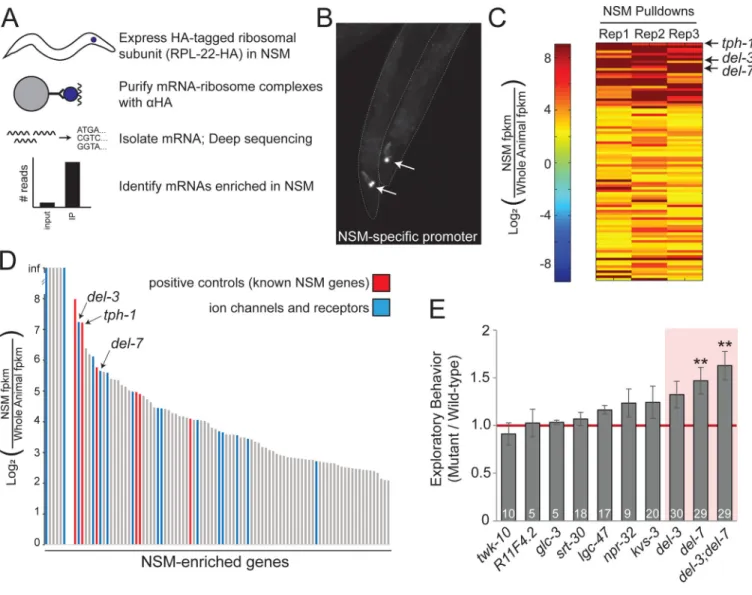
Figure 2. Identification of del-3 and del-7 as NSM-enriched ion channels.
(A) Cartoon depicting NSM TRAP. (B) Image of two NSM::rpl-22–3xHA animals, co-expressing GFP under same NSM-specific promoter. GFP-positive NSM neurons indicated by arrows. (C) Heat map showing NSM enrichment for 94 genes identified through NSM TRAP. Data are shown for each biological replicate and are sorted based on hierarchical clustering. (D) Average NSM enrichment of TRAP-identified genes. Red indicates genes with previously known NSM enrichment. Blue indicates genes with predicted
transmembrane domains. (E) Exploratory behavior of mutants lacking NSM-enriched receptors or channels, normalized to WT. A value of 1.0 (red line) indicates no change from WT. Data are shown as means ± SEM. **p<0.01, Bonferroni-corrected t-test. n is indicated in bars. See also Figure S2 and Table S1.