Publisher’s version / Version de l'éditeur:
Molecular, 10, 9, pp. M111.007765-1-M111.007765-11, 2011-05-24
READ THESE TERMS AND CONDITIONS CAREFULLY BEFORE USING THIS WEBSITE.
https://nrc-publications.canada.ca/eng/copyright
Vous avez des questions? Nous pouvons vous aider. Pour communiquer directement avec un auteur, consultez la
première page de la revue dans laquelle son article a été publié afin de trouver ses coordonnées. Si vous n’arrivez pas à les repérer, communiquez avec nous à PublicationsArchive-ArchivesPublications@nrc-cnrc.gc.ca.
Questions? Contact the NRC Publications Archive team at
PublicationsArchive-ArchivesPublications@nrc-cnrc.gc.ca. If you wish to email the authors directly, please see the first page of the publication for their contact information.
Archives des publications du CNRC
This publication could be one of several versions: author’s original, accepted manuscript or the publisher’s version. / La version de cette publication peut être l’une des suivantes : la version prépublication de l’auteur, la version acceptée du manuscrit ou la version de l’éditeur.
For the publisher’s version, please access the DOI link below./ Pour consulter la version de l’éditeur, utilisez le lien DOI ci-dessous.
https://doi.org/10.1074/mcp.M111.007765
Access and use of this website and the material on it are subject to the Terms and Conditions set forth at
Alterations in Glycopeptides Associated with Herceptin Treatment of
Human Breast Carcinoma MCF-7 and T-Lymphoblastoid Cells
Lattová, Erika; Bartusik, Dorota; Spicer, Vic; Jellusova, Julia; Perreault,
Hélène; Tomanek, Boguslaw
https://publications-cnrc.canada.ca/fra/droits
L’accès à ce site Web et l’utilisation de son contenu sont assujettis aux conditions présentées dans le site LISEZ CES CONDITIONS ATTENTIVEMENT AVANT D’UTILISER CE SITE WEB.
NRC Publications Record / Notice d'Archives des publications de CNRC:
https://nrc-publications.canada.ca/eng/view/object/?id=7033fb21-5459-425b-b26c-45cdc7dd7a18
https://publications-cnrc.canada.ca/fra/voir/objet/?id=7033fb21-5459-425b-b26c-45cdc7dd7a18
Alterations in Glycopeptides Associated with
Herceptin Treatment of Human Breast
Carcinoma MCF-7 and T-Lymphoblastoid Cells*
□
SErika Lattova´‡§¶¶, Dorota Bartusik¶储, Vic Spicer**, Julia Jellusova‡‡,
He´le`ne Perreault‡, and Boguslaw Tomanek¶§§¶¶
The therapeutic humanized monoclonal antibody IgG1 known as Herceptin® has shown remarkable antitumor effects. Although this type of therapy has increased the cancer-free survival of patients, not all tumors respond to this treatment and cancers often develop resistance to the antibody. Despite the fact that Herceptin function has been extensively studied, the precise mechanism under-lying its antitumor activity still remains incompletely de-fined. We previously demonstrated on human breast MCF-7 carcinoma and T-lymphoblastoid CEM cells that monoclonal antibody in combination with Lipoplex con-sisting of Lipofectamine mixed with plasmid DNA showed a more profound effect on cancer cell viability than anti-body alone. The analyses of N-glycans isolated from can-cer cells showed dramatic differences in profiles when cells were exposed to Herceptin. Moreover, the investiga-tion of glycosylated peptides from the same cancer cell models after treatment revealed further alterations in the post-translational modifications. Tandem mass spectra obtained from the samples treated confirmed the pres-ence of a series of glycopeptides bearing characteristic oligosaccharides as described in IgG1. However some of them differed by mass differences that corresponded to peptide backbones not described previously and more of them were detected from Herceptin treated samples than from cells transfected with Heceptin/Lipoplex. The results indicate that the presence of Lipoplex prevents antibody transformation and elongates its proper function. The bet-ter understanding of the multipart changes described in the glycoconjugates could provide new insights into the mechanism by which antibody induces regression in
cancers. Molecular & Cellular Proteomics 10: 10.1074/ mcp.M111.007765, 1–11, 2011.
Glycosylation of proteins is a ubiquitous type of post-trans-lational modification in living systems. Variations in oligosac-charide structures are associated with many normal and path-ological events such as cellular growth, host-pathogen interaction, differentiation, migration, cell trafficking, or tumor invasion (1, 2). Targeted glycosylation research has become important in the area of developing novel therapeutic ap-proaches (3–5). The structures of asparagine-linked oligosac-charides in the conserved CH2 region of the constant Fc domain of human immunoglobulin-␥ (IgG1) have been shown to affect the pharmacokinetics, antibody-dependent cellular cytotoxicity and complement-dependent cytotoxicity (6, 7). In the last decade, many recombinant antibody molecules have been licensed for the treatment of a variety of cancers and chronic diseases (8). Herceptin, also known as Trastuzumab, marketed by Genentech Inc. is one example of therapeutic IgG1 antibody. It is produced from mammalian cell culture using Chinese hamster ovary cells (9). The main oligosaccha-ride forms found in this polypeptide chain in the Fc domain at asparagine 297 are biantennary core-fucosylated complex type structures with variable terminal galactosylation (zero, one, or two galactose residues) on their nonreducing termini (10, 11). This humanized monoclonal antibody is known to effectively target breast cancer cells overexpresing the human epidermal growth factor receptor HER2/neu (12). HER2 is a cell membrane surface-bound receptor tyrosine kinase and is normally involved in the signal transduction pathways leading to cell growth and differentiation. It can be found overex-pressed in a variety tumors’ cells of epithelial origin and hematological malignancies, including acute lymphoblastic leukemia (13). When antibody binds to defective HER2 pro-tein, this protein no longer causes cells to reproduce uncon-trollably. This increases the survival of people with cancer. However, cancers usually develop resistance to trastuzumab. Unfortunately, only 25–30% of patients with HER2/neu posi-tive breast cancer respond to this antibody (14 –17). Therefore search for the potential biomarkers that could predict the efficacy of clinical outcomes is needed. More precise
inves-From the ‡Chemistry Department, University of Manitoba, 144 Dysart Road, Winnipeg, MB R3T 2N2, Canada; §The Institute of Chemistry, Centre for Glycomics, Slovak Academy of Sciences, 842 38 Bratislava, Slovakia; ¶NRC, Institute for Biodiagnostics (West), 3330 Hospital Drive NW, Calgary, Alberta T2N 4N1, Canada;储Cross Cancer Institute, Medical Physics Department, Edmonton, Alberta T6G 1Z2, Canada; **Department of Physics, University of Manitoba, Winnipeg, MB R3T 2N2, Canada; ‡‡Infectious and Inflammatory Dis-ease Center, Cancer Center, Sanford-Burnham Medical Research Institute, La Jolla, CA 92037, USA; §§University of Alberta, Oncology Department, Edmonton, Alberta T6G 1Z2, Canada
Received January 8, 2011, and in revised form, April 29, 2011 Published, MCP Papers in Press, May 24, 2011, DOI 10.1074/ mcp.M111.007765
tigation on cellular and molecular level might provide many exciting insights in understanding of mechanism resistance cancer cells to the antibody, so that antibody-based therapies can be optimized more individually (18).
We recently demonstrated how the carbohydrate moieties of two cancer cell models were affected during treatment with antibody (19). The detailed glycans profiles studied by means of mass spectrometry (MS) from the two most common can-cer cell lines— human breast MCF-7 carcinoma and T-lym-phoblastoid CEM cells before and after treatment with Herceptin showed significant differences. Dominant high-mannose structures analyzed in both original cancer cells were suppressed after treatment and instead, complex bi-and triantennary glycans were the major structures found in the treated samples. Their ratio or occurrence varied with conditions and time of exposure of the cancer cells to the antibody. The results provided very good evidence for in-volvement of glycosylation during treatment. In this regard, continuous work presented here on this subject has been aimed to the MS investigation of glycosylated peptides gen-erated by proteolytic digestions of the cancer cells before and after exposure to Herceptin or Herceptin/Lipoplex. Direct analysis of glycopeptides by tandem MS has been shown as one of the most sensitive and fast methods for a site-specific characterization of glycosylation. It can provide information on glycan composition, glycan attachment site with determi-nation of peptide sequence (20 –28), and may offer more specific biomarkers to monitor changes in the post-transla-tional modification at the onset, during cancer progression or during treatment.
EXPERIMENTAL PROCEDURES
Cell Lines and Three-Dimensional Culture Conditions—The human breast carcinoma MCF-7 cell lines were obtained from the American Type Cells Collection, Manassas, VA. T-lymphoblastic (CEM) cells were purchased from the American Type Culture Collection, Rock-ville, MD. Both cell samples were cultured in a Hollow Fiber Bioreactor device (HFB, FiberCell Systems, Frederick, MD) as previously de-scribed (29, 30). Briefly, cells were grown at 37 °C under 5% of carbon dioxide and air. The culture conditions for MCF-7 cells con-sisted of Dulbecco’s modified Eagle’s medium supplemented with 5% fetal bovine serum and 1% penicillin/streptomycin, 50 g/ml gentamycin, 15 g/L sodium bicarbonate, 10 mMsodium pyruvate, and 2 mM L-glutamine. In case of CEM cells, the RPMI 1640 culture medium was supplemented with 10% fetal calf serum and 1% pen-icillin/streptomycin solution. During 4 weeks of cultivation, cell cul-tures were supplied with fresh media. The cells were harvested from the HFB, seeded in 6-well microplates and their viability was deter-mined using the trypan blue exclusion method. Cell numbers were determined manually with a hematocytometer chamber (Hausser Sci-entific, Horsham, PA).
Treatment/Transfection—When the cells reached a concentration of 109cells per ml, treatment was performed with Herceptin
(Genen-tech Inc, San Francisco, CA) at concentration 0.01 g/ml or trans-fected with Herceptine/Lipoplex in the HFB. The day before transfec-tion, cells were maintained in serum-free growth media. Lipofectamine™2000 (LipA, InnoVita Inc., Gaithersburg, MD) was mixed with Herceptin to obtain a final concentration ratio of 3:1.
Plasmid DNA containing -Gal (InnoVita Inc., Gaithersburg, MD) was diluted in serum free media and added to the Herceptin/LipA mixture to obtain a DNA plasmid to lipid ratio of 1:9. The solution was then mixed with medium for 30 min to produce the Herceptin/Lipoplex complex. The transfection was performed with a 3 ml volume of Herceptin/Lipoplex. Cell viability was determined by trypan blue ex-clusion assay at 0 –72 h after transfection. The efficiency of transfec-tion with Herceptin/Lipoplex was determined by -gal expression and a protein concentration assay kit according to the manufacturer’s instructions (enzymatic kit, Promega) after 72 h. To avoid the pres-ence of free Herceptin, after treatment the media were pumped with a flow rate of 14 ml/min during 1 h at 37 °C, then washed twice with 1% bovine serum albumin in 3.5 ml of phosphate buffered saline and suspended in 100 l of 1% bovine serum albumin for centrifugation. Collected cells were washed with phosphate-buffered saline and collected by centrifugation. Cells were lysed by applying the tradi-tional freeze/thaw technique (31).
Trypsin Digestion—Lysed dried cells (200 g) were dissolved in 25 mMammonium bicarbonate (100 l) and digested directly with trypsin (Sigma)) at 37 °C for 18 h under nonreducing conditions or first incubated with 100 mM dithiothreitol (2.5 l) in presence of 0.1% RapiGest for 30 min at 56 °C. Subsequently, 5 l 50 mM iodoacet-amide was added and the mixture was kept in the dark at room temperature for 30 min. Next, the reaction mixture was diluted with 25 mMammonium bicarbonate (total volume 200 l) and digestion was performed with trypsin at a substrate-to-enzyme ratio of 20:1 at 37 °C for 18 h. After the incubation the digest solution was evaporated and the volume was adjusted with deionized water (20 l).
RP-HPLC Fractionation—Digested samples (5 l) were fraction-ated on a System Gold HPLC chromatograph equipped with a Sys-tem Gold 166 UV Detector and 32-Karat software (Beckman-Coulter, Canada, ON). For reversed-phase HPLC, a Vydac 218 TP54 Protein&Peptide C18 analytical column, 300-Å pore size, 0.46 ⫻ 25 cm (Separation Group, Hesperia, CA) was used. The chromatograph was equipped with a Rheodyne injector (5 l loop). The samples (5 l) were eluted with 5% acetonitrile in water as solvent A, and 90% acetonitrile (ACN)1
in 0.1% trifluoroacetic acid as solvent B at a flow rate of 0.5–1 ml/min. An elution gradient was applied from 5 to 70% ACN over 40 – 60 min. UV detection was performed at 245 nm. All fractions were collected manually, then completely dried and stored at ⫺25 °C.
Solid phase extractions on STRATA-XC
a) Glycans —PNGaseF digest was purified on a STRATA-XC col-umn (30 mg/1 ml, Phenomenex, Torrance, CA) according to a proce-dure previously described (32). Briefly, The STRATA-X-C column was washed with 100%, 50%, 0% ACN in water (5 ml of each). The PNGaseF digest (50 l) was applied onto the column with wet carrier and left to permeate. The column was then washed five times with 200 l of deionized water and the eluate containing glycans was
1The abbreviations used are: ACN, acetonitrile; ATT,
2-aza-2-thio-thymine; BSA, bovine serum albumin; CID, collision-induced dissoci-ation; DHB, dihydroxy benzoic acid; Her2/neu, human epidermal growth factor receptor; HPLC, high performance liquid chromatogra-phy; IgG, immunoglobulin-␥; MS, mass spectrometry; MALDI, matrix-assisted laser desorption ionization; MS/MS, tandem mass spec-trometry; m/z, mass-to-charge ratio; LipA, lipofectamine (the polycationic lipid 2,3-dioleoxy-N-[2(sperminecarboxymido)-ethyl]-N,Ndimethyl-1-propanaminium trifluoroacetate (DOSPA) and neutral; lipid dioleolyl phosphatidylethanolamine (DOPE) in membrane filtered water; PNGaseF, peptide N-glycosidaseF; pDNA, plasmid DNA; PHN, phenylhydrazine; PTM, posttranslational modification; QqTOF MS, quadrupole-quadrupole time-of-flight mass spectrometer.
collected into separate tubes without using any pressure. All fractions were collected, evaporated and analyzed after PHN labeling.
b) Glycopeptides—Before extraction, the column was washed with 0%, 50%, 100%, ACN in water (3 ml of each). Tryptic digest (50 l) was applied onto the column with a wet carrier and left to permeate (⬃15 min). The tube was rinsed twice with 50 l of 100% ACN and the solvent was again loaded directly onto the cartridge. The column was then washed five times with 500 l of 100% ACN, then with 500 l of 70% ACN. Glycopeptides were eluted from the column three times with 500 l of 50% ACN in deionized water and three times with 500 l of deionized water. Fractions were collected into separate tubes without using any pressure, then evaporated and completely dried.
Mass Spectrometric Analysis—Fractionated samples were recon-stituted in 7 l of deionized water and 1 l was spotted onto partially dried matrix of 2,5-dihydroxybenzoic acid (Sigma) or onto a mixture of 2-aza-2-thiothymine and phenylhydrazine hydrochloride (2:1w/w) predeposited on the surface of a MALDI target, and were air-dried. MALDI-Qq (TOF)-MS and tandem (MS/MS) experiments were carried out on the Manitoba/Sciex prototype quadrupole-quadrupole-TOF mass spectrometer operated in the positive ion mode with a mass resolving power of ⬃10,000FWHM, and accuracy 10 ppm (33). The instrument was calibrated externally using angiotensin I, substance P and ACTH over a mass range of 500 –5000 Da. Spectra were re-corded by summing 300 laser shots for each spectrum. Individual parent ions were manually selected for MS/MS. The collision energy was set between 50 –140 V as a function of the mass ion.
The peaklist was generated using MoverZ software (2001.02.13 freeware edition) available from Genomic Solutions (http://bioinfor-matics.genomicsolutions.com/MoverZDL.html). The peptide identi-ties were first assigned manually using a partial de novo approach, and then verified using the online version of Mascot search engine (version 2.3.02; Matrix Science; www.matrixscience.com) with an error tolerance on the monoisotopic ions set to 0.8 Da. The data were searched against the databases NCBInr (20110419; 236538 se-quences) and SwissProt (2011_04; 20233 sese-quences) for Homo Sa-piens (human) taxonomy. Database searches were conducted for trypsin as enzyme, with one missed cleavage permitted and variable modifications were set for carbamidomethyl, carboxymethyl, deami-dation of glutamine and asparagine, oxidized methionine, pyroglu-tamic acid from N-terminal and glupyroglu-tamic acid. All identifications were manually verified. In addition BLASTp (version 2.2.25) searches for manually assigned peptide sequences were performed (http://blast-.ncbi.nlm.nih.gov/Blast.cgi). Collision-induced dissociation (CID) spectra of peptides in figures were annotated according nomencla-ture proposed by Roepstorf and Fohlman (34).
The assignment of oligosaccharide types attached to the peptides was performed on the fragmentation pattern produced under MS/MS conditions and verified after deglycosylation (PNGaseF) and detection of glycans with phenylhydrazine (21), (35). For discrimination of glycan structures, general rules described previously were applied (36).
RESULTS
Samples of whole cell lysates obtained before and after treatment/transfection were subjected to protease digestion, following HPLC fractionation or SPE on STRATA-XC car-tridges. Each fraction was then analyzed by MALDI-MS as described in the experimental section.
Although the glycosylation pattern of monoclonal IgG1 has been studied extensively and well characterized, nonetheless it has been reported that commercially available monoclonal antibodies can differ in their lot-to-lot heterogeneity and therefore detailed analysis of global glycoprofiles is always
required (37). Thus, the same batch of Herceptin used for treatment in our study underwent a thorough analysis of gly-cans, as reported previously (19). Furthermore, a detailed analysis of glycopeptides obtained from the antibody is pre-sented here along with the investigation of glycosylated pep-tides isolated from treated samples, to validate the profiles observed.
Analysis of Glycopeptides from Herceptin—Glycopeptides
obtained from trypsin digested Herceptin were eluted on a RP-column at a flow rate of 0.5 ml/min, in the 5–15 min range. MS spectra recorded from these fractions are presented in Fig. 1. Glycosylated peptides analyzed in the first fraction produced peaks at m/z 3540.40 and 3249.31 (Fig. 1A) and according to CID spectra their composition corresponded to a peptide bearing biantennary fucosylated oligosaccharides with two and one NeuAc residue/s (supplemental Fig. S1). The latter glycopeptide (m/z 3249.31) was observed as the dom-inant peak in the next fraction, accompanied by a series of peaks, most of which corresponded to sialylated and neutral oligosaccharides having one unprocessed core-linked man-nose (m/z 2884.13, 2738.04, and 2447.00; Fig. 1B). The dom-inant glycopeptides detected in the third fraction were bearing characteristic fucosylated neutral biantennary glycans asso-ciated with IgG1 molecule and were detected at m/z 2634.06, 2796.09, and 2958.15 (Fig. 1C). Additional glycopeptides identified in the last fraction were observed at m/z 2487.99, 2405.93, 2284.87, 2246.94, and 2084.92, and their composi-tion corresponded to high-mannose (Man5GlcNAc2) and
complex afucosylated structures with no galactose at the nonreducing ends (Fig. 1D).
Most glycopeptides analyzed in Herceptin were consist-ent with a peptide of composition EEQYNSTYR (m/z 1189.51), a characteristic amino acid sequence with glyco-sylation site at asparagine 297. Deglycoglyco-sylation by PN-gaseF, modifying the peptide backbone from asparagine to aspartic acid was confirmed by MS/MS, as fragment ions indicated the sequence EEQYDSTYR (m/z 1190.51 [M⫹H]⫹; supplemental Fig. S2 and supplemental Table S2). Minor peaks (⬃3%) detected at m/z 2618.05 and 2780.11 (Table I). These glycopeptides produced fragmentation pattern with peptide backbone detected at m/z 1173.5. In our previous study during analysis of polyclonal IgG, the glycopeptide types producing peptide with m/z 1173.50 of composition EEQFNSTYR was confirmed (21). However, because of too low intensities and a lack of the fragment ions, the MS/MS spectra of these glycopeptides detected here from Herceptin were not sufficient for complete amino acids assignment.
The compositions of N-glycans analyzed here through tryp-tic glycopeptides are in good accordance with the oligosac-charide profile obtained after PNGaseF release from intact IgG1. Both approaches showed more heterogeneity in gly-cans than usually described in association with IgG1. Al-though the preparations of monoclonal antibodies are gener-ally aimed to be devoid of sialic acids (38), our data indicate
FIG. 1. MALDI-QqTOF mass spectra of glycopeptides analyzed in Herceptin after digestion with trypsin under nonreducing
condi-tions and fractionated on RP-HPLC at a flow rate 0. 5 ml/min. A, fraction with elution time 5 min; B, fraction with elution time 6 min; C,
considerable amounts of sialylated glycans (⬎5%) in Hercep-tin, even though positive mode was used for MS detection.
Analysis of Glycopeptides Obtained from Cancer Cells after Treatment—In the MALDI-QqTOF mass spectra recorded
from cancer cells treated with the antibody, peaks corre-sponding to glycopeptides were observed in a range of m/z 2000 –3600, producing single [M⫹H]⫹ions with the charac-teristic mass differences of monosaccharide residues—146, 162, 203 and 291 Da. All glycopeptides identified from MCF-7 and CEM cells after treatment with Herceptin or transfection with Herceptin/LipA are listed in Table I. The selected MALDI-QqTOF spectra recorded from RP-HPLC fractions of CEM cell
samples after Herceptin treatment are shown in Fig. 2A, 2B and compared versus a profile obtained from Herceptin di-gested and analyzed under the same conditions (Fig. 2C).
Generally, the most abundant glycopeptides were detected at m/z 2634.06 and 2796.09 in all samples. These peptides plus smaller peaks at m/z 2958.15, 2487.99, 2405.93, and 2284.87 (Fig. 2A, 2B), including peaks of sialylated glycopeptides eluted in previous fractions (m/z 3249.31 and 3540.40) were found in all treated cells. Under tandem MS conditions, these ions pro-duced typical glycopeptide fragmentation patterns (Fig. 3A). The total loss of monosaccharide moieties appeared at m/z 1189.51. The fragment ions detected at lower m/z indicated the
TABLEI
List of tryptic glycopeptides identified in cancer cells after treatments. A, CEM cells treated with Herceptin; B, CEM cells transfected with Herceptin/Lipoplex; C, MCF-7 cells treated with Herceptin; D, MCF-7 cells transfected with Herceptin/Lipoplex
CEM MCF-7
Glycopeptide (observed m/z) Oligosaccharide (composition) Peptide (m/z) A B C D
2268.87a GlcNAc
1Man2GlcNAc2Fuc 1189.51 ⫹⫹ ⫹⫹
2284.87a GlcNAc
1Man3GlcNAc2 1189.51 ⫹⫹ ⫹⫹
2405.93a Man
5GlcNAc2 1189.51 ⫹⫹ ⫹⫹
2430.96a GlcNAc
1Man3GlcNAc2Fuc 1189.51 ⫹⫹ ⫹⫹
2447.00a Gal
1GlcNAc1Man3GlcNAc2 1189.51 ⫹⫹ ⫹⫹
2487.99a GlcNAc
2Man3GlcNAc2 1189.51 ⫹⫹ ⫹⫹
2593.03a Gal
1GlcNAc1Man3GlcNAc2Fuc 1189.51 ⫹⫹ ⫹⫹
2634.06a GlcNAc
2Man3GlcNAc2Fuc 1189.51 ⫹⫹ ⫹⫹
2650.05a Gal
1GlcNAc2Man3GlcNAc2 1189.51 ⫹⫹ ⫹⫹
2738.03a NeuAc
1Gal1GlcNAc1Man3GlcNAc2 1189.51 ⫹⫹ ⫹⫹
2796.09a Gal
1GlcNAc2Man3GlcNAc2Fuc 1189.51 ⫹⫹ ⫹⫹
2884.13a NeuAc
1Gal1GlcNAc1Man3GlcNAc2Fuc 1189.51 ⫹⫹ ⫹⫹
2958.15a Gal
2GlcNAc2Man3GlcNAc2Fuc 1189.51 ⫹⫹ ⫹⫹
3087.20a NeuAc
1Gal1GlcNAc2Man3GlcNAc2Fuc 1189.51 ⫹⫹ ⫹⫹
3249.31a NeuAc
1Gal2GlcNAc2Man3GlcNAc2Fuc 1189.51 ⫹⫹ ⫹⫹
3540.40a NeuAc
2Gal2GlcNAc2Man3GlcNAc2Fuc 1189.51 ⫹⫹ ⫹⫹
2618.09a GlcNAc
2Man3GlcNAc2Fuc 1173.52 ⫹⫹ ⫹⫹
2780.11a Gal
1GlcNAc2Man3GlcNAc2Fuc 1173.52 ⫹⫹ ⫹⫹
2616.06 GlcNAc2Man3GlcNAc2Fuc 1171.53 ⫹⫹ ⫹⫹
2778.17 Gal1GlcNAc2Man3GlcNAc2Fuc 1171.53 ⫹⫹ ⫹⫹
2940.19 Gal2GlcNAc2Man3GlcNAc2Fuc 1171.53 ⫹⫹ ⫹⫹
2128.85 GlcNAc1Man3GlcNAc2 1033.50 ⫺⫹ ⫺⫺
2274.93 GlcNAc1Man3GlcNAc2Fuc 1033.50 ⫺⫹ ⫺⫺
2331.96 GlcNAc2Man3GlcNAc2 1033.50 ⫺⫹ ⫺⫺
2478.02 GlcNAc2Man3GlcNAc2Fuc 1033.50 ⫺⫹ ⫺⫺
2494.05 Gal1GlcNAc2Man3GlcNAc2 1033.50 ⫺⫹ ⫺⫺
2640.11 Gal1GlcNAc2Man3GlcNAc2Fuc 1033.50 ⫺⫹ ⫺⫺
2802.13 Gal2GlcNAc2Man3GlcNAc2Fuc 1033.50 ⫺⫹ ⫺⫺
2449.88 Man5GlcNAc2 1228.54 ⫹⫹ ⫹⫹
2323.91 GlcNAc1Man3GlcNAc2 1228.54 ⫹⫹ ⫹⫹
2469.99 GlcNAc1Man3GlcNAc2Fuc 1228.54 ⫹⫹ ⫹⫹
2527.01 GlcNAc2Man3GlcNAc2 1228.54 ⫹⫹ ⫹⫹
2673.05 GlcNAc2Man3GlcNAc2Fuc 1228.54 ⫹⫹ ⫹⫹
2689.06 Gal1GlcNAc2Man3GlcNAc2 1228.54 ⫹⫹ ⫹⫹
2835.10 Gal1GlcNAc2Man3GlcNAc2Fuc 1228.54 ⫹⫹ ⫹⫹
2851.23 Gal2GlcNAc2Man3GlcNAc2 1228.54 ⫹⫹ ⫹⫹
2997.29 Gal2GlcNAc2Man3GlcNAc2Fuc 1228.54 ⫹⫹ ⫹⫹
3288.30 NeuAc1Gal2GlcNAc2Man3GlcNAc2Fuc 1228.54 ⫹⫹ ⫹⫹
2545.00 GlcNAc1Man3GlcNAc2 1246.55 ⫹⫺ ⫹⫺
2691.04 GlcNAc2Man3GlcNAc2Fuc 1246.55 ⫹⫺ ⫹⫺
2853.11 Gal1GlcNAc2Man3GlcNAc2Fuc 1246.55 ⫹⫺ ⫹⫺
3015.20 Gal2GlcNAc2Man3GlcNAc2Fuc 1246.55 ⫹⫺ ⫹⫺
3145.28 NeuAc1Gal1GlcNAc2Man3GlcNAc2Fuc 1246.55 ⫹⫺ ⫹⫺
3306.29 NeuAc1Gal2GlcNAc2Man3GlcNAc2Fuc 1246.55 ⫹⫺ ⫹⫺
2084.92a GlcNAc
2Man3GlcNAc2Fuc 640.31 ⫹⫹ ⫹⫹
2246.94a Gal
1GlcNAc2Man3GlcNAc2Fuc 640.31 ⫹⫹ ⫹⫹
presence of the amino acid sequence EEQYNSTYR, the same sequence as detected and analyzed in Herceptin ( supple-mental Table S1). The major oligosaccharides corresponded to neutral biantennary core-fucosylated structures.
In the same fractions from treated cells, the additional peaks corresponded to compositions of new glycosylated peptides, which were detected neither in the original cells nor in the antibody itself (Fig. 2A, 2B; the m/z values in colors). Small peaks observed at m/z 2616.06, 2778.17, and 2940.21 were detected after both types of treatment (m/z values in green in Figs. 2A, 2B;supplemental Fig. S3b) and were as-sociated with a peptide of m/z 1171.53 (supplemental Fig. S4). However, because of too low intensity and lack of
fragment ions in the CID-spectra of these glycopeptides or their peptide after deglycosylation (PNGaseF), it was not pos-sible to determine the total composition of amino acid se-quence. In the case of CEM cells transfected with Herceptin/ Lipoplex and digested with trypsin under nonreducing conditions, other small peaks were observed at m/z 2128.85, 2274.93, 2331.96, 2478.02, 2494.05, 2640.11, and 2802.13 in the fraction eluted prior above discussed glycopeptides (supplemental Fig. S3a). These peaks were consistent with the mass of a peptide backbone at m/z 1033.50 and the same glycans types as found in Herceptin (Table I).
Peaks observed at m/z 2323.91, 2527.01, 2673.05, 2689.06, 2835.10, 2851.23, and 2997.25 were detected in FIG. 2. Representative MALDI-QqTOF mass spectra of glycopeptides obtained after trypsin digestion from: A, CEM cells after transfection with Herceptin/LipA for 72h; B, CEM cells after treatment with Herceptin for 72h; C, Herceptin. All peaks are as MH⫹. The
fractions from cells after both types of treatment (the m/z values are in violet in Figs. 2A, 2B). Under MALDI-MS/MS conditions, these glycopeptides lost the same glycans as seen for the antibody. Based on the glycan compositions and characteristic MS/MS pattern, the peak detected at m/z 1228.54 could be assigned to MH⫹ fragment ions corre-sponding to peptide backbone (Fig. 3B) and supplemental
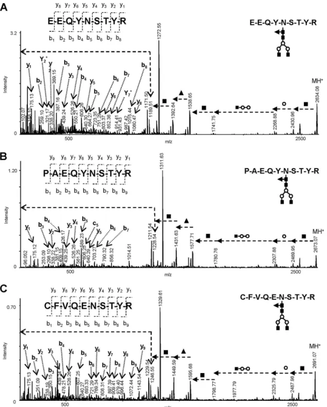
Table S3. Fragment ions detected at m/z 175.12 (y1), 298.10 (b3), 388.22 (y2), 439.25 (y3), 526.26 (y4), 589.23 (b5), 640.27 (y5), 703.27 (b6), and 790.32 (b7) indicated a peptide of se-quence PAEQYNSTYR or APEQYNSTYR or EPAQYNSTYR. The further ions observed at m/z 98.067 (b1), 804.35 (y6), and 932.40 (y7) in the CID spectrum recorded for [M⫹H]⫹ precur-sor ions with m/z 1229.54 observed after PNGase
deglyco-FIG. 3. Selected MALDI-MS/MS spectra of tryptic glycopeptides analyzed in cancer cells after treatment for [MⴙH]ⴙprecursor ions. A, m/z 2634. 01 (CEM/Her-Lipoplex); B, m/z 2673.07 (MCF-7/Her-Lipoplex; C, m/z 2691.07 (CEM/Her).
sylation, provided more evidence in favor the amino acid sequence PAEQYDSTYR (supplemental Fig. S5).
In the spectra of cells treated with Herceptin only, additional significant peaks of glycoconjugates were detected at m/z 2545.00, 2691.06, 2853.11, and 3015.20 (Fig. 2B; m/z values in blue). The compositions of these glycopeptides were again consistent with the biantennary fucosylated glycans as de-tected in IgG1, and differed by amino acid residues. For example, Fig. 3C shows MS/MS spectrum recorded for [M⫹H]⫹precursor ions at m/z 2691.07. After the initial loss of
HexNAc (m/z 2488) and Hex (m/z 2326), the next cleavage was associated with the loss of HexNAc2Hex2 residue
de-tected at m/z 1595. The subsequent loss of 146 units ob-served at m/z 1499 indicated linkage of Fuc at the GlcNAc residue attached to the asparagine. The final cleavage of 203 units produced a characteristic peak associated with a pep-tide at m/z 1246.55. The fragment ions observed next at m/z 175.12, 338.19, 439.24, 526.25, 640.30, 769.34, 897.40, 996.45, and 1143.54 could be assigned to y-type cleavages from amino acid sequence CFVQENSTYR. The existence of this peptide can be strongly supported by a number of b-type fragment ions accompanied with a characteristic loss of 28 u. The new glycopeptides discussed above did not generate MASCOT scores and to our knowledge have not been de-scribed previously; their incidence was evidently associated with the response of cancer cells to Herceptin treatment (Fig. 4).
DISCUSSION
In our work we used two cancer cell lines to study viability of cells before and after treatment with Herceptin on different conditions. Although this model of treatment does not fully
resemble to real treatment in human, it can provide important information about molecular changes associated with treat-ment. In one set of experiments the cancer cells were treated with the antibody alone for 72 h. This time showed to be the optimal time regarding decreased viability. In the second set, the same cells were exposed to the antibody combined with Lipoplex and in this case for the same time cancer cells were eradicated even more effectively. It is believe that LipA plays a direct role in plasmid DNA delivery into cell cytoplasm as well as in destabilization of the plasma membrane (39) and can allow more effective antibody targeting. Both types of these experiments showed also differences at the proteome level. The monitoring of N-glycans in cancer cells before and after exposure to the antibody showed significant alterations in glycosylation (19). Dominant high-mannose glycans (Man5–9GlcNAc2) accompanied by less abundant of
fucosy-lated tri- and tetrantennary higher sialyfucosy-lated glycans were found in both original cancer cells and after treatment these structures were replaced by complex biantennary fucosylated oligosaccharides of the same compositions as found in IgG1. A number of additional significant oligosaccharide peaks, par-ticularly corresponding to nonfucosylated triantennary galac-tosylated structures, which were present neither in the anti-body nor in the original cancer cells, were found in cells treated only with Herceptin and were not detected when Herceptin was combined with LipA. Thus, the original goal of this work was focused on the study of glycosylated peptides isolated from cancer cells after treatment and confirming gly-coprotein associated with the observed N-glycans changes.
It is expected that during treatment, antibody targeted can-cer cells are driven into apoptosis, proteins/glycoproteins present in the original cancer cells can be expected to be suppressed and IgG1 molecules would be among major biomolecules present and analyzed in treated cells. Therefore, we expected major glycopeptides analyzed in treated sam-ples to be of the same compositions as found in IgG1 (see m/z values in black in Figs. 2A–2C); their MS/MS fragmentation patterns confirmed a characteristic peptide peak at m/z 1189.51 with the amino acid sequence—EEQYNSTYR (Fig. 3A).
According glycopeptide analysis, the major oligosaccha-rides detected in all treated cells corresponded to biantennary fucosylated complex type, differing in the level of terminal galactose. Other minor forms of glycans as sialylated complex types, and other less mature structures such as high-man-nose (Man5GlcNAc2) or hybrid structure were found in all
treated samples. The comparison of glycans profiles obtained from these samples through glycopeptide analysis versus that obtained by PNGaseF release from total cell lysates showed good similarity for both types of cancer cells which were transfected with Herceptin/Lipoplex (Fig. 5B, 5C). In the ex-periments of which cancer cells were exposed to Herceptin without LipA, the glycopeptides analysis confirmed the same glycans types as above mentioned (Fig. 5D, 5E, charts in FIG. 4. The pie charts illustrating the occurrence of
glycopep-tides detected in:A, Herceptin; B, cancer cells after treatment with Herceptin for 72 h; and C, cancer cells after transfection with Her-ceptin/Lipoplex for 72 h. The proportions of the glycopeptides, dif-ferentiated according their m/z of peptide backbone, were obtained from average of normalized intensities over four sets of experiments. Glycopeptides with peptide of m/z 1033.5 were observed only from treated CEM cells digested under nonreducing conditions and were excluded from this evaluation.
black). However, none of the peptides bearing higher amount
of nonfucosylated biantennary and triantennary galactosy-lated oligosaccharides, including non-fucosygalactosy-lated trianten-nary higher sialylated structures were detected as it was observed after direct PNGaseF release from the whole cell lysates (Fig. 5D, 5E, dotted charts). In our previous study on the investigation of oligosaccharides from glycopeptides was
shown that the relative abundances of individual oligosaccha-rides were consistent with those of the glycopeptides (21). Therefore, according to glycan profiles obtained from cancer cells after treatment with Herceptin without Lipoplex, by two different approaches, the seemingly absence of glycosylated peptides bearing nonfucosylated galactosylated oligosaccha-rides could be explained by suppression of abundant
nongly-FIG. 5. Schematic diagram depicted the significant N-glycan structures analyzed: in original antibody (A); from cancer CEM (B) and MCF-7 (C) cells after exposure to Herceptin/LipA; and from CEM (D) and MCF-7 (E) cells after Herceptin treatment. Graphs show integrated MS peak intensities of the glycopeptides or glycans over the range of their isotopic envelopes, and then normalized.
cosylated peptides present in the fractions. It is also possible that masses of these glycopeptides were beyond the upper mass range set for the instrument (5000 Da). Although the identification of the glycoprotein associated with those non-fucosylated galactosylated glycans require another mass spectrometric approach or methodological procedure, evi-dently they are from other glycoprotein than IgG1, of which growth was a result of Herceptin treatment without the pres-ence of LipA. It should be emphasized that none of glycopep-tides or glycans analyzed in treated cells were detected in original MCF-7 and CEM cells.
It was surprising to learn that the MS/MS spectra acquired from new aberrant glycopeptides analyzed in all treated cells provided evidence only for the glycans of the same compo-sitions as found in IgG1, however they differed in peptide sequences. Among them, the most significant peaks corre-sponded to glycoconjugates with peptides PAEQYNSTYR and CFVQENSTYR (violet and blue m/z values in Figs. 2A, 2B). Low abundant glycopeptides detected at m/z 2802.13 and 2640.11 from CEM cells after transfection with Herceptin/LipA (supplemental Fig. S3a) have been previously observed in the study of Fc domain of immunoadhesine affected by drugs (40).
According to MS/MS fragmentation patterns all newly de-tected peptides, including those with not fully assigned amino acid sequences, showed NSTYR motif as a part of their sequence as found in IgG1 and moreover were bearing the same type glycans of the same profiles as were analyzed in the original antibody. These facts and the consistency of the data presented on two different cancer cell models suggest that all of these new glycosylated peptides described here are results of unknown transformation in the IgG1 molecule itself during treatment. As we have previously shown, transfection of cancer cells with Herceptin/Lipoplex decreased the viability of these cells to a significantly higher degree than treatment with Herceptin alone (29). Lipofectamine is known to alter the plasma membrane but it remains unclear how it enhances the effects of Herceptin.
Although further studies are needed to identify regulatory pathways that cause alterations in glycoconjugates, in the present study we have shown that more new aberrant glyco-peptides were found in cancer cells treated with Herceptin alone, than in cells exposed to Herceptin/Lipoplex. The amino acid sequences of the analyzed peptides and the glycan structures indicate that these newly described glycopeptides might be the result of intracellular degradation of Herceptin. Therefore it is worthy to study in future, whether the cotreat-ment with Lipoplex results in altered uptake of Herceptin, localization of the Herceptin/Her2 complex to different plasma membrane domains or a change in the intracellular compart-mentalization of Herceptin. Nonetheless, our results indicate that presence of LipA could play a role not only in better surface targeting, but also prevents antibody from degrada-tion and elongates its proper funcdegrada-tion during treatment. In
conclusion, the results discussed in this study indicate that treatment is more complex process and involves series path-ways on different molecular levels. The precise evaluation and better understanding of changes in glycoconjugates pre-sented here might provide new insight how to improve treat-ment involving monoclonal antibody.
Acknowledgments—We thank Prof. Mike Butler and Dr. Maureen
Spearman (Department of Microbiology, the University of Manitoba) for reading the manuscript and valuable discussion.
We have no conflicting financial interest.
* This research was supported by grants from the Natural Sciences and Engineering Research Council of Canada (NSERC), from the Canadian Foundation for Innovation (CFI), the Canada Research Chairs Program (CRC) and the Canadian Breast Cancer Alliance (CBCRA).
□S This article containssupplemental Figs. S1 to S5 and Tables S1 to S5.
¶¶ To whom correspondence should be addressed: Chemistry De-partment, University of Manitoba, 144 Dysart Road, Winnipeg, MB R3T 2N2, Canada. Tel.: 204-474-6561; Fax: 204-474-7608; E-mail: lattovae@cc.umanitoba.ca; NRC, Institute for Biodiagnotics, 3330 Hospital Drive NW Calgary, Alberta T2N 4N1, Canada. Tel.: 403-221-3222; Fax: 403-221-3230; E-mail: Boguslaw.Tomanek@nrc-cnrc. gc.ca.
REFERENCES
1. Bertozzi, C. R., and Kiessling, L. L. (2001) Chemical glycobiology. Science 291,2357–2364
2. Ogata, S., Ho, I., Chen, A., Dubois, D., Maklansky, J., Singhal, A., Ha-komori, S., and Itzkowitz, S. H. (1995) Tumor-associated sialylated an-tigens are constitutively expressed in normal human colonic mucosa. Cancer Res. 55, 1869 –1874
3. Danishefsky, S. J., and Allen, J. R. (2000) From the laboratory to the clinic: a retrospective on fully synthetic carbohydrate-based anticancer vac-cines frequently used abbreviations are listed in the appendix. Angew. Chem. Int. Ed. Engl. 39, 836 – 863
4. Parodi, A. J. (2000) Protein glucosylation and its role in protein folding. Annu. Rev. Biochem. 69, 69 –93
5. Walsh, G., and Jefferis, R. (2006) Post-translational modifications in the context of therapeutic proteins. Nat. Biotechnol. 24, 1241–1252 6. Kanda, Y., Yamada, T., Mori, K., Okazaki, A., Inoue, M., Kitajima-Miyama,
K., Kuni-Kamochi, R., Nakano, R., Yano, K., Kakita, S., Shitara, K., and Satoh, M. (2007) Comparison of biological activity among nonfucosy-lated therapeutic igg1 antibodies with three different n-linked fc oligo-saccharides: the high-mannose, hybrid, and complex types. Glycobiol-ogy 17, 104 –118
7. Suzuki, E., Niwa, R., Saji, S., Muta, M., Hirose, M., Iida, S., Shiotsu, Y., Satoh, M., Shitara, K., Kondo, M., and Toi, M. (2007) A nonfucosylated anti-her2 antibody augments antibody-dependent cellular cytotoxicity in breast cancer patients. Clin. Cancer Res. 13, 1875–1882
8. Jefferis, R. (2009) Recombinant antibody therapeutics: the impact of gly-cosylation on mechanisms of action. Trends Pharmacol. Sci. 30, 356 –362
9. Jefferis, R. (2005) Glycosylation of recombinant antibody therapeutics. Biotechnol. Prog. 21, 11–16
10. Kamoda, S., Nomura, C., Kinoshita, M., Nishiura, S., Ishikawa, R., Kakehi, K., Kawasaki, N., and Hayakawa, T. (2004) Profiling analysis of oligosac-charides in antibody pharmaceuticals by capillary electrophoresis. J Chromatogr A 1050, 211–216
11. Jefferis, R. (2009) Glycosylation of antibody therapeutics: optimisation for purpose. Methods Mol. Biol. 483, 223–238
12. Slamon, D. J., Leyland-Jones, B., Shak, S., Fuchs, H., Paton, V., Baja-monde, A., Fleming, T., Eiermann, W., Wolter, J., Pegram, M., Baselga, J., and Norton, L. (2001) Use of chemotherapy plus a monoclonal anti-body against her2 for metastatic breast cancer that overexpresses her2. N. Engl. J. Med. 344, 783–792
13. Mu¨ller, M. R., Gru¨nebach, F., Kayser, K., Vogel, W., Nencioni, A., Brugger, W., Kanz, L., and Brossart, P. (2003) Expression of her-2/neu on acute lymphoblastic leukemias: implications for the development of immuno-therapeutic approaches. Clin. Cancer Res. 9, 3448 –3453
14. Musolino, A., Naldi, N., Bortesi, B., Pezzuolo, D., Capelletti, M., Missale, G., Laccabue, D., Zerbini, A., Camisa, R., Bisagni, G., Neri, T. M., and Ardizzoni, A. (2008) Immunoglobulin g fragment c receptor polymor-phisms and clinical efficacy of trastuzumab-based therapy in patients with her-2/neu-positive metastatic breast cancer. J. Clin. Oncol. 26, 1789 –1796
15. Jones, A. (2003) Combining trastuzumab (herceptin) with hormonal therapy in breast cancer: what can be expected and why?. Ann. Oncol. 14, 1697–1704
16. Romond, E. H., Perez, E. A., Bryant, J., Suman, V. J., Geyer, C. E., Jr, Davidson, N. E., Tan-Chiu, E., Martino, S., Paik, S., Kaufman, P. A., Swain, S. M., Pisansky, T. M., Fehrenbacher, L., Kutteh, L. A., Vogel, V. G., Visscher, D. W., Yothers, G., Jenkins, R. B., Brown, A. M., Dakhil, S. R., Mamounas, E. P., Lingle, W. L., Klein, P. M., Ingle, J. N., and Wolmark, N. (2005) Trastuzumab plus adjuvant chemotherapy for oper-able her2-positive breast cancer. N. Engl. J. Med. 353, 1673–1684 17. Arnould, L., Gelly, M., Penault-Llorca, F., Benoit, L., Bonnetain, F., Migeon,
C., Cabaret, V., Fermeaux, V., Bertheau, P., Garnier, J., Jeannin, J. F., and Coudert, B. (2006) Trastuzumab-based treatment of her2-positive breast cancer: an antibody-dependent cellular cytotoxicity mechanism?
Br. J. Cancer 94, 259 –267
18. Toi, M., Horiguchi, K., Bando, H., Saji, S., and Chow, L. W. C. (2005) Trastuzumab: updates and future issues. Cancer Chemother. Pharmacol.
56 Suppl 1,94 –99
19. Lattova´, E., Tomanek, B., Bartusik, D., and Perreault, H. (2010) N-glycomic changes in human breast carcinoma mcf-7 and t-lymphoblastoid cells after treatment with herceptin and herceptin/lipoplex. J. Proteome Res.
9,1533–1540
20. Håkansson, K., Cooper, H. J., Emmett, M. R., Costello, C. E., Marshall, A. G., and Nilsson, C. L. (2001) Electron capture dissociation and infrared multiphoton dissociation ms/ms of an n-glycosylated tryptic peptic to yield complementary sequence information. Anal. Chem. 73, 4530 – 4536 21. Lattova´, E., Kapkova´, P., Krokhin, O., and Perreault, H. (2006) Method for investigation of oligosaccharides from glycopeptides: direct determina-tion of glycosyladetermina-tion sites in proteins. Anal. Chem. 78, 2977–2984 22. Go, E. P., Rebecchi, K. R., Dalpathado, D. S., Bandu, M. L., Zhang, Y., and
Desaire, H. (2007) Glycopep db: a tool for glycopeptide analysis using a “smart search”. Anal. Chem. 79, 1708 –1713
23. Carapito, C., Klemm, C., Aebersold, R., and Domon, B. (2009) Systematic lc-ms analysis of labile post-translational modifications in complex mix-tures. J. Proteome Res. 8, 2608 –2614
24. Tzur, Y., Markovich, A., and Lichtenstein, R. G. (2008) A two-dimensional array for simultaneous sequencing of n- and o-glycans and their glyco-forms on specific glycosylation sites. J. Proteome Res. 7, 1188 –1198 25. Gong, B., Cukan, M., Fisher, R., Li, H., Stadheim, T. A., and Gerngross, T.
(2009) Characterization of n-linked glycosylation on recombinant glyco-proteins produced in pichia pastoris using esi-ms and maldi-tof.
Meth-ods Mol. Biol. 534, 213–223
26. Dodds, E. D., Seipert, R. R., Clowers, B. H., German, J. B., and Lebrilla, C. B. (2009) Analytical performance of immobilized pronase for peptide footprinting and implications for surpassing reductionist glyco-proteomics. J. Proteome Res. 8, 502–512
27. Balog, C. I., Mayboroda, O. A., Wuhrer, M., Hokke, C. H., Deelder, A. M.,
and Hensbergen, P. J. (2010) Mass spectrometric identification of aber-rantly glycosylated human apolipoprotein c-iii peptides in urine from schistosoma mansoni-infected individuals. Mol. Cell Proteomics 9, 667– 681
28. Scott, N. E., Parker, B. L., Connolly, A. M., Paulech, J., Edwards, A. V. G., Crossett, B., Falconer, L., Kolarich, D., Djordjevic, S. P., Højrup, P., Packer, N. H., Larsen, M. R., and Cordwell, S. J. (2011) Simultaneous glycan-peptide characterization using hydrophilic interaction chroma-tography and parallel fragmentation by cid, higher energy collisional dissociation, and electron transfer dissociation ms applied to the n-linked glycoproteome of campylobacter jejuni. Mol. Cell Proteomics 10, M000031MCP201
29. Bartusik, D., Tomanek, B., Lattova´, E., Perreault, H., and Fallone, G. (2010) Combined treatment of human mcf-7 breast carcinoma with antibody, cationic lipid and hyaluronic acid using ex vivo assays. J Pharm Biomed
Anal 51, 192–201
30. Bartusik, D., Tomanek, B., Lattova´, E., Perreault, H., and Fallone, G. (2010) Ex vivo assays of cem cells cultured and treated in the three dimensional cultures. Biomed. Pharmacother. 64, 390 –395
31. Turk, T. M., Rees, M. A., Pietrow, P., Myers, C. E., Mills, S. E., and Gillenwater, J. Y. (1999) Determination of optimal freezing parameters of human prostate cancer in a nude mouse model. Prostate 38, 137–143 32. Lattova´, E., McKenzie, E. J., Gruwel, M. L., Spicer, V., Goldman, R., and
Perreault, H. (2009) Mass spectrometric study of n-glycans from serum of woodchucks with liver cancer. Rapid Commun. Mass Spectrom. 23, 2983–2995
33. Loboda, A. V., Krutchinsky, A. N., Bromirski, M., Ens, W., and Standing, K. G. (2000) A tandem quadrupole/time-of-flight mass spectrometer with a matrix-assisted laser desorption/ionization source: design and per-formance. Rapid Commun. Mass Spectrom. 14, 1047–1057
34. Roepstorff, P., and Fohlman, J. (1984) Proposal for a common nomencla-ture for sequence ions in mass spectra of peptides. Biomed. Mass
Spectrom. 11, 601
35. Lattova´, E., Chen, V. C., Varma, S., Bezabeh, T., and Perreault, H. (2007) Matrix-assisted laser desorption/ionization on-target method for the in-vestigation of oligosaccharides and glycosylation sites in glycopeptides and glycoproteins. Rapid Commun. Mass Spectrom. 21, 1644 –1650 36. Lattova, E., Perreault, H., and Krokhin, O. (2004) Matrix-assisted laser
desorption/ionization tandem mass spectrometry and post-source de-cay fragmentation study of phenylhydrazones of n-linked oligosaccha-rides from ovalbumin. J. Am. Soc. Mass Spectrom. 15, 725–735 37. Damen, C. W., Chen, W., Chakraborty, A. B., van Oosterhout, M., Mazzeo,
J. R., Gebler, J. C., Schellens, J. H., Rosing, H., and Beijnen, J. H. (2009) Electrospray ionization quadrupole ion-mobility time-of-flight mass spectrometry as a tool to distinguish the lot-to-lot heterogeneity in n-gly-cosylation profile of the therapeutic monoclonal antibody trastuzumab.
J. Am. Soc. Mass Spectrom. 20, 2021–2033
38. Stadlmann, J., Pabst, M., Kolarich, D., Kunert, R., and Altmann, F. (2008) Analysis of immunoglobulin glycosylation by lc-esi-ms of glycopeptides and oligosaccharides. Proteomics 8, 2858 –2871
39. Pirollo, K. F., Xu, L., and Chang, E. H. (2000) Non-viral gene delivery for p53.
Curr. Opin. Mol. Ther. 2, 168 –175
40. Jones, A. J., Papac, D. I., Chin, E. H., Keck, R., Baughman, S. A., Lin, Y. S., Kneer, J., and Battersby, J. E. (2007) Selective clearance of glycoforms of a complex glycoprotein pharmaceutical caused by terminal n-acetyl-glucosamine is similar in humans and cynomolgus monkeys.