HAL Id: tel-01806333
https://tel.archives-ouvertes.fr/tel-01806333
Submitted on 2 Jun 2018HAL is a multi-disciplinary open access
archive for the deposit and dissemination of sci-entific research documents, whether they are pub-lished or not. The documents may come from teaching and research institutions in France or abroad, or from public or private research centers.
L’archive ouverte pluridisciplinaire HAL, est destinée au dépôt et à la diffusion de documents scientifiques de niveau recherche, publiés ou non, émanant des établissements d’enseignement et de recherche français ou étrangers, des laboratoires publics ou privés.
tomato
Ruie Liu
To cite this version:
Ruie Liu. Functional analysis of active DNA demethylation in tomato. Vegetal Biology. Université de Bordeaux, 2016. English. �NNT : 2016BORD0273�. �tel-01806333�
Mlle Ruie LIU
POUR OBTENIR LE GRADE DE
DOCTEUR DE
L’UNIVERSITÉ DE BORDEAUX
Ecole doctorale des Sciences de la Vie et de la Santé
Spécialité : Biologie Végétale
FUNCTIONAL ANALYSIS OF ACTIVE DNA
DEMETHYLATION IN TOMATO
Sous la direction de : Philippe Gallusci Présentée publiquement le 29 November 2016
Membres du jury :
M Etienne Bucher : Chercheur INRA, HDR, INRA Anger, Rapporteur
M Mohamed Zouine : Maitre de conférences, HDR, INP ENSAT Université de Toulouse, Rapporteur
M Philippe Gallusci : Professeur, Université de Bordeaux, Directeur de thèse
Mme Rossitza Atanassova : Professeure, Université de Poitier, Examinatrice
M Mark Hooks : Professeur, Université de Bordeaux, Examinateur
Titre:
ANALYSE FONCTIONNELLE DE LA DEMETHYLATION D'ADN ACTIF EN TOMATERésumé
La méthylation de l'ADN génomique est l'un des principaux mécanismes épigénétiques qui conduisent à des changements stables et héréditaires de l'expression des gènes sans que cela s’accompagne de la modification de la séquence d'ADN sous-jacente. Elle fait référence à l'addition d'un groupement méthyl sur le carbone 5 des cytosines (5meC). Ces dernières années, l’étude des mécanismes régulant la mise en place et le maintien de de cette méthylation est devenu un thème de recherche importante, en raison de son rôle essentiel dans la régulation du fonctionnement du génome des plantes et des mammifères. La distribution des 5meC sur l’ensemble du génome d’un organisme, encore appelé méthylome, peut être déterminée par différentes méthodes dont le séquençage de l’ADN génomique après traitement au bisulfite de sodium (WGBS ou méthyl C séq).
Chez les végétaux, la méthylation de l’ADN peut se produire dans tous les contextes de séquence incluant les motifs symétriques CG et CHG et le contexte dissymétrique CHH (H pouvant être A, T ou C). En fonction du contexte de séquence, la méthylation des cytosines est mise en place et maintenue par trois types différents d'ADN méthyltransférase. Le maintien de la méthylation aux sites CG et CHG est assurée par l’ADN Méthyltransférase 1 (MET1) et par la Chromomométhylase, CMT3, respectivement. La chromomométhylase 2 (CMT2) est-elle impliquée dans le maintien des méthylation de type CHH, de même que les Domain Rearanged Methyltransferases (DRM). Ces dernières sont sont responsables de la mise en place de la méthylation de novo et sont guidées jusqu’à leur cible par des petits ARNs selon le mécanisme de «RNA directed DNA Methylation » (RdDM). Enfin la méthylation de l'ADN peut également être éliminée par les ADN glycosylase-lyases bifonctionnelle, également appelée les DEMETER-like DNA demethylases (DML). Chez la plante-modèle Arabidopsis, la déméthylation active de l'ADN joue un rôle essentiel dans l'empreinte maternelle et la déméthylation l’ADN génomique lors du développement de l’albumen, mais elles ne semblent pas jouer de rôle essentiel pendant le développement de la plante chez cette espèce. La méthylation de l’ADN génomique peut aussi être perdue après la réplication de l’ADN, lorsque les mécanismes devant assurer son maintien ne sont pas actifs. On parle alors de déméthylation passive de l’ADN génomique.
murissement des fruits charnus climatériques. Des études récentes ont maintenant montré que le développement et la maturation de ces fruits reposent sur la mise en place et le maintien de patrons de transcription différentielle, dont la régulation est assurée par un ensemble de processus complexes impliquant à la fois des contrôles génétiques et des régulations hormonales. Cependant, il semble que la régulation du développement et de la maturation des fruits charnus ne reposent pas basant uniquement sur les modèles génétiques, mais impliquent aussi es régulations épigénétiques. En effet, des travaux récents suggèrent que la méthylation de l'ADN pourrait également être impliquée En particulier, une diminution importante de l’abondance des 5 meC à l'échelle du génome entier et la déméthylation à certains promoteurs observés lors de la maturation des fruits de tomate.
A fin d'analyser les mécanismes moléculaires responsables de la perte de méthylation survenant lors de la maturation des fruits de tomate, le projet présenté se concentre sur l'analyse fonctionnelle des enzymes de tomate impliquées dans la déméthylation active de l'ADN génomique. Nous avons identifié 4 ADN déméthylases putatives (SlDML 1 à 4) qui contiennent toutes trois domaines hautement conservés comprenant le domaine Glycosylases qui est porteur de l’activité enzymatique et deux domaines supplémentaires A et B nécessaires à la liaison de ces enzymes à l'ADN. Dans une tentative d'étude de l'activité des protéines SlDML, les ADNcs complets codant pour les enzymes SlDML1 et 2 et ainsi que fragments de ces ADNcs codant pour des versions tronquées de la portéine SlDML2 tronquées a été clonés. La production des protéines recombinantes correspondantes dans la bactérie E Coli, n’a pas permis de démontrer leur activité biochimique.
Pour analyser les fonctions biologiques des ADN déméthylases de Tomate, des plants de tomates transgéniques altérés dans l'expression des gènes SlDML ont été générés. Ces plantes présentent de nombreuses altérations du développement, parmi lesquelles des modifications de l’organisation florale des fruits et de la forme des feuilles ainsi qu’une inhibition marquée de la maturation des fruits. En utilisant ces plantes, nous avons démontré que la déméthylation active de l'ADN est une exigence absolue pour que la maturation des fruits de tomate puisse avoir lieu. En particulier nous avons mis en évidence un lien de cause à effet direct entre la déméthylation active de l'ADN principalement médiée par une des quatres DML de tomate, SlDML 2 et la maturation des fruits. Les plantes dont l’expression du gène SlDML2 est réduite présente une inhibition de la maturation consécutive à l’hyperméthylation et la répression de l'expression des gènes codant pour les facteurs de transcription contrôlant la maturation des fruits (Ripening
l’accumulation des caroténoïdes (Phytoène Synthase 1, PSY1).
A fin de déterminer si les phénotypes des plantes transgéniques (développement affectant les fruits, les fleurs ainsi que le développement des feuilles) sont héréditaires après la perte du transgène par ségrégation, leur stabilité a été étudiée sur plusieurs générations. Dans tous les cas, les phénotypes sont perdus et les plantes après perte du transgène semblent en tout point identique à des plants de tomate sauvage, ce qui suggère une absence d'héritabilité des modifications induites par la réduction d’expression des gènes SlDML1 et 2. Cependant, nous ne pouvons exclure que certains patrons anormaux de méthylation, liés ou non aux phénotypes observés, sont néanmoins hérités. Il est possible que la perte des phénotypes ne soit pas due à une non-héritabilité d’empreintes de méthylation inappropriées à certains loci spécifiques. Cela pourrait plutôt refléter que la combinaison des états de méthylation à l’ensemble les loci nécessaire au phénotype n'est pas obtenue dans les plantes obtenues après croisement. L’analyse d’un plus grand nombre de plan, sur plusieurs générations et associé à l’analyse de la distribution des cytosines méthylées sera nécessaire pour répondre à cette question.
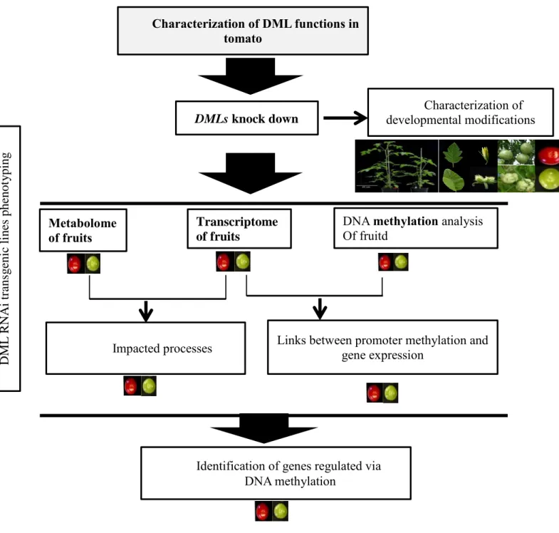
Pour déterminer, les processus contrôlés par SlDML2 dans les fruits de tomate, nous avons effectué une analyse comparative du transcriptome et du métabolome des fruits des plantes sauvages et des plantes transgéniques RNAi DML à huit étapes de développement du fruit. Ces analyses ont ensuite été corrélées aux données du méthylome de tomate déterminé à partir de fruits de tomate de la variété Ailsa craig. Ces analyses révèlent qu'en plus des gènes RIN, NOR,
CNR, PSY1 un nombre important de métabolites primaires et secondaires, et de nombreux gènes
présentent une accumulation différentielle et des patrons d'expression distincts respectivement chez les fruits transgéniques DML RNAi et chez les fruits sauvages. Par exemple, l'accumulation de caroténoïdes, la biosynthèse et la signalisation de l'éthylène, la synthèse puis la dégradation de la paroi cellulaire, mais aussi l’expression des gènes codant pour divers facteurs de transcription, et pour certains régulateurs épigénétiques, incluant une DRM, des histones dé-acétylases et différents histones déméthylases sont extrêmement affectés dans les fruits transgéniques. Ces résultats suggèrent que de nombreux gènes, y parmi lesquels ceux qui jouent des rôles essentiels pour le développement et la maturation des fruits, nécessitent d’être déméthylés pour leur expression. En conséquence, ces travaux apportent la démonstration, pour la première fois, que la déméthylation active d'ADN a des effets très globaux sur le développement et la maturation des fruits. Il est maintenant nécessaire de valider ce travail en
En conclusion, les observations présentées dans ce travail fournissent un cadre de travail permettant d’analyser les mécanismes moléculaire responsable de la déméthylation de l'ADN se produisant pendant la maturation des fruits de tomate. Ici, nous présentons une analyse complète des conséquences d’une réduction de l’expression du gène de SlDML2 sur le trancriptome et le métabolome des fruits, tout au long de leur développement. La corrélation entre les profils d’expression de gènes réalisées lors de ce travail ( variété WVA106) et les changements de la distribution de la méthylation de l’ADN telles que décrites chez la variété Ailsa craig montre qu’en plus d'un rôle général dans la régulation des gènes directement impliqués dans plusieurs voies métaboliques, plusieurs gènes codant pour des facteurs de transcription ainsi que des régulateurs épigénétiques sont également susceptibles d'être directement contrôlés par la méthylation de leur région promotrice. Cependant, nous ne pouvions pas établir une relation stricte entre la diminution de la méthylation de l'ADN et l'induction de l'expression des gènes, car de nombreux gènes présentant une diminution du niveau de méthylation de l'ADN dans leur région promotrice pendant la maturation des fruits sauvages correspondent à des gènes normalement réprimés. Ceci suggère que la méthylation active de l'ADN serait nécessaire àleur répression pendant le processus de maturation. Ainsi la relation entre la déméthylation de l'ADN et l'expression des gènes pourrait être plus complexe et ne se limiterait pas à la simple hypothèse de départ de ce travail: la déméthylation de l'ADN est nécessaire à l'expression de gènes induits au cours de la maturation. La déméthylation de active de l'ADN pourrait également être nécessaire à la répression de gènes exrimés uniquement lors des phases précoces du développement des fruits et réprimés lors du murissement.
Mots clés :
Tomate ; Déméthylation d'ADN; Mûrissement des fruits; Régions différentiellement méthylées; l'expression du gène
DEMETHYLATION IN TOMATO
Abstract
DNA methylation is one of the epigenetic mechanisms that lead to stable and heritable changes in gene expression without alteration on DNA sequence. DNA methylation refers to the addition of a methyl group to the fifth position of the cytosine ring. In recent years, DNA methylation is becoming more and more widely studied, because of its importance in mammals and plants. Methylated cytosines distribution can be determined across the genome at single-nucleotide resolution, that is methylome, using whole genome bisulfite-sequencing (BS-seq) approaches. The methylomes of an increasing number of plant species has been well described, revealing that these large-scale patterns of methylation first described for Arabidopsis are shared among flowering plants, although differences exist between plant species. In plants, cytosine methylation which occurs in all sequence context (CG, CHG, CHH, H being A, T or C) is set up and maintained by three different types of DNA methyltransferase. Methylation of symmetric CG and CHG sites can be maintained by METHYLTRANSFERASE1 (MET1) and CHROMOMETHYLASE2 (CMT2/CMT3), respectively. While maintenance of asymmetric CHH methylation relies on RNA directed DNA methylation (RdDM) or CMT2. DNA methylation can also be removed by the bifunctional DNA glycosylase-lyases, also called the DEMETER-like DNA demethylases. In the model plant Arabidopsis, active DNA demethylation plays a critical role in maternal imprinting and endosperm demethylation, but none of these functions appear to be essential for the development in this species.
Solanum lycopersicum (tomato) is an important agronomic crop and the main model to study the development and ripening process of climacteric fleshy fruit. Recent studies have now shown that the development and ripening of fleshy fruits relies on the establishment and maintenance of differential transcription patterns and complex regulatory pathways that involve both genetic and hormonal controls are operating at these developmental phases. However, it appears that a full understanding of fruit development and ripening will not be achieved based only on genetic models as suggested by recent studies, which showing an important decrease in global methylation level and demethylation at specific promoters during fruit ripening.
In order to analyze the molecular mechanisms responsible for the loss of methylation observed during tomato fruit ripening, the present project focuses on the functional analysis the tomato enzymes involved in the active demethylation of genomic DNA. As it was suggested
filled with an unmethylated cytosine through base excision pathway. As in Arabidopsis, three highly conserved domains were observed including a glycosylases domain as well as two additional domains A and B. These three domains is necessary for DNA binding and catalysis. In an attempt to study the SlDML protein activity, the tomato full length DNA glycosylase-lyases as well as different truncated recombinant proteins have been produced. Unfournatelly, none of the protein show activity in this study, a further expressional condition should be optimized. In addition, to investigate whether hypermethylated epialleles generated in the transgenic plants can be inherited after the transgene has been lost by segregation, the stability across generations of the developmental alterations affecting flower as well as leaf development was studied. As a result, T4 plants show us that the phenotypes reversed to WT phenotype once the transgene was out segregated, suggesting an absence of heritability of the modifications induced by SlDML2 knock down. However, we cannot rule out that some abnormal methylation patterns linked or not to these apparent phenotypes have been inherited. In addition, it is not known how many loci are involved in generating the flower and leaf abnormalities. Hence, it is possible that lack of phenotypes is not due to the non-heritability of the improper methylation state at specific loci. It may reflect that the correct combination of homozygous methylation state at all required loci was not obtained. Further generation and screening of more important plant population will be necessary to answer this point.
After characterizing the gene family encoding the tomato DNA demethylases, transgenic tomato plants impaired in the expression of SlDML genes have been generated. These plants present several developmental alterations, including inhibition of fruit ripening, modifications of flower, fruit and leaf shape. Using these plants, we have demonstrated that active DNA demethylation is an absolute requirement for tomato fruit ripening to occur. We show a direct cause and effect relationship between active DNA demethylation mainly mediated by one tomato DML, SlDML2, and fruit ripening. RNAi SlDML2 knockdown results in ripening inhibition via hypermethylation and repression of the expression of genes encoding ripening transcription factors (RIN, NOR, CNR) and rate-limiting enzymes of key biochemical processes (PSY1).
In recent years, the coordinated changes during tomato development and ripening was analyzed using combined transcriptome, metabolism and proteome characterization. However, it appears that a full understanding of tomato fruit development and ripening will not be achieved based only on genetic models. In addition epigenetic regulation, mainly genomic
ripening ripening. The fruit ripening defect of Cnr mutant is caused by hypermethylation of an upstream region of the CNR promoter. Zhong et al (2013) also detected that, the promoter region of several genes are demethylated during tomato fruit ripening, suggesting that DNA demethylation may play critical role during this phase of development. However, the pathways under the regulation of SlDML2 have not been comprehensively identified. With the aim to obtain a more comprehensive view of the roles of active DNA demethylation on tomato fruit development and ripening, we have performed a comparative analysis of the transcriptome and metabolome of WT and DML RNAi fruits at eight fruit development and ripening stages. These analyses was integrated with tomato epigenome determined in WT Ailsa craig plants. These analyses reveal that in addition to the four genes (RIN, NOR, CNR, PSY1) previously characterized a large number of metabolites and genes present differential accumulation and expression patterns respectively in DML RNAi transgenic fruits. Such as carotenoid, ethylene biosynthesis and signaling, cell wall synthesis and dissembling, transcription factors, and many others are extremely affected in transgenic fruits. These finding suggests that plenty of genes, including those playing essential roles for fruit development and ripening might require demethylation for their expression. Here, we present evidence for the first time that active DNA demethylation has very global effects on fruit development and ripening. Validation of this analysis will now require determining the fruit methylome of the plants impaired in active DNA demethylation.
In conclusion, the observations presented in this work provide a framework for analysis of the molecular mechanism of DNA demethylation during fruit ripening of tomato. Here, we provide a comprehensive analysis of the knock down SlDML2 on the trancriptome, metaoblom and DNA methylation in the promoter analysis. The large transcriptional reprogramming that occured in mutant during fruit ripeing was correlated alterations in DNA methylation. Here we highlight the central role of active DNA demethylation during tomato fruit ripening. In addition to a general role in the regulation of genes directly involved in several metabolic pathways, we also found that several transcription factors as well as epigenetic regulators are also likely under direct methylation control. However, we could not establish a district relationship between DNA reduction of DNA methylation and induction of gene expression, as not all DEGs containing a type-a DMRs (decreased DNA methylation during fruit ripening) do not correspond to genes normally induced in WT and repressed in transgenic plants. Some were corresponding to an opposite situation and in a few cases more complex methylation pattern
although some variations are expectable when the methylome of DML RNAi fruits will be analyzed. Hence the relationship between DNA demethylation and gene expression might be more complex than expected, and not limited to the starting hypothesis of this work: DNA demethylation is an absolute requirement for the expression of critical ripening induced genes. This is indeed clearly in this study, but the analysis presented here also suggest that DNA demethylation might also be necessary for the repression of several genes as well.
In addition, from the rencent study in Arabidopsis, ROS1 were found preferentially targets transposable elements (TEs) which are closer to protein coding genes and intergenic regions, which suggesting that ROS1 may prevent DNA methylation spreading from TEs to nearby genes. While in tomato, as our analysis, we found the methylation level of promoter of a number of genes was altered during fruit ripening, therefore, through methylome analysis, we will also get the preference of DNA methylation on TE, this analysis will give us idea that demethylation in fleshy fruit may has other distinct function as it is in Arabidopsis.
Keywords :
Tomato ; DNA demethylation ; fruit ripeing ; differently methylated regions ; gene expression
Unité de recherche
INRA-Bordeaux Aquitaine
UMR Ecophysiologie et Génomique Fonctionnelle de la Vigne Batiment Institut des Sciences de la Vigne et du Vin (ISVV) 210, chemin de Leysotte, CS 50008
Acknowledgements
This work was performed in the Ecophysiology and Functional Genomics of Vine as well as Fruit Pathology and Biology Group at INRA, University of Bordeaux from Sep.2013 to Nov.2016. The project was funded by Fulbring grant, Agence National de Recherche. I was funded by Chinese Scholorship Councile.
I would like to thank my supervisor Philippe Gallusci. I appreciate for his guidance, responsibility and positivity over these three years. He spent much time to disscuss on all the expriments I performed. His knowledge on epigenetic has been most invaluable, professional and inspiring. I also want to thank Emeline Teyssi specifically, for her discussion and guide for most experiments, as well as her particular correction in the introduction of this thesis. Both of them gave me great help on work and life. Many thanks to my group members, Linda Stammitti, Anne Bertrand, Junhua Kong, especially Linda gave me lots of useful suggestions and great help. I have been fortunate to become a member of this group.
I also can’t thank my collaborators enough, James Gavannoi for performing RNA seq and bisulifite sequencing, Alisdair Fernie and Takayuki Tohge for wide-scale metabolism analysis, Dominique Rolin for primary metabolism analysis. Yiguo Hong for VIGS silencing, Gram symour for DML expression in tomato mutants measurement. Meanwhile, I’d like to specifically thank Jame to allow me visit his lab. My eyes are widely opened on building BS-librariy during these precious three months. I also want to thanks Tohge and Alis F, who gave me idea how to do metabolism analysis and very useful guidance. Thanks to Mohamed Zouine and Elie Maza to allow me visit their lab and gaudiness on RNA seq analysis. These are precious communication. At same time, I would like to thank members in their lab, who gave me lots of suggestions and discussion. It has been a great pleasure to working with them.
My thanks to people in Ecophysiology and Functional Genomics of Vine as well as Fruit Pathology and Biology Group members, in particular to Cecil Cabassion, Michel Hernould, Pierre Baldet, for outstanding questions discussion, Esabaelle who took care my tomato plants in the last two years. Zhanwu Dai taught me network analysis and guides discussion. Classmates in these two labs were always generously sharing their knowledge and for all the stimulating and fun meetings we have had.
I can not thank enough of the members of my thesis jury, Etienne Bucher, Mohamed Zouine, Serge Delrot, Mark Hooks and Rossitza Atanassova, they gave me valuable comments and proofreading this thesis.
Mostly, I want to thank my family and friends who always support me in my life, this support is so important for me! It’s a great pleasure to have spent my time in France. Work and people here have all enriched my life and made my stay here unforgettable.
Summary
DNA methylation is one of the epigenetic mechanisms that lead to stable and heritable changes in gene expression without alteration on DNA sequence. DNA methylation refers to the addition of a methyl group to the fifth position of the cytosine ring. In recent years, DNA methylation is becoming more and more widely studied, because of its importance in mammals and plants. Methylated cytosines distribution can be determined across the genome at single-nucleotide resolution, the so-called methylome, using whole genome bisulfite-sequencing (BS-seq) approaches. The methylomes of an increasing number of plant species has been well described, revealing that these large-scale patterns of methylation first described for Arabidopsis are shared among flowering plants, although differences exist between plant species. In plants, cytosine methylation which occurs in all sequence context (CG, CHG, CHH, H being A, T or C) is set up and maintained by three different types of DNA methyltransferase. It can also be removed by the bifunctional DNA glycosylase-lyases, also called the DEMETER-like DNA demethylases. In Arabidopsis, active DNA demethylation plays a critical role in maternal imprinting and endosperm demethylation, but none of these functions appear to be essential for the development in this species.
Tomato is the main model to study the development and ripening process of climacteric fleshy fruit. Recent studies have now shown that the development and ripening of fleshy fruits relies on the establishment and maintenance of differential transcription patterns and complex regulatory pathways that involve both genetic and hormonal controls are operating at these developmental phases. However, it appears that a full understanding of fruit development and ripening will not be achieved based only on genetic models as suggested by recent studies, showing an important decrease in global methylation level and demethylation at specific pormoters during fruit ripening.
In order to analyze the molecular mechanisms responsible for the loss of methylation observed during tomato fruit ripening, the present project focuses on the functional analysis the tomato enzymes involved in the active demethylation of genomic DNA. To achieve this goal, after characterizing the gene family encoding the tomato DNA demethylases, transgenic tomato plants impaired in the expression of SlDML genes have been generated. These plants present several developmental alterations, including inhibition of fruit ripening, modifications of flower, fruit and leaf shape. Using these plants, we have demonstrated that active DNA demethylation is an absolute requirement for tomato fruit ripening to occur. We show a direct cause and effect relationship between active DNA demethylation mainly mediated by one tomato DML, SlDML2, and fruit ripening. RNAi SlDML2 knockdown results in ripening inhibition via hypermethylation and repression of the expression of genes encoding ripening transcription factors and rate-limiting enzymes of key biochemical processes. In an attempt to study the SlDML protein activity a recombinant tomato DNA glycosylase-lyases have been produced. In addition, to investigate whether hypermethylated epialleles generated in the transgenic plants can be inherited after the transgene has been lost by segregation, the stability across generations of the developmental alterations affecting flower as well as leaf development was studied.
To identify the global effect of active DNA demethylation on fruit ripening, we have compared the transcriptome and metabolome of RNAi DML plants and WT controls. This demonstrated that multiple aspects of the fruit ripening processes are affected when DNA
demethylation was impaired. Furthermore, we combined differentially methylation regions determined in Ailsa Craig which allow us identify a number of potential targets for active DNA demethylation. Validation of this analysis will now require determining the fruit methylome of the plants impaired in active DNA demethylation.
Acknowledgement ... I Summary ...II List of Figure ... VI
Chapter 1 ... 1
Introduction ... 1
I. Background: Definition of epigenetics. ... 1
1.1 Epigenetic marks ... 2
1.1.1 DNA methylation... 2
1.2 Histone posttranslational modifications... 4
2. Genome-wide distribution of methylcytosines ... 5
II. Mechanism of DNA methylation ... 8
2.1 Enzymes involved in DNA methylation in mammals. ... 8
2.2 Enzymes involved in DNA methylation in plants... 8
2.2.1 Maintenance of DNA methylation in plants ... 9
2.2.2 De novo DNA methylation in plants ... 9
2.3 Functions of DNA methylation in plants ... 11
2.3.1 Cytosine DNA methylation plays different roles during plant development 11 2.3.2 DNA methylation under environmental stress ... 14
III. DNA demethylation in plants ... 16
3.1 Passive DNA demethylation in plants ... 16
3.2 Active DNA demethylation in plants ... 16
3.2.1 Enzymes involved in DNA demethylation in plants ... 16
3.2.2 Machinery of active DNA demethylation in plants ... 16
3.3 Function of active DNA demethylation in plants... 19
3.3.1 Active DNA demethylation controls gene imprinting in the endosperm ... 19
3.3.2 Other functions of active DNA demethylation in plant ... 20
IV. The importance to screen epialleles in plants ... 23
V. Physiological changes during tomato fruit ripening... 24
5.1 Tomato fruit softening ... 25
5.3 Color change: chlorophyll degradation and carotenoid synthesis ... 28
5.4 Primary metabolites changes during tomato fruit ripening ... 29
VI. Role of DNA methylation / demethylation during fruit development and ripening ... 30
VII. Objectives of the work.………..………...……….32
Chapter 2 ... 34
Introduction ... 34
Part II. Activity test for tomato SlDMLs protein ... 60
Part III Study of the inheritance of epialleles in tomato plants after transgene segregation.. 64
Conclusion... 67
Chapter 3 ... 69
Introduction ... 69
I-Fruit Physiology and four DMLs Repression levels in transgenic fruits ... 71
II- Metabolic composition of WT and transgenic fruit ... 74
2.1 PCA analysis of metabolic compositions allows the separation of WT and transgenic fruits during ripening ... ...76
2.2 Sugars, organic acids and amino acids, pigments show different accumulation pattern in WT and transgenic fruits ... 78
2.2.1 Accumulation of sugars ... 81
2.2.2 Accumulation of amino acids ... 81
2.2.3 Accumulation of organic acids ... 81
2.2.4 Accumulation of pigments ... 82
2.3 Network analysis indicates that RNAi DML transgenic fruits display higher network density than WT ... 82
III. RNA seq analysis WT and RNAi Transgenic Lines ... 85
3.1. Summary of RNA seq data ... 85
3.2. Differential Gene Expression between WT and transgenic fruits ... 85
3.3. Comparative analysis of transgenic and WT fruits. ... 91
3.4. DEGs distribution within in each cluster ... 94
IV. Analysis of differentially methylated region, in relation to DEGs patterns. ... 98
4.2 Expression pattern of genes of the carotenoid biosynthesis pathway and association
with differentially methylated regions. ... 103
4.3 A number of Transcriptional factors from different families are associated with Differentially methylated regions ... 106
4.4 Genes Encoding Cell Wall–Biosynthesis and Modifying Protein are potentially regulated by demethylation ... 110
4.5 Genes involved in Ethylene biosynthesis, Perception and response ... 113
4.6 Other Hormone Biosynthesis and Responses in DML RNAi transgenic fruits ... 116
4.7 DMLs may interact with other Epigenetic regulators in tomato fruits ... 120
V. Conclusion and discussion ... 122
5.1 Metabolomic and RNA seq data show that DNA methylation affect multiple aspects of fruit ripening ... 122
5.1.1-Mainly fruit ripening is affected ... 122
5.1.2-Effects on primary metabolites are complex ... 123
5.1.3-Inhibition of carotenoid results from the repression of genes of the carotenoid pathway... 124
5.1.4 Cell wall ... 124
5.2 Many genes with impaired regulation in transgenic fruits are not associated with specific DMRs ... 125
5.3 Demethylation might not be strictly associated with gene induction during fruit ripening. ... 126
5.4 Some DEGs are associated with an increase in DNA methylation during fruit ripening.………...……….128
5.5 Active DNA methylation: a more general model………...128
Conclusion... 130
Chapter 4 ... 141
Discussion ... 141
Materials and methods... 149
Chapter1_ Reference ... 156
Chapter 2_ Reference ... 164
List of Figures
Fig 1. 1 Genome-wide methylation levels for different cytosine context (CG, CHG, and
CHH) in different planant species. ... 3
Fig 1. 2 Structure of a nucleosome. ... 4
Fig 1.3 N termini and C termini of the core histones and their residue-specific epigenetic modifications at four-nucleosome core histones. ... 5
Fig 1. 4 Epigenome organization in Arabidopsis and maize. ... 7
Fig 1. 5 Association of DNA cytosine methylation, TE density, small RNA and gene expression in tomato... 7
Fig 1. 6 Mechanism of RdDM in plant. ... 10
Fig 1. 7 Genes with DNA methylation variations during Arabidopsis floral development. .. 13
Fig 1. 8 Active DNA demethylation through direct base excision repair pathway in plants 17 Fig 1. 9 Working model for the IDM1-IDM2-IDL1-MBD7 complex functioning in ROS1 mediated active ... 19
Fig 1. 10 Phenotypic of epidermal patterning in the ros1 and rdd mutants and and promoter DNA methylation of EPF2 in Arabidopsis. ... 22
Fig 1. 11 Natural epimutant of Linaria vulgaris fowers and tomato CNR. ... 24
Fig 1. 12 Overview of ripening regulation in tomato fruits. ... 25
Fig 1. 13 Simplified scheme showing ethylene biosynthesis and response ... 27
Fig 1. 14 Different types of fruit peel pigment patterns in ‘Honeycrisp’ apple and Methylation levels in ‘Honeycrisp’ evaluated using bisulfite sequencing... 31
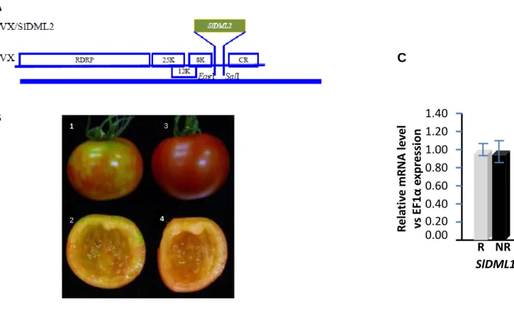
Fig 2. 1 Structures of SlDML protein and its Arabidopsis homologous proteins.. ... 60
Fig 2. 2 Structures of SlDML2 proteins along with their truncated versions used in this study ... 61
Fig 2. 3 Cloning vector and expression vector used in this study ... 61
Fig 2. 4 SlDML2- full leng, SlDML3-full length and SlDML2-845 expression and purify test... 62
Fig 2. 5 Mechanism of DNA demethylase incision assays (ref) and the acitivity test on SlDML2-845, SlDML2-962 and MspI. ... 63
Fig 2. 6 Fruit phenotype of azygous and other defects on line 1 and line 2 as well as process of screening T4 generation of line 2 and line 1 ... 65
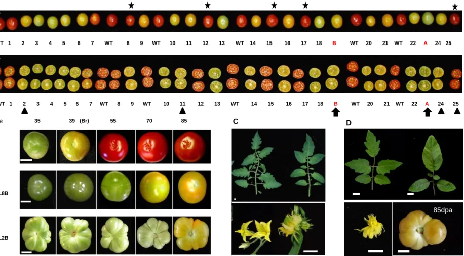
Fig 3. 1 Phenotypes of tomato DML RNAi fruits and DML expression patterns ... 73
Fig 3. 2 Characterization of metabolite content in WT1 and in transgenic RNAi fruits... .... 75
Fig 3. 3 Principal component analysis of metabolic profiles of WT1 and line 2 fruits during development and ripening. ... 77
Fig 3. 4 Clustering of metabolic profiles in WT1 and line 2Y fruits during fruit development, ripening and later stages. ... 79
Fig 3. 5 Significant metabolite changes in WT1 and line2 tomato fruits (A) and other metabolites that were measured in this study(B). ... 80
Fig 3. 6 Metabolite network of correlations in WT fruit and DML2 fruit. ... 84
Fig 3. 7 Relationship of tomato fruit pericarp related transcript expression profiles and differentially expressed genes... 87
Fig 3. 8 Difference of DEGs numbers at different stages and venne diagramm of commone DEGs between Line 2Y, Line2X and Line 8. ... 90
Fig 3. 9 Principal component analysis of RNA seq data and DEGs profiles of WT1 and line 2 fruits during development, ripening and later stages. ... 92
Fig 3. 10 clusters of DEGs between WT1 and line 2Y ... 93
Fig 3. 11 Enrichment of DEGs in each group. Gene numbers in each category were overrepresented. ... 95
Fig 3. 12 PCA of all DE genes in three groups onto the 1/2 subspace with Log2FC of Line2Y/WT. ... 102
Fig 3. 13 Expression of genes of the Carotenoid pathway in RNAi DML transgenic fruit compared to WT. ... 105
Fig 3. 14 DEG with DMR encoding Transcription factor that were repressed at least at one stage in transgenic fruit. ... 108
Fig 3. 15 Profile of DEGs involved in cell wall metabolism and fruit firmess ... 112
Fig 3. 16. Profile of Ethylene-related DEGs. ... 115
Fig 3. 17 Profile of hormone related DE genes.. ... 118
Fig 3. 18 DEGs corresponding to genes involved in DNA demethylation, DNA methylation, histone modification.. ... 121
Fig3. 19. Proposed model of fruit ripening regulation. .. ... 129
Fig 3.S 1 Characterization of metabolite content in WT1 and in transgenic RNAi fruits Line 2Y/2X ... 131
Fig 3.S 2 Characterization of metabolite content in WT2 and in transgenic RNAi fruits Line 8... 132
Fig 3.S 3 Principal component analysis of metabolic profiles of WT2 and line 8 fruits during development and ripening. ... 133 Fig 3.S 4 Metabolite network of correlations in WT fruit and DML2 fruit. ... 134 Fig 3.S 5 Row counts control in each tissue. ... 135 Fig 3.S 6 Relationship of tomato fruit pericarp related transcript expression profiles and
differentially expressed genes ... 136 Fig 3.S 7 Principal component analysis of RNA seq data and DEGs profiles of WT2 and
line 8 fruits during development, ripening and later stages. ... 137 Fig 3.S 8 Expression of genes of the Carotenoid pathway in RNAi DML transgenic fruit
compared to WT. ... 138 Fig 3.S 9 Profile of DEGs involved in cell wall metabolism and fruit firmess ... 139 Fig 3.S 10 Profile of Ethylene-related DEGs in line 8. ... 140
List of Tables
Table 1. DEGs distribuation and DMRs type in each cluster and group. ... ...95 Table 2. Enrichment of MapMan functional categories (BINs) in the DEGs associated with
type-a DMR in group 1, 2 and 3 ... 101 Table 3. DEGs with DMR that labeled with selected genes in Line 2Y ... 102 Table 4 Transcription factor with DMR as top expressed genes and RIN target genes with
DMR. ... 109
List of Supplementary Tables (provided as additional excel file online)
Table S1. Significent correlations in different WT and lines………... TabeS2. Reads obtained by RNA seq after clean and mapped percentage for all the genotype and three replicates………..
TableS3. Differentially expressed genes that only found in Line 2 (TableS3_1) or Line 8 (TableS3_2 ) and DEGs that specific differentially expressed at 46dpa (TableS3_3)………..
TableS4. Enrichment of MapMan functional categories (BINs) in the common DEGs……… TableS5. All differentially expressed Transcription Factors of common DEG………. TableS6. All differentially expressed cell wall related genes in group 1 and group 2 and 3…. TableS7. All differentially expressed genes of other hormones……….
TableS8. All differentially expressed genes involved in amino acids and Nitrite metabolism... TableS9. The ratio of all differentially expressed genes with DNA methylation level……...
Chapter1_General Introduction
DNA methylation in plants
Chapter 1
Introduction
The introduction part of this manuscript is a review of the current state of the art concerning epigenetic in plant. All epigenetic mechanisms are not detailed, and the following text mainly focuses on DNA methylation and demethylation. Histone post-translational modifications (HPTMs) are also briefly considered, because cross-talks have been described between DNA methylation and HPMTs, leading to specific combinations of epigenetic marks along the genome, as revealed by genome-wide studies (Roudier et al., 2011). The introduction part is organized into six sections. Part I of chapter I presents general notions about epigenetic marks and how they are distributed along plant genomes. Part II and III ofchapter I focus on DNA methylation and demethylation, including the description of the various components controlling these epigenetic modifications, as well as their biological functions; (II: Mechanism of DNA methylation; III: DNA demethylation in plants); PartIV of chapter I is a brief summary of the importance of epialleles in plants; Part V of Chapter I introduces tomato fruit development nd ripening and related physiological changes; Part VI summarizes the current knowledge of the role of DNA methylation / demethylation during fruit development and ripening when the work presented here was started.
I. Background: Definition of epigenetics
The definition of the term "epigenetics" has evolved over time. In the early 1940s, epigenetics was first defined by Conard Waddington as “the branch of biology which studies the causal interactions between genes and their products which bring the phenotype into being” (Waddintton, 1942; 1968). In other words, here epigenetics designs all molecular processes controlling “the expression of a genotype into a particular phenotype” (Dupont et al., 2009). Obviously, this definition is broad and not precise. It includes many different mechanisms that can modulate phenotype such as post transcriptional regulation, non-coding RNA regulation, (Holoch and Moazed 2015). Since that time, epigenetics has been redefined several times, becoming more and more specific and precise. By the middle 1990s, it has turned from causal interactions between genes and their products to chromosomal modifications that had the potential to modify gene expression during development. But today epigenetics is commonly used to precisely mean “the study of mitotically and/or meiotically heritable changes in patterns of gene expression that occur without alterations in DNA sequence” (Iwasaki and Paszkowski 2014). This definition is still evolving and was recently suggested to also include stable marks that although not heritable may lead to stable alteration of the transcriptional programing of specific cells (Avramova 2015) as
indicated by the roadmap consortium of epigenomics
(http://www.roadmapepigenomics.org/overview).
At present, it is widely accepted that posttranslational histone modifications, DNA methylation and certain non-coding RNA-mediated epigenetic regulations (Holoch and Moazed 2015) constitute epigenetic mechanisms which are critically important in modulating the structure
of chromatin. Chromatin, which is only found in eukaryotic cells, designs a complex and organized structure made of proteins, DNA and RNA. The structural unit of chromatin, the nucleosome, consists of 146 bp of DNA wrapped around a protein core made of 4 histones dimers. The chromatin allows the organization and compaction of the genetic material into the nucleus. Along each chromosome, chromatin is organized into transcriptionally active less condensed euchromatin, and transcriptionally inactive highly condensed heterochromatin. But chromatin structure is highly dynamic, and may undergo changes during development or in response to environmental signals. Because epigenetic mechanisms govern these modifications in chromatin structure, they impact DNA accessibility for all DNA-template processes including gene transcription (Lauria and Rossi 2011), DNA recombination (Choi and Henderson 2015) and transposition (Mirouze and Vitte 2014). In the following text, only the role of epigenetic regulations in gene expression is described, the other processes using DNA as a template are not discussed.
The epigenetic regulation of the genome activity relies on different mechanisms. Some of these mechanisms involve chromatin modifiers, which are responsible for covalent modifications in chromatin, including DNA methylation, and histone post-translational modifications (HPTMs), the so-called epigenetic marks. Other epigenetic mechanisms involve chromatin remodelers, which non-covalently modify chromatin structure by changing the nucleosome position, destabilizing nucleosomes, or substituting histone variants to the canonical histones. Both chromatin modifiers and chromatin remodelers usually function in concert, to modify chromatin structure.
1.1 Epigenetic marks
Two types of epigenetic marks have been described, DNA methylation and histone post-translational modifications corresponding both to covalent modifications, affecting respectively the DNA molecules, and the different histone proteins.
1.1.1 DNA methylation
Although DNA covalent modifications have been described since 1948, it was first suggested that these modifications may modulate gene expression much later in 1969 (Hotchkiss 1948;Griffith and Mahler 1969). DNA methylation refers to the addition of a methyl group to the fifth position of the cytosine ring. This covalent modification is found in procaryotes (Adhikari and Curtis 2016) and initially existed in most of the eukaryotic including plants, fungi, protists and animals (Zemach et al., 2013). But it appears that the ability to methylate DNA was lost in some organisms. For example, the genomes of the budding yeast Saccharomyces cerevisiae and of the nematode worm Caenorhabditis elegans do not contain methylated cytosine (Colot and Rossignol 1999). DNA methylation is considered as a very stable mark that is maintained by well described mechanisms (Law and Jacobsen 2010; Matzke and Mosher 2014;) and which can be removed by a variety of mechanisms depending on the organism considered (Piccolo and Fisher ;Chinnusamy and Zhu 2009;Kohli and Zhang 2013). Recently, another pattern of DNA modification, DNA hydroxymethylation was only found in mammals and was shown to be an intermediate to DNA demethylation. In addition, 5-hydroxymethylcytidine (5hmC) is also though to play regulatory roles in gene expression (Song and Pfeifer, 2016). More recently, DNA N6-adenine methylation (6mA) was also proposed to become a new epigenetic mark in eukaryotes although it is detected
at very low amount. The possible regulatory function of 6mA mark was reviewed by Luo et al (2015) (Luo et al., 2015).
In mammals, DNA methylation mainly happens in the symmetrical CpG context, which occupies approximate 70-80% of CG throughout the genome (Law and Jacobsen 2010). However, recent publications have described that DNA methylation in non CG context (mCH) was also observed in embryonic stem cells, and adult mammalian somatic cells, such as mammalian brain cells. Genome wide methylomes show that the content of mCH in fetal brain cells is very low, but abundant in human adult brain tissue. This increase in mCH is correlated with tissue-specific functions (Pinney 2014;Schultz et al., 2015). This suggests that, in addition to mCG that plays major roles in mammals development, mCH appears to have important functions during the formation of specific tissues. In plants, the cytosine methylation patterns are distinct: cytosine methylation can occur in all sequence contexts, in CG, CHG symmetrical contexts, and in non-symmetrical CHH context (where H=A, T or C). The distribution of mC between the different sequence contexts varies between plants. For example, in Arabidopsis methylation occurs predominantly at the CG context (CG:55%; CHG:23%; CHH:22%) (Zhang et al., 2006;Lister et
al., 2008), whereas Zhong et al (2013) found that in tomato, CHH is the major context for mC (CG:28%; CHG:23%; CHH:49%) (Zhong et al., 2013).However in most plants, the methylation level in CG context is always higher than in CHG and CHH contexts (calculated as the number of methylated sites over the total number of sites in a genome, i.e., mCG/total CG sites). This indicates that methylation predominantly occurs in CG context compared with other contexts. For example, Niederhuth et al (2016) found that mCG is always the highest among the three cytosine contexts by comparing 34 different angiosperm species, although there is a large variation in methylation levels in each cytosine context in different species (Fig 1.1) (Niederhuth et al., 2016).
Fig 1. 1 Genome-wide methylation levels for different cytosine contexts (CG, CHG, and CHH) in different plant species. Cytosine methylation levels in 34 different plant species. Figure A was adapted from Niederhuth et al (2015); Cytosin methylation level and genome size in different species. Figure A and Figure B showed CG methylation level is highest in all the species were measured. Figure B was adapted from Mirouze et al (2014).
B DNA m et h y la tio n lev el A
1.2 Histone posttranslational modifications
Histones are basic proteins that are essential for the packaging of DNA into chromatin. The nucleosome, which is the structural chromatin unit, consists of 146 bp of DNA wrapped around an octameric histone core made of 4 histones dimers. Among the five major families of histones that have been described H2A, H2B, H3 and H4 are the core histone proteins, while H1 is known as the linker histone (Fig 1.2) (Luger et al., 1997;Georgopoulos 2002).
Fig 1. 2 Structure of a nucleosome. The assembly of DNA into a compact structure termed chromatin is essential for packaging the genome into the cell nucleus. Å: angstroms. Figure was adapted from Georgopoulos (2002).
Histone posttranslational modifications (HPTMs) include acetylation, methylation, phosphorylation, sumoylation as well as ubiquitination and occur at amino acid residues (lysines, histidine, etc) located mainly in the amino terminal tail of histone that protrudes from the nucleosome (Fig 1.3). The Histone PTM’s diversity is multiplied by the fact that different amino acid residues can be modified in each single histone, and that some modifications occur at various levels. For example, the lysine K4 of histone H3 may be mono-, di-, or tri-methylated. Histone marks are associated with either activation or repression of gene transcription. For example histones H3 and H4 acetylation, and histone H3 methylation of lysine K4 are associated with gene activation(for a review, see (Lauria and Rossi 2011)). HPTM can affect chromatin structure in two different ways (Bowman and Poirier 2015). First all marks except methylation modify the net charge of the histones, and might alter the interactions between nucleosomes or between DNA and histones within a single nucleosome. For example … Second, HPTMs constitute signals that are read by other proteins, often organized as protein complexes, able to influence chromatin structure, or to directly regulate gene expression. Indeed the signal recognized by these regulatory proteins may correspond to individual marks, or to a combination of different HPTMs. The information
provided by the HPTMs constitutes the so-called histone code whose existence was first postulated by Jenuwein and Allis (Jenuwein and Allis 2001).
Fig 1.3 N termini and C termini of the core histones and their residue-specific epigenetic modifications at four nucleosome core histones. H2A, H2B, H3 and H4 represent four-nucleosome histones. Different shapes with letters represent different histone marks as indicated. Figure referenced from Graff et al (2008)
2. Genome-wide distribution of methylcytosines
Whole genome bisulfite-sequencing (WGBS) approaches enable determination of methylcytosines distribution across the genome at single-nucleotide resolution, revealing the so-called methylomes (Laird PW 2010).
In mammals, DNA methylation is spread over the entire genome, with the exception of dense clusters known as CpG islands often found near gene promoters (Pinney 2014)).
In plants, a majority of DNA methylation occurs at transposable elements (TE) and repetitive sequences that are clustered in heterochromatin in centromeric, and pericentromeric regions, but that may also be found in euchromatin (Chan et al., 2005). TEs and other repeats are methylated in all possible contexts (CG, CHG and CHH), and this methylation has been shown to be essential for the repression of transposons transcription and mobility. The genome wide profiling of the Arabidopsis methylome has also shown that the methylation pattern of genes is complex and can be located in various part of genes (Zhang et al., 2006). Hence, in Arabidopsis, 61.5% of the genes were entirely unmethylated. When present DNA methylation can occur either in the promoter region (5.2% of the Arabidopsis genes) and/or gene bodies (33.3% of the genes). Promoter methylation was associated with genes presenting differential expression pattern, whereas gene body methylation, which is mainly restricted to CGs, is prevalent in constitutively expressed genes with moderate to high transcription level. Hence unlike methylation at transposons, CG
methylation in gene bodies does not seem to cause silencing (Lister et al., 2008). Furthermore in
met1 mutants, which lack virtually all CGs methylation (see below), the expression of
body-methylated genes did not appear to be systematically increased when compared to unbody-methylated genes (Zhang et al., 2006; Law and Jacobsen 2010). Indeed the function of body methylation in plants remains to be further investigated (For a review, see Bewick et al (2017)).
The methylomes of an increasing number of plant species are now being described, revealing that these large-scale patterns of methylation first described for Arabidopsis are shared among flowering plants, but some differences also exist (Springer et al., 2016). For example, whereas in Arabidopsis intergenic regions are mostly short and devoid of methylation, this is not true for rice or maize where these regions are dominated by transposons and methylated (Fig 1.4 ) (Springer et
al., 2016). Other differences are related to the repartition of the methylation in the 3 different
sequence contexts. For example, in Arabidopis CHG and CHH methylation often occur together and are mostly located at transposons together with CG methylation, but this is not the case in species such as rice, maize or tomato. In maize, where the genome-wide CHH methylation levels are quite low, most transposons lack elevated CHH methylation (West 2014). The analysis of the maize methylome furthermore revealed limited regions often located close to genes, characterized with high CHH methylation and low level of CG and CHG methylation (Gent et al., 2013). Li et al (2015) raised the hypothesis that these so-called CHH islands may act as epigenetic insulators, preserving the silencing of transposons from activity of nearby genes (Gent et al., 2013;Li et al., 2015a). In rice, CHH methylation is mainly located in euchromatic regions where it essentially targets small TE such as miniature inverted transposable elements (MITEs), which are located with high frequency at the 5’ and 3’ end of protein-coding genes (Zemach et al., 2010). The analysis of tomato methylomes also revealed such enrichment in CHH methylation in the 5’ regions of genes, associated with MITEs (Zhong et al., 2013), although in tomato there is also a substantial level of CG and CHG methylation in the same regions (Fig 1.5). Interestingly two recent studies in maize have shown the association between the insertion of a MITE in the promoter region of the Vegetative to generative transition 1 (Vgt1), a specific regulatory gene and a characteristic trait, early flowering time (Castelletti et al., 2014) or drought tolerance (Mao et al., 2015). In both case a correlation has been established between the presence of the MITE in the promoter, an increase in promoter CHH methylation, and a decrease in gene expression, suggesting that TE insertion can influence neighboring genes expression via an effect on the chromatin state of their promoter regions. Another difference concerns gene body methylation, whereas rice and Arabidopsis correspond only to CG methylation, however, in maize, it also contains CHG methylation .
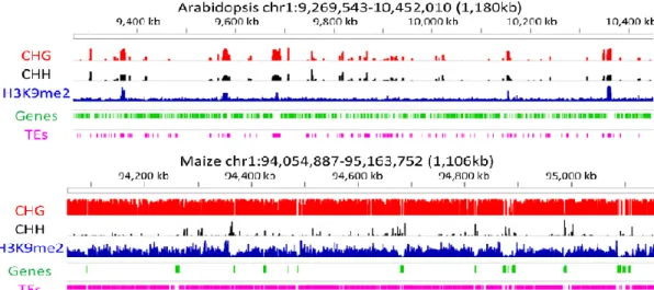
Fig 1. 4 Epigenome organization in Arabidopsis and maize. The organization of genes (green) and TEs (pink), the relative abundance of three chromatin modifications, CHG DNA methylation (red), CHH DNA methylation (black), and H3K9me2 methylation (blue). Figure is adapted from Springer et al (2016).
Fig 1. 5 Association of DNA cytosine methylation, TE density, small RNA and gene expression in tomato. Genes were classified into 5 groups based on their expression level in tomato fruit at breaker (group 5: highest, group 1: lowest). (A) Distribution of miniature inverted transposable elements (MITEs) in the regions 2 kb upstream and downstream of TSS and PAS (bin size = 100 bp). (B) Distribution of 24nt small RNAs. (C) Distribution of CG methylation. (D) Distribution of CWG methylation. (E) Distribution of CCG methylation. (F) Distribution of CHH methylation. TSS: transcription start site; PAS: polyadenylation site. Figure is adapted from Zhong et al (2013).
II. Mechanism of DNA methylation
DNA methylation is catalyzed by enzymes called DNA methyltransferases (DMTs). Different DMTs have been characterized in both mammals and plants, which are involved, either in maintenance of DNA methylation during cell divisions, or in the establishment of new DNA methylation patterns (the so called de novo methylation)
2.1 Enzymes involved in DNA methylation in mammals.
Maintenance of DNA methylation in mammalsIn mammals four DNA methyltransferases (DNMTs) have been characterized that are highly conserved. DNMT1 maintains DNA methylation at hemi-methylated DNA after DNA replication during cell division. It is the most abundant DNMTs in adult cells. DNMT3A and NNMT3B are involved in establishing de novo DNA methylation, as they don’t require hemi-methylated DNA to bind. DNMT3-like (DNMT3L) is another member of the DNMT3 family, but it has no enzymatic activity by itself. This enzyme binds to DNMT3A or DNMT3B and enhances their catalytic activity (Pinney 2014). In mammals, DNMT1 is the principal enzyme that can mediate the maintenance of CG methylation. This enzyme is required for embryonic development and survival of somatic cells in mice. It has been well summarized that DNMT1 doesn’t work alone, but work with some accessory proteins. For example, ubiquitin like PHD and RING finger 1 (UHRF1) were recently shown to be key regulators for maintenance of DNA methylation. The uhrf1 mutant is indeed characterized by a severe decrease in DNA methylation. The current model for UHFR1 action is as follows: UHRF1 recognizes hemi-methylated DNA via its SET and RING-associated (SRA) domains and H3K9me3 via its TUDOR and PHD domains; UHFR1 ubiquitylates H3K23/H3K18 to facilitate the environment for DNMT1 binding. Then DNMT1 binds ubiquitylated H3K23 inducing a conformational change in DNMT1 which promotes its activation (Nishiyama et al., 2016). In addition, UHRF1 interacts with DNMT3A and DNMT3B, which suggests a role for UHRF1 in de novo methylation. Maintenance of DNA methylation also requires the chromatin remodeling factor Lymphoid Specific Helicase1, but the precise role of LSH1 in DNA methylation remains unknown (Nishiyama et al., 2016).
2.2 Enzymes involved in DNA methylation in plants.
In plants, four DNA methyltransferase classes have been characterized. DNA methyltransferase 1 (MET1) which is the homologue of DNMT1 maintains methylation at CG sites. CHROMOMETHYLASE3 (CMT3) is a plant specific enzyme that maintains CHG methylation and requires histone H3 methylation at the lysine K9 to be recruited at its target sites (Yang et al., 2016). De novo DNA methylation in the different sequence contexts is mediated by two enzymes, one is the homologue of the DNMT3 methyltransferases, DOMAINS REARRANGED METHYLTRANSFERASE 2 (DRM2) and another one is CMT2 (Matzke, M. A. and R. A. Mosher, 2014).
2.2.1 Maintenance of DNA methylation in plants
It has been well documented that, in plants, MET1 is responsible for CG methylation maintenance (Kankel, M. W., et al. 2003). The mechanism of maintenance of CG methylation is highly conserved between plants and mammals. MET1 can’t work alone but requires additional proteins; recruitment of MET1 at target sites requires two different SRA proteins, VARIANT IN METHYLATION (VIM) and Decrease in DNA Methylation 1 (DDM1). However, in plants whether these proteins behave in a similar way as in mammals, needs further validation (Kankel, M. W., et al. 2003).
CHG methylation is maintained by the plant specific enzyme, CMT3 (chromomethylase 3), and requires the H3K9 methyltransferases KRYPTONITE (KYP/SUVH4), SUVH5 and SUVH6 (Lindroth et al., 2001). Genome-wide profiling of H3K9Me2 and DNA methylation showed that these marks are highly correlated (West et al., 2014). CMT3 mutant displayed a dramatic loss of DNA methylation as also observed in a suvh4 mutant, SUPPRESSOR OF VARIEGATION 3-9 HOMOLOGUE 4 a histone methyltransferase that is largely responsible for H3K9 dimethylation (Cedar and Bergman 2009;Du et al., 2014). Furthermore, two other H3K9 histone methyltransferases, SUVH5 and SUVH6 also contribute to global levels of CHG methylation (Ebbs and Bender 2006). Hence, in Arabidopsis CMT3 is recruited to specific sites by binding dimethyl K9 histone H3 (H3K9Me2) (Du et al., 2015). Reciprocally, KYP binds CHG methylated motives through its SRA domain (Johnson et al., 2007) thereby establishing a self-reinforcement loop between CHG methylation and H3K9 dimethylation.
2.2.2 De novo DNA methylation in plants
In plants, de novo methylation is mediated by RNA directed DNA methylation, a process also called RdDM (Law and Jacobsen 2010), which is also responsible of maintenance of CHH methylation. RdDM is mainly dependent on the methyltransferases, DOMAINS REARRANGED METHYLTRANSFERASE1 (DRM1) and DRM2, and it is always associated with 24nt siRNA, which direct DNA methylation at their homologous regions (For a review, see(Matzke and Mosher 2014)). Alternatively de novo methylation may rely on the chromatin remodeler DDM1 (DECREASE IN DNA METHYLATION 1), together with the CHROMOMETHYLASE 2 (Zemach et al., 2013;Stroud et al., 2014).
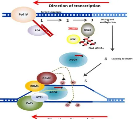
A number of components of the RdDM pathway have been recently identified in Arabidopsis leading to the proposal of a model for this complex epigenetic mechanism (Fig 1.6) (Gallusci et al., 2016). RdDM depends on specialized transcriptional machinery and involves at least two steps: 24-nt siRNA biogenesis and siRNA-guided de novo methylation (for a review, see (Matzke and Mosher 2014;Zhou and Law 2015).
Fig 1. 6 Mechanism of RdDM in plants. RNA transcripts are generated from repetitive sequences (transposons and others) by an RNA polymerase known as PolI V. RNA-DEPENDENT RNAPOLYMERASE (RDR) then converts the RNA to double stranded transcripts. These are processed into 24-nucleotide small RNAs (siRNAs) by DICER-LIKE3 (DCL3). These are methylated at their ends by HUAENHANCER1 (HEN1) and the guide strand complementary to the genomic DNA, that will be the target of the RdDM, is incorporated into ARGONAUTE (AGO4). AGO4 is recruited through interactions with PolV and KOWDOMAIN-CONTAINING TRANSCRIPTION FACTOR1 (KTF1). RNA-DIRECTEDDNA METHYLATION1 (RDM1) links AGO4 and DOMAINS REARRANGED METHYLTRANSFERASE2 (DRM2), which catalyzes de novo methylation of DNA. Figure is adapted from Gallusci et al (2016).
The classic RdDM pathway is initiated by recruitment of Polymerase IV (Pol IV), a plant specific DNA dependent RNA polymerase to the appropriate regions of the genome, including TEs and intergenic regions to transcribe a single strand RNA. The recruitment of Pol IV to target sequences is not fully understood. For a large subset of the RdDM targets, Pol IV recruitment necessitates a homeodomain protein, SAWADEE HOMEODOMAIN HOMOLOG 1 (SHH1) which recognizes chromatin enriched with unmethylated H3K4 and H3K9me2 and interacts with Pol IV (Law et al., 2013;Zhang et al., 2013b). The long single strand RNAs produced by Pol IV are rapidly converted into double strand RNAs (dsRNAs) by RNA DEPENDENT RNA POLYMERASE 2 (RDR2). The generation of dsRNAs also involves the putative chromatin remodeling protein CLASSY 1 (CLSY1), but the role of this factor remains unknown. The dsRNAs are then processed into 24-nt siRNAs by Dicer-like 3 ribonuclease III enzyme (DCL3). The double-stranded 24-nt siRNAs are transferred to the cytoplasm and loaded into the Argonaute (AGO) protein AGO4 to form a silencing complex. The silencing complex is transferred back to the nucleus with the help of AGO4, and siRNAs are targeted back to DNA repeats through sequence homology.
The siRNA-guided de novo methylation requires then another plant-specific DNA-dependent RNA polymerase, Pol V, and some associated factors. Pol V generates long intergenic non coding RNAs from target loci. The AGO-loaded siRNAs pair with this Pol V scaffold RNAs, and recruit the de novo DNA methyltransferase DOMAINS REARRANGED METHYLTRANSFERASE 2 (DRM2) which catalyzes de novo DNA methylation at the target locus. Pol V transcription and association with chromatin are facilitated by the DDR complex. This complex comprises the putative chromatin remodeler DEFECTIVE IN RNA-DIRECTED DNA METHYLATION 1 (DRD1), DEFECTIVE IN MERISTEM SILENCING 3 (DMS3), and RNA-DIRECTED DNA METHYLATION 1 (RDM1), which has been shown to interact with both AGO4 and DRM2, and to bind to methylated single-stranded DNA. Some other RdDM components may also be needed to complete this process, including some histone-modifying enzymes that remove active marks.
Recently, alternative RdDM pathways have been suggested. For example Yang et al (2016) found that the majority of the RdDM loci don’t require DCL proteins and 24nt siRNA, but rather 25-50 nt RNAs Pol IV-dependent small RNAs (P4 RNAs,) that may act as trigger RNAs to initiate DNA methylation following the RdDM pathway (Yang et al., 2016).
RdDM has been shown to be inhibited by heterochromatin, which is enriched in larger transposons. Furthermore lack of DRM2 causes a relatively modest decrease in CHH methylation, demonstrating that the majority of CHH methylation does not depend on RdDM. Indeed most CHH methylation at heterochromatic sequences is mediated by another pathway, requiring the chromomethylase CMT2 and DDM1 and depending on linker histone H1 (Zemach et al., 2013).
In Arabidopsis both the DDM1/CMT2 and the RdDM/DRD1 pathways mediate nearly all transposon CHH methylation. Hence both pathways act together to inhibit transposon mobility. But this scheme may not be valid in all plant species. For example in rice, the Osdrm2 mutation was shown to lead to a near complete loss of CHH methylation (Tan et al., 2016). Hence, in rice, almost all CHH methylation seems to be established by OsDRM2. Furthermore OsDDM1 is required for the facilitation of OsDRM2-mediated CHH methylation. These results suggest that de
novo DNA methylation though mediated by similar pathways, can vary between plant species (Tan et al., 2016).
2.3 Functions of DNA methylation in plants
In eukaryotes, cytosine DNA methylation is a conserved and stable epigenetic mark that plays essential roles in the silencing of transposable elements (TEs) and genes (Law and Jacobsen 2010). A number of articles have reviewed that cytosine methylation is critical for diverse biological process, including the establishment and maintenance of tissue specific gene expression patterns, genomic imprinting and X chromosome inactivation (Laird PW, 2003; Duymich et al., 2016).
2.3.1 Cytosine DNA methylation plays different roles during plant development
The genome-wide distribution of methylcytosines is subjected to dynamic changes during development
The distribution of methylcytosines has been analyzed at a genome-wide level in different plants and developmental contexts, including endosperm development (Hsieh et al., 2009; Zemach