HAL Id: ensl-00182503
https://hal-ens-lyon.archives-ouvertes.fr/ensl-00182503
Submitted on 26 Oct 2007
HAL is a multi-disciplinary open access
archive for the deposit and dissemination of
sci-entific research documents, whether they are
pub-lished or not. The documents may come from
teaching and research institutions in France or
abroad, or from public or private research centers.
L’archive ouverte pluridisciplinaire HAL, est
destinée au dépôt et à la diffusion de documents
scientifiques de niveau recherche, publiés ou non,
émanant des établissements d’enseignement et de
recherche français ou étrangers, des laboratoires
publics ou privés.
A heterochromatin protein 1 homologue in
Caenorhabditis elegans acts in germline and vulval
development
Florence Couteau, Fréderic Guerry, Francesca Palladino, Fritz Muller
To cite this version:
Florence Couteau, Fréderic Guerry, Francesca Palladino, Fritz Muller. A heterochromatin protein
1 homologue in Caenorhabditis elegans acts in germline and vulval development. EMBO Reports,
EMBO Press, 2002, pp.235-241. �ensl-00182503�
A heterochromatin protein 1 homologue in
Caenorhabditis elegans acts in germline and
vulval development
Florence Couteau, Fréderic Guerry
1, Fritz Müller
1& Francesca Palladino
+Laboratory of Molecular and Cellular Biology, UMR5665/CNRS, Ecole Normale Supérieure de Lyon, F-69364 Lyon, France and 1Institute of Zoology, University of Fribourg, CH-1700 Fribourg, Switzerland
Received November 29, 2001; revised December 21, 2001; accepted January 11, 2002
Proteins of the highly conserved heterochromatin protein 1 (HP1) family have been found to function in the dynamic organization of nuclear architecture and in gene regulation throughout the eukaryotic kingdom. In addition to being key players in heterochromatin-mediated gene silencing, HP1 proteins may also contribute to the transcriptional repression of euchromatic genes via the recruitment to specific promoters. To investigate the role played by these different activities in specific developmental pathways, we identified HP1 homologues in the genome of Caenorhabditis elegans and used RNA-mediated interference to study their function. We show that one of the homologues, HPL-2, is required for the formation of a functional germline and for the development of the vulva by acting in an Rb-related pathway. We suggest that, by acting as repressors of gene expression, HP1 proteins may fulfil specific functions in both somatic and germline differen-tiation processes throughout development.
INTRODUCTION
Heterochromatin protein 1 (HP1) proteins are non-histone chromosomal proteins that directly contribute to the higher-order packaging of chromatin by binding to modified histones (Jones et al., 2000; Rice and Allis, 2001). All HP1 proteins are structurally related and characterized by the presence of two conserved domains, an N-terminal chromo domain (CD) separated by a variable hinge region from a C-terminal related domain termed the chromo shadow domain (CSD). The CD is responsible for binding to methylated histone H3, while the CSD is required for dimerization and protein–protein interactions in the nucleus (Brasher et al., 2000; Rice and Allis, 2001).
The best-characterized member of this family, HP1a, is the product of the Drosophila Su(var)2-5 locus, initially identified as a key player in heterochromatic position-effect silencing (Eissenberg
et al., 1990) and more recently shown to be required for the
normal transcriptional activity of both heterochromatic and euchromatic genes (Lu et al., 2000; Hwang et al., 2001). Recent evidence from mammalian cells is consistent with a role for HP1 proteins in the transcriptional repression of euchromatic genes via the recruitment to specific promoters by co-repressor proteins including Rb (Jones et al., 2000; Nielsen et al., 2001). However, it remains to be established whether, in vivo, HP1 targeting is a general phenomena or limited to certain tissues. Furthermore, the late larval lethality associated with the
Drosophila Su(var)2-5 mutation has precluded studies on the
specific function of HP1 in defined developmental processes (Eissenberg and Hartnett, 1993). To gain insight into the function of HP1 proteins throughout development, we have undertaken a genetic analysis of HP1-like proteins from Caenorhabditis
elegans. We present evidence that one of the proteins, HPL-2,
plays an essential function in germline development and in the differentiation of vulval tissues by acting in an Rb-related transcriptional repressor pathway.
RESULTS
Localization of C. elegans HP1 homologues
in germline and somatic cells
We named the two C. elegans HP1 homologues HPL-1 and HPL-2 (Figure 1A and B). hpl-2 gives rise to two alternatively spliced transcripts, hpl-2a and hpl-2b, predicted to encode two
236 EMBO reports vol. 3 | no. 3 | 2002
F. Couteau et al.
scientific report
proteins that are identical throughout the CD and hinge region,but differ in their C-terminal ends. Of the two transcripts, only HPL-2a contains an intact CSD including all the conserved residues essential for dimerization of this domain (Brasher et al., 2000), while HPL-2b contains an additional 126 amino acids with no significant homology to any other protein of known function.
Northern blot analysis confirmed the presence of the two hpl-2 transcripts in approximately equal amounts in a mixed stage population of worms (data not shown). To study the in vivo localization of the HPL proteins, we constructed GFP-tagged versions of full-length HPL-1 and HPL-2 (Figure 1C). Both fusion proteins could be detected in the nuclei of most, if not all, cells of adult animals (data not shown). For hpl-2::gfp, strong expression was observed in all embryonic nuclei starting at the 20–24-cell stage and persisted through to adulthood. In addition, hpl-2::gfp expression could be weakly detected in germ cells, developing oocytes and embryos starting at the two-cell stage, before the onset of zygotic transcription, suggesting that the protein is maternally inherited (Figure 2; data not shown). DAPI staining on fixed samples confirmed that the fusion protein co-localizes with DNA. While HPL-2–GFP was found to be uniformly associated with all condensed meiotic and mitotic chromosomes (Figure 2, A–F), in interphase nuclei expression was often found in regions immediately adjacent to, but not overlapping, condensed chromatin (Figure 2, G–I). These results suggest that HPL-2 localization may be cell cycle regulated.
Inactivation of hpl-2 results in sterility
The function of hpl-1 and hpl-2 was investigated through RNA interference (RNAi) (Fire et al., 1998). While hpl-1(RNAi) did not result in any obvious phenotype at any of the temperatures tested, at 25°C, 24–53% F1 progeny of hpl-2(RNAi) animals were sterile and at a low frequency showed an everted vulva (evl) phenotype (Table I). Given that injection of double-stranded (ds)RNA corresponding to a 3′ region unique to hpl-2b (see Methods) failed to give any detectable phenotype, the sterility could result either from specific inactivation of the shorter hpl-2a transcript or the simultaneous inactivation of both transcripts (data not shown). Co-injection of dsRNAs corresponding to both
hpl-1 and hpl-2 increased sterility among the F1 progeny of
injected animals to 60–100%. This synergism suggests that, although hpl-1 alone is not necessary for a functional germline, it is partially redundant with hpl-2 for this function.
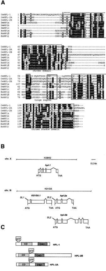
Fig. 1. (A) Alignment of amino-acid sequences of the HP1 proteins from human
(HP1α, -β and -γ), Drosophila (HP1a, -b and –c) and C. elegans (HPL-1, -2a and -2b) by the Clustal W method followed by manual editing. The conserved CD and CSD are boxed in. Note that HPL-2B diverges from other HP1-like proteins in the C-terminal part of the CSD. Black and grey boxes indicate identical and conserved residues, respectively. Asterisks indicate the 5′ acidic stretch of residues found in Drosophila HP1a and all human homologues but absent from C. elegans proteins. Heavy-lined boxes within hinge region denote the previously described bipartite nuclear localization sequence (Smothers and Henikoff, 2001), which is missing from the C. elegans homologues. (B) Genomic structure of the C. elegans hpl-1 and hpl-2 genes.
hpl-2a (K01G5.2a) and hpl-2b (K01G5.2b) arise from alternative splicing of
a single transcript that is part of an operon including the upstream gene K01G5.1, predicted to encode a ring zinc finger protein of the C3HC4 type. The AUG start codon for hpl-2 is found 120 bp downstream from the stop codon of K01G5.1, and RT–PCR analysis confirmed the presence of an SL2 transpliced leader sequence on the hpl-2 transcript. The ATG start and stop codons and the SL1 and SL2 transpliced leader sequences are shown. Boxes correspond to exons, connecting lines to introns. (C) Schematic representation of the HPL-1 and HPL-2 proteins, showing CD and CSD domains and the GFP insertion site in the fusion proteins used in this study.
In C. elegans, a mechanism dependent on chromatin context is responsible for silencing repetitive transgene arrays in the germline (Kelly et al., 1997). Homologues of the Polycomb family of transcriptional repressors, encoded by the mes genes, and histone H1.1 have been shown to be required for this mechanism as well as germline development (Seydoux and
Strome, 1999; Jedrusik and Schulze, 2001). Given that HP1 proteins have been implicated in somatic position-effect silencing in both Drosophila and mammals (Jones et al., 2000), we decided to test whether the germline phenotypes observed in
hpl-2(RNAi) animals might reflect a role for HPL-2 in the
germline silencing process. For this purpose, we made use of a transgene array carrying multiple tandem copies of a plasmid encoding a GFP-tagged version of an ubiquitously expressed
C. elegans gene (let-858) (Kelly and Fire, 1998), which is
expressed in somatic lineages but subjected to silencing in the germline. While in control animals carrying the let-858 reporter we were unable to detect any fluorescence in the germline at 25°C (n = 100), 68% (n = 76) of the F1 progeny of hpl-2(RNAi) worms showed germline desilencing in one or both gonad arms (Figure 3, compare C with D). hpl-1(RNAi), in contrast, failed to derepress the let-858 reporter (n = 50). Therefore, like the MES proteins and histone H1.1, HPL-2 is required for germline silencing, presumably through an influence on chromatin.
mes mutant animals are sterile due to the degeneration of
germ cells (Seydoux and Strome, 1999), and inactivation of H1.1 by RNAi results in similar defects in germline development (Jedrusik and Schulze, 2001). To determine whether hpl-2(RNAi) sterile animals also show germ-cell degeneration, we performed
hpl-2(RNAi) in animals expressing a GFP::H2B histone fusion
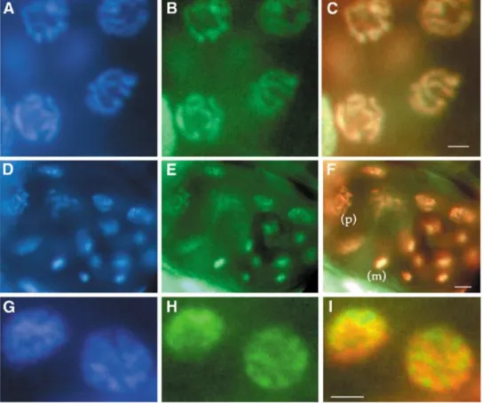
Fig. 2. Localization of HPL-2::GFP in the soma and germline. (A, D, G) DAPI fluorescence; (B, E, H) GFP fluorescence; (C, F, I) merged image of DAPI and GFP with
DAPI in false red and GFP in green. (A–C) HPL-2::GFP is present in the germline and associated with the DNA of condensed nuclei in pachytene. (D–F) Twenty-eight-cell
stage embryo with HPL-2::GFP expression in all nuclei. Nuclei in metaphase (m) and prophase (p) are indicated. (G–I) Nuclei of intestinal cells in interphase.
Scale bars: 2 µm (A–C), 4 µm (D–F), 1 µm (G–I).
Table I. Depletion of hpl-2 by RNAi results in sterility
Numbers refer to percentages with the range of results from multiple RNAi experiments (at least four). Numbers in parentheses refer to the to the total number of F1 animals scored.
aThese animals had a protruding vulva.
Experiments were carried out at 20 or 25°C, as indicated. RNA injected % Sterile % evla none (n = 1600; 25°C) 0 0 hpl-1 (n = 500; 20°C) 0 0 hpl-1 (n = 641; 25°C) <0.1 0 hpl-2 (n > 1000; 20°C) <1 0 hpl-2 (n = 1852; 25°C) 24–53 1–5 hpl-1+hpl-2 (n = 500; 20°C) 0 0 hpl-1+hpl-2 (n = 261; 25°C) 60–100 6–16
238 EMBO reports vol. 3 | no. 3 | 2002
F. Couteau et al.
scientific report
allowing direct visualization of germline nuclei in the gonad of live animals. In adult wild-type animals, the germline develops in a defined and largely invariant manner. Moving from the distal region proximally, germ cells proliferate, enter meiosis and differentiate into oocytes in the loop region and proximal gonad (Schedl, 1997). Oocytes are fertilized as they pass through the spermatheca into the uterus. While in hpl-2(RNAi) animals we did not detect any gross defects in chromosome morphology and meiotic progression appeared normal, in the proximal region of the gonad we noted several defects (Figure 4, compare A with B). Oocytes were abnormally shaped and were often found to pile up in the uterus. Nuclei of oocytes in the proximal region of the gonad and uterus were also often enlarged and misshaped, and they showed increased GFP staining suggestive of endomitosis (Figure 4D; data not shown). The fact that endomitotic oocytes were observed in the gonad arm as well as the uterus suggests that they are not simply the result of a failure to be fertilized by sperm upon passage through the spermatheca (Iwasaki et al., 1996). Instead, we suggest that the phenotypes observed might reflect a defect in oocyte maturation.
HPL-2 is required for vulval development
by acting in the synMuvB pathway
In hpl-2 (RNAi) animals, we occasionally observed the ectopic induction of vulval tissues (data not shown). We therefore decided to test whether hpl-2 might play a role in vulval development. In C. elegans, the vulva is derived from the descendants of three out of six equivalent vulva precursor cells (VPCs), P5.p, P6.p and P7.p (Fay and Han, 2000). The three other VPCs, P3.p, P4.p and P8.p, normally adopt a cell fate giving rise to non-vulval cells, which fuse to the hypodermal syncytium. A conserved LET-23 RTK/Ras/MAP kinase signalling cascade is responsible for inducing the P(5–7).p cells to adopt a vulval fate by overcoming inhibitory signals from two functionally redundant sets of genes, known as synMuvs. The synMuv genes fall into two classes, referred to as A and B, that define two functionally redundant pathways. Animals carrying both a class A
and a class B mutation exhibit a multivulva (Muv) phenotype because P3.p, P4.p and P8.p adopt induced vulval fates, while animals carrying one or more mutations of the same class have a wild-type vulva (reviewed in Ferguson and Horvitz, 1989). Interestingly, a number of proteins with class B synMuv activity are homologous to proteins implicated in transcriptional repression, chromatin modification and nucleosome remodelling. These include LIN-35 and LIN-53, which are homologues of mammalian pRb and RbAp48, respectively, HDA-1, a homologue of the histone deacetylase HDAC1, and CHD-4, the homologue of mammalian Mi-2 (Fay and Han, 2000; Solari and Ahringer, 2000; von Zelewsky et al., 2000). This conservation prompted us to ask whether hpl-1 and hpl-2 have properties of synMuv genes. We found that while injection of hpl-1 dsRNA in either a synMuvA or a synMuvB background failed to produce a Muv phenotype at any of the temperatures tested, injection of hpl-2 dsRNA in three synMuvA mutant backgrounds, lin-15(n767),
lin-38(751) and lin-8(n111), resulted in a highly penetrant Muv
phenotype at 25°C, with 95–100% of all animals showing multiple ectopic inductions of vulval tissue (the Muv phenotype; Tables II and III). At 20°C, the penetrance of the Muv phenotype was significantly reduced. The temperature sensitivity of the Muv phenotype presumably reflects the uncovering or induction of a heat-sensitive process (Golden and Riddle, 1984; Melendez and Greenwald, 2000). These results show that hpl-2 acts in the synMuvB pathway. An intact RTK/Ras/Map kinase pathway is essential for expression of the synMuv phenotype (Ferguson
et al., 1987). We therefore asked whether the let-23(sy1)
vulvaless phenotype is epistatic to the phenotype observed in
hpl-2(RNAi)lin-15A(RNAi) animals (75% Muv, n = 50). let-23(sy1)
animals injected with hpl-2 and lin-15A dsRNAs failed to produce a Muv phenotype and were vulvaless (n = 30). These results show that RTK/Ras pathway activity is required for the Fig. 3. Desilencing of a repetitive let-858::gfp transgene in the germline of F1
offspring of hpl-2(RNAi) animals. Germline is outlined by brackets. (C) In control animals, GFP fluorescence can be strongly detected in somatic nuclei but not in the germline. (D) In hpl-2(RNAi) animals, GFP fluorescence is detected in all germline nuclei and in oocytes (arrowheads). (A and B) Nomarski images
of the same animals as in (C) and (D). Fig. 4. Germline development is abnormal in hpl-2(RNAi) animals. Nomarski(DIC) images of the gonad of wild-type (A) and hpl-2(RNAi) (B) animals. Note that the proximal region of the gonad (arrowheads in A and B) in hpl-2(RNAi) animal is filled with enlarged, abnormal oocytes. (C and D) Fluorescent images of the same worms as in (A) and (B) in which DNA is visualized by histone H2B::GFP. Note the intense fluorescence (arrowheads in D) corresponding to accumulation of DNA in the enlarged nuclei seen in (B). Scale bar, 50 µm.
hpl-2(RNAi)synMuvB Muv phenotype and that, like other
synMuvB genes, hpl-2 plays a role in repressing the Ras signalling pathway.
While inactivation of hpl-2 in various synMuvA backgrounds invariably resulted in a highly penetrant Muv phenotype, injection of hpl-2 dsRNA in different synMuvB backgrounds resulted in various phenotypes depending on the synMuvB allele used (Tables II and III). hpl-2 RNAi in either a lin-36(n766) or
lin-53(n833) background failed to produce any detectable
phenotype at any temperature tested, while co-injection of both
hpl-2 and lin-15B dsRNAs resulted in a significant number of
Muv animals at 25°C (Tables II and III). Three other synMuv strains, lin-9(n112), lin-35(n745) and lin-37(n758), are less fertile and smaller than the wild type at 25°C (Ferguson and Horvitz, 1989; our observations). Injection of hpl-2 dsRNAs in these backgrounds at 25°C exacerbated these phenotypes and provoked larval lethality. Nonetheless, amongst the surviving adults we could detect a significant portion of animals showing
ectopic vulval inductions (Tables II and III; data not shown). Furthermore, at 20°C, 41–61% of lin-35 hpl-2(RNAi) animals (n = 157) and 20–44% of lin-37 hpl-2(RNAi) animals (n = 148) were sterile and showed slowed growth. These results suggest that hpl-2 may also contribute to the synMuvA pathway, as reported previously for other Muv genes (Melendez and Greenwald, 2000; Solari and Ahringer, 2000), and interact with members of the synMuvB pathway in other essential develop-mental pathways.
DISCUSSION
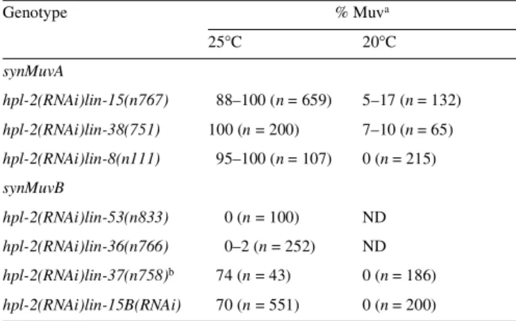
We have shown that the C. elegans HP1 homologue HPL-2 is required in two specific developmental pathways: the formation of a functional germline and vulval cell-fate specification. Although it is not clear whether the sterility and germline derepression we observe upon inactivation of hpl-2 are directly linked phenomena, based on the expression of HPL-2 in the germline and the role of HP1 proteins in gene regulation via chromatin structure, we suggest that HPL-2 may fulfil a repressive function dependent on the heterochromatin-like properties of the germline. This repression could contribute to the maintenance of a functional, undifferentiated germline, a function shared with the C. elegans Polycomb homologues and the histone H1.1. In the soma, HPL-2 is also required for the development of the vulva. Our data show that hpl-2 plays a key role in the synMuvB pathway. While hpl-2 also contributes partially to the synMuvA pathway, it does not appear to act as a typical class A synMuv gene. The synMuvB pathway includes homologues of the mammalian proteins Rb, RbAp48, HDAC1 and, as shown more recently, DP and E2F (Lu and Horvitz, 1998; Ceol and Horvitz, 2001). A model for synMuv function proposes that DP and E2F homologues may tether a subset of synMuv proteins to specific promoters via interaction with LIN-35Rb. Based on the observation that hpl-2 appears to play a role in transcriptional repression in the germline and interact genetically with lin-35Rb, an attractive extension of this model is that the recruitment of HPL-2 by LIN-35 Rb contributes to the repression of vulval specification genes by the local packaging of E2F-target genes into condensed, transcriptionally inactive heterochromatin-like domains, by their recruitment to heterochromatic compartments of the nucleus or by the recruitment of other repressive functions. The recent report of a physical association between HP1 and Rb possibly contributing to the transcriptional Table II. hpl-2 has properties of a synMuv gene
aAnimals were examined under the dissecting microscope for the presence of anterior and posterior pseudovulvae and scored as Muv if they showed two or more pseudovulvae in addition to the normal vulva. Numbers in parentheses refer to the total number of F1 counted.
b% Muv animals among surviving adults; hpl-2 RNAi in this background at 25°C resulted in slow growth and larval lethality.
Experiments were carried out at 20 or 25°C, as indicated. ND, not done. Genotype % Muva 25°C 20°C synMuvA hpl-2(RNAi)lin-15(n767) 88–100 (n = 659) 5–17 (n = 132) hpl-2(RNAi)lin-38(751) 100 (n = 200) 7–10 (n = 65) hpl-2(RNAi)lin-8(n111) 95–100 (n = 107) 0 (n = 215) synMuvB hpl-2(RNAi)lin-53(n833) 0 (n = 100) ND hpl-2(RNAi)lin-36(n766) 0–2 (n = 252) ND hpl-2(RNAi)lin-37(n758)b 74 (n = 43) 0 (n = 186) hpl-2(RNAi)lin-15B(RNAi) 70 (n = 551) 0 (n = 200)
Table III. Vulval induction phenotype caused by hpl-2(RNAi)
Induction of individual VPCs was determined at 25°C by scoring detachment from the cuticle at the L4 stage. Numbers refer to percentages with the range of results from multiple RNAi experiments (at least four).
aTaken from von Zelewsky et al. (2000).
Induction of individual VPCs (%)
P3.p P4.p P5.p P6.p P7.p P8.p n
Wild type 0 0 100 100 100 0 manya
hpl-2(RNAi)lin-15A(n767) 71 100 100 100 100 80 49
hpl-2(RNAi)lin-15B(RNAi) 82 87 100 100 100 79 61
240 EMBO reports vol. 3 | no. 3 | 2002
F. Couteau et al.
scientific report
repression of euchromatic genes in mammalian cells supportssuch a model (Nielsen et al., 2001). Drosophila and mammalian HP1 proteins have been shown to be localized within distinct nuclear domains corresponding to either heterochromatic or euchromatic regions (Minc et al., 1999; Smothers and Henikoff, 2001). Although heterochromatin has not been defined cytologically in C. elegans, our localization studies suggest that, like the
Drosophila and human HP1 proteins, HPL-2 may also be
associated with specific nuclear subdomains in the interphase nucleus and contribute to chromatin-mediated repression in these regions. Further work in C. elegans will be aimed at under-standing exactly how HP1 proteins contribute to the repression of specific genes in both somatic and germline differentiation processes throughout development.
METHODS
Construction of hpl-1::gfp and hpl-2::gfp. hpl-1::gfp was constructed by in-frame insertion of the gfp fragment from pPD102.33 (A. Fire, personal communication) into the EcoRI site in the first exon of hpl-1. The resulting construct, pFP04, contains 3.6 kb upstream of the ATG start codon and 960 bp downstream of the stop codon of hpl-1. To construct hpl-2::gfp, a 7 kb fragment from cosmid K01G5 containing the entire hpl-2 operon and 3.9 kb of sequence upstream was subcloned into pBSKS (Stratagene). The resulting construct was digested with
PvuII and religated to leave a single BamHI site available in the
third exon of hpl-2. The BamHI gfp fragment from pPD102.33 5 was inserted in-frame into this unique site to generate pFG2.
Transgenic worms were generated using 20 µg/ml of pFP04 or pFG2 and 150 µg/ml of pRF4 as described previously (Mello et al., 1991). Stable lines in which the hpl-2::gfp transgene had been integrated into the genome were generated by the standard method (Jin, 1999) using an exposure to 30 mJ UV light (λ = 254 nm) using a Bio-Rad UV-crosslinker. Stable lines were backcrossed twice to eliminate extraneous mutations arising from the UV treatment.
RNAi. RNAi experiments were carried out as described by Fire et al. (1998), using cDNAs yk432 (hpl-1) and yk470 (hpl-2b)
(kindly provided by Yuji Kohara) as templates for in vitro transcription reactions with T7 and T3 polymerase. dsRNA specific to hpl-2b was synthesized from a PCR fragment corresponding to nucleotides 464–907 of hpl-2b obtained using the following primers: T3, TTATTAACCCTCACTAAAGCGTTC-GTCGACGCTTACGGCGAATT; and T7, TAATACGACTCACTA-TAGTTAT GAGTTTCTTGGGAACAAGAGA. lin-15B-specific RNAi was prepared as described previously (von Zelewsky et al., 2000). dsRNA was injected at a concentration of 400 µg/ml. For all experiments, injected mothers were placed on seeded plates for 12 h to allow the RNAi effect to take place, then transferred to fresh plates at 24 and 36 h. The F1 progeny of these plates was scored for viability, Muv phenotype or germline derepression. As a control for the effectiveness of RNAi, we tested whether expression of the reporter gene hpl-2::gfp was silenced in an
hpl-2(RNAi) background. We found that hpl-2::gfp hpl-2(RNAi)
worms failed to express gfp, confirming that the RNAi treatment is effective.
Microscopy and image acquisition. For GFP and DAPI co-localization studies, worms were washed in M9 medium and fixed in EtOH at 4°C for 10 min. After a second wash in M9,
worms were mounted in DAPI solution (1 µg/ml). Fixation in paraformaldehyde gave similar results. All observations were performed using a Zeiss Axioplan II microscope. Images were acquired with a CoolSnap CCD camera (Roper Scientific) and pseudocoloured and merged using Adobe Photoshop.
ACKNOWLEDGEMENTS
Nematode strains were obtained from the Caenorhabditis Genetics Center. We thank the C. elegans Genome Sequencing Consortium for sequence information, the C. elegans Genetic Center for strains, Bill Kelly for the let-858::gfp reporter strain, Yuji Kohara for cDNAs, Alex Hajnal for strains and helpful discussion, and Eric Gilson, Michel Labouesse and Monique Zetka for comments on the manuscript. F.C. was supported by the Ligue Nationale contre le Cancer and F.P. by the Région Rhône-Alpes and SNF grants 3100 Trade Union 040776.94 and 3100-056953.99.
REFERENCES
Brasher, S.V. et al. (2000) The structure of mouse HP1 suggests a unique mode of single peptide recognition by the shadow chromo domain dimer.
EMBO J., 19, 1587–1597.
Ceol, C.J. and Horvitz, R.H. (2001) dpl-1 DP and efl-1 E2F Act with lin-35 Rb to antagonise Ras signaling in C. elegans vulval development.
Mol. Cell, 7, 461–473.
Eissenberg, J.C. and Hartnett, T. (1993) A heat shock-activated cDNA rescues the recessive lethality of mutations in the heterochromatin-associated protein HP1 of Drosophila melanogaster. Mol. Gen. Genet., 240, 333–338. Eissenberg, J.C., James, T.C., Foster-Hartnett, D.M., Hartnett, T., Ngan, V.
and Elgin, S.C. (1990) Mutation in a heterochromatin-specific chromosomal protein is associated with suppression of position-effect variegation in
Drosophila melanogaster. Proc. Natl Acad. Sci. USA, 87, 9923–9927.
Fay, D.S. and Han, M. (2000) The synthetic multivulval genes of C. elegans: functional redundancy, Ras-antagonism, and cell fate determination.
Genesis, 26, 279–284.
Ferguson, E.L. and Horvitz, H.R. (1989) The multivulva phenotype of certain
Caenorhabditis elegans mutants results from defects in two functionally
redundant pathways. Genetics, 123, 109–121.
Ferguson, E.L., Sternberg, P.W. and Horvitz, H.R. (1987) A genetic pathway for the specification of the vulval cell lineages of Caenorhabditis elegans.
Nature, 326, 259–267. [Erratum appears in Nature, 327, 82.]
Fire, A., Xu, S., Montgomery, M.K., Kostas, S.A., Driver, S.E. and Mello, C.C. (1998) Potent and specific genetic interference by double-stranded RNA in Caenorhabditis elegans [see comments]. Nature, 391, 806–811. Golden, J.W. and Riddle, D.L. (1984) A pheromone-induced developmental
switch in Caenorhabditis elegans: temperature-sensitive mutants reveal a wild-type temperature-dependent process. Proc. Natl Acad. Sci. USA, 81, 819–823.
Hwang, K.K., Eissenberg, J.C. and Worman, H.J. (2001) Transcriptional repression of euchromatic genes by Drosophila heterochromatin protein 1 and histone modifiers. Proc. Natl Acad. Sci. USA, 98, 11423–11427. Iwasaki, K., McCarter, J., Francis, R. and Schedl, T. (1996) emo-1, a
Caenorhabditis elegans Sec61p γ homologue, is required for oocyte
development and ovulation. J. Cell Biol., 134, 699–714.
Jedrusik, M.A. and Schulze, E. (2001) A single histone H1 isoform (H1.1) is essential for chromatin silencing and germline development in
Caenorhabditis elegans. Development, 128, 1069–1080.
Jin, Y. (1999) Transformation. In Hope, I.A. (ed.), C. elegans: A Practical
Approach. Oxford University Press, Oxford, UK, pp. 69–96.
Jones, D.O., Cowell, I.G. and Singh, P.B. (2000) Mammalian chromodomain proteins: their role in genome organisation and expression. BioEssays, 22, 124–137.
Kelly, W.G. and Fire, A. (1998) Chromatin silencing and the maintenance of a functional germline in Caenorhabditis elegans. Development, 125, 2451–2456.
Kelly, W.G., Xu, S., Montgomery, M.K. and Fire, A. (1997) Distinct requirements for somatic and germline expression of a generally expressed Caernorhabditis elegans gene. Genetics, 146, 227–238. Lu, B.Y., Emtage, P.C., Duyf, B.J., Hilliker, A.J. and Eissenberg, J.C. (2000)
Heterochromatin protein 1 is required for the normal expression of two heterochromatin genes in Drosophila. Genetics, 155, 699–708. Lu, X. and Horvitz, H.R. (1998) lin-35 and lin-53, two genes that antagonize
a C. elegans Ras pathway, encode proteins similar to Rb and its binding protein RbAp48. Cell, 95, 981–991.
Melendez, A. and Greenwald, I. (2000) Caenorhabditis elegans lin-13, a member of the LIN-35 Rb class of genes involved in vulval development, encodes a protein with zinc fingers and an LXCXE motif. Genetics, 155, 1127–1137.
Mello, C.C., Kramer, J.M., Stinchcomb, D. and Ambros, V. (1991) Efficient gene transfer in C. elegans: extrachromosomal maintenance and integration of transforming sequences. EMBO J., 10, 3959–3970. Minc, E., Allory, Y., Worman, H.J., Courvalin, J.C. and Buendia, B. (1999)
Localization and phosphorylation of HP1 proteins during the cell cycle in mammalian cells. Chromosoma, 108, 220–234.
Nielsen, S.J. et al. (2001) Rb targets histone H3 methylation and HP1 to promoters. Nature, 412, 561–565.
Rice, J.C. and Allis, C.D. (2001) Histone methylation versus histone acetylation: new insights into epigenetic regulation. Curr. Opin. Cell
Biol., 13, 263–273.
Schedl, T. (1997) Developmental genetics of the germ line. In Riddle, D.L., Blumenthal, T., Meyer, B.J. and Priess, J.R. (eds), C. elegans II. Cold Spring Harbor Laboratory Press, Cold Spring Harbor, NY, pp. 241–269.
Seydoux, G. and Strome, S. (1999) Launching the germline in Caenorhabditis
elegans: regulation of gene expression in early germ cells. Development,
126, 3275–3283.
Smothers, J.F. and Henikoff, S. (2001) The hinge and chromo shadow domain impart distinct targeting of HP1-like proteins. Mol. Cell. Biol., 21, 2555–2569. Solari, F. and Ahringer, J. (2000) NURD-complex genes antagonise Ras-induced
vulval development in Caenorhabditis elegans. Curr. Biol., 10, 223–226. von Zelewsky, T., Palladino, F., Brunschwig, K., Tobler, H., Hajnal, A. and
Muller, F. (2000) The C. elegans Mi-2 chromatin-remodelling proteins function in vulval cell fate determination. Development, 127, 5277–5284.