HAL Id: tel-00829101
https://tel.archives-ouvertes.fr/tel-00829101
Submitted on 2 Jun 2013
HAL is a multi-disciplinary open access archive for the deposit and dissemination of sci-entific research documents, whether they are pub-lished or not. The documents may come from teaching and research institutions in France or abroad, or from public or private research centers.
L’archive ouverte pluridisciplinaire HAL, est destinée au dépôt et à la diffusion de documents scientifiques de niveau recherche, publiés ou non, émanant des établissements d’enseignement et de recherche français ou étrangers, des laboratoires publics ou privés.
Smooth Muscle : Expression, Function and Mechanism
Kui Zhai
To cite this version:
Kui Zhai. Cyclic Nucleotide Phosphodiesterases (PDEs) in Smooth Muscle : Expression, Function and Mechanism. Agricultural sciences. Université Paris Sud - Paris XI; Institute of Biophysics (Pékin), 2012. English. �NNT : 2012PA114853�. �tel-00829101�
Joint Thesis Supervision between
UNIVERSITÉ PARIS-SUD
DOCTORAL SCHOOL:
INNOVATION THÉRAPEUTIQUE: DU FONDAMENTAL A L’APPLIQUÉ
PÔ LE: PHYSIOPATHOLOGIE MOLECULAIRE ET CELLULAIRE
DISCIPLINE: PHYSIOPATHOLOGIE MOLECULAIRE ET CELLULAIRE
&
INSTITUTE OF BIOPHYSICS (CHINESE ACADEMY OF SCIENCES)
YEAR 2012 - 2013 SÉRIE DOCTORAT N°
DOCTORAL THESIS
defended on November 20th, 2012 by
Kui ZHAI
Cyclic Nucleotide Phosphodiesterases (PDEs) in Smooth Muscle:
Expression, Function and Mechanism
Director of thesis: Véronique LEBLAIS Professeur des Universités (Université Paris-Sud, France) Co-director of thesis: Guangju JI Professeur (Chinese Academy of Science, Beijing, Chine) Composition of the jury:
President and reviewer: Qing-Hua LIU Professeur (South-Central University for Nationalities, Wuhan, Chine) Reviewer: Claire LUGNIER DR émérite CNRS (Université de Strasbourg)
Examiners: Rodolphe FISCHMEISTER DR Inserm (Université Paris-Sud, France)
Acknowledgements
This thesis was based on experimental work done in INSERM UMR-S 769, LabEx LERMIT, Faculté de Pharmacie, Université Paris-Sud and in National Laboratory of Biomacromolecules, Institute of Biophysics, Chinese Academy of Sciences. In 2008, I was fortunate to receive a doctoral grant from French Embassy which I am truly grateful to. Additional support was provided by Université Paris-Sud and Institute of Biophysics, which are highly appreciable.
My deepest gratitude goes firstly and foremost to my directors Prof. Véronique LEBLAIS and Prof. Guangju JI. Their professional guidance, constant encouragement and support kept me all the way on track during my scientific research. I address my special thanks to them for their supervision and guidance over the past years.
I would also express heartfelt gratitude to Dr. Rodolphe FISCHMEISTER, Director of Inserm UMR-S 769, for giving me the opportunity to work in his lab, sharing his endless knowledge in physiology and pharmacology, and creating a pleasant working atmosphere.
I would like to thank all my wonderful colleagues for teaching me techniques, offering experimental tips. I sincerely thank Fabien HUBERT for his help in all experiments. I would like to express gratitude to Dr. Grégoire VANDECASTEELE, Dr. JérômeLEROY and Patrick LECHÈNE for their help on FRET and path clamp experiments; to Valérie NICOLAS for her help on confocal microscope; to Yassine SASSI on cell isolation and culture; to Delphine MIKA, Françoise BOUSSAC, Cristina MOLINA-ESPINOSA, Zeineb HAJ SLIMANE, Philippe MATEO, Julia SCHITTL, Audrey VARIN, Sophie SEURON, Yanyun WU, Congyan PAN, Bin WEI, Xu ZHANG, Lin MIAO, Yan CHANG, Qi YUAN, Yingxiao CHEN, Yinglong TANG, Lei GU and Zhiguang YANG for their all kind help. It gave me many pleasures to work with them.
I sincerely thank Dominique FORTIN for her help. It is my great pleasure to spend Christmas with your family and to visit Royan. I am also grateful to Jérôme
PIQUEREAU and Stéphanie RIMBAUD for giving me a chance to visit their beautiful hometown.
Special thanks to my friends Jinguo WANG, Sibo LI, Dejiu ZHANG, Xiaoli TIAN and Lin XIA for their help and encouragements.
Last but not least, I am especially indebted to my beloved parents and wife for everything they have done for me. Without their love and encourage, I would never have the chance to finish the thesis.
Kui ZHAI Beijing, 2012.10
I
Index
Index ... I
List of figures ... IV
List of tables... V
Glossary and abbreviations ... VI
1 Introduction ... 1
1.1 Physiology of the smooth muscle ... 2
1.1.1 Characteristics of the smooth muscle cells... 2
1.1.2 Cardiovascular system ... 5
1.1.2.1 Overview ... 5
1.1.2.2 Morphology of vascular smooth muscle (VSM) ... 5
1.1.2.3 Sympathetic innervation ... 6
1.1.3 Lower urinary tract ... 7
1.1.3.1 Overview ... 7
1.1.3.2 Morphology of urinary bladder ... 8
1.1.3.3 Spontaneous activity of the bladder ... 9
1.1.4 Excitation-contraction-coupling (ECC) in the SM ... 11
1.1.4.1 Mechanisms of contraction in the SMC ... 11
1.1.4.2 The electromechanical coupling ... 13
1.1.4.3 The pharmacomechanical coupling ... 13
1.2 The cAMP/β-adrenergic signaling pathway ... 15
1.2.1 β-ARs ... 16
1.2.2 G protein ... 16
1.2.3 Adenylyl cyclase (AC) ... 18
1.2.4 cAMP targets ... 18
1.2.4.1 PKA ... 18
1.2.4.2 Epac ... 20
1.2.4.3 Cyclic nucleotide-activated ion channels ... 20
1.2.5 Mechanisms of cyclic nucleotide-induced relaxation ... 22
1.2.5.2 Hyperpolarization of the SMC ... 25 1.2.5.3 Decrease in MLC20 phosphorylation... 27 1.3 PDEs family ... 29 1.3.1 PDE1 ... 30 1.3.2 PDE2 ... 33 1.3.3 PDE3 ... 36 1.3.4 PDE4 ... 39 1.3.5 PDE5 ... 42 1.3.6 PDE6 ... 44 1.3.7 PDE7 ... 45 1.3.8 PDE8 ... 46 1.3.9 PDE9 ... 47 1.3.10 PDE10 ... 48 1.3.11 PDE11 ... 49
1.4 Cyclic AMP compartmentation ... 50
1.4.1 Overview ... 50
1.4.2 Innovative methods for subcellular compartmentation analysis ... 52
1.4.3 Role of PDEs in cAMP compartmentation ... 54
1.4.3.1 PDEs and cAMP compartmentation in cardiac myocyte ... 54
1.4.3.2 cAMP compartmentation in SMC ... 58
2 Objectives of the thesis ... 60
3 Materials and methods ... 61
3.1 Materials ... 61
3.1.1 Drugs and reagents ... 61
3.1.2 Cell culture reagents ... 61
3.1.3 Antibodies ... 62
3.2 Methods used in the study of rat aortic SMCs (RASMCs) ... 63
3.2.1 Animals ... 63
3.2.2 Isolation and culture of RASMCs ... 63
3.2.3 Immunocytochemistry ... 65
3.2.4 Cyclic AMP-PDE activity assay ... 66
3.2.5 FRET imaging ... 69
3.3 Methods used in the study of rat bladder SMCs (RBSMCs) ... 71
3.3.1 Animals ... 71
III
3.3.3 Ca2+ imaging on neonatal RBSMCs ... 72
3.3.4 Ca2+ imaging on intact neonatal rat bladder ... 73
3.3.5 PCR experiments on bladder tissue ... 73
3.3.6 Western blot experiments on bladder tissue ... 77
3.4 Data analysis ... 78
4 Results and discussion ... 79
4.1 PDEs and cAMP compartmentation in cultured RASMCs ... 79
4.1.1 Introduction ... 79
4.1.2 Paper І ... 80
4.1.3 Main results and conclusion ... 97
4.2 PDEs in rat bladder SM ... 98
4.2.1 Role of PDEs in regulating phasic contractions of the neonatal rat bladder ... 98
4.2.1.1 Introduction ... 98
4.2.1.2 Paper ІІ ... 99
4.2.1.3 Role of PLB in PDEs mediated effects ... 140
4.2.1.4 Main results and conclusion ... 142
4.2.2 PDEs expression and function in neonatal and adult rat bladder ... 143
4.2.2.1 Comparison of PDEs expression in neonatal and adult rat bladder ... 143
4.2.2.2 Comparison of PDE3 function and expression in neonatal and adult rat bladder ... 145
4.2.3 Main results and conclusion ... 148
5 Conclusion and perspectives ... 149
5.1 PDEs and cAMP compartmentation in cultured RASMCs ... 149
5.2 PDEs in rat bladder SM ... 152
List of figures
Figure 1.1. ... 2 Figure 1.2. ... 3 Figure 1.3. ... 4 Figure 1.4 ... 6 Figure 1.5 ... 7 Figure 1.6 ... 8 Figure 1.7 ... 8 Figure 1.8 ... 10 Figure 1.9 ... 12 Figure 1.10 ... 15 Figure 1.11 ... 23 Figure 1.12 ... 25 Figure 1.13 ... 27 Figure 1.14 ... 29 Figure 1.15 ... 36 Figure 1.16 ... 52 Figure 4.1 ... 141 Figure 4.2. ... 143 Figure 4.3. ... 146 Figure 4.4. ... 147V
List of tables
Table 1.1 ... 31 Table 3.1 ... 61 Table 3.2 ... 61 Table 3.3 ... 62 Table 3.4 ... 62 Table 3.5 ... 63 Table 3.6 ... 71 Table 3.7 ... 74 Table 3.8 ... 74 Table 3.9 ... 75 Table 3.10 ... 76 Table 3.11 ... 76 Table 3.12 ... 77 Table 4.1 ... 144Glossary and abbreviations
Abbreviation Full name
007 8-(4-Chlorophenylthio)-2'-O-methyladenosine-3',5'-cyclic monophosphorothioate, Sp-isomer
AC Adenylyl cyclase
AKAP A kinase anchor protein
BAY BAY-60-7550
BK channels Large conductance Ca2+-activated K+ channels
BRL BRL 50481
BSA Bovine serum albumin
CaM Ca2+/calmodulin
cAMP Cyclic 3',5'-adenosine monophosphate
cDNA Complementary deoxyribonucleic acid
CFP Cyan fluorescent protein
cGMP Cyclic 3',5'-guanosine monophosphate
CGP CGP-20712A methanesulfonate salt
Cil Cilostamide
CNG Cyclic nucleotide-gated channel
DAPI 4',6-diamidino-2-phenylindole
DEPC Diethyl-pyrocarbonate
DMEM Dulbecco's Modified Eagle Medium
DMSO Dimethylsulfoxide
dNTP Deoxyribonucleotide triphosphate
DTE Dithioerythritol
DTT DL-Dithiothreitol
Epac cAMP-responsive rap1 guanine nucleotide exchange factor
VII
FRET Fluorescence resonance energy transfer
GAPDH Glyceraldehyde 3-phosphate dehydrogenase
GEF Guanine nucleotide exchange factor
H-89
HCN channels
N-[2-(p-Bromocinnamylamino)ethyl]-5-isoquinolinesulfonamide· 2HCl hydrate
Hyperpolarization-activated cyclic nucleotide-modulated channels HEPES 4-(2-hydroxyethyl)-1-piperazineethanesulfonic acid
IBMX 3-isobutyl-1-methylxanthine
IBTX Iberiotoxin
ICER Inducible cyclic AMP early repressor
ICI ICI 118,551 hydrochloride
KATP channels ATP-sensitive K+ channels
LTCC L-type Ca2+ channels
LUT Lower urinary tract
MAPK Mitogen-activated protein kinase
mg Milligram
MIMX 8-methoxymethyl-3-isobutyl-1-methylxanthine
min Minute
ml Milliliter
MLC20 Myosin light chain 20
MLCK MLC kinase
MLCP MLC phosphatase
MyBP-C Myosin binding protein-C
NCX Na+/Ca2+-exchanger
PAGE Polyacrylamide gel electrophoresis
PCR Polymerase chain reaction
PDE 3',5'-cyclic nucleotide phosphodiesterase
PGE1 Prostaglandin E1
PIP2 PLB
Phosphatidylinositol 4,5-bisphosphate Phospholamban
PLC Phospholipase C
PMCA Plasma membrane Ca2+-ATPase
PMSF Phenylmethylsulfonyl fluoride
qRT-PCR Quantitative real time-polymerase chain reaction
RASMC Rat aorta SMC
RNA Ribonucleic acid
Rnase Ribonuclease
Ro Ro-20-1724
ROCK Rho kinase
RT-PCR Reverse transcriptional polymerase chain reaction
RyR Ryanodine receptor
SDS Sodium dodecyl sulfate
SERCA Sarcoplasmic reticulum Ca2+-ATPase
sGC Soluble guanylyl cyclase
SMC Smooth muscle cell
SM-MHC Smooth muscle-myosin heavy chain
SNAP S-nitroso-N-acetylpenicillamine
SOCC SR
Store-operated calcium channels Sarcoplasmic reticulum
t1/2off Time from the peak to obtain half recovery
t1/2on Time to half peak
TEMED N,N',N'-tetramethyl-ethane-1,2-diamine
tmax Time to peak
TNFα Tumor necrosis factor α
Tris Tris-(hydroxy methyl)-amino methane
YFP Yellow fluorescent protein
1 Introduction
1.1 Physiology of the smooth muscle ... 2
1.2 The cAMP/β-adrenergic signaling pathway ... 15
1.3 PDEs family ... 29
1 Introduction
Cyclic AMP (3'-5'-cyclic adenosine monophosphate; cAMP), the first identified second messenger, regulates a number of physiological processes such as the contractile tone of cardiac and smooth myocytes (1). Since the discovery that cAMP acts as a second messenger, interest in this molecule and its companion, cyclic GMP (3'-5'-cyclic guanosine monophosphate; cGMP), has grown. In the past 60 years, research into the field of second messengers has led to major progresses in the knowledge of transmembrane signaling transduction, receptor/effector coupling and protein kinase cascades. The breadth and impact of these works are reflected by six different Nobel prizes (2).
The intracellular levels of cyclic nucleotides are tightly controlled by their synthesis through adenylyl cyclases (ACs) and guanylyl cyclases (GCs) and by their degradation through phosphodiesterases (PDEs) (3). Cyclic nucleotide PDEs have been categorized into 11 families according to sequence homology, enzymatic properties, and sensitivity to inhibitors (1). Recently, PDEs have been identified as key players in limiting the spread of cAMP and cGMP, and in shaping and organizing intracellular signaling microdomain (4).
Smooth muscle (SM) forms a continuous layer that lines the walls of the hollow organs of the body including blood vessels and urinary bladder. The key physiological feature of SM is its ability to contract and relax (5). Moreover, SM cells (SMCs) perform other functions, which become progressively more important during vessel remodelling in physiological conditions such as pregnancy and exercise, or after vascular injury (6).
My thesis is focused on the role of different PDE subtypes in controlling cAMP signaling in two distinct SM tissues, the rat aorta and the rat bladder.
2
1.1 Physiology of the smooth muscle
SMs form sheets, bundles, or sheaths around other tissues in almost every organ and play an important role in various body systems (7). For example, in cardiovascular system, SM encircling blood vessels control the distribution of blood and help to regulate blood pressure. In urinary system, SM in the walls of small blood vessels alters the rate of filtration in the kidneys. Layers of smooth muscle in the walls of the ureters transport urine to the urinary bladder, and the contraction of the SM in the wall of the urinary bladder expel urine out of the body.
According to the contractile patterns, SM is broadly classified into two types: phasic and tonic (8). As shown in Figure 1.1, phasic SM displays rhythmic contractile activity and is characteristic of the gastrointestinal and urogenital systems including bladder SM. In contrast, tonic SM is continuously contracted and is characteristic of the large arteries and veins such as the thoracic aorta.
Figure 1.1 The contractile pattern of the SM.
1.1.1 Characteristics of the smooth muscle cells
SMCs are relatively long and slender, ranging from 5 to 10 µm in diameter and from 30 to 200 µm in length. Each cell is spindle shaped and has a single, centrally located
nucleus. SMCs lack myofibrils and sarcomeres. As a result, this tissue has no striations and is called nonstriated muscle. Thick filaments are scattered throughout the sarcoplasm of a SMC. The thin filaments are attached to dense bodies, which are structures distributed throughout the sarcoplasm in a network of intermediate filaments composed of the desmin protein (9). The dense bodies and intermediate filaments anchor the thin filaments so that when sliding occurs between thin and thick filaments, the cell shortens (7).
The primary function of SMCs is to contract in response to various pharmacological and/or mechanical stimuli (10). Generally, Ca2+ signal plays a critical role in the contractile process of a SMC. When stimulated by mediators such as acetylcholine (ACh) in bladder, intracellular Ca2+ concentration quickly increases and initiates contraction which shortens the cell (Figure 1.2); when the stimuli disappear, intracellular Ca2+ is recycled or extruded to the extracellular medium and the cell relaxes (Figure 1.2).
Figure 1.2 Contractile property of a single bladder smooth myocyte. Upper panel: the changes of Ca2+ concentration in a SMC was lively monitored in the presence of Vehicle (A), ACh (B), or refresh (C). Lower panel: the morphological changes of the SMC were simultaneously recorded in the presence of Vehicle (D), Agonist (E), or Refresh (F). Bar = 10 µM.
4
Besides their contractile property, SMCs also perform other functions, which become progressively more important during vessel remodelling in physiological conditions such as pregnancy and exercise, or after vascular injury (6). In these cases, SMCs synthesize large amounts of extracellular matrix components and increase their capacity of proliferation and migration. Contractile and synthetic SMCs, which represent the two ends of a spectrum of SMCs with intermediate phenotypes, have clearly different morphologies (Figure 1.3).
Figure 1.3 Ultrastructural characteristics of contractile and synthetic SMCs. Adapted from Rensen et al., 2007 (6).
Contractile SMCs are elongated, spindle-shaped cells, whereas synthetic SMCs are less elongated (Figure 1.3). Synthetic SMCs contain a high number of organelles involved in protein synthesis; whereas these are largely replaced by contractile filaments in contractile SMCs. Moreover, synthetic and contractile SMCs have different proliferative and migratory characteristics. Generally, synthetic SMCs exhibit higher growth rates and higher migratory activity than contractile SMCs.Now it is well established that during cell culture, vascular SMCs undergo a phenotypic
switch from a contractile/quiescent to a proliferative/synthetic phenotype, miming the phenotype of a cell isolated from an injured vessel (11).
1.1.2 Cardiovascular system
1.1.2.1 Overview
The cardiovascular system has five general classes of blood vessels (7). Arteries carry blood away from the heart. As they enter the peripheral tissues, arteries branch repeatly, and the branches decrease in diameter. The smallest arterial branches are called arterioles. From the arterioles, blood moves into capillaries, where diffusion takes place between blood and interstitial fluid. From the capillaries, blood enters into small venules, which unite to form larger vein that return blood to the heart.
1.1.2.2 Morphology of vascular smooth muscle (VSM)
The walls of arteries and veins have three distinct layers: the tunica intima, tunica media, and tunica externa (Figure 1.4) (7).
The tunica intima is the inner layer of a blood vessel. It includes the endothelium and a surrounding layer of connective tissue with a variable number of elastic fibers. In arteries, the outer margin of the tunica intima contains a thick layer of elastic fibers called the internal elastic membrane.
The tunica media is the middle of a blood vessel. It contains concentric sheets of SM tissue in a framework of loose connective tissue. The collagen fibers bind the tunica media to the tunica intima and tunica externa. The tunica media is commonly the thickest layer in a small artery. It is separated from the surrounding tunica externa by a thin band of elastic fibers called the external elastic membrane. The SMCs of the tunica media encircle the endothelium that lines the lumen of the blood vessel. When these SMCs contract, the vessel decreases in diameter, and when they relax, the diameter increases. Large arteries also contain layers of longitudinally arranged SMCs.
6
Figure 1.4 Scheme of arteries and Veins. Adapted from Martini et al., 2011 (7).
The unica externa is the outer layer of a blood vessel. It is a connective tissue sheath. In arteries, it contains collagen fibers with scattered bands of elastic fibers. In veins, it is generally thicker than the tunica media and contains networks of elastic fibers and bundles of SMCs. The connective tissue fibers of the tunica externa typically blend into those of adjacent tissues, stabilizing and anchoring the blood vessel.
1.1.2.3 Sympathetic innervation
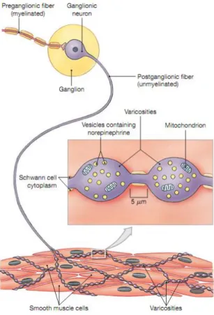
VSM is primarily under the control of sympathetic innervation. As shown in Figure 1.5, when stimulated, sympathetic preganglionic neurons release ACh at synapses with postganglionic neurons. The effect of ACh on the postganglionic neurons is always excitatory. These postganglionic neurons then release neurotransmitters at specific target organs such as SMs. Most sympathetic ganglionic neurons release norepinephrine (NE) at their varicosities. The NE released by varicosities affects its targets until it is reabsorbed by varicosities and inactivated by the enzyme monoamine oxidase (MAO). A small part of NE diffuses out of the area or is broken down by the enzyme catechol-O-methyltransferase (COMT) in surrounding tissues. The effects of sympathetic stimulation result primarily from the interactions of NE with adrenergic receptors.
Figure 1.5 Sympathetic innervation of SMCs. When stimulated, sympathetic preganglionic neurons release ACh at synapses with ganglionic neurons. Then neurotransmitters such as NE are released from varicosities and act on SMCs. Finally, these neurotransmitters are recycled or degraded. Adapted from Martini et al.,2011 (7).
1.1.3 Lower urinary tract
1.1.3.1 Overview
The lower urinary tract (LUT) is composed of the urinary bladder and the outflow tract (Figure 1.6) (12). It has two important functions: the urine storage and emptying. During filling stage, the bladder is relaxed and the outflow tracts offer a high resistance; during emptying stage the outflow resistance falls and the bladder generates a high wall tension to raise intravesical pressure by the contraction of the detrusor SMCs.
8
Figure 1.6 Schematic representation of the lower urinary tract. From Fry et al., 2010 (12).
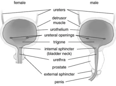
1.1.3.2 Morphology of urinary bladder
The bladder is composed of two parts: the body and the base (Figure 1.7). The bladder body is located above the ureteral orifices. The base contains the trigone, the uretero-vesical junction, the deep detrusor, and the anterior bladder wall (13).
Figure 1.7 Scheme of the bladder. Adapted from Anderson et al., 2004 (13).
The bladder is a hollow SM organ, which is comprised of a mucous membrane and the detrusor SM covered partly by peritoneal serosa and partly by fascia. The urinary bladder is composed of three layers: the cells of the outer and inner layers tend to be
oriented longitudinally, and those of the middle layer tend to be oriented circularly. The individual detrusor SMCs are typical SMCs, similar to those in other muscular organs. They are long, spindle-shaped cells with a central nucleus. These cells are several hundreds µM long and 5-6 µM wide, when they are fully relaxed (Figure 1.2) (14).
1.1.3.3 Spontaneous activity of the bladder
The significant feature of the bladder is its ability to generate considerable spontaneous contractile and electrical activities that are observed from isolated bladders, multicellular detrusor preparations and even from isolated cells (15-17). In addition, the spontaneous activity is much greater in animals and patients with overactive bladders (16, 18, 19). Spontaneous activity is resistant to tetrodotoxin (TTX), which blocks Nav channels activity and related action potential, and is regulated by muscarinic agonists (17, 20), L-type Ca2+ channels (LTCC) blockers (21), and NO-cGMP pathway (17). Several hypotheses are postulated to interpret the spontaneous activity, which are not mutually exclusive. The three major of them are the neurogenic, the myogenic and the urotheliogenic hypotheses.
A. The neurogenic hypothesis
This hypothesis was firstly put forward by de Groat (22). Normal storage of urine is dependent on the spinal reflex and the tonic inhibitory systems. The spinal reflex systems mediate the urethral outlet through activating sympathetic and somatic pathways. The tonic inhibitory systems in the brain suppress the parasympathetic excitatory outflow to the urinary bladder. Voiding is a much more complex reflex, which is coordinately mediated by the inhibition of sympathetic-somatic pathways and the activation of spinobulbospinal parasympathetic pathways. Bladder overactivity may be induced by damages of the brain and the central inhibitory pathways or sensitization of the peripheral afferent terminals in the bladder. Therefore, this hypothesis may represent a distinct population of patients with overactive bladders. Furthermore, transmitter leaking from the motor fibers is able to generate
10
small local contractions or increases of tone (13). B. The myogenic hypothesis
The myogenic hypothesis suggests that the spontaneous activity is associated to the excitability and intercellular coupling of the SMCs with other myocytes (23) or interstitial cells lead to the larger contractions (24). It has been reported that spontaneous activity is increased in single isolated cells of overactive bladder (16). Ikeda et al. showed that gap junction expression was increased in lamina propria myofibroblasts and urothelial cells from the spinal cord-injured adult bladder, which is required for the coordinated activity (19). These cells are supposed to modulate and coordinate activity of detrusor bundles facilitating the large, slower overactive-bladder contractions (25). Interstitial cells may also be involved in the regulation of spontaneous activity, as they are innervated by afferent nerves labeling for NO synthase and also possess cGMP activity (12).
C. The urotheliogenic hypothesis
This hypothesis indicates that spontaneous activity originates in the suburothelial layer of interstitial cells and then propagates to the detrusor where spontaneous contractions generated.
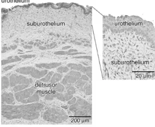
As shown in Figure 1.8, the urothelium is a transitional epithelium interfacing directly with the bladder lumen. Below this is a suburothelium containing a dense network of capillaries and afferent nerves, and also a network of interstitial cells connected by gap junctions (12). Hawthorn et al. reported that the presence of an intact urothelium on isolated bladder strips attenuated contractions induced by carbachol but not KCl (26). The negative inotropic reagent has been demonstrated to be a diffusible agent but its identity is at present unknown (26). Through optical imaging experiments, spontaneous activity is shown to arise in the suburothelium and spread to the detrusor layer (27).
1.1.4 Excitation-contraction-coupling (ECC) in the SM
ECC refers to the chain of processes linking a stimulus to the contractile response of a muscle. Two major types of ECC have been described in the SM: an electromechanical and a pharmacomechanical coupling (28).
1.1.4.1 Mechanisms of contraction in the SMC
The contractile/relaxing state of the SMC is under the control of the level of phosphorylation of the 20-kDa light chain of myosin (MLC20). MLC20 phosphorylation promotes actin-myosin crosslinking and subsequent contraction by increasing myosin ATPase activity. Thus, an increase in MLC20 phosphorylation generates contraction of the SMC, and conversely the dephosphorylation of MLC20 induces its relaxation. Two key enzymes are involved in the control of MLC20 phosphorylation: the MLC kinase (MLCK), a Ca2+/CaM-activated kinase, and the MLC phosphatase (MLCP), a serine/threonine protein phosphatase type I.
12
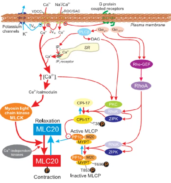
Figure 1.9 Schematic representation of major pathways involved in excitation-contraction coupling in SM. CPI-17: PKC-potentiated inhibitory protein; MLC20: 20 kDa light chain of myosin; MYPT: myosin phosphatase target subunit; Rho-GEF: GDP-GTP exchange factor; RhoK: Rho kinase (or ROCK); ROC: receptor-operated channels; SAC: stretch-activated channels; VDCC: voltage dependent Ca2+ channels. Adapted from Sanders et al., 2008 (10).
As shown in Figure 1.9, the primary mechanism leading to the SMC contraction is an increase in cytosolic Ca2+ concentration which binds to calmodulin to increase the activity of MLCK (10). A second mechanism of contraction is independent of an increase in cytosolic Ca2+ concentration, but involves a decrease in MLCP activity. MLCP activity is essentially controlled by the small GTPase RhoA and its target Rho kinase (ROCK) (10). Phosphorylation of myosin-binding subunit of MLCP (MYPT 1)
by ROCKs leads to the inhibition of MLCP, which prevents MLC dephosphorylation and hence SM contractility.
1.1.4.2 The electromechanical coupling
During electromechanical coupling, the increase of Ca2+ concentration is associated with changes of the membrane potential (Em), which opens voltage-gated channels
located on the plasma membrane and thereby allow the entry of Ca2+ from the outside medium. Two types of calcium channels (L and T type) are thought to play a key role in electromechanical coupling (9). The T-type or low-voltage activated Ca2+ channels have been observed in a variety of SMCs (9). They are activated at a low Em of
around -50 mV and attain their maximum around -20 mV. These channels have a rather low conductance and are more readily inactivated. The L-type or high-voltage avtivated Ca2+ channels carry the majority of the Ca2+-inward current in the SMCs (9). They show a threshold for activation around -40 mV and are fully activated at a slightly positive Em. They have a higher conductance and are sensitive to inhibition by
such classical organic Ca2+ channels blockers as dihydropyridines and phenylalkylamines (9).
1.1.4.3 The pharmacomechanical coupling
Pharmacomechanical coupling means the stimulation of contraction without necessary changes of the Em (9). This process is dependent on both intracellular Ca2+ influx through receptor-activated Ca2+ channels and Ca2+ release through intracellular Ca2+ stores. In addition, Rho/ROCK signaling pathway acts by altering the Ca2+ sensitivity of the contractile machinery (29).
A major pathway for pharmacomechanical Ca2+ release is the phosphatidylinositol cascade. In brief, receptor activation, in particular Gq protein coupled receptors,
initiates the hydrolysis of the lipid phosphatidyl inositol 4, 5-bisphosphate (PIP2) by phospholipase C (PLC) to yield diacylglycerol (DAG) and inositol 1,4,5-trisphosphate (IP3). DAG and IP3 both act as second messengers and are thought to control a
14
variety of cellular processes (9). The endoplasmic reticulum is the physiologically important reservoir in SMCs. IP3 diffuses into the cytosol to the membrane of the endoplasmic reticulum, where it activates its receptor, the IP3R, which allows Ca2+ release into the cytosol.
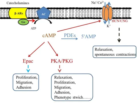
1.2 The cAMP/β-adrenergic signaling pathway
Beta-adrenoceptors (β-ARs)-mediated signaling pathway, one of the major signaling pathways in mammalian cells, mediates several physiological and pathological processes of SMs. The paradigm of this signaling pathway is summarized in Figure 1.10. Classically, circulating catecholamines or catecholamines released from sympathetic nerves bind to the β-ARs and then activate a stimulatory guanine nucleotide-binding protein (Gs), which in turn activates AC that synthesizes cAMP from ATP. Elevated intracellular cAMP activates several distinct downstream targets including the cAMP-dependent protein kinase (PKA), cyclic nucleotide-gated (CNG) ion channels and exchange protein activated by cAMP (Epac). Finally, cAMP is degraded by PDEs into an inactive nucleotide 5'AMP (3) or transported by active efflux transporters, namely the multidrug resistance-associated proteins (MRPs) MRP4 and MRP5 (30).
Figure 1.10 A scheme of β-AR signaling pathway in SM. In brief, noradrenaline binds to β-AR and activate Gs-AC-cAMP pathway, resulting in a series functional changes of SM. CNG: cyclic nucleotide-gated channels; HCN: hyperpolarization-activated CNG channels; Epac: exchange protein directly activated by cAMP.
16
1.2.1 β-ARs
β-ARs belong to the G-protein coupled receptors (GPCR) family. Their characteristic structure consists of seven trans-membrane domains, with an extracellular N-terminal tail, three intra- and three extra-cellular loops, and an intracellular C-terminal tail (31). In the 1960’s, β-ARs were pharmacologically subdivided into two subtypes, β1- and
β2-ARs, on the basis of their functional responses to various adrenergic activating and
inhibitory agents (32). The genes encoding these two receptors were cloned in 1987 and 1986, respectively. In 1989, a third subtype of β-AR was also identified and named as β3-AR (33). The β1- and β2-ARs share 48.9% homology, whereas β3-AR
exhibits 50.7% and 45.5% homology with the other two subtypes, respectively (34). β-ARs are widely expressed in many different types of blood vessels, where they mediate catecholamine-induced relaxation (35) through releasing intracellular second messenger cAMP. Classically, β2-ARs are the predominant subtype in the vasculature
(35, 36),but the involvement of each β-AR subtype varies according to the vascular bed and species. For instance, β1-ARs seem to be predominant in coronary and
cerebral arteries (37). β-ARs are widely expressed in the urinary bladder where they play an important role in the storage of urine. At mRNA level, all three β-ARs are detectable in the bladder. In rat bladder, the abundance of the three subtypes is similar (38), whereas in human bladder, >95% of β-AR mRNAs belong to the β3-subtype (39).
At the protein level, β1- and β2-ARs have been identified by radioligand binding in the
bladder of humans (40) and several animals species (41). However, the expression of β3-AR protein in the bladder has not been detected.
1.2.2 G protein
The heterotrimeric G proteins, which are key regulators in GPCR-related transduction pathways, are composed of 3 subunits (α, β, and γ) (42). So far, at least 20 Gα, 5 Gβ,
and 11 Gγ subtypes of G proteins subunits have been identified (43). The Gα subunits
remarkably among the G proteins, with the Gβ subunits exhibiting higher sequence
similarities than the Gγ group. The Gα subunits are a family of 39-52 kDa proteins that
have been divided, on the basis of their amino acid sequences, into 4 classes: Gαs, Gαi,
Gαq, and Gα12. Gβγ is tightly complexed in dimer which dissociates only under
denaturing conditions. It serves to increase the affinity of Gα for its receptors and to
regulate several effectors, either directly or in conjunction with Gα subunits.
Now, it becomes clear that β1-ARs are exclusively coupled to Gαs protein, whereas
β2-ARs are coupled to both Gαs and Gαi proteins. In 1993, Xiao and her colleagues
firstly described that a β2-AR-stimulated cAMP production is dissociated from the
regulation of myofilament and sarcoplasmic reticulum functions (44). A functional compartment of cAMP signaling that is due to the activation of β2-AR coupled to Gi
and/or Go was postulated to explain this phenomenon (45). Later, a switching of the
β2-ARs coupling in which the phosphorylated and internalized β2-ARs tend to lose
affinity for Gs and gain affinity for Gi was reported (46). Based on this observation,
Daaka et al. suggested that PKA-mediated heterologous desensitization of the β2-ARs
pathway may actually serve as an independent physiological signaling route rather than as an escape circuit for the receptor from unabated stimulation by agonists (46). Recently, Baloğlu et al. showed that β-AR mainly activates Gs protein in aorta from
young rat, whereas it also activates Gi in aorta from old rats, contributing to impaired
vasodilator β-adrenergic responses in rat aorta during maturation (47). Vascular β2-AR
coupling to Gi proteins also occurs in physiological situations. For example, Banquet
et al. indicated that activation of the β2-AR elicits an endothelial nitric oxide synthase
(eNOS)-dependent relaxation of mouse pulmonary artery via a Gi/o-Src
kinase-PI3K/Akt pathway (48). β3-ARs have also been shown to be coupled to Gi
proteins in human ventricule myocytes (49). Their stimulation produces negative inotropic effects through activation of a nitric oxide synthase (NOS) pathway (50). In rat thoracic aorta, β3-AR-mediated vasorelaxation is NOS-dependent but Gi/o
18
1.2.3 Adenylyl cyclase (AC)
ACs are the enzymes that synthesize cAMP from adenosine triphosphate (ATP). To date, 9 membrane-bound AC isoforms and 1 soluble AC isoform have been characterized (53) and are divided into three groups. Group 1 consists of Ca2+/calmodulin (CaM)-stimulated enzymes that are activated synergistically by Gαs
and Ca2+/CaM and inhibited by Gαi (types AC 1, 3, and 8). Group 2 represents
isoforms that are activated synergistically by Gαs and Gβγ (types AC 2, 4, and 7).
Group 3 is composed of the isoforms that are inhibited by Gαi and Ca2+ (types AC 5
and 6). Additionally, AC9 has been characterized as a distinct (or atypical) isoform with restricted expression (53). ACs are widely expressed in several tissues with different pattern (54). In cultured rat aortic SM cells (RASMCs), the mRNAs encoding 3 isoforms (AC3, AC5 and AC6) were detected, and the corresponding proteins are expressed in both caveolin-rich and noncaveolin domains (55). The transcripts of AC4 and AC8 were also expressed in these cells (56). There is increasing evidence for specific functional roles of these distinct isoforms of AC in vascular SMCs. Accordingly, Gros et al. demonstrated that overexpression of AC1 and AC3 isoforms inhibit cell proliferation of cultured RASMC, whereas overexpression of AC6 only enhances their cAMP-mediated cellular arborization, consistent with a model of the association of specific isoforms of AC in discrete functional compartments, resulting in isoform-selective regulation of cellular growth versus cytoskeletal organization (57).
1.2.4 cAMP targets
1.2.4.1 PKA
Protein Kinases are phosphotransferases that catalyze the transfer of the γ-phosphoryl group of ATP to an amino acid side chain of the basic sequences in the presence of Mg2+. PKA is composed of 4 separate subunits, 2 catalytic (C) and 2 regulatory (R)
subunits. PKAs have been classified into two classes, PKA I and PKA II, according to the nature of the R subunit. Three C subunit genes (Cα, Cβ, and Cγ) and four different R subunit genes (RIα, RIβ, RIIα, and RIIβ) have been identified (58). The binding of 2 molecules of the activating ligand cAMP to each R subunit induces conformational changes that lead to the dissociation of the holoenzyme into its constituent C and R subunits. The free active C subunit can then affect a range of diverse cellular events by phosphorylating an array of protein substrates, including enzymes, receptors, ion channels and transcriptional factors (59).
Extensive studies have been performed to demonstrate the expression pattern of R and C subunits. Singh et al. showed that, among the three major vascular layers (the intima, the media, and the adventitia), over 90% of the total protein kinase activity is observed in the middle layer (60). DEAE-cellulose chromatography of the soluble enzyme revealed the existence of two major forms of PKA, type I and type II, with the type II representing 60% of the total enzymatic activity (61). Cα1 and Cβ1 protein variants are expressed almost in every tissue type, whereas the Cαs protein is exclusively expressed in sperm cells. Cγ is only expressed in human testis (62). Poole et al. showed that strong PKA-RI is detected in the majority, if not all, myenteric neurons throughout the gastrointestinal tract and SMCs are also immunoreactive for PKA, with labelling observed in the cytoplasm and nucleus of cells in both the longitudinal and circular muscle layers of guinea-pigs (63). Recent studies showed that PKAIα, PKAIIα, and PKAIIβ are observed in the human prostate (64) and cavernous arteries (65).
In SM, as well as in the other tissues, elevation of cAMP is capable of activating the cGMP-dependent protein kinase (PKG) (58). Although PKG is relatively specific for cGMP over cAMP (affinity ratio: 50-100 fold), basal cAMP concentration in SM is usually higher than that of cGMP (five to six fold higher in pig coronary artery), which allows cross-activation when cAMP is moderately elevated (66).
20
1.2.4.2 Epac
In 1998, two distinct groups identified a novel family of cAMP sensor protein, named Epac (exchange protein directly activated by cAMP) or cAMP-GEF (cAMP-regulated guanine exchange factor) (67, 68). So far, two isoforms of Epac, Epac1 and Epac2, have been identified, which are encoded by two distinct genes. Epac1 is most prominent in the brain, heart, kidney, pancreas, spleen, ovary, thyroid and spinal cord, whereas Epac2 is restricted and most prominent in discreet regions of the brain, as well as the adrenal glands, liver, SM and pancreatic islets of Langerhans (68, 69). Only Epac1 isoform was detected in SMC from rat aorta (70), as well as in human endothelial cells (HUVEC) (71). Epac proteins are expressed at many locations within the cell, including the cytosol, the nucleus as well as the nuclear and plasma membranes. Because of their cellular localization and molecular partners, Epac proteins activate different downstream effectors (72). Epac1 and Epac2 are multidomain proteins constituted by a regulatory N-terminal domain containing 1 (Epac1) or 2 (Epac2) binding domains for cAMP and a catalytic C-terminal domain (73).
Several cellular processes have been proposed to involve in Epac-mediated mechanisms in the SM, including vascular SM relaxation (74), adhesion of microvascular SMCs to fibronectin (75), anti-inflammatory role of cAMP in chronic obstructive pulmonary disease (76), human airway SM phenotype plasticity (77, 78), inhibition of airway SMC proliferation (79), and VSMC migration (80).
1.2.4.3 Cyclic nucleotide-activated ion channels
Cyclic nucleotide-activated ion channels are composed of two families: the cyclic nucleotide-gated (CNG) channels and the hyperpolarization-activated cyclic nucleotide-modulated (HCN) channels. These two families exhibit high sequence similarity and belong to the superfamily of voltage-gated potassium channels. Both channels are activated by cyclic nucleotide binding. In addition, whereas HCN channels are activated by voltage, CNG channels are virtually voltage independent.
A. CNG channels
In 1985, CNG channels were firstly discovered on the plasma membrane of the outer segment of vertebrate rod photoreceptors, where they play a critical role in phototransduction (81). So far, there are six vertebrate CNG channel subunits: CNGA1, CNGA2, CNGA3, CNGA4, CNGB1, and CNGB3 (82). These subunits can assemble in a variety of combinations to produce tetrameric channels. Although all six CNG channel subunits share significant sequence homology only three of these subunit types, CNGA1, CNGA2, and CNGA3, can form homomeric channels in heterologous expression systems. CNGA4, CNGB1, and CNGB3 do not form functional homomeric channels but can coassemble with other subunits to form functional heteromeric channels.
CNG channels are widely expressed in several SM tissues across species. Specifically, CNGA1 was found to be abundantly expressed in the endothelium layer, and also expressed in VSM layer but at a much lower level in guinea pig arteries (83). In contrast, strong expression of CNGA2 channel was detected in both the endothelium and SM layers of human arteries (84). Recently, CNGA1 channels were shown to be expressed in rat urethra (85). Functionally, SM CNG channels play an important role in nerve-mediated nitrergic relaxation of rat urethra (85) and thromboxane A2-induced
contraction of rat small mesenteric arteries (86). B. HCN channels
HCN-channels constitute a related family of channels with different physiological role from that of CNG channels. HCN channels are unique among vertebrate voltage-gated ion channels, in that they have a reverse voltage-dependence that leads to activation upon hyperpolarization. In addition, these channels are directly regulated by cAMP. The current generated by HCN channels has been called the Ih (hyperpolarization), Iq
(queer), or If (funny) current (87).
The HCN channel family, like the CNG channel family, comprises several subunit types. There are four known HCN subunit isoforms, HCN1-HCN4, which combine to form tetrameric channels (87). These channels have been identified in SM. For
22
example, Greenwood and Prestwich indicated that HCN2, HCN3, and HCN4 mRNA are expressed in portal vein SMCs where they play an important role in regulating spontaneous contractions (88). Recently, He et al. showed that all 4 HCN channel isoforms exist in the bladder, and they affect the bladder excitation, presumably via bladder interstitial cells of cajal (ICCs) (89).
1.2.5 Mechanisms of cyclic nucleotide-induced relaxation
cAMP and cGMP are main messengers that mediate relaxation under physiological conditions. As cAMP shares most of its relaxant mechanisms with cGMP, both cyclic nucleotide will be considered in this section.
So far, at least 3 distinct mechanisms are thought to mediate the dilator effect of cyclic nucleotides and their dependent protein kinases: (1) the decrease in the cytosolic Ca2+ concentration; (2) the hyperpolarization of the SMC membrane potential; (3) the reduction in the sensitivity of the contractile machinery by uncoupling contraction from MLC phosphorylation (90).
1.2.5.1 Cytosolic Ca2+ concentration modulation in the SMC
The first mechanism proposed for cyclic nucleotide-induced relaxation of SM is the reduction of free intracellular cytosolic Ca2+ levels. As shown in Figure 1.11, multiple mechanisms are involved in the modulation of Ca2+ concentration.
A. Decreased Ca2+ release from the endoplasmic reticulum
The endoplasmic reticulum (ER) is a huge intracellular Ca2+ store, playing a critical role in the homoestasis of cytosolic Ca2+ concentration and the SM tone. There are two major types of Ca2+ release channels on the ER: IP3Rs and ryanodine receptors (RyRs). The IP3R channel is able to release a large quantity of Ca2+ to the cytosol, which is thought to mediate the initial phase of agonist-induced contraction. The IP3R is phosphorylated by forskolin, an agent that directly activates AC and increases cAMP level (91). The sensitivities of these phosphorylations to various protein kinase
inhibitors suggest that phosphorylation of the IP3Rs is induced in response to increases of both cAMP and cGMP in intact cells (91). Thus, it is conceivable that both cAMP and cGMP signaling pathways regulate IP3R activity through activation of PKG and the resultant phosphorylation of the IP3R (Figure 1.11) in some types SMs.
Figure 1.11 Mechanisms involved in the reduction of cytosolic Ca2+ concentration induced by cyclic nucleotides and their dependent protein kinases. Green arrows imply stimulation, and red arrows imply inhibition. IP3R: Inositol trisphosphate receptor; LTCC: L-type Ca2+ currents; NCX: Na+/Ca2+ exchanger; PIP2: Phosphatidylinositol 4,5-bisphosphate; PLC: Phospholipase C; PMCA: Plasma membrane Ca2+-ATPase; SR: Sarcoplasmic reticulum. Adapted from Morgado et
al., 2012 (90).
B. Increased Ca2+-sequestration into the ER
Sarcoplasmic reticulum Ca2+-ATPase (SERCA) is a transport protein in the ER that serves to recycle the cytosolic Ca2+ into the ER. Phospholamban (PLB) reversibly inhibits the activity of SERCA (90). Phosphorylation of PLB by PKA or PKG, at the Ser 16 residue, relieves its inhibition on SERCA, thus increasing its ATPase activity and the rate of Ca2+ uptake into the ER (90) (Figure 1.11). Both cGMP- and cAMP-elevating agents are reported to induce SM relaxation through stimulation of Ca2+ uptake into the ER via PLB phosphorylation (92).
24
C. Decreased influx of extracellular Ca2+
In SM, there are at least two types of plasmalemmal Ca2+ channels recognized as relevant entry routes of extracellular Ca2+: store-operated calcium channels (SOCCs) and voltage-dependent Ca2+ channels.
The Ca2+ entry by SOCCs is activated by the emptying of intracellular Ca2+ stores and acts to replenish these stores (93). Basically, two mechanisms have been proposed to explain SOCCs (93). The first mechanism is that diffusible ‘‘calcium influx factor’’ released from intracellular organelles activates calcium-independent phospholipase A, which in turn generates lysophospholipids that activates SOCCs at the plasma membrane. The second model was more recently postulated. In brief, the ER Ca2+ sensor stromal interaction molecule І (STIM I) activates Orai I which is a transmembrane protein of the plasma membrane. The STIM І-Orai I complexes are formed in a spatially restricted manner at the junctions of ER and plasma membranes and SOCCs are activated in the vicinity.
The voltage-dependent Ca2+ channels include L-type and T-type Ca2+ channels. In most SMCs, LTCCs are the most numerous channels and probably the most major route for calcium influx (9). Experiments in mice with SM-specific inactivation of the LTCC gene revealed that these channels are key players in the hormonal regulation of blood pressure and development of myogenic tone (94). Voltage-gated Ca2+ channels are modulated by several signaling systems, including cyclic nucleotides. Data on the modulation of LTCC in VSMCs by cyclic nucleotides are sometimes contradictory but, in general, is accepted that cyclic nucleotides inhibit LTCC (Figure 1.11) (90).
D. Increased efflux of intracellular Ca2+ through the Plasma Membrane
Ca2+-ATPase (PMCA)
PMCA mediates extrusion of Ca2+ into the extracellular space and plays a major role in reducing excessive cytosolic Ca2+ in the resting condition or in mediating the vasodilator action of various endogenous agents (Figure 1.11). PMCA uses energy from ATP hydrolysis to produce Ca2+ efflux in exchange for 2H+ influx against a high electrochemical gradient across the plasma membrane. When CaM is absent, the
CaM-binding domain interacts with cytosolic regions of the enzyme, inhibiting its activity (i.e., autoinhibition). Phosphorylation of some sites in the CaM binding regions by several protein kinases, such as PKA, PKG, PKC, and Ca2+/CaM-dependent protein kinase results in the activation of the PMCA pump (90). E. Increased efflux of intracellular calcium through stimulation of the
Na+/Ca2+-exchanger (NCX)
The NCX is driven by the transmembrane Na+ gradient and maintained by the Na+ pump (Na+/K+-ATPase). It normally transports 1 Ca2+ out in exchange for the entry of 3 Na+. Both cAMP and cGMP are reported to regulate the NCX in the SM. Furukawa et al. showed that cGMP stimulates NCX in VSMCs in primary culture (95). By using wild type (WT) and transgenic (TG) mice that specifically over-express NCX1.3 in SM, Karashima et al. showed that the forskolin-induced decreases in intracellular Ca2+ concentration and tension are much greater in aortas from TG mice than in those from WT mice (96). These results directly indicated that NCX is involved in the forskolin-induced reduction of intracellular Ca2+ concentration and tension in the mouse thoracic aorta (96).
1.2.5.2 Hyperpolarization of the SMC
In the SMC, cyclic nucleotides can target different types of K+ channels (Figure 1.12), which are the mediators of Em and play a critical role in maintaining the SM tone.
Figure 1.12 Modulation of K+ channels by cyclic nucleotide-dependent protein kinases in SM. Green arrows imply stimulation; red arrows imply inhibition. From Morgado et al., 2012 (90).
26
A. Voltage-gated K+ channels (Kv channels)
The Kv channels activation is thought to be involved in the vasodilator response to several endogenous and exogenous substances. It has been reported that β-AR stimulation and forskolin activate Kv currents through cAMP/PKA in rabbit VSMCs (97, 98). On the other hand, NO was reported to induce the dephosphorylation of Kv proteins through cGMP and SHP-1 tyrosine phosphatase (99).
B. Large conductance Ca2+-activated K+ channels (BK channels)
The BK channels are activated by cytosolic Ca2+. These channels have been indicated to be involved in the relaxant response induced by many endogenous vasodilators (e.g., adenosine, prostacyclin, NO) and to play an important role as a negative feedback mechanism to limit membrane depolarization and vasoconstriction (100-105).
C. ATP-sensitive K+ channels (KATP channels)
A hallmark feature of KATP channels is their inhibition by micromolar concentrations
of intracellular ATP (Figure 1.12). Therefore, any pathophysiological condition that damages the ATP mitochondrial generation would increase the opening of KATP
channels and thereby cause vasodilation. In VSMC, the KATP channels are stimulated
by many pharmacological and endogenous vasodilators that activate these channels by increasing cAMP concentration (90).
D. Inward rectifier K+ channels (Kir channels)
The Kir channels regulate the membrane potential in SMCs from several types of resistance arteries and may be responsible for external K+-induced dilations. The existence of currents through these channels has been reported in a variety of VSM, including coronary, cerebral, and mesenteric arteries, and may be preferentially expressed in small rather than large arteries. These Kir channels conduct inward K+ currents much more readily than they conduct outward K+ current, due to their
activation by hyperpolarization rather than depolarization. Kir channels, like other K+ channels, may also be modulated by several vasodilators (e.g., adenosine, and sodium nitroprusside) and cAMP/PKA pathway has been reported to be involved in some Kir-mediated vasodilation (90).
1.2.5.3 Decrease in MLC20 phosphorylation
MLC20 phosphorylation promotes the contraction of the SMC, and conversely a decrease in MLC20 phosphorylation promotes its relaxation. This may occur either through inhibition of MLCK activity or through increase in MLCP activity (Figure 1.13).
Figure 1.13 Role of MLC20 in the regulation of the contractile state in SM. Adapted from Morgado et al., 2012 (90).
A. Inhibition of MLCK activity
The activity of MLCK is primarily mediated by the Ca2+/CaM complex. Elevation of cAMP levels inhibits phosphorylation of MLCK through a PKA-dependent pathway. This decreases the affinity of MLCK for the Ca2+/CaM complex and thereby reduces myofilament Ca2+ sensitivity (9). There is no direct evidence for PKG-dependent phosphorylation of MLCK. However, cross-activation of PKA by cGMP is reported to
28
participate in inhibition of MLCK activity, and subsequent vasodilation (9). B. Increase in MLCP activity
Multiple of evidences show that PKG could activate MLCP, thereby decreasing MLC20 phosphorylation and SMC contraction (Figure 1.13) (9). It has been shown that cGMP-dependent activation of MLCP involves direct phosphorylation of the regulatory subunit (MYPT-1) of MLCP (90).
It has also been reported that cGMP-induced inhibition of Ca2+ sensitization can be directly mediated by RhoA/ROCK pathway. Recently, Epac has been proposed to mediate the relaxation induced by cAMP in several SMs through downregulation of RhoA (74).
In conclusion, cAMP acts as a second messenger regulating various functions of SMs. Firstly, cAMP induces the relaxation of many different types of SMs via distinct mechamisms and regulates the SM tone to adapt the enviroment changes around them. Second, cAMP is an important regulator of the proliferation and migration of a SMC. Thus cAMP is especially critical in some diseases of SM such as vascular damage.
1.3 PDEs family
PDEs are a large family of enzymes that specifically hydrolyse the second messengers cAMP and/or cGMP into their inactive forms, the non-cyclic nucleotides 5'-AMP and 5'GMP, respectively (3).
Figure 1.14 Degradation of cyclic nucleotides by PDEs. From Keravis et al., 2005 (106).
Almost immediately after the discovery of cAMP, Butcher and Sutherland described the cyclic nucleotide PDE activity (107). During the 1970s and 1980s, research into the biochemical characterization and functional role of PDEs has grown. Biochemical characterizations of PDE activities were obtained by anion exchange chromatography of tissue cytosolic fractions that allowed the dissociation of various fractions of PDE activities. These fractions were differentiated by their substrate specificity and sensitivity to Ca2+/CaM. Based on the elution order, these fractions were numbered. For example, PDE I represented mainly CaM-activated PDE activity. More and more PDE characterizations were found in tissues and cell extracts. As properties, functional roles as well as distributions of PDE activity differed from one to other tissue, the confusion appeared in the literature concerning the PDE nomenclature. In addition, the identification of inhibitors, activators, or ligands that act preferentially
30
on 1 isozyme allowed discrimination of new PDE isozymes and lead investigators to give new names for various PDE isozymes, such as ROI-PDE (rolipram-inhibited PDE), CGI-PDE (cGMP-inhibited PDE), CaM-PDE (calmodulin-activated PDE), cGS-PDE (cGMP-stimulated PDE), and cGB-PDE (cGMP-binding PDE). Through application of cloning and polymerase chain reaction (PCR) techniques, a number of different PDEs gene families have also been identified, and the first five PDE families were designated by roman numerals as PDE-I through PDE-V.
To solve the confusion concerning PDE nomenclature, Beavo and other investigators initiated an official nomenclature for PDE isozymes (108). Therefore, an international nomenclature of PDE was established. For example, for RNPDE1A2: Ratus Norvegicus (RN) represents species; PDE1 shows the gene family; A represents the individual gene product within the family; the final Arabic numeral (2) represents the splice variant.
Based on the primary amino acid sequence as well as kinetic and regulatory properties, 11 PDE families derived from 21 genes are now identified and named PDE 1 to 11 (1). In spite of the large number of PDE isoforms, all of them have an N-terminal regulatory domain (R domain) and a C-terminal catalytic domain (C domain). PDE4 is specifically characterized by regulatory features in its C domain. PDEs share a conserved C domain, but amino acid sequence outside this region differs markedly. The N-terminal region of PDEs diverges widely among PDEs in structure and size, and contains sequences that are target sites for various regulators, including binding sites for CaM or cGMP, phosphorylation sites for protein kinases (CaMK, PKA, PKG, PKC and PKB), and sites for dimerization or membrane association (109). Some PDEs are highly specific for hydrolysis of cAMP (PDEs 4, 7, and 8) or cGMP (PDEs 5, 6 and 9), while others hydrolyze both cAMP and cGMP (110-112).
1.3.1 PDE1
The PDE1 family is known as Ca2+/CaM-dependent PDE. Three different gene products have been cloned: PDE1A, PDE1B and PDE1C (113). PDE1A and PDE1B
exhibit higher affinity for cGMP than cAMP, whereas PDE1C hydrolyses cGMP and cAMP with equal efficiency (108).
Table 1.1 Characterisitics of PDE families. Adapted from Lugnier et al., 2006 (109). PDEs Substrate Property Selective inhibitor
PDE1 cAMP/cGMP Ca2+/CaM-activated Nimodipine, MIMX PDE2 cAMP/cGMP cGMP-stimulated EHNA, Bay 60-7550 PDE3 cAMP/cGMP cGMP-inhibited Cilostamide, Milrinone PDE4 cAMP cGMP-insensitive Rolipram, Ro 20-1724
Roflumilast
PDE5 cGMP PKA/PKG- Zaprinast, DMPPO
phosphorylated E4021, Sildenafil PDE6 cGMP Transducin-activated Zaprinast, DMPPO,
E4021, Sildenafil PDE7 cAMP Rolipram-insensitive BRL 50481, ICI242 PDE8 cAMP Rolipram-insensitive PF-04957325
IBMX-insensitive
PDE9 cGMP IBMX-insensitive BAY 73-6691, PF-04447943
PDE10 cAMP/cGMP Unknown PQ-10
PDE11 cAMP/cGMP Unknown Unknown
A. Variants and tissue expression pattern
PDE1A protein is highly expressed in the brain. However, PDE1A variants differ greatly in their tissue expression. PDE1A1 and PDE1A4 mRNAs show a broad tissue distribution. However, the mRNAs of other variants exhibit highly specific expression, for instance PDE1A5 and PDE1A6 are detected only in brain or thyroid and PDE1A10 was detected only in testis. So far, only two variants of PDE1B have been identified. PDE1B1 mRNA was found predominantly in the human brain, where its expression was correlated with the regions showing extensive D1 dopamine receptor mRNA. PDE1B1 is also expressed in the heart and skeletal muscle. Five variants of
32
PDE1C encoding four different proteins have been identified from several different species. PDE1C1 mRNA extensively expressed in the brain and heart and seems to be the major type highly expressed in the mouse cerebellar granular cells. In olfactory epithelium PDE1C2 is the high-affinity CaM-PDE. PDE1C1 and PDE1C4/5 mRNA were found in the testis (1). In vascular SMCs, the isoforms of PDE1 depend on the phenotype state and the species. Recently, Lakics et al. showed that all three isoforms of PDE1 were identified in human bladder tissue, among them PDE1C was majorly expressed (114).
B. Regulation
The transcripts and protein of PDE1A are increased by long-term nitrate treatment in rat aortas, leading to nitrate tolerance (115). PDE1B is known as an early response gene and its expression is up-regulated by the treatment of granulocyte macrophage colony stimulating factor in differentiating monocytes (116). PDE1C expression is observed in proliferative phenotypes of human arterial SMCs but not in quiescent SMCs (117). Phosphorylation has also been shown as a manner for the regulation of PDE1 as shown in Figure 1.15 (3).
C. Function
PDE1B-deficient mice show increased locomotor activity and deficits in spatial learning (118). It is reported that LTCC and other cardiac targets are regulated by PDE1 isoforms (119). In a rat model of pressure overload by banding aorta, PDE1 activity is shown to be up-regulated in left ventricular tissue, suggesting that the increase in cGMP-PDE activity in response to pressure overload plays an important role in neutralizing cGMP action in cardiac tissue (120). The functional role of PDE1 in VSM may be to modulate the contractile tone by reducing cGMP in response to a Ca2+ signal. Using a method designed to estimate the association of Ca2+/CaM with PDE in intact tissue, it was shown that the exposure of intact coronary artery strips to histamine increased the association of PDE1 with Ca2+/CaM, indicating increased PDE1 activity (121). Recently, several novel functions of PDE1 have been reported.
Cai et al. found that PDE1C plays a critical role in regulating collagen homeostasis during pathological vascular remodeling (122). Inhibition of PDE1 activity was shown to markedly attenuat β-catenin/TCF signaling by down-regulating β-catenin protein (123). Miller et al. suggested that induction of PDE1A plays a critical role in cardiac fibroblast activation and cardiac fibrosis, and targeting PDE1A may lead to regression of the adverse cardiac remodeling associated with various cardiac diseases (124). Xin et al. found that PDE1 inhibition relaxes the detrusor SM by raising cellular cAMP levels, that subsequently stimulates RyRs which lead to BK channel activation, membrane potential hyperpolarization, and decrease in intracellular Ca2+ levels (125).
D. Inhibitors
So far, no truly selective PDE1 inhibitors are available. Nonselective PDE inhibitors such as theophylline and 1-methyl-3-isobutylxanthine (IBMX) inhibit PDE1 activity. Modification of IBMX at the C-8 position to 8-methoxymethyl-IBMX (MIMX) does not affect the IC50 of the compound for PDE1 (low micromolar) but increases the
selectivity for PDE1 over the other PDEs by 30- to 50-fold (126). Several drugs are reported to inhibit basal and CaM-activated PDE1 activity (109), including nimodipine (127), vinpocetine (IC50 from 8 to 50 µM) (128), and other compounds
(KS505a, bepril, flunarizine, amiodarone and SCH 51866) (109). In 2005, Snyder et al. developed a new PDE inhibitor, IC224, which inhibits basal and CaM-activated PDE1 subtypes with an IC50 of 0.08 µM and a selectivity over other PDEs >100-fold
(129). More recently, the flavonoid dioclein, which inhibits PDE1 activity with Ki value of about 0.6 µM, was shown to induce the relaxation of human saphenous vein through a PKG-dependent mechanism (130).
1.3.2 PDE2
The PDE2 family is known as cGMP-stimulated PDE because it is specifically stimulated by cGMP that binds to the GAF domain (Figure 1.15) located in