HAL Id: hal-02503732
https://hal.archives-ouvertes.fr/hal-02503732
Submitted on 10 Mar 2020HAL is a multi-disciplinary open access archive for the deposit and dissemination of sci-entific research documents, whether they are pub-lished or not. The documents may come from teaching and research institutions in France or abroad, or from public or private research centers.
L’archive ouverte pluridisciplinaire HAL, est destinée au dépôt et à la diffusion de documents scientifiques de niveau recherche, publiés ou non, émanant des établissements d’enseignement et de recherche français ou étrangers, des laboratoires publics ou privés.
Interplay between Orientation at Electrodes and Copper
Activation of Thermus thermophilus Laccase for O2
Reduction
Vivek Pratap Hitaishi, Romain Clément, Ludovica Quattrocchi, Philippe
Parent, David Duché, Lisa Zuily, Marianne Ilbert, Elisabeth Lojou, Ievgen
Mazurenko
To cite this version:
Vivek Pratap Hitaishi, Romain Clément, Ludovica Quattrocchi, Philippe Parent, David Duché, et al.. Interplay between Orientation at Electrodes and Copper Activation of Thermus thermophilus Laccase for O2 Reduction. Journal of the American Chemical Society, American Chemical Society, 2020, 142 (3), pp.1394-1405. �10.1021/jacs.9b11147�. �hal-02503732�
1
Interplay between orientation at electrodes and copper activation
of Thermus thermophilus laccase for O
2reduction
Vivek Pratap Hitaishi,†,‡ Romain Clément,†,‡ Ludovica Quattrocchi,†,‡ Philippe Parent,§ David Duché,∥ Lisa Zuily,†,‡ Marianne Ilbert,†,‡ Elisabeth Lojou,†,‡* Ievgen Mazurenko†,‡*
† Aix Marseille Univ, CNRS, BIP UMR 7281, 31 Chemin Aiguier, CS 70071, 13402 Marseille Cedex 09, France
‡ Aix Marseille Univ, CNRS, IMM FR 3479, 31 Chemin Aiguier, CS 70071, 13402 Marseille Cedex 09, France
§ Aix Marseille Univ, CNRS, CINAM UMR 7325, Campus de Luminy, 13288 Marseille Cedex 09, France
∥Aix Marseille Univ, Université de Toulon, CNRS, IM2NP UMR 7334, 13397 Marseille, France
Corresponding authors e-mails : [email protected]; [email protected]
KEYWORDS: Enzyme; catalysis; electrochemistry; copper; cuprous oxidase; multicopper oxidase; laccase; carbon nanotube; self-assembled monolayer;
This document is the unedited Author’s version of a Submitted Work that was subsequently accepted for publication in Journal of the American Chemical Society, copyright © American Chemical Society after peer review. To access the final edited and published work see https://pubs.acs.org/doi/10.1021/jacs.9b11147
ABSTRACT:
Multicopper oxidases (MCOs) catalyze the oxidation of a variety of substrates while reducing
oxygen into water through four copper atoms. As an additional feature, some MCOs display an
enhanced activity in solution in the presence of Cu2+. This is the case of the hyperthermophilic laccase HB27 from Thermus thermophilus, the physiologic role of which is unknown. As a
particular feature, this enzyme presents a methionine rich domain proposed to be involved in
copper interaction. In this work, laccase from T. thermophilus was produced in E. coli, and the
effect of Cu2+ on its electroactivity at carbon nanotube modified electrodes was investigated. Direct O2 electroreduction is strongly dictated by carbon nanotube surface chemistry in
accordance with the enzyme dipole moment. In the presence of Cu2+, an additional low potential cathodic wave occurs, which was never described earlier. Analysis of this wave as a function
2
of Cu2+ availability allows us to attribute this wave to a cuprous oxidase activity displayed by the laccase and induced by copper binding close to the Cu T1 center. A mutant lacking the
methionine-rich hairpin domain characteristic of this laccase conserves its copper activity
suggesting a different site of copper binding. This study provides new insight into the copper
effect in methionine rich MCOs, and highlights the utility of the electrochemical method to
investigate cuprous oxidase activity and to understand the physiological role of these MCOs.
INTRODUCTION
Enzymes from multicopper oxidase (MCO) family oxidize a wide variety of substrates with the
concomitant reduction of oxygen into water. Some are particularly well described, namely
laccases, bilirubin oxidases (BODs) and copper efflux oxidases (CueO). They all contain four
copper atoms, spatially organized in two sites. The electrons are transferred from the Cu type 1
(T1) substrate-binding site to a trinuclear center, composed of one T2 Cu and two T3 Cu, where
O2 is reduced into water.1,2 Most laccases have a low substrate specificity allowing them to
catalyze the oxidation of various aromatic substrates and thus being potentially involved in
3
Thermus thermophilus HB27 Laccase (Tt Lac) is a 53 kDa protein3 identified in this extremely
thermophilic bacterium and first crystallized in 2011.4 A particular hairpin located near Cu T1 and accommodating 6 out of 13 methionines available in the enzyme sequence was identified.5 Methionine rich region is a particular feature of the proteins involved in copper homeostasis,
notably CueO from E. coli which is a MCO sharing 30.8% identity with Tt Lac.3 CueO confers cells resistance in copper stress environment, to protect cells it is believed to act as an efficient
copper oxidase able to detoxify the periplasm from toxic cuprous ions.6–8 The Met-rich region of CueO is supposed to hamper the binding of bulky organic substrates, such as
2,2’-azino-bis-(3-ethylbenzthiazoline-6-sulfonate (ABTS)), a model substrate of laccases.9 In agreement,
deletion of the Met-rich domain induced a 10-fold increase in the activity in solution towards
ABTS.9,10 As a notable property of Tt Lac, shared with CueO, its activity in solution was shown to be enhanced by the presence of exogenous cupric ions. Actually, a fifth Cu atom (sCu for
substrate Cu) was revealed in the structure of CueO in the presence of Cu2+, located at one extremity of the Met-rich helix close to Cu T1.11,12 This copper is labile, and the occupancy of
the sCu-site strongly depends on the environment conditions, e.g. on the affinity of the buffer
for Cu2+.13 The dissociation constant for Cu+ at the sCu-site was 4 orders of magnitude smaller
than that of Cu2+, and CueO would function as laccase, i.e. phenol oxidase, only when a copper atom occupies the sCu-site. Later, at least two other potential Cu-binding sites, located in the
Met-rich helix and much more exposed to the solvent than the sCu-site, were identified.12,14
These Cu-binding sites are situated less than 13 Å from the sCu-site or from each other, a
distance that is compatible with electron transfer (ET).15 They were proposed to play a role in copper detoxification, performing Cu+ fixation and oxidation by ET to the Cu T1 buried in the protein.
E. coli CueO has been studied on various electrochemical interfaces, although far less reports
4
electrochemistry of CueO adsorbed on a pyrolytic graphite electrode.16 The catalysis of O2
reduction was observed in the absence of redox mediators, a process called direct electron
transfer (DET) process in contrast with mediated one. Direct catalysis of O2 reduction occurred
with an onset of 0.35 V vs Ag/AgCl at pH 5, suggesting that this enzyme should be classified
as middle redox potential MCO due to the methionine coordination of Cu T1 center.1 No effect of Cu2+ on the catalysis was detected, and an ET pathway directly from the electrode to the Cu T1 was proposed.16 Non catalytic redox signals attributed to Cu T1 were only observed for the Met-rich helix deletant of CueO, suggesting that Cu T1 was more accessible after helix
deletion.9 However, the non catalytic redox wave at +0.28 V vs Ag/AgCl attributed to Cu T1
was later observed for WT E. coli CueO adsorbed on more porous carbon aerogel.17 The electrochemical studies realized thereafter on CueO did not take into consideration or even
mentioned the Met-rich helix.18–21
Even less studies reported the behavior of Tt Lac on electrochemical interfaces. The first study
was realized in 2011 with Tt Lac drop casted from an agarose solution on glassy carbon
electrodes.26 Activity was tentatively measured at pH 4.5 in the presence of ABTS, but no clear catalytic current was observed. Harry Gray et al. reported the direct wiring of Tt Lac on Ketjen
black modified by pyrenebutyric acid.27 In the absence of redox mediators and additional copper, the catalysis of O2 reduction occurred with an onset of 0.34 V vs Ag/AgCl at pH 5. This
value matched the redox potential of Cu T1 whose non catalytic cathodic and anodic peaks
were observed at 0.3 and 0.36 V vs Ag/AgCl respectively. Further, the same group reported the
kinetic modeling of O2 reduction by Tt Lac.28 They showed that the average distance of Cu T1
to the electrode for Tt Lac immobilized on Ketjen black was 24-28 Å. No influence of copper
addition was investigated.
This literature survey emphasizes the complexity of action of exogenous copper for these two
5
between cuprous and phenol oxidase activities observed in different conditions. The similarities
in the behavior and the structures of Tt Lac and E. coli CueO suggest a similar mechanism of
copper-related phenomena, yet not completely deciphered for either of them from a biochemical
point of view. The studies of these phenomena from an electrochemical point of view for both
E. coli CueO and Tt Lac are lacking. The goal of this paper is to link biochemical studies of the
copper effect to electrochemical ones in order to get new insight in the mechanism of Tt Lac
activation by copper. To this end, we take benefit of Tt Lac different electrochemical behavior
depending on the charge of nanostructures used for enzyme immobilization on the electrode.
We first determine the key parameters that drive the electrochemical wiring of Tt Lac, and
propose an electrostatic model for oriented enzyme immobilization at the electrode interface.
Then, and for the first time as far as we are aware, we report the electrochemical detection and
analysis of the cuprous oxidase activity of Tt Lac in the presence of increasing amounts of Cu2+, and as a function of enzyme orientation. The influence of the Met-rich domain in Tt Lac is
considered using the wild type (WT) enzyme and a hairpin deletant of Tt Lac. We propose two
different ET pathways between Tt Lac and the electrode, one linked to the direct wiring of Tt
Lac through the Cu T1, and another linked to the ET from the electrode to Cu T1 through the
exogenous copper cation bound to protein.
EXPERIMENTAL SECTION
Chemicals and materials. Ethanol analytical grade 96% (v/v),
2,2′-azino-bis(3-ethylbenzothiazoline-6-sulfonic acid) (ABTS), 6-mercaptohexanoic acid (6-MHA), cysteamine
(CYST), bathocuproine disulfonate anion (BCS), ethylenediaminetetraacetic acid (EDTA),
6-mercapto-1-hexanol (OH), N-methyl-2-pyrrolidone (NMP), sodium fluoride 99% (NaF),
sodium acetate (NaAc), acetic acid (CH3COOH), sodium hydroxide 97 % (NaOH), sulfuric
acid 95-98 % (H2SO4), copper (CuSO4), zinc (ZnSO4), nickel (NiSO4) and calcium (CaSO4)
6
Alfa Aesar. All chemicals for protein purification are high purity products from Sigma-Aldrich.
Sodium acetate buffer solutions were prepared by mixing NaAc and acetic acid in an
appropriate ratio to obtain desired pHs and a final buffer concentration of 100 mM. DEPP buffer
was prepared by adding H2SO4 to DEPP solution (pK1 = 4.48; pK2 = 8.58) until the desired pH
is reached. All solutions were prepared with Milli-Q water (18.2 MΩ cm). BOD from Bacillus
pumilus (Bp BOD) was a gift from Dr Nicolas Mano (CRPP, Bordeaux, France).29 Two types
of carbon nanotubes (CNT) were used for enzyme immobilization. Carboxylic-functionalized
multiwalled carbon nanotubes (hereafter denoted as Neg-CNTs) were purchased from NanoLab
Inc. (U.S.A.) and dispersions (2 mg/mL) were prepared in Milli-Q water by sonicating for 1
hour. Amino-functionalized multiwalled carbon nanotubes (hereafter denoted as Pos-CNTs)
were purchased from DropSens (Spain) and dispersions (2 mg/mL) were prepared in
NMP:Milli-Q water (1:1) by sonicating for 4 hours.
Cloning, expression and purification of Tt Lac. The gene encoding for Tt Lac (strain T. thermophilus HB27) was amplified from T. thermophilus chromosome with primers PRC1 and
PRC2 (Table S1). The plasmid pet21b (Addgene, Cambridge, USA) was linearized with
primers PRC3 and PRC4 (Table S1). Tt Lac gene was inserted into pet21b by ligation using the
SLIC method described previously.30 The plasmid obtained will be called pet21b-Ttlac. Site directed mutagenesis (pet21b-TtlacC445A) was performed with the QuickChange®
Site-Directed Mutagenesis Kit (Stratagene, San Diego, USA) with primers PRC5 and PRC6
described in Table S1. For the hairpin mutant (pet21b-Ttlachairpin), primers PRC7 and PRC8
were used to linearize the plasmid while depleting the hairpin region. BamHI restriction sites
were added at the extremity of each primer. After BamHI digestion of the PCR product, ligation
was performed. Plasmids were transformed in E. coli DH5α and for protein production into E.
coli Origami™ 2(DE3) strains (Merck Millipore, Burlington, USA). Plasmids were sequenced
7
required plasmid were grown in Luria-Bertani (LB) media (Merck, Darmstadt, Germany) with
200 µg/mL ampicillin, 25 µg/mL kanamycin and 25 µg/mL tetracycline.
E. coli Origami™ 2(DE3) cells containing pet21b-Ttlac wild type or mutants were grown at
37°C in aerobic condition up to OD600nm 0.6. 500 µM IPTG was then added to induce gene
expression. Temperature was decreased to 22°C, 500 µM CuSO4 was added and cells were
further incubated for 24 hours in aerobic condition followed by 24 hours in anaerobic condition
as described by Durao and Gounel.29,31 Cells were harvested by centrifugation at 7200 g and frozen at -80°C.
Cell pellet was resuspended at 4°C in sodium phosphate buffer 50 mM pH 7.4 containing 500
mM NaCl and 10 mM imidazole (Buffer 1). Protease inhibitor, lysozyme and DNase were
added. Cells were sonicated in ice for 1 minute with 60 s intervals (5 cycles) (Sonic & Material
Inc, Bioblock, Danbury, USA). Tt Lac wild type or mutants were purified from the soluble
fraction after two successive centrifugations (10,500 x g for 30 minutes and 140,000 x g for 30
minutes). The supernatant was loaded onto a His-Trap HP 1 ml column (GE Healthcare,
Chicago, USA). Proteins were eluted with a step gradient of imidazole (50 mM, 250 mM and
500 mM imidazole). Tt Lac was eluted at 250 mM imidazole. Purified proteins were dialyzed
against sodium phosphate buffer 100 mM pH 7.4 (Buffer 2) (dialysis tubing: 12-15 kDa pore
size, Medicell Membrane Ltd, London, UK) during 1 night, and concentrated (Vivaspin®,
30kDa, Sartorius, Göttingen, Germany). SDS PAGE gel was performed to check the protein
purity.
To remove the His-tag of Tt Lac, 1.5 mg/ml of purified protein was incubated in 20/1
concentration (mg/mg) of protease TEV (purified as described in SI) during one night at 4°C in 25 mM sodium phosphate buffer pH 8, 200 mM NaCl and 5 mM β-mercaptoethanol. After
8
digestion, Tt Lac with deleted His-tag was loaded onto a Ni-NTA column and collected in the
unbound fractions while TEV protease was eluted at 500 mM imidazole.
Solution-based enzymatic assays. Enzymatic assays were performed at 30°C with
spectrophotometer microplate reader (Spark 10M, Tecan, Männedorf, Swiss) by measuring
absorption at 420 nm. Tests were performed with ABTS. Three technical replicates were
performed. Copper activation was measured with 20 mM ABTS in 50 mM acetate buffer pH 5,
with different CuSO4 concentrations. Kinetic activity parameter was quantified with or without
CuSO4 with 0.25 µM Tt Lac WT, in 50 mM NaAc buffer pH 5, with different ABTS
concentrations. ABTS kinetic parameters of the enzyme were calculated by Sigma-Plot (Systat
Software Inc., San Jose, USA) with Michaelis-Menten equation with one site saturation: Vi =
(Vmax × [S]0) / (KM + [S]0) where Vi = initial speed, Vmax = maximal initial speed, KM =
Michaelis constant and [S]0 = ABTS concentration.
Electrode Preparation. Two working electrodes were used throughout the study. A
polycrystalline gold electrode (geometric surface of 0.008 cm2) was purchased from Bio-Logic
Science Instruments. It was treated by thiol solutions as described in 32. A pyrolytic graphite electrode (PG electrode, geometric surface of 0.071 cm2) either bare or modified with different CNTs was also used. PG electrode cleaning and further surface modification was adapted as in
33. Briefly, the PG electrode was polished by a wet emery paper (1200), sonicated in 30%
ethanol solution for cleaning and dried prior to any further modification. 2 µL of Pos-CNT or
Neg-CNT dispersion was drop casted on the pre-cleaned PG electrode and dried at 60°C.
Modified electrodes show good adhesion and mechanical stability of the CNTs deposit which
was confirmed by the stable catalytic response along with the electrochemical experiment.
For enzyme adsorption, unless otherwise indicated, both polycrystalline gold or PG electrodes
9
enzyme solution in 100 mM NaAc buffer at pH 5. Then enzyme modified electrode was gently
washed with the same buffer to remove the loosely adsorbed enzymes, and transferred to the
electrochemical cell containing 100 mM NaAc buffer pH 5 for further electrocatalytic
experiments.
Electrochemistry measurements. Electrochemical measurements (Cyclic voltammetry (CV),
and chronoamperometry) were performed in a standard 3-electrode cell (comprising a
polycrystalline gold or a PG as a working electrode, a Hg/Hg2SO4 reference electrode (sat.
K2SO4) and a Pt-wire auxiliary electrode) using a potentiostat from Autolab PGSTAT30
controlled by Nova software (Eco Chemie). All potentials are quoted vs Ag/AgCl reference
electrode (sat. KCl) by adding 430 mV to the measured potential, they can further be converted
to the values versus normal hydrogen electrode (NHE) by adding 197 mV. Current densities
are reported to the geometrical surface of the electrodes. The cell was thermostated at 30 °C
and oxygen was continuously bubbled into the cell throughout the experiments, unless
otherwise specified. The anaerobic experiments were performed in the glove box.
CNT characterization. XPS experiments were performed on the SUMO UHV experimental
setup for photoemission and surface infrared. The data were recorded under ultra-high vacuum
using a Resolve 120 hemispherical electron analyzer (PSP Vacuum) and an
unmonochromatized X-ray source (Mg Kα at 1253.6 eV, PSP Vacuum) operated at 124 W at
an incidence angle of 30° with respect to the analyzer axis. A drop of CNT dispersion was
deposited on a gold coated glass substrate, dried for 30 minutes at 50°C, then introduced into
the experiment under UHV. The spectra were deconvoluted using Origin software.
Zeta-potential measurements were made with Zetasizer from Malvern Panalytical in 100 mM NaAc
buffer at desired pH using a disposable cell.
Structure analysis. Pymol 2 (Schrödinger, New-York, USA) was used to draw protein
10
distribution was calculated using a Pymol plugin APBS Electrostatics. The structure was
prepared and atom charges and radiuses were assigned with PDB2PQR 34 using AMBER force field and PROPKA35 to calculate the titration states at a particular pH. Ionic strength of 0.15 M and pH 5 were used by default in these calculations.Dipole moments were determined from the
pqr output files using the Protein Dipole Moments Server.36 Consurf server (http://consurf.tau.ac.il/2016/) was used to determinate the amino acid conservation degree.37 Phyre238 was used to model protein structure of mutants. Clustal Omega and ESPript 3 were used to perform sequence alignment.39,40
RESULTS AND DISCUSSION
Key parameters driving DET with Tt Lac. In MCOs, the usual entry site of electrons for
catalysis was proved to be the Cu T1, which is the site where the physiological substrate binds.
To get the highest interfacial ET rate, following Marcus theory, many works previously showed
that the Cu T1 environment has to be placed at the shortest distance to the electrode surface.1
Careful consideration of the 3D structure of the enzyme is required to determine the key
parameters for its orientation on the electrode interface yielding direct electron transfer (DET).
Figure 1 shows the crystallographic structure of Tt Lac with the surface amino acid residues
colored as a function of their charges. The Cu T1 center is buried in the protein shell lying at
the closest distance of ca. 11 Å from the surface region that shows a net negative charge under
the Met-rich hairpin (magenta). On the opposite face of the molecule a net positive patch is
present, inducing a significant dipole moment (878 D at pH 5, 965 D at pH 7) pointing opposite
from the Cu T1. As a result, it is expected that immobilizing the enzyme on negatively charged
interfaces will repel the Cu T1 surrounding, thus disfavoring DET, in a similar way as reported
11 Figure 1: (top panel) 3D structure of Tt Lac (PDB 2XU9) showing the hairpin domain (magenta), T1 and T2/T3 Cu centers (blue spheres) and all Met sulfurs (yellow spheres). (bottom panel) Electrostatic charges at the surface of Tt Lac in the same orientation as on top panel at pH 5 with the following color code: positive charges in blue, negative charges in red, neutral in white. The positive end of the dipole moment vector is shown as a yellow stick and it is positioned in the image plane.
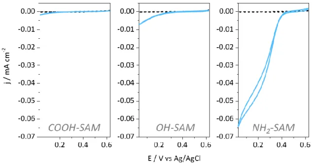
To firmly prove the orientation-driven DET process, the effect of electrode surface charges on
DET was first evaluated by adsorption of Tt Lac on thiol-based self-assembled-monolayers
(SAMs). Figure 2 gives the CVs obtained under O2 atmosphere with Tt Lac adsorbed on
carboxylic (COOH)-, hydroxyl (OH)-, or amino (NH2)-terminated SAMs. At pH 5, according
to their respective pKa, these thiols generate a negative, neutral hydrophilic and positive
12
positive at pH 5.4 Nevertheless, almost no catalytic signal on COOH-SAM, and only a weak catalytic current can be observed on OH-SAM. On the contrary, catalytic signals with current
densities in the range of 60 µA.cm-2 developed on NH2-SAMs. The onset potential for O2
reduction on positive SAMs is +440 mV vs Ag/AgCl at pH 5. This value reflects the Cu T1
redox potential typical of a Met axial ligand.41
Figure 2: CV responses of O2 reduction by Tt Lac adsorbed on different SAM-modified gold electrodes:
6-MHA (COOH-SAM), MH (OH-SAM), and CYST (NH2-SAM). Dashed lines represent
SAM-modified gold electrodes without Tt Lac. 0.1 M NaAc buffer pH 5, scan rate 5 mV s-1.
SPR and ellipsometry measurements highlight the presence of an enzyme layer with a thickness
close to a monolayer whatever the SAM chemistry (Figure S1), meaning that the absence of
catalytic current on COOH-SAM is not linked to an electrostatic repulsion preventing enzyme
adsorption on the electrode surface. The favored DET process on the positive SAM and the
absence of any DET on the negative one confirm the hypothesis based on the structure
examination. Our results also emphasize the very similar electrochemical behavior of Tt Lac
13 Tt Lac adsorbed on carbon nanotubes with a positive zeta potential. Carbon nanomaterials
are often more suited for the enhancement of catalytic currents due to high surface area and the
involvement of hydrophobic interactions with CNT-walls favoring the enzyme adsorption.42 Tt Lac was first adsorbed on a deposit of CNTs presenting a positive zeta potential, thus expected
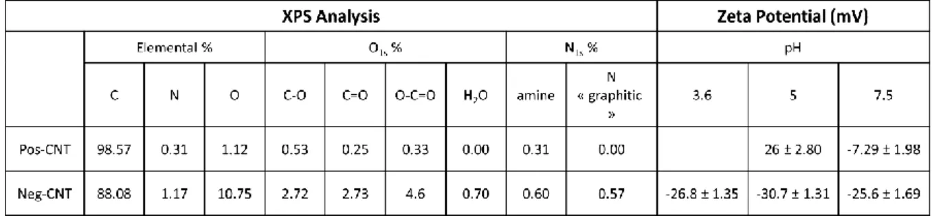
to favor enzyme orientation for DET. As reported in Table 1, this first CNT material (named
thereafter Pos-CNT) contains less than 0.33% of carboxylic functionalities and comparable
amount of amino functionalities, (Figure S2) yielding a positive zeta potential of +26 mV at pH
5.
Table 1: Quantitative comparison of XPS data and values of zeta potential at different pHs for Pos-CNT and Neg-Pos-CNT.
Accordingly, with Tt Lac immobilized by adsorption on Pos-CNT, a DET catalytic process
under O2 occurs at an onset potential very close to the one observed on NH2-SAM (Figure 3).
No catalytic current can be observed in the absence of Lac, or under N2. Despite pronounced
DET process, we were unable to clearly isolate the non-catalytic signals of Tt Lac in anaerobic
conditions (Figure S3), probably because of low coverage and large distance between T1 Cu
14 Figure 3: CVs at Pos-CNT in the presence of Tt Lac under O2 (blue line) or N2 (black dashed line).
Electrode rotation speed 800 rpm, scan rate 5 mV s-1. Inset: Relationship between Tt Lac relative activity
and pH for O2 catalytic reduction at the Pos-CNT.
Varying the rotation rate highlights no limitation of the catalytic current by mass transfer at
observed current densities (Figure S4A). The relationship between the limiting catalytic current
and pH displays a bell shape, with an optimum at pH close to 5, in agreement with the optimal
pH determined by classical spectrophotometric assays (Figure 3, inset).3
The protein was produced with a His-tag at C-terminal end of the polypeptide. As a control, the
protein with His-tag cleaved by TEV-protease was also studied on the Pos-CNT deposit. It
displays a very similar behavior with the His-tagged enzyme, suggesting that this region is not
involved in the electrode recognition (Figure S4B).
Tt Lac adsorbed on carbon nanotubes with a negative zeta potential. The second type of
15
1.12%), mainly in the form of carboxylic functions (4.6%). The direct consequence is the large
negative value of zeta potential at pH 5 (-30.7 ± 1.35 mV) (Table 1). These CNTs will be named
Neg-CNTs. In agreement with our finding on SAMs, virtually no DET can be observed when
Tt Lac was immobilized by adsorption on Neg-CNT (Figure 4).
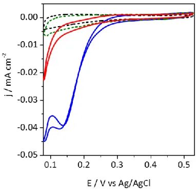
Figure 4: CVs for O2 reduction on Neg-CNT in the absence of Tt Lac (black dashed line), in the presence
of Tt Lac (blue solid line) and in the presence of Tt Lac and 1 mM ABTS (red solid line). 0.1 M NaAc buffer pH 5, scan rate 5 mV s-1. Inset: chronoamperometry of O
2 reduction at 0.2 V in the absence (black
dashed line) and in the presence of Tt Lac (blue solid line); 1 mM ABTS was added at 200 s and 400 s as indicated by arrows.
Adding redox mediators in solution is another way to connect artificially the enzymes, and to
give access to the proportion of enzymes in an unfavored orientation for DET. However, using
Tt Lac for O2 reduction, only extremely weak MET currents below 5 µA cm-2 were recorded in
the presence of ABTS, the classical redox mediator used in MCO spectrophotometric activity
16
solution-based enzymatic assay at 30 °C (Table S2), i.e. 100 times higher than the KM of another
MCO B. pumilus BOD43 that displays pronounced MET current in the same conditions (Figure S5). The high KM value and low kcat of Tt lac for ABTS (kcat/KM ~ 102 M-1 s-1) suggest its low
efficiency for this substrate oxidation and explains the weak MET. In analogy with E. coli
CueO, it also possibly reflects a limited access of ABTS to the substrate binding site because
of the hairpin presence. We also tested other mediators of different charge and hydrophobicity
that could present better affinity for Tt Lac (ferrocenemethanol, ferrocenecarboxylic acid,
DMP). None of them allowed higher MET currents to be observed.
Effect of Cu2+ on Tt Lac electrochemical behavior on Neg-CNT. One structural specificity
of Tt Lac is the presence of the Met-rich hairpin. For CueO, such Met-rich domain organized
in a helix was proposed to allow the enzyme to be involved in copper detoxification through
the binding of exogenous copper atoms and subsequent oxidation of Cu+ to the less toxic Cu2+.4 The Met-rich helix was also proposed to cover the substrate binding site thus restricting access
of bulky substrates like ABTS, while the bound copper would promote ET from the substrate
to Cu T1.12
We confirmed in the present work the previously reported enhancement of the
spectrophotometric activity of Tt Lac measured in solution in the presence of ABTS with
increasing addition of Cu2+ (Figure S6).3 The KM for ABTS was decreased to 1.6 mM in the
presence of Cu2+ and the activity was enhanced ca. 3 times (Table S2). Whether the absence of
direct ET process on Neg-CNT could be alleviated by addition of exogenous Cu2+ as occurring in solution is one fundamental issue to be investigated. Cupric salts were added in the NaAc
buffer solution, and the electrochemical signal under O2 was recorded with Tt Lac immobilized
on Neg-CNT. A typical CV is given in Figure 5 for 500 µM Cu2+ concentration. A cathodic signal, with no anodic counterpart, is occurring on the first and following cycles with an onset
17
it vanishes after addition of fluoride, known to inhibit O2 catalytic reduction by MCOs, in the
solution. A control experiment shows that Cu2+ in the absence of Tt Lac is reduced at a potential at least 150 mV lower on the Neg-CNT based electrode (Figure S7).
Figure 5: CVs under O2 of Tt Lac adsorbed on Neg-CNT without (black dashed curve) or in the presence
of 500 µM CuSO4 (blue solid curve). CV under N2 of Tt Lac adsorbed on Neg-CNT in the presence of
500 µM CuSO4 is given as a green dashed curve. The red curve is obtained after 40 mM NaF addition
in the oxygenated buffer containing CuSO4. 0.1 M NaAc buffer pH 5, scan rate 5 mV s-1.
To get more insight in this new catalytic process, increasing additions of Cu2+ into the buffer
18 Figure 6: (A) CVs under O2 with Tt Lac adsorbed on Neg-CNT in the presence of increasing CuSO4
concentrations from zero to 5 mM. (B, C) Dependence of the half wave potential and peak current density on CuSO4 concentration. 0.1 M NaAc buffer pH 5, scan rate 5 mV s-1.
Within a concentration range from 100 µM to 5 mM Cu2+, the typical Cu2+-dependent catalytic wave appears with a half wave potential which is shifted anodic linearly with log[Cu2+] with a slope of 62 mV (Figure 6C). For each Cu2+ concentration, the CV curve is characterized by a
peak current, followed by the cathodic current increase in relation to the direct reduction of
Cu2+ independent of the enzyme (Figure S7A). Within the current density range, there is no effect of the rotation rate on the shape of the catalytic wave nor on the magnitude of the peak
19
as Cu2+ and Cu+ chelators respectively were added in the buffer solution containing Cu2+, the Cu-related catalytic wave disappeared (Figure S8). As a control experiment, we also
demonstrated that the Cu2+-dependent catalytic wave is not linked to Cu associated to the His-tag on the protein. Actually, as shown in Figure S8C, the catalytic wave persisted even with the
non His-tag protein. In conclusion, the Cu2+-dependent catalytic signal is clearly associated to the availability of free non-complexed Cu2+ in solution.
Based on these experimental data, several hypotheses able to clarify the exact process behind
Cu2+-induced activation might be formulated: i) Cu2+ binding to Tt Lac lowers the electrostatic repulsion of the negative Cu T1 region allowing the enzyme to be re-orientated in a way more
favorable for DET through Cu T1 center; ii) Cu2+ bound to Tt Lac forms a complex acting as a catalyst itself for oxygen reduction without participation of Cu T1/T2/T3 centers; iii) Cu2+
bound to Tt Lac participates into ET forming an additional electron relay between the electrode
and Cu T1 (or Cu T2/T3); iv) the observed wave is related to the cuprous oxidase activity
previously proposed for MCOs such as Tt Lac.
In the first hypothesis, Cu2+ ions would be acting as a shield of large negative charge near Cu T1 center allowing the latter to approach closer to the electrode surface for ET. Actually, Kano
and coworkers found an increase in the catalytic performance when adding Ca2+ with E. coli CueO deposited on negative CNTs25, which they attributed to the di-cations bridging the T1 Cu to the CNTs. To test this possibility, similar concentrations of Ca2+, Zn2+ or Ni2+ were added in
the buffer solution instead of Cu2+. No effect on the electrochemical signal could be observed (Figure S9). Note also that these cations were shown to be useless in the activation of Tt Lac in
solution.3
In the second hypothesis, we investigated the possibility of Cu2+ complexed by some Tt Lac residues to act as a molecular catalyst for oxygen reduction without Cu T1-T3 centers
20
involvement. Indeed, some copper complexes are known to act as catalysts for oxygen
reduction, and we observed a catalytic process on Neg-CNT-modified electrode in the presence
of Cu2+ but starting at potentials at least 0.15 V lower than the one in the presence of Tt Lac (Figure S7B). Alternatively, considering the low potential at which the Cu2+-dependent
catalytic process occurs, direct reduction of O2 through the T2/T3 Cu site might occur, thus
bypassing the intramolecular ET from Cu T1 to TNC. Such pathway was previously
demonstrated using Didymocrea sp. J6 Lac immobilized on gold nanoparticles.44 To rule out the two possibilities of non-enzymatic molecular copper complex implication and direct ET
through the TNC, a Cu T1 deletant of Tt Lac was constructed in which Cys445 was replaced
by Ala. In agreement with previous studies,45,46 this cysteine is essential for T1 copper binding and the purified mutant was white-colored due to the absence of Cu T1, and inactive. ICP and
UV-Vis analyses showed that it still contains 2.3 atoms of Cu organized as T2/T3 center (Table
S2, Figure S10A), and CD spectrum confirmed that the mutant mostly retained its secondary
structure (Figure S10B). We thus conclude that the mutation was successful although the
absence of Cu T1 partially impacted the protein folding. After adsorption of this mutant on
Neg-CNT and addition of Cu2+ in solution, no catalysis could be observed (Figure 7), suggesting that the Cu2+-dependent catalytic process proceeds necessarily through the laccase Cu T1 center and that no molecular Cu-complex is implicated in the catalysis.
21 Figure 7: CVs of Tt Lac C445A adsorbed on Neg-CNT in the absence (black dashed line) and presence (red line) of 1 mM CuSO4. The blue line represents the CV for Neg-CNT with Tt Lac WT in the presence
of 1 mM CuSO4. 0.1 M NaAc buffer pH 5, scan rate 5 mV s-1, O2 atmosphere.
Both two remaining hypothesis imply Cu2+/Cu+ binding at a coordination site close to the Cu T1 center allowing ET to it (Scheme 1A,B). In the case iii, the catalytic cycle starts with Cu2+ binding to this coordination site, yet with low affinity.13 Following this binding step, an additional relay is formed in the proximity of the electrode, bridging it with the Cu T1 center
and enabling the reduction of O2 through the newly formed ET pathway. Similar mechanism of
copper action was suggested in previous publications studying E. coli CueO or Tt Lac activities
in solution,13 but it has never been described in electrochemical studies. In the case iv the extremely small amounts of Cu+ formed at the electrode at potentials more than 200 mV higher than the formal Cu2+/Cu+ potential would be immediately bound by a high affinity coordination site in Tt Lac. The enzyme then exhibits its cuprous oxidase activity and oxidizes Cu+ to Cu2+
22
while transferring electrons to oxygen molecules through the classical T1-T2/T3 chain. The
Cu2+ is released to the solution and is able to be reduced at the electrode again thus completing the catalytic cycle. The final result of both mechanisms would be ET from the electrode to
oxygen by means of copper ions and the enzyme molecule, the difference being only the relative
affinities of the coordination site for Cu2+ and Cu+ and whether the first ET step happens to Cu-enzyme complex or not (Scheme 1).
Scheme 1. Proposed mechanisms of copper-induced catalytic wave formation on Neg-CNTs-Tt Lac: (A) copper – electron relay according to the hypothesis iii and (B) copper – substrate according to the hypothesis iv.
In a previous study relative to cuprous oxidase activity of CueO in solution, the authors fixed
Cu2+ ion at the proposed coordination site by using MOPS, a buffer with low affinity to Cu2+/Cu+.13 We couldn’t use MOPS for the desired pH-range, but we tested another buffer
based on diethylpiperazine which is not able to form any complexes with transition metals due
to steric hindrance.47 Unlike the CueO work, we were unable to fix the Cu2+ at an additional coordination site whatever the buffer used. After electrode rinsing and transfer to a copper-free
23
buffer, the Cu-related catalytic wave vanishes (Figure S11). This suggests rather weak binding
of the Cu2+ and tends to privilege the hypothesis iv as a more probable explanation of the observed electrocatalytic process.
What we observe therefore is the cuprous oxidase activity of Tt Lac initiated by substrate
generation on the electrode. The fact that the catalytic wave onset is observed at potentials 150
mV more positive than the copper reduction on the bare electrode suggests a high affinity of Tt
Lac for Cu+ since only negligible amount of Cu2+ is reduced at this potential. A similar apparent shift of the mediated catalysis potential was observed recently for M. thermophilum cellobiose
dehydrogenase and ferrocene monocarboxylic acid when the concentration of the latter was
increasing.48 From the extent of this shift, the authors concluded the catalytic concentration of the mediator necessary to sustain a given current. In our case, the precise analysis is not possible
since Cu2+/Cu+ redox process is complicated by Cu+-disproportionation and complexation, but we can roughly estimate that Tt Lac starts to demonstrate its cuprous oxidase activity when Cu+ concentration is in the nanomolar range (SI).
The validity of such pathway is further attested by the response analysis in the absence of
oxygen (Figure S12). In these conditions, the catalytic wave is absent but a cathodic current
increase is still observed with the increase of Cu2+ concentration, this time with an anodic counterpart (Figure S12B). The potential of the cathodic peak is in the agreement with the onset
of the Cu-related catalytic wave and it demonstrates a similar shift with Cu2+ concentration
(Figure 6). According to the hypothesis iv, the cathodic peak in the presence of enzyme can be
explained by a facilitated reduction of Cu2+ into Cu+ due to the further complexation of the latter with Tt Lac. The anodic counterpart appearing only in the anaerobic atmosphere thus
corresponds to the inverse process in the conditions when the enzyme cannot perform catalytic
oxidation of the bound Cu+. The more pronounced cathodic peak of the wild type Tt Lac in
24
due to the presence of oxygen traces in the hydrophobic CNT-network. To eliminate any
catalysis involvement, the C445A Tt Lac mutant, in which the Cu T1 is absent (Figure 7), was
studied in the absence of O2 with increasing Cu2+ concentrations. The Cu+ binding is still
possible for such mutant, and CV peaks similar to the WT appeared in the anaerobic conditions
(Figure S12C). The cathodic peak is less pronounced and its charge equals to the anodic one
due to the absence of catalysis implication. The presence of these high potential Cu-related
peaks for both Tt Lac WT and C445A mutant confirms the existence of a high affinity Cu+ binding site for this enzyme.
Another feature of the catalytic wave, which resembles to an inactivation/activation process,
may be attributed to an inhibition process induced by Cu+ produced in excess on the electrode surface at low potentials. This type of inhibition was previously observed with other enzymes
immobilized on electrodes. As illustrations, H+ reduction by hydrogenases was inhibited by H2,49 the reduction product, or nitrate reduction by nitrate oxidase was inhibited by nitrate,50
leading in both cases to similar electrochemical signal shape as we have recorded in the
presence of Cu2+ in this work. Besides, the inhibition by Cu+ concentrations higher than 0.1 mM was reported earlier for CueO.9
ET pathways within Tt Lac immobilized on Pos-CNTs. The next question is whether Cu2+
addition may also affect the DET signal obtained when Tt Lac is immobilized on Pos-CNT.
Increasing Cu2+ amounts were thus added in the buffer solution and the CVs were run under O 2
25 Figure 8: CV responses under O2 by Tt Lac adsorbed on Pos-CNT with increasing concentrations of
CuSO4 from 0 to 5 mM. Dashed curve represents the same electrode in the absence of CuSO4 under N2.
0.1 M NaAc buffer pH 5, scan rate 5 mV s-1.
Starting from the DET signal for O2 reduction by Tt Lac adsorbed on Pos-CNT, a decrease of
the DET catalytic current is observed with increasing Cu2+ concentrations, especially visible at higher potentials. In the same time, the Cu2+-dependent catalytic peak appears, similar to the
one observed on Neg-CNT, also decreasing and shifting in potential with increasing Cu2+ concentrations. As a control, Ca2+ addition into the solution up to 5 mM has no effect on the DET process (Figure S13). When transferring the Pos-CNT-Tt Lac bioelectrode in a Cu2+-free
26
buffer, not only the Cu2+-dependent catalytic wave disappeared, but the DET signal was recovered (Figure S14). To explain the apparent competition between DET and Cu-related
processes observed for Tt Lac on Pos-CNT, one should recall that in DET mode the electrons
are transferred directly from the electrode to the T1 Cu center. In the presence of Cu2+, cuprous
oxidase activity can take place, and Cu+ formed on the electrode can bind to the enzyme and also transfer electrons to the T1 center thus creating an alternative pathway competing with
DET (Scheme 2). This observation may explain the decrease of the DET-current with Cu2+ addition and concomitant increase of the Cu-related wave. On the other hand, we don’t expect
100% of adsorbed enzymes to be in DET orientation.51 These enzymes adsorbed in the
orientation not favorable to DET process may still contribute to the cuprous oxidase activity
since Cu+ formed on the electrode can freely diffuse giving raise to the characteristic Cu-related catalytic wave.
27
Involvement of the Met-rich domain in the electrocatalysis. Now that we ascribed the
appearance of the electrochemical wave upon copper addition to cuprous oxidase activity of Tt
Lac, the exact position of Cu-binding remains to be found. It was suggested that Met-rich
regions of proteins are involved in metal binding and transport.52,53 In this aspect, the Met-rich
hairpin functionally resembles the Met-rich helix of E.coli CueO. If the Cu2+-dependent catalytic process is linked to the Met-rich hairpin of Tt Lac, such catalytic process should not
occur in a multicopper oxidase which does not contain any Met-rich domain. B. pumilus BOD
is an excellent control for that aim, because it does not yield any DET on Neg-CNT as Tt Lac.
As shown in Figure S15, the addition of Cu2+ up to 20 mM does not allow the Cu2+-dependent
catalytic signal to be observed when using B. pumilus BOD.
We therefore constructed a deletant of Tt Lac, named -hairpin Lac, where we deleted a part of
the sequence between residues A292 and G308 that constitutes the hairpin and contains 6
methionines. The -hairpin Lac conserved 82% activity compared to the WT and was still able
to be activated by copper addition in the ABTS solution assay. Its KM was insignificantly
increased compared to WT falling within the range of the experimental error (Table S2). These
results drastically differ from those obtained with the Met-rich helix deletant of CueO which
showed a 40-fold increase of ABTS-oxidizing activity and disappearance of copper activation
in solution.19 We therefore conclude that, unlike in the case of CueO helix, the hairpin of Tt Lac
doesn’t present a steric hindrance for the ABTS binding and the low ABTS-oxidizing activity
of Tt Lac must be explained by other reasons, i.e. low affinity. Interestingly, MD simulations
of Tt Lac and analogous hairpin-deletant earlier demonstrated a similar manner of ABTS
docking to both molecules despite hairpin absence thus confirming our experimental
28
In agreement with the solution assay, the -hairpin Lac behaved similarly to the wild type when
immobilized on Neg-CNTs or Pos-CNTs (Figure S16). On Neg-CNTs no DET process could
be observed, and a catalytic wave similar to the WT was obtained in the presence of Cu2+. The absence of DET is understandable because the charge in the vicinity of Cu T1 remains negative
in -hairpin Lac. However, the persistence of the Cu2+-dependant catalytic process despite the deletion of the Met-rich hairpin confirms that the hairpin domain is not required for Tt Lac
copper-activated catalytic process.
Actually, in CueO related studies, it was underlined that besides Cu binding sites in the
Met-rich helix another sCu binding site is located beyond the helix and formed by M355, D360,
D439, and M441 residues. It is situated along a distance of 7.5 Å to the Cu T1 and linked to it
through a hydrogen-bond that might be involved in an ET pathway.11 Such site wasn’t previously identified in Tt Lac.3 Our sequence alignment suggests however that a similar Cu-binding site cannot be excluded taking into account that at least three from four residues of Tt
Lac (D353, D390, M391) are well aligned with the Cu-binding residues of CueO (Figures S17
and S18). These three residues were not altered by the hairpin deletion (Figure S17) which may
explain the conservation of the copper activation process in this mutant.
CONCLUDING REMARKS
We demonstrated that thanks to a particular surface charge distribution and using different
CNTs, Tt Lac can be adsorbed on the electrode surface in an orientation either favoring DET
through T1 or virtually precluding it. Inspired by a related MCO, E.coli CueO, we used this
feature to study electrochemically the influence of Cu2+ on the activity of Tt Lac. The electrode with Tt Lac adsorbed in the no-DET orientation exhibits a low-potential cathodic catalytic wave
upon Cu2+ addition into the electrochemical cell. This wave was not reported earlier in any studies on either E. coli CueO or Tt Lac. We ascribe it to a cuprous oxidase activity of Tt Lac
29
wave appears and gradually suppresses the direct ET through Cu T1 when Tt Lac is adsorbed
in the favorable orientation. We rely on these data to propose the mechanism of copper binding
and oxidation by Tt Lac with electrons transferred to Cu T1.
The cuprous oxidase activity of Tt Lac is apparently responsible for the enhancement of the
spectrophotometric activity for ABTS oxidation observed earlier in solution assay in the
presence of increasing amounts of Cu2+.3 This phenomenon should be discussed apart since there is no Cu+ present in the assay, contrary to the electrochemical system where it is generated on the electrode. In the case of E. coli CueO, such enhancement was shown to be caused by the
occupation of sCu site by a copper atom required for phenol oxidase activity. This additional
copper atom would mediate ET between buried Cu T1 and the organic substrate. Apparently,
the same behavior is valid for Tt Lac in the cuvette where the presence of labile Cu2+ at
hypothetical sCu site allows to enhance ABTS oxidizing activity. We speculate that higher
specificity of this site for Cu+ than Cu2+ leads to the increase of redox potential of sCu2+/sCu+ couple, which becomes able to withdraw electrons from ABTS and pass them further to Cu T1.
In the solution assay, it cannot be distinguished whether the increase of oxidized ABTS
concentration reflecting enzyme activity is related to electrons transferred first from ABTS to
sCu or to Cu T1. We propose that using protein voltammetry we are able to discriminate
whether electrons enter the enzyme directly from the electrode or by means of exogenous
copper. Once exogenous copper is present, it opens a possibility for the new pathway via
binding and oxidizing Cu+ at sCu-site. Electrochemically, the appearance of this new electron pathway is translated into the change of reduction potential and decrease of DET current, if any,
through Cu T1. Another advantage of voltammetry is the fact that small amounts of Cu+ can be produced in a controllable manner at the electrode in the vicinity of enzyme molecules and react
with them before entering into Fenton or disproportionation reactions. We thus propose
30
As such, the Cu-related mechanism of Tt Lac is homologous to the cuprous oxidase activity of
E.coli CueO, suggesting that both enzymes might fulfill similar physiological roles of copper
detoxification in vivo. However, unlike E.coli CueO mutant with deleted Met-rich region, the
removal of the corresponding Met-rich hairpin of Tt Lac did not lead to an enhanced phenol
oxidase activity while copper activation persisted. This suggests certain divergence in the
behavior of these two enzymes, notably non-involvement of Met-rich hairpin of Tt Lac in
copper binding and oxidation. We anticipate that future site-directed mutagenesis will allow to
elucidate the details and differences of their mechanisms.
SUPPORTING INFORMATION
Protease TEV purification; SPR, ellipsometry, CD spectroscopy, ICP-OES methods
description; calculation of Tt Lac affinity for Cu+; loading of Tt Lac on SAM-modified gold electrodes; XPS spectra of CNTs; CVs of Tt Lac in anaerobic conditions; CVs of Tt Lac at
different rotation speeds; CVs of His-tag-cleaved Tt Lac, CVs of Bp BOD before and after Cu2+
addition; enhancement of Tt Lac activity as a function of Cu2+ concentration; CVs of different concentrations of Cu2+ at Neg-CNT in the presence and absence of Tt Lac and O2; effect of
chelators and other cations (Ca2+, Zn2+, Ni2+); UV-Vis and CD spectrum of C445A mutant; CVs in DEPP buffer and CVs of electrode transfer to a copper-free buffer; CVs of Tt Lac WT and
C445A mutant on Neg-CNTs in anaerobic conditions and in the presence of Cu2+; CVs of the
Δ-hairpin mutant; sequences alignment and structures superposition of Tt Lac and E.coli CueO;
oligonucleotides used; kinetic and thermodynamic parameters of Tt Lac and mutants.
ACKNOWLEDGMENT
This work was supported by ANR (ENZYMOR-ANR-16-CE05- 0024) and Aix-Marseille
31
Pascale Infossi and Proteomic Analysis Center (IMM, CNRS, Marseille) for experimental
assistance, Dr Ling Peng from CINAM (Marseille) for access to zetasizer facility, Dr N. Mano
(CRPP, Bordeaux) for the kind gift of Bp BOD, Dr Marie-Thérèse Giudici-Orticoni for fruitful
discussions and Dr Anne de Poulpiquet for critical reading of the manuscript.
REFERENCES
(1) Mano, N.; De Poulpiquet, A. O2 Reduction in Enzymatic Biofuel Cells. Chem. Rev.
2018, 118 (5), 2392–2468. https://doi.org/10.1021/acs.chemrev.7b00220.
(2) Jones, S. M.; Solomon, E. I. Electron Transfer and Reaction Mechanism of Laccases.
Cell. Mol. Life Sci. 2015, 72 (5), 869–883. https://doi.org/10.1007/s00018-014-1826-6.
(3) Miyazaki, K. A Hyperthermophilic Laccase from Thermus Thermophilus HB27.
Extremophiles 2005, 9 (6), 415–425. https://doi.org/10.1007/s00792-005-0458-z.
(4) Serrano-Posada, H.; Valderrama, B.; Stojanoff, V.; Rudiño-Piñera, E. Thermostable Multicopper Oxidase from Thermus Thermophilus HB27: Crystallization and Preliminary X-Ray Diffraction Analysis of Apo and Holo Forms. Acta Crystallogr. Sect.
F. Struct. Biol. Cryst. Commun. 2011, 67 (Pt 12), 1595–1598.
https://doi.org/10.1107/S174430911103805X.
(5) Serrano-Posada, H.; Centeno-Leija, S.; Rojas-Trejo, S. P.; Rodríguez-Almazán, C.; Stojanoff, V.; Rudiño-Piñera, E. X-Ray-Induced Catalytic Active-Site Reduction of a Multicopper Oxidase: Structural Insights into the Proton-Relay Mechanism and O2-Reduction States. Acta Crystallogr. Sect. D Biol. Crystallogr. 2015, 71, 2396–2411. https://doi.org/10.1107/S1399004715018714.
(6) Rensing, C.; Grass, G. Escherichia Coli Mechanisms of Copper Homeostasis in a Changing Environment. FEMS Microbiol. Rev. 2003, 27 (2–3), 197–213. https://doi.org/10.1016/S0168-6445(03)00049-4.
(7) Tree, J. J.; Kidd, S. P.; Jennings, M. P.; McEwan, A. G. Copper Sensitivity of CueO Mutants of Escherichia Coli K-12 and the Biochemical Suppression of This Phenotype.
Biochem. Biophys. Res. Commun. 2005, 328 (4), 1205–1210.
https://doi.org/10.1016/J.BBRC.2005.01.084.
(8) Rensing, C.; McDevitt, S. F. The Copper Metallome in Prokaryotic Cells. In Metallomics
and the Cell; Banci, L., Ed.; Metal Ions in Life Sciences; Springer Netherlands:
Dordrecht, 2013; Vol. 12, pp 417–450. https://doi.org/10.1007/978-94-007-5561-1. (9) Kataoka, K.; Komori, H.; Ueki, Y.; Konno, Y.; Kamitaka, Y.; Kurose, S.; Tsujimura, S.;
Higuchi, Y.; Kano, K.; Seo, D.; Sakurai, T. Structure and Function of the Engineered Multicopper Oxidase CueO from Escherichia Coli-Deletion of the Methionine-Rich Helical Region Covering the Substrate-Binding Site. J. Mol. Biol. 2007, 373 (1), 141– 152. https://doi.org/10.1016/j.jmb.2007.07.041.
32
(10) Kurose, S.; Kataoka, K.; Otsuka, K.; Tsujino, Y.; Sakurai, T. Promotion of Laccase Activities of Escherichia Coli Cuprous Oxidase, CueO by Deleting the Segment Covering the Substrate Binding Site. Chem. Lett. 2007, 36 (2), 232–233. https://doi.org/10.1246/cl.2007.232.
(11) Roberts, S. A.; Wildner, G. F.; Grass, G.; Weichsel, A.; Ambrus, A.; Rensing, C.; Montfort, W. R. A Labile Regulatory Copper Ion Lies near the T1 Copper Site in the Multicopper Oxidase CueO. J. Biol. Chem. 2003, 278 (34), 31958–31963. https://doi.org/10.1074/jbc.M302963200.
(12) Singh, S. K.; Roberts, S. A.; McDevitt, S. F.; Weichsel, A.; Wildner, G. F.; Grass, G. B.; Rensing, C.; Montfort, W. R. Crystal Structures of Multicopper Oxidase CueO Bound to Copper(I) and Silver(I): Functional Role of a Methionine-Rich Sequence. J. Biol. Chem.
2011, 286 (43), 37849–37857. https://doi.org/10.1074/jbc.M111.293589.
(13) Djoko, K. Y.; Chong, L. X.; Wedd, A. G.; Xiao, Z. Reaction Mechanisms of the Multicopper Oxidase CueO from Escherichia Coli Support Its Functional Role as a Cuprous Oxidase. J. Am. Chem. Soc. 2010, 132 (6), 2005–2015. https://doi.org/10.1021/ja9091903.
(14) Cortes, L.; Wedd, A. G.; Xiao, Z. The Functional Roles of the Three Copper Sites Associated with the Methionine-Rich Insert in the Multicopper Oxidase CueO from E. Coli. Metallomics 2015, 7 (5), 776–785. https://doi.org/10.1039/C5MT00001G.
(15) Page, C. C.; Moser, C. C.; Chen, X.; Dutton, P. L. Natural Engineering Principles of Electron Tunnelling in Biological Oxidation-Reduction. Nature 1999, 402 (6757), 47– 52. https://doi.org/10.1038/46972.
(16) Miura, Y.; Tsujimura, S.; Kamitaka, Y.; Kurose, S.; Kataoka, K.; Sakurai, T.; Kano, K. Bioelectrocatalytic Reduction of O 2 Catalyzed by CueO from Escherichia Coli Adsorbed on a Highly Oriented Pyrolytic Graphite Electrode. Chem. Lett. 2007, 36 (1), 132–133. https://doi.org/10.1246/cl.2007.132.
(17) Miura, Y.; Tsujimura, S.; Kurose, S.; Kamitaka, Y.; Kataoka, K.; Sakurai, T.; Kano, K. Direct Electrochemistry of CueO and Its Mutants at Residues to and Near Type I Cu for Oxygen-Reducing Biocathode. Fuel Cells 2009, 9 (1), 70–78. https://doi.org/10.1002/fuce.200800027.
(18) Kontani, R.; Tsujimura, S.; Kano, K. Air Diffusion Biocathode with CueO as Electrocatalyst Adsorbed on Carbon Particle-Modified Electrodes. Bioelectrochemistry
2009, 76 (1–2), 10–13. https://doi.org/10.1016/J.BIOELECHEM.2009.02.009.
(19) Kataoka, K.; Kogi, H.; Tsujimura, S.; Sakurai, T. Modifications of Laccase Activities of Copper Efflux Oxidase, CueO by Synergistic Mutations in the First and Second Coordination Spheres of the Type I Copper Center. Biochem. Biophys. Res. Commun.
2013, 431 (3), 393–397. https://doi.org/10.1016/J.BBRC.2013.01.040.
(20) Sugimoto, Y.; Kitazumi, Y.; Tsujimura, S.; Shirai, O.; Yamamoto, M.; Kano, K. Electrostatic Interaction between an Enzyme and Electrodes in the Electric Double Layer Examined in a View of Direct Electron Transfer-Type Bioelectrocatalysis. Biosens.
Bioelectron. 2015, 63, 138–144. https://doi.org/10.1016/j.bios.2014.07.025.
33
Rate Analysis of a Site-Specifically Wired Copper Oxidase. Phys. Chem. Chem. Phys.
2018, 20, 6159–6166. https://doi.org/10.1039/C8CP00041G.
(22) Zhang, L.; Cui, H.; Zou, Z.; Garakani, T. M.; Novoa-Henriquez, C.; Jooyeh, B.; Schwaneberg, U. Directed Evolution of a Bacterial Laccase (CueO) for Enzymatic Biofuel Cells. Angew. Chemie - Int. Ed. 2019, 58 (14), 4562–4565. https://doi.org/10.1002/anie.201814069.
(23) Climent, V.; Fu, Y.; Chumillas, S.; Maestro, B.; Li, J.-F.; Kuzume, A.; Keller, S.; Wandlowski, T. Probing the Electrocatalytic Oxygen Reduction Reaction Reactivity of Immobilized Multicopper Oxidase CueO. J. Phys. Chem. C 2014, 118 (29), 15754– 15765. https://doi.org/10.1021/jp5034382.
(24) Chumillas, S.; Maestro, B.; Feliu, J. M.; Climent, V. Comprehensive Study of the Enzymatic Catalysis of the Electrochemical Oxygen Reduction Reaction (ORR) by Immobilized Copper Efflux Oxidase (CueO) From Escherichia Coli. Front. Chem. 2018,
6, 358. https://doi.org/10.3389/fchem.2018.00358.
(25) Xia, H.; Kitazumi, Y.; Shirai, O.; Ozawa, H.; Onizuka, M.; Komukai, T.; Kano, K. Factors Affecting the Interaction between Carbon Nanotubes and Redox Enzymes in Direct Electron Transfer-Type Bioelectrocatalysis. Bioelectrochemistry 2017, 118, 70– 74. https://doi.org/10.1016/J.BIOELECHEM.2017.07.003.
(26) Liu, X.; Gillespie, M.; Ozel, A. D.; Dikici, E.; Daunert, S.; Bachas, L. G. Electrochemical Properties and Temperature Dependence of a Recombinant Laccase from Thermus Thermophilus. Anal. Bioanal. Chem. 2011, 399 (1), 361–366. https://doi.org/10.1007/s00216-010-4345-9.
(27) Agbo, P.; Heath, J. R.; Gray, H. B. Catalysis of Dioxygen Reduction by Thermus Thermophilus Strain Hb27 Laccase on Ketjen Black Electrodes. J. Phys. Chem. B 2013,
117 (2), 527–534. https://doi.org/10.1021/jp309759g.
(28) Agbo, P.; Heath, J. R.; Gray, H. B. Modeling Dioxygen Reduction at Multicopper Oxidase Cathodes. J. Am. Chem. Soc. 2014, 136 (39), 13882–13887. https://doi.org/10.1021/ja5077519.
(29) Gounel, S.; Rouhana, J.; Stines-Chaumeil, C.; Cadet, M.; Mano, N. Increasing the Catalytic Activity of Bilirubin Oxidase from Bacillus Pumilus: Importance of Host Strain and Chaperones Proteins. J. Biotechnol. 2016, 230, 19–25. https://doi.org/10.1016/j.jbiotec.2016.04.035.
(30) Jeong, J.-Y.; Yim, H.-S.; Ryu, J.-Y.; Lee, H. S.; Lee, J.-H.; Seen, D.-S.; Kang, S. G. One-Step Sequence- and Ligation-Independent Cloning as a Rapid and Versatile Cloning Method for Functional Genomics Studies. Appl. Environ. Microbiol. 2012, 78 (15), 5440–5443. https://doi.org/10.1128/AEM.00844-12.
(31) Durão, P.; Chen, Z.; Fernandes, A. T.; Hildebrandt, P.; Murgida, D. H.; Todorovic, S.; Pereira, M. M.; Melo, E. P.; Martins, L. O. Copper Incorporation into Recombinant CotA Laccase from Bacillus Subtilis: Characterization of Fully Copper Loaded Enzymes. JBIC
J. Biol. Inorg. Chem. 2008, 13 (2), 183–193.
https://doi.org/10.1007/s00775-007-0312-0.
34
Lecomte, S.; Ilbert, M.; de Poulpiquet, A.; Lojou, E. Electrostatic-Driven Activity, Loading, Dynamics, and Stability of a Redox Enzyme on Functionalized-Gold Electrodes for Bioelectrocatalysis. ACS Catal. 2018, 12004–12014. https://doi.org/10.1021/acscatal.8b03443.
(33) Mazurenko, I.; Monsalve, K.; Rouhana, J.; Parent, P.; Laffon, C.; Goff, A. Le; Szunerits, S.; Boukherroub, R.; Giudici-Orticoni, M.-T.; Mano, N.; Lojou, E. How the Intricate Interactions between Carbon Nanotubes and Two Bilirubin Oxidases Control Direct and Mediated O2 Reduction. ACS Appl. Mater. Interfaces 2016, 8 (35), 23074–23085. https://doi.org/10.1021/acsami.6b07355.
(34) Dolinsky, T. J.; Nielsen, J. E.; McCammon, J. A.; Baker, N. A. PDB2PQR: An Automated Pipeline for the Setup of Poisson-Boltzmann Electrostatics Calculations.
Nucleic Acids Res. 2004, 32 (Web Server issue), W665-7.
https://doi.org/10.1093/nar/gkh381.
(35) Søndergaard, C. R.; Olsson, M. H. M.; Rostkowski, M.; Jensen, J. H. Improved Treatment of Ligands and Coupling Effects in Empirical Calculation and Rationalization of p K a Values. J. Chem. Theory Comput. 2011, 7 (7), 2284–2295.
https://doi.org/10.1021/ct200133y.
(36) Felder, C. E.; Prilusky, J.; Silman, I.; Sussman, J. L. A Server and Database for Dipole Moments of Proteins. Nucleic Acids Res. 2007, 35 (Web Server issue), W512-21. https://doi.org/10.1093/nar/gkm307.
(37) Ashkenazy, H.; Erez, E.; Martz, E.; Pupko, T.; Ben-Tal, N. ConSurf 2010: Calculating Evolutionary Conservation in Sequence and Structure of Proteins and Nucleic Acids.
Nucleic Acids Res. 2010, 38 (Web Server), W529–W533.
https://doi.org/10.1093/nar/gkq399.
(38) Kelley, L. A.; Mezulis, S.; Yates, C. M.; Wass, M. N.; Sternberg, M. J. E. The Phyre2 Web Portal for Protein Modeling, Prediction and Analysis. Nat. Protoc. 2015, 10 (6), 845–858. https://doi.org/10.1038/nprot.2015.053.
(39) Madeira, F.; Park, Y. mi; Lee, J.; Buso, N.; Gur, T.; Madhusoodanan, N.; Basutkar, P.; Tivey, A. R. N.; Potter, S. C.; Finn, R. D.; Lopez, R. The EMBL-EBI Search and Sequence Analysis Tools APIs in 2019. Nucleic Acids Res. 2019, 47 (W1), W636–W641. https://doi.org/10.1093/nar/gkz268.
(40) Robert, X.; Gouet, P. Deciphering Key Features in Protein Structures with the New ENDscript Server. Nucleic Acids Res. 2014, 42 (W1), W320–W324. https://doi.org/10.1093/nar/gku316.
(41) Xu, F.; Palmer, A. E.; Yaver, D. S.; Berka, R. M.; Gambetta, G. A.; Brown, S. H.; Solomon, E. I. Targeted Mutations in a Trametes Villosa Laccase. J. Biol. Chem. 1999,
274 (18), 12372–12375. https://doi.org/10.1074/jbc.274.18.12372.
(42) Scherbahn, V.; Putze, M. T.; Dietzel, B.; Heinlein, T.; Schneider, J. J.; Lisdat, F. Biofuel Cells Based on Direct Enzyme-Electrode Contacts Using PQQ-Dependent Glucose Dehydrogenase/Bilirubin Oxidase and Modified Carbon Nanotube Materials. Biosens.
Bioelectron. 2014, 61, 631–638. https://doi.org/10.1016/j.bios.2014.05.027.
35
2012, 96 (2), 301–307. https://doi.org/10.1007/s00253-012-4312-9.
(44) Dagys, M.; Laurynėnas, A.; Ratautas, D.; Kulys, J.; Vidžiūnaitė, R.; Talaikis, M.; Niaura, G.; Marcinkevičienė, L.; Meškys, R.; Shleev, S. Oxygen Electroreduction Catalysed by Laccase Wired to Gold Nanoparticles via the Trinuclear Copper Cluster. Energy
Environ. Sci. 2017, 10 (2), 498–502. https://doi.org/10.1039/C6EE02232D.
(45) Zeng, J.; Geng, M.; Liu, Y.; Xia, L.; Liu, J.; Qiu, G. The Sulfhydryl Group of Cys138 of Rusticyanin from Acidithiobacillus Ferrooxidans Is Crucial for Copper Binding.
Biochim. Biophys. Acta - Proteins Proteomics 2007, 1774 (4), 519–525.
https://doi.org/10.1016/j.bbapap.2007.02.008.
(46) Roger, M.; Sciara, G.; Biaso, F.; Lojou, E.; Wang, X.; Bauzan, M.; Giudici-Orticoni, M.-T.; Vila, A. J.; Ilbert, M. Impact of Copper Ligand Mutations on a Cupredoxin with a Green Copper Center. Biochim. Biophys. Acta - Bioenerg. 2017, 1858 (5), 351–359. https://doi.org/10.1016/j.bbabio.2017.02.007.
(47) Kandegedara, A.; Rorabacher, D. B. Noncomplexing Tertiary Amines as “better” Buffers Covering the Range of PH 3-11. Temperature Dependence of Their Acid Dissociation Constants. Anal. Chem. 1999, 71 (15), 3140–3144. https://doi.org/10.1021/ac9902594.
(48) Meneghello, M.; Al-Lolage, F. A.; Ma, S.; Ludwig, R.; Bartlett, P. N. Studying Direct Electron Transfer by Site-Directed Immobilization of Cellobiose Dehydrogenase.
ChemElectroChem 2019, 6 (3), 700–713. https://doi.org/10.1002/celc.201801503.
(49) Fourmond, V.; Baffert, C.; Sybirna, K.; Dementin, S.; Abou-Hamdan, A.; Meynial-Salles, I.; Soucaille, P.; Bottin, H.; Léger, C. The Mechanism of Inhibition by H2 of H2-Evolution by Hydrogenases. Chem. Commun. 2013, 49 (61), 6840. https://doi.org/10.1039/c3cc43297a.
(50) Jacques, J. G. J.; Burlat, B.; Arnoux, P.; Sabaty, M.; Guigliarelli, B.; Léger, C.; Pignol, D.; Fourmond, V. Kinetics of Substrate Inhibition of Periplasmic Nitrate Reductase.
Biochim. Biophys. Acta - Bioenerg. 2014, 1837 (10), 1801–1809.
https://doi.org/10.1016/J.BBABIO.2014.05.357.
(51) Mazurenko, I.; Monsalve, K.; Infossi, P.; Giudici-Orticoni, M.-T.; Topin, F.; Mano, N.; Lojou, E. Impact of Substrate Diffusion and Enzyme Distribution in 3D-Porous Electrodes: A Combined Electrochemical and Modelling Study of a Thermostable H 2 /O 2 Enzymatic Fuel Cell. Energy Environ. Sci. 2017, 10, 1966–1982. https://doi.org/10.1039/C7EE01830D.
(52) Xue, Y.; Davis, A. V; Balakrishnan, G.; Stasser, J. P.; Staehlin, B. M.; Focia, P.; Spiro, T. G.; Penner-Hahn, J. E.; O’Halloran, T. V. Cu(I) Recognition via Cation-π and Methionine Interactions in CusF. Nat. Chem. Biol. 2008, 4 (2), 107–109. https://doi.org/10.1038/nchembio.2007.57.
(53) Doerrer, L. H. Cu in Biology: Unleashed by O2 and Now Irreplaceable. Inorganica
Chim. Acta 2018, 481, 4–24. https://doi.org/10.1016/j.ica.2017.11.051.
(54) Bello, M.; Valderrama, B.; Serrano-Posada, H.; Rudiño-Piñera, E. Molecular Dynamics of a Thermostable Multicopper Oxidase from Thermus Thermophilus HB27: Structural Differences between the Apo and Holo Forms. PLoS One 2012, 7 (7), e40700.