HAL Id: hal-03142196
https://hal.inria.fr/hal-03142196
Submitted on 17 Feb 2021
HAL is a multi-disciplinary open access
archive for the deposit and dissemination of
sci-entific research documents, whether they are
pub-lished or not. The documents may come from
teaching and research institutions in France or
abroad, or from public or private research centers.
L’archive ouverte pluridisciplinaire HAL, est
destinée au dépôt et à la diffusion de documents
scientifiques de niveau recherche, publiés ou non,
émanant des établissements d’enseignement et de
recherche français ou étrangers, des laboratoires
publics ou privés.
Cardiac Motion Modeling with Parallel Transport and
Shape Splines
Nicolas Guigui, Pamela Moceri, Maxime Sermesant, Xavier Pennec
To cite this version:
Nicolas Guigui, Pamela Moceri, Maxime Sermesant, Xavier Pennec. Cardiac Motion Modeling with
Parallel Transport and Shape Splines. International Symposium on Biological Imaging, Apr 2021,
Nice, France. �hal-03142196�
CARDIAC MOTION MODELING WITH PARALLEL TRANSPORT AND SHAPE SPLINES
Nicolas Guigui
†Pamela Moceri
?†Maxime Sermesant
†Xavier Pennec
††
Universit´e Cˆote d’Azur, Inria, Epione, Sophia-Antipolis, France
?UR2CA, Universit´e Cˆote d’Azur, Facult´e de M´edecine, Nice, France
ABSTRACT
In cases of pressure or volume overload, probing cardiac function may be difficult because of the interactions between shape and deformations. In this work, we use the LDDMM framework and parallel transport to estimate and reorient deformations of the right ventricle. We then propose a nor-malization procedure for the amplitude of the deformation, and a second-order spline model to represent the full cardiac contraction. The method is applied to 3D meshes of the right ventricle extracted from echocardiographic sequences of 314 patients divided into three disease categories and a control group. We find significant differences between pathologies in the model parameters, revealing insights into the dynamics of each disease.
Index Terms— LDDMM, Shape Analysis, Cardiac Mod-elling
1. INTRODUCTION
Spatio-temporal shape analysis is of growing importance in the study of cardiac diseases. In particular, the assessment of cardiac function requires the measurement and analysis of cardiac motion beyond scalar indicators such as ejection frac-tion or area strain. The case of the Right Ventricle (RV), is of particular interest as it has been shown to have a large capac-ity to adapt to overload by remodelling [1], raising the issue of disentangling the deformation from the initial anatomy.
A powerful and popular viewpoint based on shape regis-tration is that of Riemannian Geometry and the Large Defor-mations Diffeomorphic Metric Mapping (LDDMM) frame-work, modelling non-linear transformations as elements of the group of diffeomorphisms of the embedding space, to en-sure smooth and invertible mappings between shapes, and us-ing parallel transport to build a reference-centred represen-tation of all the patients’ trajectories. Parallel transport is a natural geometric tool to normalise deformations that is con-sistent with registration. Two main approaches have been proposed. The first consists in estimating a mean trajectory, and representing each individual’s trajectory as a translation of this mean evolution by a subject-specific perturbation, that is parallel transported to each time point [2, and references therein]. The other strategy [3, 4, and references therein],
which will be developed in this paper, consists in first rep-resenting each trajectory with respect to its own reference, and then transporting it to a global reference –the atlas– thus normalizing the individual deformations.
However, the computation of parallel transport in the dif-feomorhism group is challenging, as no closed form solutions exist and the numerical methods lack stability or result in over-smoothing. Moreover, [5] showed that parallel transport in LDDMM does not conserve global properties such as scale or volume changes. In the case of cardiac deformations, the magnitude of the temporal deformation is comparable to that of the subject’s reference to atlas deformation, and substan-tial volume changes are observed. Thus the lack of scale-invariance is crucial. In this work, we investigate the effect of a straightforward scaling of the transported deformation, in order to preserve the ejection fraction of the RV.
Furthermore, we leverage recent results on the Pole Lad-der [6], a method that consists in approximating the parallel transport of a tangent vector along a geodesic. This method was first proposed by [3], for brain data in the context of Alzheimer’s disease. We use it, in the context of LDDMM, to associate a deformation of the atlas to each shape. This allows to build patient-specific trajectories all starting from the atlas. The patient-specific trajectories are then summarized by fitting a spline regression, which postulates a second-order dynamic model [7] whose parameters are estimated with an optimization procedure. This allows to compactly represent all the patients’ trajectories in the same space, and to proceed with linear statistics.
2. MOTIVATION: THE RIGHT VENTRICLE UNDER PRESSURE
With this framework, we study the RV under pressure or vol-ume overload due to different diseases: Pulmonary Hyperten-sion (PHT), Tetralogy of Fallot (ToF) and Atrial Septal De-fect (ASD) and seek to characterize their impact on the con-traction of the RV, during the systolic phase of the cardiac cycle. Previous work on these pathologies demonstrated dif-ferences in RV function between the ASD group and the ToF group, despite comparable shape remodelling [8]. Moreover, [9] showed that the Area Strain (AS), i.e. the relative change of area of each cell of the mesh that represents the RV, was
a strong predictor of survival in the PHT group. [10] studied the interactions between AS and shape descriptors.
Both studies suffered from a low power due to a small co-hort against high-dimensional markers. Thanks to the use of sparse control points to parameterize the LDDMM and spline deformations, our descriptors are more compact and translate into increased statistical power.
We use 3D meshes extracted from 314 echocardiographic sequences from patients examined at the CHU of Nice. The meshes were extracted with a commercial software (4D RV Function 2.0, TomTec Imaging Systems, GmbH, DE) with point-to-point correspondences across time and patients. These are formed by 938 points and 1872 triangles. All the shapes were realigned with a subject-specific rigid-body deformation. An atlas was computed from the end diastolic meshes of the control group, after alignment.
3. METHOD 3.1. The LDDMM framework
The LDDMM framework encompasses both algorithms for shape matching (a.k.a. registration) and a Riemannian geo-metric structure on the space of shapes. The former allows to compute shape descriptors, and to parameterize diffeomor-phisms. The latter, provides a distance to compare defor-mations, and an associated notion of parallelism to transport them. We present here the formulation of [11] and its imple-mentation in [12], focusing on the case of landmarks.
A natural and efficient computational construction of feomorphisms is obtained by flows associated to ordinary dif-ferential equations (ODEs) ∂φt(·) = vt[φt(·)], with the
ini-tial condition φ0 = Id. The time-dependent vector field vt
can be interpreted as the instantaneous speed of the points during deformation, and must verify certain regularity con-ditions to ensure that solutions to the ODE are indeed dif-feomorphisms. An efficient way to enforce these conditions is to consider vector fields obtained by the convolution of a number Nc of momentum vectors carried by control points:
vt(x) =P Nc k=1K(x, c (t) k )µ (t)
k , where K is the Gaussian
ker-nel: K(x, y) = exp(−kx−ykσ2 2). The (closure of the) set of
such vector fields forms a reproducing kernel Hilbert space, with the associated norm kvk2
K =
P
i,jK(ci, cj)µ T iµj. The
total cost, or energy of the deformation can be defined as R1
0 kvtk 2 Kdt.
It can be shown that the momentum vectors that mini-mize this energy, considering c(0)k , c(1)k , k = 1 . . . Nc fixed,
together with the equation driving the motion of the control points, follow a Hamiltonian system of ODEs:
( ˙ ck(t) =PjK(c (t) k , c (t) j )µ (t) j ˙ µk(t) = −Pj∇1K(c (t) k , c (t) j )µ (t)T k µ (t) j (1)
A diffeomorphism φ1is thus uniquely parameterized by the
initial conditions c(0)k , µ(0)k , k = 1 . . . Nc, and a shape
reg-istration criterion between a template T and target S can be defined as
C(c, µ) = kS − φc,µ1 (T )k22+ α2kvc,µ0 k 2
K. (2)
where α is a regularisation parameter that penalises large de-formations. Minimizing C therefore amounts to finding the transformation that best deforms T to match S. The gradi-ent of C can be computed through automatic differgradi-entiation, to perform gradient descent. The optimal value of C defines a distance between φ1and the identity. In fact this distance
derives from an invariant Riemannian metric on the group of diffeomorphisms and the path φtis a minimizing geodesic for
this metric. By considering the action of diffeomorphisms on shapes, it projects to a distance between the shapes S and T . 3.2. Scaled parallel transport with the pole ladder Along with a distance, the Riemannian metric provides a no-tion of parallel transport. It is defined by an ODE that al-lows to transport a set of momentum vectors along a path of diffeomorphisms φt ([4, section 13.3.3]). However, this
differential equation is hard to solve in practice and alterna-tive methods have been proposed for the case of transporting along a geodesic. We here leveraged recent results on the con-vergence properties of the Pole Ladder [6] to propose a new implementation within the LDDMM framework. We solve the registration problem (4) between the end-diastolic (ED) shape and each time frame ti, then use the Pole Ladder to
transport this deformation to the atlas, and reconstruct a cor-responding shape at time ti.
However, there is a substantial correlation between the magnitude of the systolic deformation and the End Diastolic (ED) volume (ρ = 0.42 in the data-set considered in this pa-per). As the parallel transport is isometric, this deformation may be too large for the atlas. An example of the obtained end-systolic (ES) frame is shown on Figure 1 for a patient whose RV volume is greater that that of the atlas, which re-sults in an unrealistic ES frame. A clinically relevant quantity that should be conserved is the Ejection Fraction (EF), de-fined as the relative volume change, as it is a straightforward indicator of cardiac function. We then introduce a parameter λ such that scaling the magnitude of each intra-subject de-formation after parallel transport conserves the EF relative to the ED frame. This parameter is optimized by gradient de-scent on each patient. We validate this scaling by probing the conservation of the area strain, and the ES ejection fraction. 3.3. Spline regression for shape evolution
The registration framework described in section 3.1 estimates a geodesic path between two shapes (the equivalent of a uni-form motion). For a trajectory such as the contraction of the cardiac RV, one may expect to find second-order dynamics,
Fig. 1. Example: transparent ED mesh over coloured ES mesh. Left: Original patient with extreme volume, Middle: after parallel transport, unrealistic deformation, Right: after scaling of the parallel transport.
making a (first-order) geodesic regression ill-suited. We thus propose to use the second-order model defined in [7] to ac-count for the motion of the RV during systole. The second-order terms utcan be interpreted as random external forces
smoothly perturbing the trajectory around a mean geodesic. They modify the continuous-time system of equations (2) as follows: ∀t ∈ [0, 1], ( ˙ ck(t) =PjK(c (t) k , c (t) j )µ (t) j ˙ µk(t) = −Pj∇1K(c (t) k , c (t) j )µ (t)T k µ (t) j + u (t) k (3)
If we consider a discrete sequence of observation times t1 = 0, t2, . . . td = 1 and configurations xt1, . . . , xtd, one
seeks to find the path φtthat minimizes the new cost
CS(c, µ, ut) = 1 α2d d X i=1 kxti− φti(xt0)k 2 2 + Z 1 0 ku(t)k2dt + kvc,µ 0 k 2 K. (4)
In practice, the ODEs (1) and (3) are discretized in n time steps and an integration method such as Euler or Runge-Kutta is used. We define all the patients trajectories between t = 0 and t = 1, and use the same discretization for all the pa-tients to ensure that u0, . . . , uNcare estimated at
correspond-ing times. Along with µ(0), these are estimated by gradient descent as in the case of registration. We use a kernel band-width σ = 15 in all the experiments, and 60 control points for all the deformations of the atlas. The initial control points are fixed for the entire data-set so that the initial momenta can be compared consistently. They have been optimized to register the atlas on all the transported ES frames.
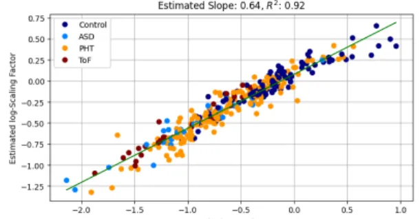
Fig. 2. Scaling Parameter λ with respect to the ED volume. 4. RESULTS
4.1. AS and EF conservation
As expected, the scaling coefficient is closely related to the ED volume. We use a linear regression to identify a linear re-lation between log(λ) and log(Vref/VED). This is displayed
on Figure 2.
We validate our scaled parallel transport algorithm by as-sessing the conservation of scalar quantities of interest: the ejection fraction (EF) and area strain (AS). Both quantities are defined as the relative change of respectively volume or area during systole. The AS is a local quantity computed on each cell of the mesh. We compute the root mean squared error (RMSE) at each cell, and report here the mean over all cells.
We compute the EF on the original data, and compare it with the values computed on the shapes obtained by paral-lel transport (PT), and scaled paralparal-lel transport (SPT). The RMSE are displayed on Table 1. Interestingly, it is possi-ble to obtain a low error on the EF after the scaled transport, but this does not preserve the AS. This shows that although these quantities are related, they carry different information and that AS depends on the initial shape, itself related to the pathology.
Original Values RMSE PT RMSE SPT AS -0.24 ± 0.08 0.18 0.13 EF 0.42 ± 0.13 0.13 0.04 Table 1. Validation of parallel transport with EF and AS. 4.2. Groupwise differences on the splines
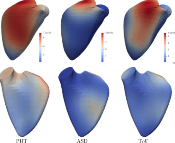
Secondly, we study the differences between the diseases on the spline deformations. These are parameterized by the initial momentum µ, and by the discretized external forces (u(0), . . . , u(1)). We perform a Hotelling multivariate test to compare each disease to the control group, and perform a Bonferroni correction for multiple testing, to maintain type I error risk at α = 0.05. The results for the momentum are displayed Figure 3 for the ASD, ToF and PHT groups and show significant differences between each disease and the control group. The differences observed near the tricuspid
PHT ASD ToF
Fig. 3. Group Mean momentum where it significantly differs from the control group. Red indicates stronger impact, while blue indicates no impact (no difference). Top row: free wall. Bottom row: septum
valve mainly reflect the difference of magnitude of the defor-mations, and one should be cautions before drawing further conclusions as the quality of the mesh may vary near this region. However it is interesting to notice that very little dif-ferences are observed for the ASD group, which corroborates previous results [8]. Similarly, only small differences are observed on the septum, showing that the shape differences usually observed on the PHT group have been filtered out. This makes the differences observed on the free wall interest-ing and other markers such as the circumferential strain will be studied to confirm these effects.
The second-order terms give insight into the dynamic dif-ferences between the groups. Indeed, significant difdif-ferences were found again for the ToF and PHT groups, but the loca-tions and orientaloca-tions vary across time. This will be studied more in depth in future work.
5. COMPLIANCE WITH ETHICAL STANDARDS This study was performed in line with the principles of the Declaration of Helsinki. Approval was granted by the CPP Sud M´editerran´ee V (2017-A02077-46).
6. ACKNOWLEDGMENTS
This work was partially funded by the ERC grant Nr. 786854 G-Statistics from the European Research Council under the European Union’s Horizon 2020 research and innovation program. It was also supported by the French government through the 3IA Cˆote d’Azur Investments ANR-19-P3IA-0002 managed by the National Research Agency. The authors are grateful to the OPAL infrastructure from Universit´e Cˆote d’Azur for providing resources and support. They warmly thank Nicolas Duchateau for his preparatory work on the data and his helpful comments. The authors have no conflict of interest to declare.
7. REFERENCES
[1] J. Sanz et al., “Anatomy, Function, and Dysfunction of the Right Ventricle: JACC State-of-the-Art Review,” Journal of the American College of Cardiology, vol. 73, no. 12, pp. 1463–1482, Apr. 2019.
[2] J.-B. Schiratti, S. Allassonni`ere, O. Colliot, and S. Dur-rleman, “A Bayesian Mixed-Effects Model to Learn Trajectories of Changes from Repeated Manifold-Valued Observations,” Journal of Machine Learning Re-search, vol. 18, no. 133, p. 33, 2017.
[3] M. Lorenzi and X. Pennec, “Efficient Parallel Transport of Deformations in Time Series of Images: From Schild to Pole Ladder,” Journal of Mathematical Imaging and Vision, vol. 50, no. 1, pp. 5–17, Sep. 2014.
[4] L. Younes, Shapes and Diffeomorphisms, ser. Applied Mathematical Sciences. Berlin, Heidelberg: Springer Berlin Heidelberg, 2019, vol. 171.
[5] M. Niethammer and F.-X. Vialard, “Riemannian metrics for statistics on shapes : Parallel transport and scale in-variance,” 2013.
[6] N. Guigui and X. Pennec, “Numerical Accuracy of Lad-der Schemes for Parallel Transport on Manifolds,” Jul. 2020.
[7] A. Trouv´e and F.-X. Vialard, “Shape splines and stochastic shape evolutions: a second order point of view,” Quarterly of Applied Mathematics, vol. 70, no. 2, pp. 219–251, 2012.
[8] P. Moceri et al., “3D right ventricular shape and strain in congenital heart disease patients with right ventricular chronic volume loading,” European Heart Journal -Cardiovascular Imaging, 2020.
[9] P. Moceri et al., “Three-dimensional right-ventricular regional deformation and survival in pulmonary hy-pertension,” European Heart Journal - Cardiovascular Imaging, vol. 19, no. 4, pp. 450–458, Apr. 2018. [10] M. Di Folco, P. Clarysse, P. Moceri, and N. Duchateau,
“Learning interactions between cardiac shape and de-formation: application to pulmonary hypertension,” in STACOM Workshop, Held in Conjunction with MICCAI 2019, ser. Lecture Notes in Computer Science, Shenzen, China, 2019.
[11] S. Durrleman et al., “Morphometry of anatomical shape complexes with dense deformations and sparse parame-ters,” NeuroImage, vol. 101, pp. 35–49, Nov. 2014. [12] A. Bˆone, M. Louis, B. Martin, and S. Durrleman,
“De-formetrica 4: an open-source software for statistical shape analysis,” Sep. 2018.