HAL Id: hal-02347129
https://hal.archives-ouvertes.fr/hal-02347129
Submitted on 5 Nov 2019
HAL is a multi-disciplinary open access
archive for the deposit and dissemination of
sci-entific research documents, whether they are
pub-lished or not. The documents may come from
teaching and research institutions in France or
abroad, or from public or private research centers.
L’archive ouverte pluridisciplinaire HAL, est
destinée au dépôt et à la diffusion de documents
scientifiques de niveau recherche, publiés ou non,
émanant des établissements d’enseignement et de
recherche français ou étrangers, des laboratoires
publics ou privés.
stabilization
Denis Combes, Didier Le Ray, François M Lambert, John Simmers, Hans
Straka
To cite this version:
Denis Combes, Didier Le Ray, François M Lambert, John Simmers, Hans Straka.
An intrinsic
feed-forward mechanism for vertebrate gaze stabilization. Current Biology - CB, Elsevier, 2008,
�10.1016/j.cub.2008.02.018�. �hal-02347129�
Magazine R241
transport; both proteins belong to the RND superfamily of proteins, originally defined by bacterial channel and transporter proteins involved in resistance, nodulation and division. These similarities have led to the suggestion that Ptc might function by transporting antagonists or agonists of Smo across the plasma membrane. Two lines of evidence have provided support for such roles of Ptc: in one study the secretion of pro-vitamin D3, which can act as an inhibitor of Smo, was shown to be promoted by Ptc activity. On the other hand, cholesterols and oxysterols have been shown to act as Smo agonists, prompting the suggestion that Ptc might regulate Smo by transporting these lipids away from Smo. Neither of these contrasting mechanisms addresses directly the control of Smo localisation by Ptc.Intriguingly, Ptc itself has now been found to shuttle to and from the primary cilia in response to Hh activity. In contrast to Smo, Ptc localises to the cilia in the absence of Hh signal, but is removed from them on binding to Hh. Thus, the primary cilia act not only as a centre for the regulation of the intracellular components of the pathway but also as a sensor for the extracellular ligand.
The analysis of Hh signalling has given us many new insights into how cells sense and respond to signals and has illuminated our understanding of how such signals are deployed to generate cellular diversity during development. The sheer variety of its effects still poses important questions about the molecular
basis of the differential response of cells to varying levels and duration of signalling activity and the nature of the differing competence of cells to respond to the same signal. Key unresolved issues include the biochemical function of Ptc and the way in which it regulates Smo activity as well as the role of the primary cilium in sensing and responding to the signal. Despite the novelties of Hh signalling, there are still some striking similarities between it and the other systems deployed by metazoans, most notably the Wnt pathway. In this respect, none of these signalling pathways seems unique; and the
loss from nematodes of genes encoding key components of the Hh pathway — including Smo, SuFu and Fused— indicates that not all are indispensable for multicellular development.
Further reading
Bürglin, T.R., and Kuwabara, P.E. (2006). Homologs of the Hh signalling network in C. elegans.
WormBook, ed. The C. elegans Research Community, WormBook, doi/10.1895/ wormbook.1.76.1, http://www.wormbook.org. Guerrero, I., and Chiang, C. (2006). A conserved
mechanism of Hedgehog gradient formation by lipid modifications. Trends Cell Biol. 17, 1–5. Hooper, J.E., and Scott, M.P. (2005).
Communicating with Hedgehogs. Nat. Rev. Mol. Cell Biol. 6, 306–317.
Saha, K., and Schaffer, D.V. (2006). Signal dynamics in Sonic hedgehog tissue patterning. Development 133, 889–900.
Wang, Y., McMahon, A.P., and Allen, B.L. (2007). Shifting paradigms in Hedgehog signalling. Curr. Opin. Cell Biol. 19, 159–165.
Institute of Molecular and Cell Biology, Proteos, 61 Biopolis Drive, Singapore 138673. E-mail: [email protected] Hh targets CiA Fu Cos2 Hh targets Sufu PKAGSK3 CK1 U CiA Slmb CiR CiA Fu Cos2 PKA CK1 CiA Fu Cos2 PKA CK1 Slmb P P P P P CiR CiA Proteasome P -Hh +Hh GSK3 GSK3 Current Biology Figure 4. Control of Hh target gene transcription by Ci.
In the absence of Hh (left), phosphorylation of the full-length, transcription-activating form of Ci (CiA) by kinases in the Cos-2 complex, results in recruitment of the F-box protein Slimb that pro-motes ubiquitination of CiA and its partial proteolysis by the proteasome to yield the truncated, repressor form, CiR. Hh promotes dissociation of the Cos2 complex (right), in part through Fu-mediated phosphorylation of Cos2, protecting CiA from phosphorylation and hence proteolysis. Association of CiA with SuFu modulates its entry into the nucleus.
An intrinsic
feed-forward mechanism
for vertebrate gaze
stabilization
Denis Combes1, Didier Le Ray1, François M. Lambert2,
John Simmers1 and Hans Straka2 Accurate perception of the visual world plays a major role in animal survival. All vertebrates, whether running, swimming or flying, are confronted with the effects of their locomotor actions on the ability to perceive their surrounding environment [1]. The potential consequences of self-generated body motion include head movements that cause retinal image displacement with a resultant degradation of visual information processing. In order to maintain visual acuity during locomotion, retinal image drift must be counteracted by dynamic compensatory eye and/or head-adjustments that derive from sensory-motor transformations of vestibulo-ocular, optokinetic and proprioceptive inputs [2]. Here we report that efference copies of rhythmic neural signals produced by locomotor pattern-generating circuitry within the spinal cord of larval Xenopus laevis are conveyed to the brainstem extraocular motor nuclei and potentially contribute to gaze stabilization during locomotion. Appropriate spinal network-extraocular motor coupling not only persisted during actual undulatory tail movements in semi-intact preparations, but also during fictive locomotion in isolated brainstem- spinal cords without any movement-derived sensory inputs. This suggests that inherent feed-forward signalling may be used in combination with sensory feed-back to counteract the visual consequences of tadpole self- motion, with major implications for understanding gaze control in general.
Experiments were conducted on pre-metamorphic Xenopus tadpoles (stage 55 [3]; see
Supplemental Data available on- line with this issue) in which strong lateral head displacements occur during undulatory swimming in freely- behaving animals. High- speed
video recordings established that these left–right head excursions during swimming were accompanied by synchronous oscillations of the eyes in the opposite direction
in each half cycle, thereby countering the alternating head displacements (Figure 1A, left). As in vertebrates generally [4], such eye counter- rotations are orchestrated Left-right extraocular coupling
D Spino-extraocular coupling L-MR R-LR L-LR 100 ms B A C L-LR R-LR L-MR L-sr 15 R-sr 15 L-LR L-MR R-LR R-MR Ta il mo vemen t Spinal roots Extraocular ne rv es Extraocular ne rv es 50ms Left Right L-LR R-LR L-MR R-MR L-sr R-sr Vestibular nuclei Spinal networks ? ? Tail muscle contraction Extraocular motor nuclei Visual networks Vestibular afferents Proprioceptive signals Locomotor efference copy left
lateral rectus lateral rectusright Right medial rectus Left medial rectus R-sr / R-LR L-sr / R-LR 0/1 0.5 0/1 0.5 L-MR / R-LR 0/1 0.5 L-LR / R-LR 0/1 0.5 Left Right Current Biology
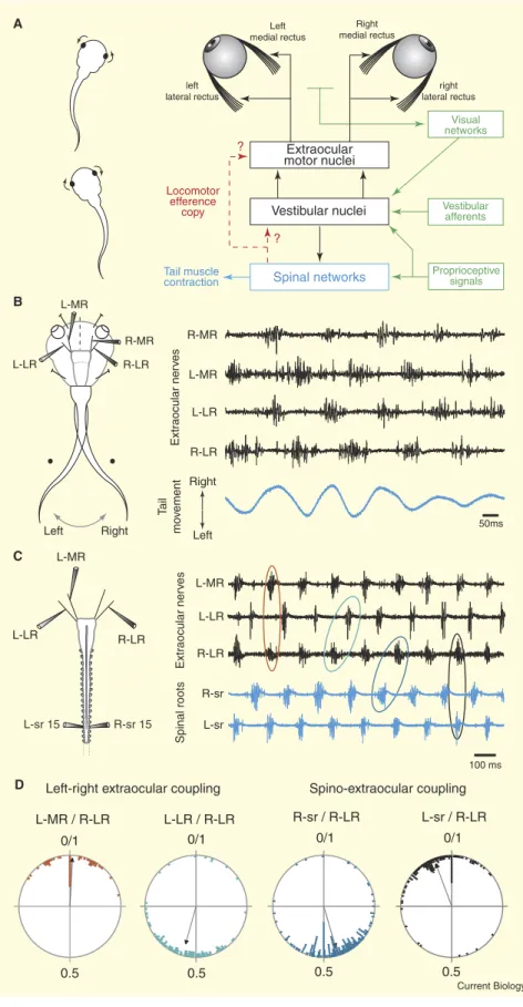
Figure 1. Spinal cord-driven signalling for compensatory eye movements in swimming Xenopus tadpoles.
(A) Left: in stage 55 tadpoles, horizontal head rotations resulting from left–right tail swim-ming movements are associated with op-positely-directed conjugate eye movements. Right: schematic of the neural organisation of sensory-motor networks responsible for compensatory eye movements and pos-sible interactions with locomotor generating circuitry in the spinal cord. (B) Rhythmic activation of extraocular motoneu-rons during horizontal-plane swimming in a semi-intact head-fixed preparation with freely-moving tail (left). Simultaneous extra-cellular recordings (right) from bilateral me-dial rectus (MR) and lateral rectus (LR) motor nerves during spontaneous tail movements (lower trace in blue). Extraocular motor activ-ity was coordinated with tail oscillations such that the right (R) MR and left (L) LR motoneu-rons discharged in-phase with each directed tail excursion, whereas L-MR and R-LR motoneurons were activated dur-ing each left-directed movement. Note that both otic capsules containing the semicir-cular canals and otolith organs had been re-moved and the optic nerve was transected. (C) Left: spontaneous ‘fictive’ swimming in a completely isolated brainstem/spinal cord preparation. Right: simultaneous recordings from bilateral LR and the L-MR extraocular motor nerves, along with the left and right motor roots of the 15th spinal cord segment
(blue traces). Activity in the extraocular mo-tor nerves remained strictly coordinated with alternating left/right locomotor bursts in the spinal roots (sr). (D) The relative tim-ing of extraocular motor bursts durtim-ing fictive swimming was appropriate for conjugate eye movements subserving gaze stabiliza-tion. Circular plots show phase relationships between onsets of locomotor-related burst activity in extraocular and spinal motor nerves of four in vitro preparations (phase of 0 or 1 corresponds to burst synchrony; phase of 0.5 corresponds to burst alternation; see also Table S1 in the Supplemental Data). Synchro-nous bursts in the MR and LR motor nerves on opposite sides — for example, L-MR/ R-LR; see red ellipse in (C) — are consist-ent with conjugate eye movemconsist-ent, while al-ternating bursts in homologous extraocular nerves — for example L-LR/R-LR; green ellipse in (C) — correspond to alternating left-right eye motion. The alternation between ipsilateral spinal root and LR activity — for example, R-sr/R-LR; blue ellipse in (C) — and the synchrony between opposite sides — for example, L-sr/R-LR; black ellipse in ( C) — would cause eye movements that counteract head displacements.
Magazine R243
by the hindbrain vestibular nuclei (Figure 1A, right) which transform the sensory signals arising from self-motion into appropriate motor commands for the extraocular muscles [5].
To determine a possible central nervous contribution to these gaze- stabilizing movements, we employed semi-intact preparations in which tail undulations in the horizontal plane could occur, while the head was held stationary and the brainstem exposed for recording from selected bilateral motor nerves to the extraocular muscles. During spontaneous swimming in such head- fixed, tail- free preparations, discrete bursts of action potentials occurred in extraocular motor nerves controlling horizontal eye movements which were timed to the trajectory of actual tail bending (Figure 1B). The synergistic pair of medial and lateral rectus motor nerves that normally drive left- directed conjugate eye rotations [5] were active in- phase with each right- directed tail movement, while the antagonistic nerve pair controlling right-directed eye movements became active as the tail swung towards the left. Importantly, the tail oscillation- timed discharge in these extraocular nerves persisted after removal of the labyrinthine endorgans on both sides (as in Figure 1B), thereby confirming that their cyclic activity could not have arisen from the sensory detection of any residual head movement.
Furthermore, the locomotory- timed activation of extraocular motoneurons persisted in completely isolated brainstem-spinal cord preparations and therefore in the absence of all movement- related sensory feedback. During spontaneous ‘fictive’ swimming in such in vitro preparations [6] (Figure 1C), the horizontal extraocular motor nerves were cyclically active in time with spinal ventral root bursts that drive the alternating left–right muscle contractions responsible for undulatory tail movements in the intact animal. Discharge in left medial and right lateral rectus motoneurons occurred conjointly with spinal locomotor bursts on the left side of the cord, whereas during the opposite phase of the fictive swim cycle, the right medial and left lateral rectus motoneurons were co-ordinately
active with bursts in right-sided spinal roots (Figure 1D, right). This strict temporal relationship, which corresponded closely to that seen in semi-intact preparations (Figure 1B), was thus appropriate for producing conjugate eye movements (Figure 1D, left) that during swimming in the intact animal would counteract oppositely-directed head displacements resulting from left–right tail oscillations.
These findings provide compelling evidence that rhythmic locomotor signals generated within the tadpole spinal cord are used as an internal prediction of the disruptive consequences of body movements for visual processing during swimming. This adds a further dimension to our understanding of ocular motor control, since hitherto, sensory-motor transformations have been generally thought to be exclusively responsible for compensatory eye movements during vertebrate self-motion [2]. However, copies of spinal locomotor output offer a convenient substrate for initiating eye adjustments in the fastest possible way, pre-empting the slower reactionary engagement of the various movement-encoding sensory pathways that would serve to ensure the gain and precision of the gaze-stabilizing response. Whether the ascending locomotor signals are conveyed directly to their extraocular motor targets or, more likely, are integrated with converging sensory inputs within the vestibular nuclei (Figure 1A, right) remains to be determined.
This scheme also differs from the traditional view of the role of motor efference copies [7] or corollary discharges [8] in guiding adaptive behaviour, where such predictive internal signals are subtracted from self-generated sensory inputs to counter any unwanted consequences of the animals own actions [1]. During swimming in Xenopus tadpoles, copies of spinal locomotor commands are evidently engaged in minimizing the visual consequences of self-action, but here, these feed-forward signals are used in combination with sensory feedback information. Moreover, this spinal drive presumably matches the change in visuomotor demands associated with the developmental transition in locomotor strategy during metamorphosis, when
undulatory swimming in tadpoles is progressively superseded by bilaterally-synchronous hind- limb kick propulsion in the post- metamorphic froglet [6]. Because maintaining visual acuity during self-motion is a problem faced by aquatic and terrestrial vertebrates alike, it is probable that spinal locomotor circuitry also accesses the brainstem gaze control pathways in other vertebrates, including those confronted with more complex visual disturbances resulting from their flexible necks and/or limb- based locomotion.
Our work on the simpler and more tractable nervous system of Xenopus tadpoles now aims to understand the mechanistic basis of gaze stabilization during locomotion, and in particular, to elucidate
how the sensory signalling of three-dimensional body motion interacts with spinal feed-forward commands at the neural network and cellular levels.
Supplemental data
Supplemental data are available at http:// www.current-biology.com/cgi/content/ full/18/6/R241/DC1
References
1. Cullen, K.E. (2004). Sensory signals during active versus passive movement. Curr. Opin. Neurobiol. 14, 698–706.
2. Angelaki, D.E., and Hess, B.J. (2005). Self-motion-induced eye movements: effects on visual acuity and navigation. Nat. Rev. Neurosci. 6, 966–976.
3. Nieuwkoop, P., and Faber, B. (1956). Normal Tables for Xenopus Laevis (Amsterdam: North Holland Publishing Company).
4. Walls, G.L. (1962). The evolutionary history of eye movements. Vision Res. 2, 69–80. 5. Straka, H., and Dieringer, N. (2004). Basic
organization principles of the VOR: lessons from frogs. Prog. Neurobiol. 73, 259–309. 6. Combes, D., Merrywest, S.D., Simmers,
J., and Sillar, K.T. (2004). Developmental segregation of spinal networks driving axial- and hindlimb-based locomotion in metamorphosing Xenopus laevis. J. Physiol. Lond. 559, 17–24.
7. von Holst, E., and Mittelstaedt, H. (1950). Das Reafferenzprinzip. Naturwissenschaften
37, 464–476.
8. Sperry, R.W. (1950). Neural basis of the spontaneous optokinetic response produced by visual inversion. J. Comp. Physiol. Psychol. 43, 482–489.
1Universités Bordeaux 2, 1; Centre National
de la Recherche Scientifique, Unité Mixte de Recherche 5227, Bordeaux 33076, France.
2Université Paris Descartes, Faculté de
Médecine, Centre National de la Recherche Scientifique, Unité Mixte de Recherche 7060, Paris 75006, France.