Amino acid regulation of mTORC1 by
Liron Bar-Peled B.S. Biochemistry University of Georgia
SUBMITTED TO THE DEPARTMENT OF BIOLOGY IN PARTIAL FULFILLMENT OF THE REQUIREMENTS FOR THE DEGREE OF
DOCTOR OF PHILOSOPHY IN BIOLOGY AT THE
MASSACHUSETTS INSTITUTE OF TECHNOLOGY
SEPTEMBER 2013 I
© Liron Bar-Peled. All rights reserved.
The author herby grants to MIT permission to reproduce and to distribute publicly paper and electronic copies of this thesis document in whole or in part in any
medium now known or hereafter created. Signature of Author: _
Department of Biology June 14, 2013 Certified by:
David M. Sabatini Member Whitehead Institute Professor of Biology
Thesis Supervisor Accepted by:
/ ' Stephen P. Bell
Professor of Biology Chair, Committee for Graduate Students
Amino acid regulation of mTORC1 by
Liron Bar-Peled
Submitted to the Department of Biology on June 14, 2013 in Partial Fulfillment of the Requirements of the Degree of Philosophy in Biology
Abstract
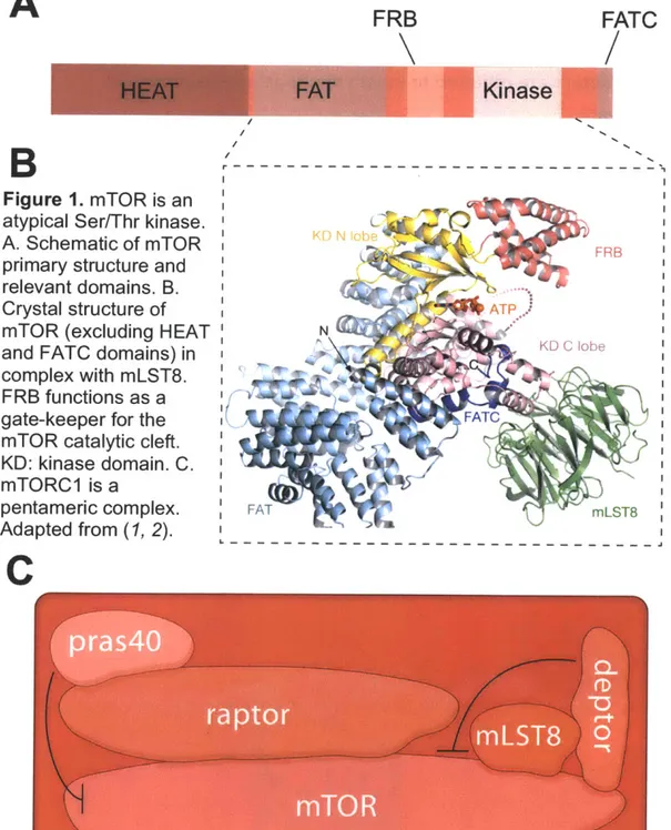
Mammalian target of rapamycin complex I (mTORC1) is an atypical Ser/Thr kinase that regulates cellular and organismal growth. Accordingly, mTORC1 has substantial roles in regulating insulin sensitivity and lifespan, and when deregulated, it is implicated in the pathogenesis of common cancers.
mTORC1 responds to a diverse set of stimuli, including growth factors, oxygen availability, energy and amino acid levels in order to control essential anabolic and catabolic processes. Amino acids promote mTORC1 shuttling to the
lysosomal surface, its site of activation. This translocation is mediated by a family of heterodimeric GTPases known as the Rags that reside on the lysosomal surface. Unique among the small GTPases, the Rags are obligate heterodimers: the highly related RagA and RagB are functionally redundant and bind to RagC or RagD, which are also very similar to each other. Amino acids regulate the
binding of nucleotides to RagB, such that amino acid stimulation increases its GTP loading, leading to the recruitment and binding of mTORC1. In the work described here, we identify two Rag interacting complexes termed 'Ragulator' and 'GATOR' that form a lysosome based signaling platform that controls the activity of the Rags. We find that Ragulator is a pentameric complex that is both necessary and sufficient to determine the intracellular localization of Rags.
Moreover, we find that Ragulator functions as a guanine nucleotide exchange factor (GEF) for RagA and RagB stimulating GTP-loading, a key event in the amino-acid dependent activation of mTORC1. Additionally, we describe the function of GATOR, an octomeric complex that is defined by two distinct
subcomplexes termed GATOR1 and GATOR2. We find that GATOR2 functions as a positive regulator of mTORC1 whereas GATOR1 negatively controls this pathway. Epistasis analysis reveals GATOR2 functions upstream of GATOR1, which inhibits the Rags through its GTPase activating protein (GAP) activity towards RagA and RagB. GATOR1 components are mutated in gliolbastoma and ovarian tumors and GATOR1 deficient cancer cells are hypersensitive to the mTORC1 inhibitor rapamycin. Thus, we define the molecular mechanisms regulating the function of Rags and propose a model for the activation of the mTORC1 pathway by amino acids.
Acknowledgements
David Sabatini is everything I could have asked for in a mentor: insightful, adventurous, creative, meticulous and frank. My meetings with David were usually impromptu and happened at my bench. While they would begin with science (where his infectious curiosity is evident) they would often meander into other areas such as cycling and politics. I owe David a great deal of gratitude for
helping me to develop into the scientist that I am today.
Throughout my years as a graduate student Hidde Ploegh and lain Cheeseman have served as my thesis committee members. Though both have incredibly busy schedules, they have always left a door open for spontaneous meetings and their advice has helped propel my research forward. I would also like to thank Matt Vander Heiden and Brendan Manning whose work I've long admired for serving on my thesis defense committee.
When I first arrived in the Sabatini lab, Yasemin Sancak guided me through the ropes of molecular biology. Not only have we had wonderful collaborations but a lasting friendship, which I am truly thankful for. I was also very fortunate to have Lynne Chantranupong as a bay mate for these past two years and I am delighted to have had the opportunity to work with her during this time. I've had fruitful collaborations with Roberto Zoncu and Larry Schweitzer, whose scientific ideas have guided me in new directions. More often than not,
room 359 has felt like home and this could only made possible by Yasemin, Lynne, Nada, Nora, Tony, Tim, Zhi, Shuyu and Naama. I would also like to thank Dohoon Kim for his generosity and our lengthy conversations that were rarely about science. Edie Valeri and Kathleen Ottina have kept the lab running and
made my research possible. Finally, the Sabatini lab, would not be such a special place to work in if it were not for its members and I would like to thank them all, past and present.
I met many of my graduate school classmates before we were even officially enrolled at MIT and in the past six years we've shared the ups and downs of research. I have made some lasting friends at MIT and I have greatly enjoyed both our scientific and non scientific adventures. I would especially like to thank: Mohini Jangi, Keren Higleldorf, Zach Whitfield, David Weinberg, Lauren
Surface, Emily Rosowski, Sejal Vyas and Albert Alaamada for their continued friendship and advice.
I cannot even begin to thank my family (Aba, Ema, Tal and Yael) for providing unconditional love and support. My parents have always been there for me and I cherish their wisdom. My first research experience was in my father's lab, and it was here that I received my first training in, and appreciation for
biochemistry. My father continues to serve as a great inspiration and is one of the reasons I will pursue a career in science.
Finally, there is my fiancee Fei. I cannot imagine making it through these past few years without her. Fei's compassion and joy that has kept me going through the perils of research in lab and has been a steadying force with the ever increasing stress of research. I am extraordinarily lucky to have met Fei and I am grateful for her companionship on the journey ahead.
Table of Contents
Abstract... 3
Acknow ledgem ents ... 4
Table of Contents... 7
Chapter 1: Introduction ... 10
The Discovery of TOR ... 10
The two faces of TOR... 12
m T O R C 1 ... ... 1 3 m T O R C 2 ... 1 4 Pharmacological Inhibitors of mTOR... 15
Making a cell grow: Downstream processes regulated by mTORCI... 16
Protein synthesis ... 16
S 6 K ... . . . ... .... ... 16
4EBPs .. ... ... 17
Translation of TOP mRNAs... 18
Ribosome biogenesis ... 18
Metabolism ... 18
Lipid biosynthesis... 19
Mitochondrial biogenesis and Glycolysis ... 20
Pyrim idine biosynthesis... 20
Autophagy ... 21
Lysosome biogenesis... 21
Regulating the Regulator: Signaling pathways upstream of mTORC1 ... 21
Rheb... 22
Tuberous sclerosis complex... 22
Growth factor and Cytokine signaling ... 23
Energy sensing... 24
Oxygen sensing ... .25
Amino acid sensing ... .. 26
The Rag GTPases ... 27
mTORC1 and Disease... 29
Cancer..._ 29 Diabetes... 30
Aging ... ----...---... 30
Immune disorders... 31
Introduction to the work presented in this thesis... 32
Figures... 34
References... ... .... 36
Chapter 2: Ragulator-Rag mediated translocation of mTORC1 to the lysosomal surface is necessary for its activation by amino acids ... 48
Summary... ... ... ... ... ... 49
Introduction... ... ... 50
Results... ... 52
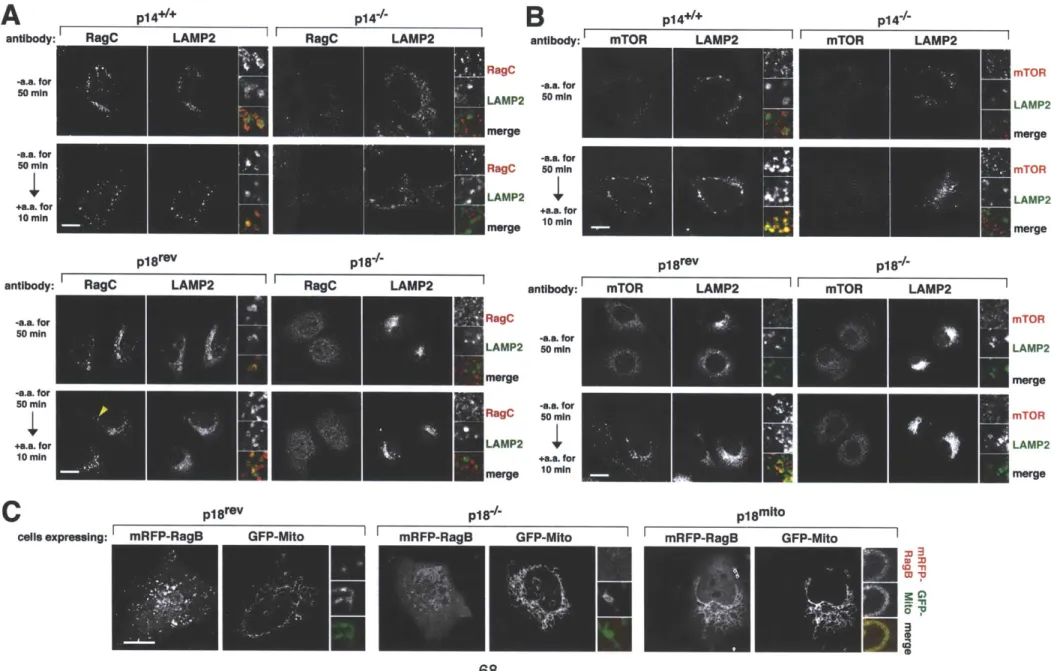
Amino acids cause the translocation of mTORC1 to lysosomal membrane, where the Rag GTPases are already present ... 52
The translocation of mTORC1 to lysosomes does not depend on growth factors, Rheb or mTORCI activity... 55
The trimeric Ragulator complex interacts with the Rag GTPases an co-localizes
with them on lysosomal membranes ... 55
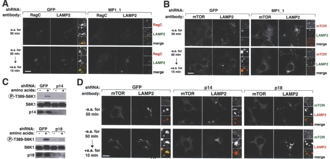
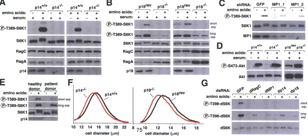
Ragulator localizes the Rag proteins to the lysosomal surface and is necessary for the amino acid-dependent recruitment of mTORC1 to the same compartment ... 6 7 Ragulator is necessary for TORC1 activation by amino acids in mammalian and Drosophila cells ... ... 72
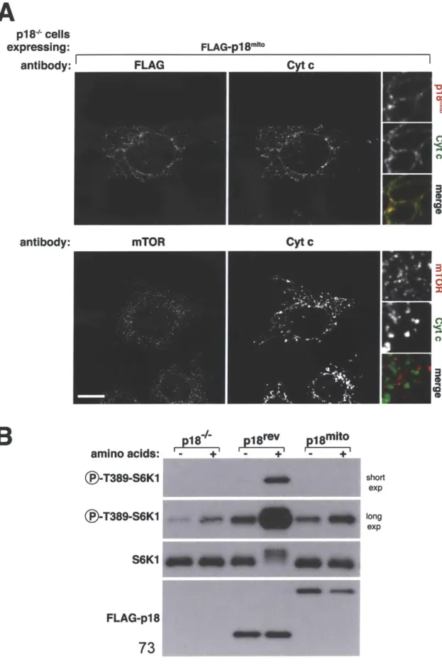
Forced targeting of mTORC1 to the lysosomal surface eliminates the amino acid sensitivity of the mTORC1 pathways ... 79
Forced targeting of mTORC1 to the lysosomal surface eliminates the requirement in mTORC1 signaling for Rag and Ragulator, but not Rheb function ... 8 8 D iscussio n ... 94
Materials and Methods ... 98
A cknow ledgem ents ... 104
R eferences ... 105
Ragulator is a GEF for the Rag GTPases that signal amino acid levels to m T O R C 1... 109
S um m ary ... 110
Introd uctio n ... 111
R es u lts ... 113
HBXIP and C7orf59 encode components of an expanded Ragulator complex 113 HBXIP and C7orf59 are necessary for TORC1 activation by amino acids in mammalian and Drosophila cells... 116
Localization of the Rag GTPases and mTOR to the lysosomal surface requires H BX IP and C 7orf59 ... 120
Amino acids regulate the Rag-Ragulator interaction ... 125
Ragulator preferentially interacts with nucleotide-free Rag GTPases ... 125
Ragulator is a GEF for RagA and RagB ... 136
The v-ATPase controls Ragulator function in cells ... 143
D iscussio n ... 148
M aterials and M ethods ... 152
A cknow ledgem ents ... 159
R eferences... 160
A tumor suppressor complex with GAP activity for the Rag GTPases that signal amino acid sufficiency to mTORCI ... 166
S um m ary ... 167
Introd uctio n ... 168
Results and Discussion ... 169
M aterials and M ethods ... 195
A cknow ledgem ents ... 208
R eferences ... 209
Chapter 5: Future Directions and Discussion... 211
Why do the Rags function as heterodimers... 211
How does the v-ATPase regulate the activity of Ragulator ... 213
Is there a broader significance to the regulated interaction between Rags and R ag u lato r ... 2 14 How Does GATOR2 regulate GATOR1... 214
Conclusions ... 216
References ... ... 217
Chapter 1 Introduction
Cell growth is a fundamental biological process and at its simplest form entails the accumulation of biomass- an increase in carbohydrates, lipids, proteins and nucleic acids. Not surprisingly, the pathways regulating growth control are conserved across the vast swath of eukaryotic life. A master regulator of cell growth can be though of as a rheostat, gauging environmental and
intracellular conditions and in turn modulating pathways responsible for growth. When conditions are favorable for cellular growth (adequate supplies of nutrients and low stress levels) anabolic programs such as translation, transcription and metabolism are activated leading to an increase in cell size. However, when growth-promoting conditions are reversed, a cell must rapidly shut down these anabolic programs and instead promote restorative catabolic programs in order to survive.
Today we know this master growth regulator to be the evolutionarily conserved target of rapamycin (TOR) pathway. In the twenty years since its discovery, the study of this pathway has transformed the field of growth
regulation; not only providing a deeper molecular understanding of growth control but making important advances in the treatment of numerous human diseases. In this introductory chapter, I will provide a signaling centric description of the TOR
pathway, with an emphasis on areas relevant to the presented work.
The Discovery of TOR
Our understanding of the TOR pathway began with the discovery of rapamycin, a polyketide macrolide produced by a soil bacteria on Rapa Nui, a small sunny island in the middle of the south Pacific (3). Rapamycin was initially characterized as a powerful fungicide inducing a striking G, arrest in
some human cancer cell lines. This spurred initial interest in its application as a chemotherapeutic- an idea that would only be realized decades later in the treatment of advanced renal cell carcinoma (5, 6). Notably, rapamycin treatment of a variety of cell types resulted in a dramatic reduction in cell size, implicating its role in growth control (3). Rapamycin was also appreciated as a potent immunosuppressant (7), efficiently blocking IL-2 mediated lymphocyte
proliferation at low nanogram doses (8), while maintaining immune surveillance
(8) and thus avoiding additional complications associated with other
immunosuppressants. Today, rapamycin (also known as serolimus) is a mainstay in organ transplantation (9) and rapamycin-coated stents are used during
angioplasties to inhibit restenosis of arteries (10). These therapeutic properties of rapamycin fueled great interest in understanding the molecular mechanisms underlying its function.
Initial insights into the cellular targets of rapamycin came from
Saccharomyces cerevisiae. Elegant genetic screens in this organism uncovered
that mutations in three genes completely blocked the antiproliferative effects of rapamycin (11, 12). The first gene identified was FRP1, which encodes the evolutionarily conserved prolyl-hydroxylase FKBP12 that forms a stable complex with rapamycin in cells (11). While knockout of FRP1 mediates complete
resistance to rapamycin-induced G1 arrest, loss of FKBP12 did not have an overt growth phenotype. This suggests that while FRP1 is necessary for the function of rapamycin it was not the relevant cellular target (12).
The two remaining genes contained dominant active mutations resulting in corresponding protein products that cannot interact with the FKBP12-rapamycin complex. The two genes are over 7000 base pairs in size and were renamed target of rapamycin 1 and target of rapamycin 2 (TOR1 and TOR2, respectively)
(12). Deletion of TOR1 results in smaller cells and is synthetic lethal with
rapamycin treatment, yet proves dispensable for yeast viability. In contrast deletion of TOR2 is lethal (13). These results indicated that although TOR1 and TOR2 share substantial homology they also have non-overlapping functions.
While genetic approaches in yeast identified the genes that replicate the cellular effects of rapamycin, it would be biochemical approaches in mammalian cell lines that would uncover this compound's physical target.
In parallel efforts to uncover the function of rapamycin in mammalian cells, multiple research groups undertook protein purification campaigns to identify FKBP12-rapamycin interacting proteins. This yielded the discovery of a massive protein (289 kDa) of unknown function that was named the apropos mammalian TOR or mTOR (14-16). Unlike budding yeast, higher eukaryotes as well as
Saccharomyces pombe only contain one ortholog of TOR.
mTOR is a founding member of the phosphatidylinositol-3 kinase related kinase (PIKK) family which also includes ATM, ATR, DNA-PK and SGM-1L (17). The PIKKs are characterized by their large size and a C-terminal Ser/Thr kinase domain which resemble the catalytic domain of the lipid kinase P13K (18). A sixth member of the PIKK family, TRRAP, lost its kinase activity. The mTOR
consensus phosphorylation motif is poorly defined, compared to other members of the PIKK family, making bioinformatic identification of mTOR substrates difficult. Two recent phospho-proteomic studies revealed that the mTOR
phosphorylation site is enriched at the +1 position for proline, aliphatic (Leu, Val) and hydrophobic (Phe, Tyr, Trp, His) residues (19, 20). The PIKK family
members also share additional domains including a FRAP, ATM and TRRAP (FAT) domain and a C-terminal FAT domain that together flank the kinase domain of mTOR and are both required for its activity (17, 21). At the N-terminus of mTOR are two Huntington, elongation factor 3, the A subunit of phosphatase 2A and TOR (HEAT) domains necessary for protein-protein interactions (22).
Finally, sandwiched between the TOR kinase domain and FAT domain is the FKBP12-rapamycin binding (FRB) domain (23, 24), which covers the catalytic cleft and functions as a gate keeper for substrate entry (25) (Figure 1, A and B). The two faces of TOR
In cells, mTOR does not function on its own but rather in two distinct multicomponent complexes commonly referred to as mTOR complex 1 and mTOR complex 2 (mTORC1 and mTORC2, respectively) (6). These complexes are conserved to yeast, where TORC1 is nucleated by either TOR1 or TOR2 and TORC2 only by TOR2 (26). The identification of these two complexes explains the original observations that TOR1 and TOR2 have non-overlapping functions.
mTORC1
mTORC1 is a pentameric complex (Figure 1C), which homodimerizes to form a 2 MDa rhomboid. This massive complex contains a central cavity that is
presumed to be important for substrate phosphorylation (27). Affinity purifications of mTOR (the mainstay for finding mTORC1 complex members) identified
regulatory associated protein of mTOR (raptor) as an mTOR binding protein (28,
29). Raptor is the defining member of mTORC1 and contains multiple WD40
interaction domains and HEAT motifs making it a highly effective protein scaffold. Raptor not only serves as a docking site for many mTORC1 substrates through its interaction with their TOR signaling (TOS) motifs (30, 31) but is also essential for the intracellular localization of mTORC1 (32, 33). mTORC1 is known as the rapamycin sensitive complex and is rapidly inactivated by acute rapamycin treatments. The rapamycin-FKPB12 complex is thought to block mTORC1 function by disrupting the mTOR-raptor interaction (27) in addition to preventing substrate entry into its catalytic cleft (25). Intuitively, mTOR is essential for life and mice null for this protein die during implantation (34). Similarly, mouse embryos lacking raptor expire shortly after e6.5 (35).
The second mTORC1 member identified was mLST8 (36), whose budding yeast ortholog (LST8) was originally characterized as a gene that was synthetic lethal with the COPII and nucleopore member Sec13 (37). Like raptor and many GATOR proteins (see chapter 4), mLST8 also contains a WD40 domain and in cultured mammalian cells is required for proper mTORC1 function (36). Deletion of mLST8 is lethal at embryonic day e10.5 (35), however loss of this protein does
not alter mTORC1 activity in cells obtained from knockout animals, further
complicating our understanding of its function (35). Proline rich Akt substrate of 40 kDa (PRAS40) is one of two endogenous mTORC1 inhibitors and interacts directly with raptor to inhibit mTORC1 kinase activity both in vitro and in vivo (38,
39). As its name suggests, PRAS40 is phosphorylated by Akt (40), which
promotes its dissociation from raptor and relieves its inhibitory activity towards mTORC1 (38). DEPTOR rounds out the remaining members of mTORC1. Like PRAS40, DEPTOR also inhibits mTORC1 kinase activity (41).
In yeast, TORC1 contains two othrologs of its mammalian counterpart: KOG1 the ortholog of raptor and LST8 (26). Additionally, TORC1 also contains the yeast specific TC089, whose function appears important for cell wall
maintenance (42). Curiously, unlike other TORC1 components (with the exception of TOR2), deletion of KOG1 is lethal illustrating its non-redundant functions in yeast (26). As described below, mTORC1 serves as a master
regulator of cell and organismal growth. mTORC2
Unlike mTORC1, the cellular functions of mTORC2 remain poorly
understood. Like mTORC1, mTORC2 is also a zaftig of a complex, however it is currently unknown whether it adopts a higher order structure. Rapamycin-insensitive companion of mTOR (rictor) is an essential component of mTORC2
(43, 44), and serves as a scaffold that is required for the phosphorylation and
activation of the mTORC2 substrates: Akt, PKCa and SGK1 (35, 45, 46). The rictor-mTOR interaction is not perturbed by short-term rapamycin treatments, thus defining mTORC2 as the rapamycin insensitive complex (43). However, in certain cancer cell lines, prolonged treatment with rapamycin disrupts mTORC2 stability leading to a decrease in Akt phosphorylation (47). In other cell lines Akt phosphorylation is increased upon rapamycin treatment due to inhibition of the S6K-IRS1 feedback loop discussed below. Like raptor, rictor is necessary for murine viability, however mice lacking rictor die at e10.5 compared to the much earlier death for raptor-null mice (35). In addition to rictor, a troika of additional mTORC2 specific components exist: protein associated with rictor 1 or 2
(PROTOR1, PROTOR2) (48) and mammalian stress-activated protein kinase interacting protein 1 (mSIN1) (49, 50). Binding of PROTOR1 or PROTOR2 to rictor is mutually exclusive and the function of their remains elusive. mSIN1 is required for mTORC2 stability and Akt phosphorylation (49). mLST8 and
DEPTOR are shared with mTORC2 and maintain their functions in this complex as well (41, 43).
Yeast TORC2 contains three orthologs of mTORC2: AVO1 (mSIN1) (51), AVO3 (Rictor) and LST8 (26). Additionally, AVO2 is also a TORC2 component, but its function is poorly understood (26). While mTORC1 promotes cell growth, mTORC2 is responsible for cell proliferation and survival, making this pathway an enticing therapeutic target.
Pharmacological Inhibitors of mTOR
While rapamycin serves as both a useful discovery tool and
pharmacological agent, it has long been appreciated that this allosteric inhibitor only partially blocks mTOR kinase activity. Recent pharmacological screens resulted in several ATP-competitive inhibitors (Torin, PP242 and PP30) that specifically target the mTOR kinase domain (52-54). With median inhibitory concentrations (IC50) in the low nanomolar range toward mTORC1 in vitro and in cells, these catalytic inhibitors have begun to reveal many novel functions for mTORC1 (19, 55, 56). Importantly, this new class of mTOR inhibitors also disrupts the function of mTORC2, notably the phosphorylation of Akt (52). Thus, these catalytic inhibitors may be more clinically beneficial than rapamycin in treatment of certain cancers. Indeed, recent pre-clinical studies revealed a striking decrease in P13K driven tumors after treatment with catalytic site inhibitors when compared to rapamycin treatment (57),(58). However, the full therapeutic benefits of these mTOR catalytic inhibitors will only be realized upon identification of biomarkers that can predict which tumors are addicted to
deregulation of the mTORC1 and mTORC2 pathways.
The research presented in this thesis pertains to mTORC1 and the remainder of the introduction will describe signaling pathways upstream and downstream of this complex as well as the deregulation of this pathway in human disease.
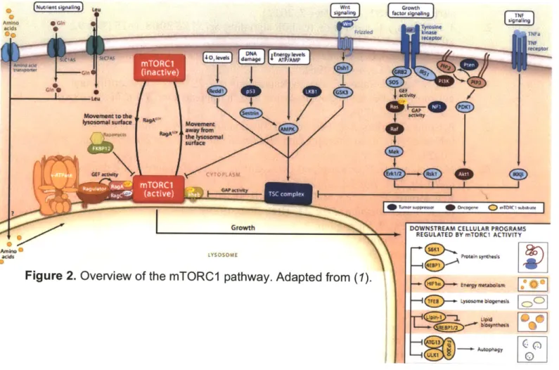
Making a cell grow: Downstream pathways regulated by mTORC1. As a master regulator of growth, mTORC1 increases cell biomass by promoting anabolic programs including translation, transcription, mitochondrial biogenesis, and lipid and nucleic acid biosynthesis, while inhibiting catabolic pathways such as autophagy (Figure 2).
Protein synthesis
Proteins account for roughly 50% of the dry weight of a cell, making regulation of protein synthesis by mTORC1 critical to cell growth (59),(60). mTORC1-mediated translational control is best understood at the level of its effectors S6 Kinasel (S6K1) and elF-4E binding proteins (4EBPs) and by
promoting ribosome biogenesis and translation of transcripts containing a 5' terminal oligopyrimidine (TOP) sequence.
S6K
Early investigations with rapamycin revealed that phosphorylation of the ubiquitous S6 ribosomal protein was sensitive to this drug (61-63). S6 forms part of the 40S ribosomal subunit and is phosphorylated on multiple residues by the
protein kinase A, G, C (AGC) family member S6K1 (64, 65), a direct mTORC1 substrate (66, 67). In mammalian cells S6K1 exists as a two isoforms: cytosolic
p70 and nuclear p85. A second S6K, S6K2, was discovered inadvertently by the generation of the S6K1-'- mouse (68). Although it is a substrate of mTORC1 and
partially overlaps with S6K1 function, the cellular role of S6K2 is unclear (68). mTORC1 regulates S6K1 activity by phosphorylating its C-terminal loop (66). This forms a docking site for another AGC kinase, PDK1, which subsequently
phosphorylates and activates S6K1 (69). How the phosphorylation of S6 by S6K regulates translation remains controversial. Cells taken from transgenic mice in which all five phosphorylation sites on S6 have been mutated to Ala are smaller
and insensitive to further reductions in cell size in response to rapamycin
treatment (70). This demonstrates that mTORC1-dependent cell size regulation is directly dependent on S6 phosphorylation. Paradoxically these mutant cells show an increase in protein synthesis caused by an increase in translation initiation (70), underscoring a poorly defined feedback loop.
In addition to S6 phosphorylation, S6K1 exerts its effects on translation by phosphorylation of eIF4B (71), which promotes its association with the pre-initiation complex and stimulates the translation pre-initiation helicase eIF4A. S6K1 further supports translation initiation, by relieving the PDCD4-mediated inhibition of eIF4A. S6K1 directly phosphorylates PDCD4 (72), which is then ubiquitinated and undergoes proteasomal degradation. S6K1 also impinges on elongation through its phosphorylation and inhibition of eEF2K (73). Although not directly
part of protein synthesis, S6K1 increases nascent transcript splicing by activating the exon junction protein SKAR, which in combination with S6K1 enhances splicing efficiency (74).
4EBPs
The rate-limiting step in protein synthesis is translation initiation. Hence, multiple layers of regulation exist for this step, many of which, including cap-dependent translation, are controlled by mTORC1,. Covalent modification of a majority of nascent transcripts with a 7-methyl guanosine cap on the 5' end ensures message stability. The initiation factor eIF4E binds to the capped mRNA and recruits eIF4G1 facilitating translation initiation. 4EBPs sequester eIF4E preventing its interaction with the initiation scaffold eIF4G1 and formation of the initiation complex (59). Phosphorylation of 4EBPs by mTORC1 prevents 4EBPs from binding to eIF4E thus allowing the initiation complex to be assembled (75,
76). In mammals, three 4EBP proteins exist: 4EBP1 and 4EBP2 appear to have
redundant functions as a single deletion of either does not increase total protein
synthesis (77), while the function of 4EBP3 is cryptic. MEFs collected from double 4EBP1 and 4EBP2 knockout mice are completely resistant to mTORC1-dependent translational regulation (55) implying that 4EBP1 and 2 are the major translational effectors for this pathway.
Translation of TOP mRNAs
mTORC1 also impacts protein synthesis by modulating the translation of a subset of mRNAs containing a TOP motif (59). TOP motifs consist of pyrimidine rich tracts, located in the 5' UTR of a transcript and are enriched in mRNAs encoding protein synthesis factors (59). How mTORC1 selectively regulated the translation of TOP mRNAs remained largely unknown until two groups (including our own) applied high-throughput ribosome profiling techniques to this question
(55, 78). These studies revealed that elF4E binds selectively to TOP mRNAs and
mTORC1 promotes translation of these messages by inhibiting the function of 4EBP1 and 4EBP2 (55). Both studies also provided an unparalleled view of the
translational landscape regulated by mTORC1 and identified new regulatory elements including TOP-like (55) and pyrimidine rich translational element (PRTE) (78) motifs important for translation control.
Ribosome biogenesis
At the most basic level, translational output can be influenced by the total number of ribosomes. Early studies with rapamycin showed a dramatic decrease in the transcription of ribosomal RNA, ribosomal proteins and tRNAs (79).
Subsequent studies clarified this relationship by identifying the TIF-1A as a downstream effector of mTORC1. mTORC1 promotes the nuclear localization of TIF-1A (80), which functions as a co-factor for RNA polymerase I that is critical for rRNA biosynthesis. Additionally, mTORC1 phosphorylates and inhibits MAF1
(81, 82), an inhibitor of RNA polyemerase 111, which is required for 5S rRNA and tRNA transcription.
In addition to regulating protein synthesis, mTORC1 plays a critical role in the synthesis of other macromolecules such as lipids (83) and nucleic acids (84). mTORC1 also exerts its control over glycolytic metabolism and energy
production by modulating key metabolic transcription factors such as HIFla. Lipid Biosynthesis
A growing cell demands an ever-increasing supply of lipids to maintain continued expansion of the plasma membrane and organogenesis. Sterol response element binding protein 1 (SREBP1) functions as a master
transcriptional regulator of lipid and cholesterol biosynthesis (85). SREBP1 lies dormant in the ER membranes until a decrease in sterol levels or activation of the insulin pathway stimulates its proteolytic processing and ensuing release from the ER. Activated SREBP1 translocates to the nucleus and promotes expression of key genes required for lipid biosynthesis, cholesterol biosynthesis and the oxidative arm of the pentose phosphate pathway (85). mTORC1
regulates SREBP1 nuclear translocation through its effector S6K1, which is required for efficient SREBP1 -dependent gene expression (86-88). Additionally, mTORC1 activates SREBP1 by inhibiting Lipin-1, a lipid phosphatase and suppressor of SREBP1 signaling (56). By directly phosphorylating Lipin-1, mTORC1 prevents its nuclear shuttling, where Lipin-1 promotes redistribution of SREBP1 to the nuclear lamina resulting in a decrease in SREBP1 activity (56). Interestingly, depletion of SREBP1 results in smaller cells (89), suggesting that one mechanism by which mTORC1 regulates cell size is through lipid/sterol synthesis.
When mammals undergo starvation, ketone bodies are used as an alternative energy currency for the brain. In a recent study from our lab (90),
mTORC1 was found to inactivate hepatic ketogenesis by inhibiting the function of PPARa, a nuclear receptor that controls the expression of genes required for fatty acid and ketone body synthesis (91). mTORC1, via S6K2 activation (92), promotes the nuclear accumulation of the histone deacetylase (HDAC) nCoR1
(90), which blocks the transcriptional activity of PPARa.
Mitochondrial biogenesis and Glycolysis
The anabolic programs driven by mTORC1 are tremendously energy consuming (i.e. the synthesis of one peptide bond requires hydrolysis of four phosphoanhydride bonds). It is therefore not surprising that mTORC1
accommodates these energy demands by promoting mitochondrial function and biogenesis through a concerted increase in mitochondrial DNA content and in expression of oxidative phosphorylation genes (93). mTORC1 is thought to promote the interaction of PGC-1c, a master regulator of mitochondrial
biogenesis, with the transcription factor YinYang 1 (94). However this model must be reconciled with the fact that mTORC1 does not reside in the nucleus (32, 33). Thus, additional mTORC1-dependent mechanisms likely exist to increase
mitochondrial biogenesis.
HIFla is a transcription factor induced under hypoxic conditions to help a cell cope with reduced oxidative phosphorylation (95, 96). By increasing the expression of key glycolytic genes and glucose transporters and driving pyruvate flux towards lactate, HIF1a guarantees a steady supply of ATP under low oxygen conditions (97). mTORC1 promotes ATP production and glycolytic flux by
positively regulating the transcription and translation of HIF1aC (98-101). Pyrimidine biosynthesis
Two recent studies uncovered a critical role for mTORC1 in the regulation of pyrimidine biosynthesis, a key component of nucleic acids. By utilizing
metabolic profiling, Manning and colleagues discovered that mTORC1
upregulates synthesis of N-carbomyl-aspartate, a critical substrate required for pyrimidine biosynthesis (102). Through a phosphoproteomics approach, Hall and colleagues converged on S6K1 as the key intermediary between pyrimidine
biosynthesis and mTORC1 (103). The Manning and Hall labs went on to demonstrate that S6K1 phosphorylates and activates CAD, the rate-limiting enzyme in pyrimidine biosynthesis, responsible for N-carbomyl-aspartate
production, potentially linking mTORC1 activation to progression through S-phase.
Autophagy
While enabling anabolic programs to increase cellular growth, mTORC1 shuts down catabolic programs such as autophagy to prevent a futile cycle of synthesis and degradation. Autophagy is best characterized as a cellular recycling program. It encompasses the sequestration and break down of cytosolic proteins (microautophagy) and organelles (macroautophagy) to
replenish the cell with key macromolecules under nutrient starvation (104). Upon mTORC1 inhibition, double membrane vesicles originating from the ER engulf cytosolic proteins and organelles and fuse with lysosomes where degradative enzymes rapidly digest these cellular products into simpler molecules (105). mTORC1 primarily regulates autophagy by inhibiting key regulators of this process. Specifically, mTORC1 phosphorylates and inhibits the kinase complex of ULK1 -ATG13-FIP200 (106-108), required for autophagasome formation. Recently, mTORC1 was also shown to activate DAP1 a negative regulator of autophagy (109).
Lysosome biogenesis
As discussed below, the lysosomal surface is the site of mTORC1 activation. It is therefore not surprising that mTORC1 has a critical role in lysosome biogenesis through its regulation of transcription factor EB (TFEB). mTORC1 directly phosphorylates TFEB1 promoting 14-3-3 protein binding and its sequestration in the cytoplasm (110-112). When mTORC1 is inactivated, TFEB is rapidly dephosphorylated, sheds its 14-3-3 proteins and shuttles to the nucleus. There, TFEB controls the expression of lysosome maintenance (113) and autophagasome formation genes (114), resulting in an increase in lysosome number, size and function.
Regulating the Regulator: Signaling pathways upstream of mTORCI
As a master regulator of cell growth mTORC1 must be keenly aware of the nutrient conditions inside the cell as well as those in the extracellular milieu., The pathway has therefore evolved to sense a wide variety of inputs including energy, amino acid and oxygen levels, genotoxic stress, and metazoan specific signals such as growth factors and hormones (Figure 2). All upstream inputs that funnel onto mTORC1 can be divided into two branches: those that modulate the activity of its main activator ras homolog enriched in brain (Rheb) and those required for its movement to the lysosomal surface, where it is activated. Rheb
The small GTPase Rheb was initially identified as a protein upregulated in rat brains during seizures (115). Its connection to the TORC1 pathway was first discovered in flies, where gain of function screens in the fly eye placed dRheb upstream of dTORC1 and established its role as a positive regulator of this pathway (116, 117). Subsequent biochemical studies in mammalian cells indicated that GTP-bound Rheb functions as a potent stimulator of mTORC1 kinase activity (38). In vivo, the localization of Rheb is just as important as its nucleotide state in activating this pathway. Like other small GTPases, Rheb relies on a lipid modification (farnesylation) (118) for its proper localization to late endosomes/lysosomes (119). Mutation of the cysteine required for C-terminal Rheb farnesylation, reduces mTORC1 activity (119), whereas flooding the cytoplasm by overexpressing Rheb, uncouples mTORC1 activation from its lysosomal localization (33, 120). Curiously, while Rheb is absolutely required for mTORC1 activation in all metazoans and the yeast S. pombe (121), it is not required for pathway activation in S. cerevisiae (122). This difference likely stems from the need to integrate new signaling inputs such as growth factors (see below) in metazoans but not in cerevisiae.
Tuberous sclerosis complex
Mutation in the genes encoding tuberous sclerosis complex 1 and 2 (TSC1 and TSC2, TSC1/2) were originally identified as the causative agents in
the hamaratomatous syndrome of the same name (123, 124). Pioneering studies in flies and mammalian cells defined TSC1/2 as a negative upstream regulator of TORC1 (125, 126). The discovery of Rheb and its function
downstream to TSC1/2, heralded biochemical investigations which revealed that TSC2 harbors GTPase activating protein (GAP) activity toward Rheb (127-129). TSC2 utilizes an asparagine thumb to accelerate Rheb GTP hydrolysis, however key residues utilized by other small GTPases for hydrolysis are not conserved in Rheb, suggesting a novel mechanism by which GTP is hydrolyzed (130).
Recently, TBC1 D7 was identified as a third member of TSC, required for maintaining the TSC1-TSC2 interaction. Like TSC1 and TSC2, depletion of TBC1 D7 increases mTORC1 activity, however this member has yet to be found mutated in patients (131). To inactivate Rheb, TSC1/2 localizes to the lysosomal surface, a process thought to be dependent on TSC2 (132).
The identification of the Rheb-TSC1/2 axis greatly clarified the regulation of mTORC1 and revealed that TSC1/2 functions as a central hub for a multitude of different inputs that converge onto this pathway (133).
Growth factor and Cytokine signaling
Binding of growth factors such as insulin or EGF to their cognate receptor tyrosine kinases triggers complex signal transduction pathways ultimately leading to the activation of Ras and P13K. GTP bound Ras activates a kinase cascade turning on the RSK and ERK kinases, which phosphorylate and inactivate TSC2
(134, 135). Active P13K generates 3,4,5-phosphoinositol (PIP3) recruiting Akt to
the plasma membrane where it is activated by PDK1. Akt both directly and indirectly activates mTORC1 by phosphorylating and inhibiting TSC2 (126,
136-138) and PRAS40 (38). To temper the activating signals generated by Ras and
P13K, cell rely on the tumor suppressors NF1 (139) and PTEN (140) whose function as a Ras GAP and a PIP3 phosphatase, respectively, inhibit
downstream pathway signaling. Notably, cancers driven by mutations in these tumor suppressors are marked by hyperactivation of mTORC1. The mTORC1 pathway has also evolved its own safety valve in the form of two negative
feedback loops with insulin receptor signaling. Active S6K1 phosphorylates IRS1, a key component of the insulin receptor, leading to its degradation and
attenuation of PIP3 signaling (141, 142). In the second feedback loop, mTORC1 activates GRB10 a negative regulator of the insulin receptor (19, 20). These two negative feedback loops have important ramifications for diabetes and cancer (see below).
The WNT and NFKB pathways are the remaining extracellular signaling pathways that funnel onto TSC1/2. Binding of the WNT ligand to its cognate GPCR results in the inhibition of GSK3, which activates TSC2 (143). Conversely, the death ligand TNFa activates the IKK kinase, which phosphorylates TSC1 leading to the inactivation of the complex (144). It has become apparent that phosphorylation of TSC1 or TSC2 serves as the preeminent form of TSC1/2
regulation. Phosphorylation either activates or inactivates this complex (143, 145), with the latter being modulated by complex destabilization or 14-3-3 binding to TSC2 which prevents its localization with Rheb (126, 132, 138).
Energy sensing
Unlike growth factor signaling which activates mTORC1, a drop in energy levels as detected by a decrease in the ratio of ATP:AMP negatively regulates this pathway. AMP kinase (AMPK) functions as a master sensor of cellular energy levels (146). AMP competes with ATP for a binding pocket in
the regulatory y subunit of the obligate heterotrimer. Upon AMP binding the y subunit undergoes a conformational change that activates the catalytic a subunit
(147). When active, AMPK phosphorylates TSC2 (145) as well as raptor (148)
and both events down regulate mTORC1 activity. Because mitochondria produce the majority of ATP in the cell, the regulation of AMPK is intimately connected to the function of this organelle. Reducing ATP levels by treating cells with the diabetic drugs metformin or phenformin (complex I inhibitors) (149-152), the anti-aging polyphenol resveratrol (F1Fo mitochondrial ATPase inhibitor) (152) or by
suppressor LKB1 also stimulates AMPK through direct phosphorylation of its P regulatory subunit of AMPK (153, 154). Finally, genotoxic stress (DNA damage) regulates AMPK through p53-mediated up regulation of sestrins, which in turn activate AMPK (155).
Oxygen sensing
A short-term drop in oxygen levels is sufficient to inhibit mTORC1 (156), a process that is independent of AMPK function. Genetic screens in flies identified the orthologs of regulated in development and DNA damage response 1
(REDD1) as a protein induced under short term hypoxic conditions that functions downstream of P13K signaling but upstream of TSC (157, 158). REDD1 is
thought to activate TSC, by binding 14-3-3 proteins that otherwise are destined to sequester TSC2 away from Rheb (158, 159). How REDD1 senses oxygen is currently is unknown.
Amino acid sensing
Early investigations revealed that amino acids were required to stimulate protein synthesis in rat skeletal muscles (160). Later studies confirmed that a mixture of all 20 amino acids directly activated the mTORC1 pathway and along with growth factor signaling was absolutely required for the phosphorylation of S6K1 and the 4EBPs (76, 161). Whether all amino acids, one particular amino acid or an amino acid byproduct is being sensed by the mTORC1 pathway remains to be elucidated. Seminal studies established that leucine as well as arginine were necessary for mTORC1 activation but were not sufficient to activate mTORC1 in cells deprived of all amino acids (76). Dissecting the amino acid signal is further complicated by the fact that some plasma membrane amino acid transporters require additional amino acids to pump their cargo into the cytoplasm (162); thus blurring the singular importance of any one amino acid. The development of cell-free assays used to measure amino acid activation of mTORC1 promises to help answer this outstanding question (163).
Amino acid stimulation of the TORC1 pathway is evolutionarily conserved to yeast where it is reduced to the more primitive form of nitrogen sensing
(164-166). Depending on the yeast strain, growth on nitrogen poor substrates
inactivates TORC1. This leads to a transcriptional up regulation of key metabolic enzymes required for production of glutamine as well as a shuttling of amino acid permeases to the plasma membrane to help the cell scavenge for nitrogen rich sources (164). In flies, mutation of amino acid transporter genes pathetic,
minidiscs and slimfast decrease animal size (167-169), indicating the necessity
of amino acid signaling during development.
Although it was clear for over a decade that amino acids were required for mTORC1 activation, precisely how they functioned remained a mystery. Recently, our lab along with others have identified that amino acids regulate the
intracellular localization of mTORC1 (32, 33). When cells are starved of amino acids, mTORC1 is found in a poorly defined cytoplasmic compartment. Upon amino acid stimulation mTORC1 rapidly shuttles to the lysosomal surface where it is presumed to interact with Rheb (32). The raptor component of mTORC1 is critical to its lysosomal targeting (32, 33) and curiously it also contains a domain shared by many vesicle coat proteins (170). Targeting mTORC1 constitutively to the lysosmal surface eliminates the need for the amino acid signal to activate the pathway as does localizing this complex along with Rheb to the plasma
membrane (170). Thus, the purpose of the amino acid signal is to bring mTORC1 and Rheb together. In yeast, TORC1 is localized to the vacuole and does not shuttle in response to amino acids (171). The lack of a functional Rheb homolog in budding yeast likely makes TORCI movement unnecessary.
Where amino acid sensing occurs is still a matter of debate. While extracellular amino acids must enter the cell to activate mTORC1 during amino acid starvation (162), the use of the translation inhibitor cyclohexamide to
generate intracellular pools of amino acids reveals that sensing occurs inside the cell and not at the plasma membrane (32). Clarifying the site of sensing, Zoncu et al. used cell free reconstitution assays to establish that amino acid sensing
initiates from within the lysosomal lumen (163). Disruption of the lysosomal membrane with detergents or ionophores also inhibits amino acid sensing, however changes in luminal pH appear dispensable for this process. Luminal amino acid sensing was further corroborated in cells by over-expression of the PAT1 transporter, a lysosomal amino acid exporter which drains the lysosomal lumen of amino acids and efficiently turns off mTORC1 signaling even in the presence of extracellular amino acids (163). Collectively these studies demonstrate that lysosomal accumulation of amino acids is critical for the sensing mechanism.
While it was long believed that the amino acid signal funneled through the TSC1/2-Rheb axis, the development of TSC2~'- mice proved otherwise. mTORC1 remained sensitive to amino acid regulation in MEFs obtained from these
animals (172, 173), implicating an alternative pathway. The identification of the Rag GTPases as mTORC1 interacting proteins (32, 174) completely changed our understanding of how this pathway senses amino acids.
The Rag GTPases
Rags are unique among all small GTPases as they function as obligate heterodimers (32, 174-177). Mammalian cells contain four Rag GTPases: RagA and RagB are functionally redundant and bind to the highly similar RagC and RagD (175-177). While RagA, RagC and RagD are ubiquitously expressed, RagB expression is restricted to the brain, suggesting a specialized function for this protein (177). It is currently unknown if a preferred Rag heterodimer exists, however the existence of only two Rags (RagA and RagC) in all other eukaryotes (with the exception of plants that do not encode these genes) suggests a
functional redundancy for the other Rags. This hypothesis will be tested with the generation Rag knockout mice. The yeast ortholog of RagA/B is GTR1 whereas GTR2 is the ortholog of RagC/D (171, 178, 179). The GTRs were first connected to the TORC1 pathway in a screen that identified negative regulators of
macroautophagy (178). While the GTRs are not essential genes, their deletion is synthetic lethal in the presence of rapamycin (171), a phenotype that extends to
other TORC1 components. The crystal structures of the GTR1-GTR2 reveals that the two G proteins are tied together by their C-terminal roadblock domains (180,
181); a domain that is curiously found in four other components of the amino acid
sensing pathway (Chapter 3) (182). In the GTP loaded state, the G domain of the GTRs face away from each other, however when GTR2 is bound to GDP its G domain undergoes a dramatic rearrangement and leans into the G domain of
GTRI (180, 181). The functional significance of this rearrangement remains to be
determined (Chapter 5), nevertheless, the nucleotide bound state of the Rags is key to their function (discussed below).
Loss of function studies in mammalian, fly and yeast cells established that the Rags GTPases were critical in mediating the amino acid signal to mTORC1
(32, 171, 174). In cells depleted of Rags, mTORC1 cannot translocate to the
lysosomal surface (33). Rags interact with raptor in an amino acid dependent manner, and their localization to the lysosomal surface defines their role as a docking site for mTORC1 on this organelle (32). Unlike other small GTPases, the Rags do contain lipid modifications that are commonly used to indicate a
protein's intracellular home. As will be discussed in Chapters 2 and 3, the Rags rely on the pentameric Ragulator complex for their localization (33, 182) whose function is conserved to the EGO complex in yeast (171, 179).
Metabolic labeling studies have demonstrated that during amino acid starvation RagB is bound to GDP and upon amino acid stimulation GDP is exchanged for GTP (32). Rag mutants thought to mimic different nucleotide bound states demonstrate that a GTP-locked RagA heterodimer strongly interacts with raptor whereas the Rag complex with the opposite nucleotide bound state does not (32). In mice or cells expressing RagA or RagB GTP mutants, mTORC1 is constitutively localized to the lysosomal surface and the pathway is insensitive to regulation by amino acids (32, 183). Rags do not directly sense amino acids and the identification of a RagA/B guanine nucleotide exchange factor (GEF) (Chapter 3) (182) and GAP (Chapter 4) illustrates that a
complex signal transduction pathway lies between the amino acid signal and these GTPases.
mTORC1 and disease
Given the ubiquity of the cellular processes under the control of mTORC1 and the multitude of signaling pathways that regulate its activity, it is not
surprising that this pathway is often deregulated in numerous human diseases (6). The most prominent examples of mTORC1 deregulation in disease are discussed below.
Cancer
The genetic hamaratomatous syndrome TSC, characterized by large benign tubers was the first and is the best described cancer prone syndrome driven by aberrant mTORC1 signaling (184). Deletion or loss-of-function mutations in either TSC1 or TSC2 underlie a majority of these cases. Although this syndrome is rare (1:6000 live births) the location of tumors in the lungs, brain, kidney and heart can disrupt normal physiological operations leading to severe complications (184). Treatment of TSC induced subependymal giant cell astrocytomas with the rapamycin analog everolimus leads to a near complete remission of this tumor. A related cancer caused by TSC1/2 loss and exclusively affecting women is lymphangiomyomatosis (LAM) characterized by lung cysts that destroy the lung parenchyma. The disease is fatal because even after lung transplantation the cysts return (185). Recent clinical trials with rapamycin, although preliminary, appear to be promising in the treatment of LAM (186).
In addition to TSC, other hamaratomatous cancers are defined by loss of tumor suppressors that lie upstream of mTORC1 and include: Peut-Jeghers
(LKB1) (186), Neurofibromatosis (NF1) (187), Cowden syndrome (PTEN) (186) and Birt-Hoog-Dube syndrome caused by mutations in Folliculin a tumor
suppressor proposed to be part of the mTORC1 pathway (188). Finally, expression of DEPTOR is also found to be unregulated in multiple myelomas
(41). By inhibiting mTORC1 activity, DEPTOR relieves the negative feedback
loop between S6K1 and IRS1 allowing for continued activation of Akt (41). Functioning downstream of many classical oncogenic pathways such as the P13K/Pten/Akt and the Ras/Raf pathways, aberrant mTORC1 activation is a staple in many cancers (6, 41, 189). Although mTORC1 promotes cell growth through a variety of mechanisms, regulation of translation represents an
especially important oncogenic avenue for this signaling pathway. Depletion of the protein synthesis inhibitors 4E-BPs is pro-proliferative (190) and is important for tumorigenicity (191), whereas over-expression of 4EBP-1 in Akt driven lymphoma reduces tumor volume (58). Moreover, elF4E has emerged as an important oncogene by promoting the translation of factors important for cell survival and proliferation such as cyclin D and c-Myc (190, 192). Finally, the up
regulation and stabilization of HIFla by mTORC1 (98-101) promotes
angiogenesis by increasing VEGF expression and adapting tumors to a hypoxic environment (101).
Diabetes
Targeting core components of the mTORC1 pathway in transgenic mice has revealed a prominent role for mTORC1 in diabetes (6). Mice with a
conditional knockout of raptor in adipose tissue have increased blood glucose tolerance, insulin sensitivity and resistance to diet induced obesity (193). Given the requirement of mTORC1 for adipogenesis (83), these mice are leaner owing to a decrease in adipose tissue. These results are mirrored in whole body S6K1 knockout mice as well (194). Conversely, TSC2 deficient cells are extraordinarily insulin and IGF1 resistant (141) and these results are explained by the S6K-lRS1 and GRB10 negative feedback loops.
Aging
For over a century, caloric restriction has been demonstrated to extend lifespan in numerous model organisms including mice and rhesus monkeys (195,
through mTORC1 although the molecular mechanisms underlying this pathway have yet to be established. Furthermore, depletion of key mTORC1 components in yeast, worms, flies and mice all show an increase in lifespan (196, 197). In worms, TORC1 appears to regulate longevity through its control of the Pha4 and
Skn1 transcription factors (198, 199). Excitingly, treatment of middle-aged mice with rapamycin significantly increased their lifespan (-16% in females and 8% in
males) (200).
Immune disorders
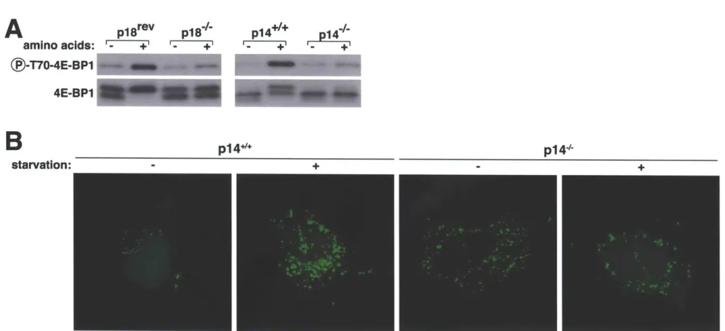
The discovery of rapamycin as a potent immunosuppressant highlighted the importance of the mTORC1 pathway in the immune system (3). While rapamycin is known to be a potent inhibitor of many immune cells, only recently was a primary immunodeficiency linked to this pathway. Mutation in the 3'UTR of the transcript encoding a Ragulator protein (p14) results in decreased protein expression and causes a severe reduction in neutrophil counts (201).
Interestingly, patients diagnosed with this disorder were below the first percentile in height compared to their age-matched peers (201) and cells isolated from these patients display a marked reduction in mTORC1 activity (33). This syndrome represents the first in what promises to be a large class of immune disorders with aberrant mTORC1 signaling underlying their etiology.
Introduction to the work presented in the thesis
Initial studies first described the stimulation of mTORC1 by amino acids over fifteen years ago, but for much of this time how amino acids regulate this pathway has remained enigmatic. The discovery that the Rag GTPases mediate the amino acid signal to mTORC1 represents a significant step forward not only in our grasp of mTORC1 signaling but also its role in normal and diseased states.
For my doctoral thesis I studied how amino acids control the function of the Rags by addressing the following questions:
1. How do the Rags localize to the lysosomal surface?
Because the Rags do not contain lipid modifications to direct their intracellular localization, we hypothesized that additional proteins must directly interact and tether them to the lysosomal surface. By purifying the Rags under several conditions, we identified five proteins (p14, MP1, p18, HBXIP and
C7orf59) that form a novel complex, which we named 'Ragulator'. Ragulator is found at the lysosomal surface and specifically interacts with the Rags on this
organelle. Loss of function and mislocalization studies revealed that Ragulator is both necessary and sufficient for Rag localization. In cells null for or highly
depleted of Ragulator proteins mTORC1 is no longer found at the lysosomes and remains inactive.
2. What factors are required to activate Rags upon amino acid stimulation? Several key experiments revealed that the Rag-Ragulator interaction is regulated by amino acids suggesting that Ragulator may also control the
nucleotide loading of the Rag GTPases. The Rags pose a unique experimental challenge for identifying factors that regulate the nucleotide state of a single Rag given that they exist as obligate heterodimers. To circumvent this problem, we developed several methods that allowed us to analyze the nucleotide binding state of one Rag at a time. This led to the discovery of Ragulator as a GEF for RagA and RagB. GEFs lead to the activation of their cognate GTPase by increasing GTP binding through displacement of GDP from the GTPase.
Interestingly, the GEF activity of Ragulator is not localized to one Ragulator subunit, but requires the complete pentameric complex, indicating that Ragulator
belongs to a new family of multi-protein GEFs.
3. What factors are required to inactivate Rags upon amino acid withdrawal?
To identify negative regulators of the Rags, we purified them in the
presence of a chemical crosslinker that preserves transiently interacting proteins. This approach led to the identification of a complex of eight Rag-interacting proteins that we refer to as 'GATOR' (GAP Activity TOwards Rags). GATOR is defined by two distinct subcomplexes, GATOR1 and GATOR2, where GATOR1 negatively regulates mTORC1 while GATOR2 positively regulates this pathway by inhibiting GATOR1. GATOR1 directly interacts with the Rag GTPases and inhibits their function through its GAP activity towards RagA and RagB. Upstream negative regulators of mTORC1 are commonly mutated in cancer and, indeed, in a subset of glioblastoma and ovarian tumors, we find inactivating mutations in GATOR1 genes. Moreover, in cancer cell lines missing GATOR1 components the mTORC1 pathway is insensitive to amino acid starvation and hypersensitive to treatment with the mTORC1 inhibitor rapamycin.
A
FRB
FATC
Kinase
B
Figure 1. mTOR is an atypical Ser/Thr kinase. A. Schematic of mTOR
primary structure and relevant domains. B. Crystal structure of mTOR (excluding HEAT and FATC domains) in complex with mLST8. FRB functions as a gate-keeper for the mTOR catalytic cleft. KD: kinase domain. C. mTORC1 is a pentameric complex. Adapted from (1, 2).
C
- - -FRB ATP... N o ~KID C lobe FATC FAT M LST8f! v'izled "o
4 o ievek d=
Growth
LYSOSOME
Figure 2. Overview of the mTORC1 pathway. Adapted from (1).
DOWNSTREAM CELLULAR PROGRAMS REGULATED BY mTORC1 ACTIVITY
Protein iymflheah ED ynwwmek.ns - - A O
[El
10 35 Ammfo ,iNuuen
sin
4'n
(
\
ky _w teceper Afekio 0 r -- 0 C"" I IReferences
1. L. Bar-Peled, D. M. Sabatini, SnapShot: mTORC1 signaling at the lysosomal surface. Cell 151, 1390 (Dec 7, 2012).
2. T. E. Harris, J. C. Lawrence, Jr., TOR signaling. Sci STKE 2003, rel5 (Dec 9, 2003).
3. R. T. Abraham, G.
J.
Wiederrecht, Immunopharmacology of rapamycin. AnnuRev Immunol 14, 483 (1996).
4. S. N. Sehgal, H. Baker, C. Vezina, Rapamycin (AY-22,989), a new antifungal antibiotic. II. Fermentation, isolation and characterization. jAntibiot (Tokyo) 28, 727 (Oct, 1975).
5. A. Gomez-Pinillos, A. C. Ferrari, mTOR signaling pathway and mTOR inhibitors in cancer therapy. Hematol Oncol Clin North Am 26, 483 (Jun, 2012).
6. M. Laplante, D. M. Sabatini, mTOR signaling in growth control and disease.
Cell 149, 274 (Apr 13, 2012).
7. R. E. Morris, E. G. Hoyt, M. P. Murphy, R. Shorthouse, Immunopharmacology of FK-506. Transplant Proc 21, 1042 (Feb, 1989).
8. S. Sakaguchi, T. Yamaguchi, T. Nomura, M. Ono, Regulatory T cells and immune tolerance. Cell 133, 775 (May 30, 2008).
9. R. W. Yatscoff, D. F. LeGatt, N. M. Kneteman, Therapeutic monitoring of rapamycin: a new immunosuppressive drug. Ther Drug Monit 15, 478 (Dec,
1993).
10. R. Wessely, New drug-eluting stent concepts. Nat Rev Cardiol 7, 194 (Apr, 2010).
11. Y. Koltin et al., Rapamycin sensitivity in Saccharomyces cerevisiae is
mediated by a peptidyl-prolyl cis-trans isomerase related to human FK506-binding protein. Mol Cell Biol 11, 1718 (Mar, 1991).
12.
J.
Heitman, N. R. Movva, M. N. Hall, Targets for cell cycle arrest by the immunosuppressant rapamycin in yeast. Science 253, 905 (Aug 23, 1991). 13. J. Kunz et al., Target of rapamycin in yeast, TOR2, is an essentialphosphatidylinositol kinase homolog required for G1 progression. Cell 73, 585 (May 7, 1993).
14. E. J. Brown et al., A mammalian protein targeted by Gl-arresting rapamycin-receptor complex. Nature 369, 756 (Jun 30, 1994).
15. D. M. Sabatini, H. Erdjument-Bromage, M. Lui, P. Tempst, S. H. Snyder, RAFT1: a mammalian protein that binds to FKBP12 in a rapamycin-dependent
fashion and is homologous to yeast TORs. Cell 78, 35 (Jul 15, 1994). 16. C. J. Sabers et al., Isolation of a protein target of the FKBP12-rapamycin
complex in mammalian cells.j Biol Chem 270, 815 (Jan 13, 1995).
17. H. Lempiainen, T. D. Halazonetis, Emerging common themes in regulation of PIKKs and P13Ks. Emboj 28, 3067 (Oct 21, 2009).
18. G. Manning, D. B. Whyte, R. Martinez, T. Hunter, S. Sudarsanam, The protein kinase complement of the human genome. Science 298, 1912 (Dec 6, 2002).