This paper is available online free of all access charges (see http://jxb.oxfordjournals.org/open_access.html for further details)
RESEARCH PAPER
Is xylem of angiosperm leaves less resistant to embolism
than branches? Insights from microCT, hydraulics, and
anatomy
Matthias Klepsch1,*,†, Ya Zhang1,*, Martyna M. Kotowska2,3, Laurent J. Lamarque4,5, Markus Nolf6,
Bernhard Schuldt3, José M. Torres-Ruiz4,7, De-Wen Qin8, Brendan Choat6, Sylvain Delzon4, Christine Scoffoni9, Kun-Fang Cao8 and Steven Jansen1
1 Institute of Systematic Botany and Ecology, Albert-Einstein-Allee 11, Ulm University, D-89081 Ulm, Germany 2 Department of Biological Sciences Faculty of Science, Macquarie University, NSW 2109, Australia
3 Plant Ecology, Albrecht von Haller Institute for Plant Sciences, University of Göttingen, Untere Karspüle 2, D-37073 Göttingen,
Germany
4 BIOGECO, INRA, University of Bordeaux, 33615 Pessac, France 5 EGFV, INRA, University of Bordeaux, 33882 Villenave d’Ornon, France
6 Hawkesbury Institute for the Environment, University of Western Sydney, Richmond, New South Wales 2753, Australia 7 Université Clermont-Auvergne, INRA, PIAF, 63000 Clermont-Ferrand, France
8 Guangxi Key Laboratory of Forest Ecology and Conservation, College of Forestry, Guangxi University, Daxuedonglu 100, Nanning,
Guangxi 530004, PR China
9 Department of Biological Sciences, California State University, Los Angeles, 5151 State University Drive, Los Angeles, CA 90032, USA
* Joint first authors.
† Correspondence: matthias.klepsch@uni-ulm.de
Received 15 August 2018; Editorial decision 28 August 2018; Accepted 28 August 2018 Editor: Howard Griffiths, University of Cambridge, UK
Abstract
According to the hydraulic vulnerability segmentation hypothesis, leaves are more vulnerable to decline of hydraulic conductivity than branches, but whether stem xylem is more embolism resistant than leaves remains unclear. Drought-induced embolism resistance of leaf xylem was investigated based on X-ray microcomputed tomography (microCT) for Betula pendula, Laurus nobilis, and Liriodendron tulipifera, excluding outside-xylem, and compared with hydraulic vulnerability curves for branch xylem. Moreover, bordered pit characters related to embolism resistance were investi-gated for both organs. Theoretical P50 values (i.e. the xylem pressure corresponding to 50% loss of hydraulic conduct-ance) of leaves were generally within the same range as hydraulic P50 values of branches. P50 values of leaves were similar to branches for L. tulipifera (−2.01 versus −2.10 MPa, respectively), more negative for B. pendula (−2.87 ver-sus −1.80 MPa), and less negative for L. nobilis (−6.4 verver-sus −9.2 MPa). Despite more narrow conduits in leaves than branches, mean interconduit pit membrane thickness was similar in both organs, but significantly higher in leaves of
B. pendula than in branches. This case study indicates that xylem shows a largely similar embolism resistance across
leaves and branches, although differences both within and across organs may occur, suggesting interspecific vari-ation with regard to the hydraulic vulnerability segmentvari-ation hypothesis.
Keywords: Bordered pit, branch, embolism, hydraulic segmentation, leaf, microCT, pit membrane, xylem.
This is an Open Access article distributed under the terms of the Creative Commons Attribution License (http://creativecommons.org/licenses/by/4.0/), which permits unrestricted reuse, distribution, and reproduction in any medium, provided the original work is properly cited.
Introduction
Ever since the pioneering experiments by 18th and 19th century botanists, it has been well known that leaves play a crucial role in long-distance water transport of plants (Hales, 1727; Böhme, 1893;
Strasburger, 1893). As water transitions from liquid to vapour in leaf tissues, it generates an important pull, transmitted to the xylem water column through the cohesive strength of the liquid water. This results in a negative pressure in the hydraulic pathway from roots to stems and leaves (Tyree and Ewers, 1991; Buckley et al., 2017). Although there is general agreement based on a large body of evidence for the cohesion–tension theory (Dixon, 1914;
Scholander et al., 1965; Dixon and Tyree, 1984; Holbrook et al., 1995; Pockman et al., 1995), an important, controversial ques-tion concerns the temporal and spatial frequency of embolism in xylem conduits along the entire xylem pathway at the whole-plant level (e.g. Jansen and Schenk, 2015; Venturas et al., 2017).
Understanding how often and at what water potential xylem embolism formation occurs in planta is important for evaluating the risk of hydraulic failure, plant adaptations to drought, and future species distribution patterns (Sack et al., 2016). Generally, the pressure required to induce 50% loss of xylem hydraulic conductivity (P50, MPa) is a useful trait in comparing embolism resistance across species and different organs (Choat et al., 2012;
Bouche et al., 2016b). Given concerns about potential artefacts associated with manipulating a transport system under negative pressure (Jansen and Nardini, 2014; Torres-Ruiz et al., 2014), various novel methods have been developed over the past years to improve the accuracy of the results, such as direct obser-vation of embolism in conduits (Windt et al., 2006; Brodribb et al., 2016b; Choat et al., 2016). The high resolution and opti-mal phase contrast obtained with X-ray computed tomog-raphy are especially useful to study the dynamic and temporal aspects of embolism occurrence and spread across xylem in vivo (Brodersen et al., 2013; Choat et al., 2015; Bouche et al., 2016b;
Scoffoni et al., 2017b; Nolf et al., 2017).
According to the initial interpretation of the hydraulic seg-mentation hypothesis, distal organs (e.g. leaves) in a tree become hydraulically isolated from the more proximal stems (e.g. main stem) due to the drop in water potential along the hydraulic continuum across organs, but with similar vulnerability to xylem embolism between distal and proximal segments (Zimmermann, 1983). This hypothesis, however, was later modified as the ‘hydraulic vulnerability segmentation’ hypothesis, suggesting that distal organs show a higher vulnerability to xylem embolism, which may contribute to the hydraulic safety of the proximal, more permanent, high-investment tissues of stems (Tyree and Ewers, 1991; Pivovaroff et al., 2014; Johnson et al., 2016; Wolfe et al., 2016; Zhu et al., 2016). Although a recent meta-analysis showed that leaves in 60 out of 73 species were more vulnerable to decline of hydraulic conductivity than stems (Scoffoni and Sack, 2017), it remains unclear whether or not this pattern is caused by leaves exhibiting more vulnerable xylem to embolism than branches or stems. Indeed, leaf hydraulic conductance (Kleaf) quantifies the efficiency of water movement through both the leaf xylem and outside-xylem pathways, and recent studies have suggested that outside-xylem pathways could be especially vul-nerable to dehydration (Trifiló et al., 2016; Scoffoni et al., 2017a).
Recent work based on direct observation of xylem embol-ism in leaf veins and branches within a single species showed a clear difference in vulnerability to embolism between dis-tal and proximal organs in grapevine (Charrier et al., 2016;
Hochberg et al., 2016), a difference between roots, stems, and leaves of olive (Rodriguez-Dominguez et al., 2018), but a lack of segmentation in Pinus pinaster and tomato (Bouche et al., 2016a; Skelton et al., 2017). This may indicate that the hydraulic vulnerability segmentation hypothesis does not hold universally, or that hydraulic segmentation is not determined by a difference in xylem embolism resistance but by anatomical segmentation at the branch–petiole tissue level (André et al., 1999) and/or the outside-xylem tissue (Trifiló et al., 2016;
Scoffoni et al., 2017a).
Since xylem anatomy plays an important role in determin-ing hydraulic safety (e.g. Lens et al., 2011; Hacke, 2014; Jansen and Nardini, 2014), detailed anatomical observations using light and electron microscopy are highly appropriate to complement experimental measurements and observations. Drought-induced embolism is especially associated with the structure of bordered pits, which represent openings in the secondary cell walls between neighbouring vessels and tracheids (Tyree and Sperry, 1989;
Choat et al., 2008). In angiosperms, the thickness of intervessel pit membranes of freshly embedded wood samples was found to be closely associated with embolism resistance, although a func-tional, mechanistic understanding of this relationship remains unclear (Jansen et al., 2009; Li et al., 2016; Zhang et al., 2017). While some attention has been paid to intra-tree variation of xylem in gymnosperms, especially with respect to conduit diam-eter and pit dimensions (Domec et al., 2009; Bouche et al., 2016b;
Losso et al., 2018), it is unclear if angiosperm pit membranes of leaf xylem show any significant difference in their ultrastructure and pit membrane thickness compared with branch xylem.
The aim of this study was to visualize embolism formation under different levels of drought stress in leaf veins (mid-rib, second and third vein order) of three angiosperm tree species using X-ray microcomputed tomography (microCT). Because earlier microCT observations showed leaves to be relatively strongly resistant to xylem embolism (Bouche et al., 2016a; Scoffoni et al., 2017a), we hypothesize that leaf and branch xylem will show similar resistances to embol-ism, contrary to what the hydraulic vulnerability segmenta-tion hypothesis predicts. If leaves and branches were to show no substantial differences in their embolism resistance, pit membrane thickness may not differ between branches and leaves. Anatomical observations could thus provide additional support to test the vulnerability segmentation hypothesis between leaves and branches.
Material and methods
Leaf vulnerability to embolism Plant material
Branches from two temperate (Betula pendula L. and
nobilis L.) angiosperm species were collected between 4 and
8 October 2016. All branches showed green leaves without autumn colours and were collected from a single specimen per species. For B. pendula, 16 branches were collected from a 5 m-tall tree growing at the Paul-Scherrer-Institute (Villingen, Switzerland). The 26 branches of L. nobilis studied came from a 1.5 m potted plant cultivated at the Botanical Garden of Ulm University, and 24 samples of L. tulipifera were from a 2 m-tall sapling bought at a local plant nursery in Brugg (Switzerland). Branches with a length of 30–70 cm were cut in air and imme-diately transferred to the laboratory at the TOMCAT beamline of the Swiss Light Source (SLS; Villigen, Switzerland). The cut branches had a diameter of ca 1 cm and included three to five growth rings. Samples were bench-dehydrated over 3–4 d to induce a wide range of xylem water potentials between −0.5 and −9.5 MPa.
Since all measurements were based on the same individ-ual per species (n=1 specimen), intraspecific variation was excluded. At the same time, however, this approach indicated that the anatomical observations and quantification of xylem embolism were not completely independent from each other. Therefore, no generalization from a single specimen to the species level can be made.
X-ray microtomography
Leaves from cut branches were scanned at the TOMCAT beam-line (X02DA) of the Swiss Light Source over a total of 72 h between 4 and 8 October 2016. The main advantage of work-ing with cut branches was that these could be mounted on the beamline’s samples holder, while this was not possible for the intact trees. Moreover, cut branches dried relatively quickly and allowed us to perform scans over a wide range of xylem water potentials. Although working with cut branches may result in artificial xylem embolism in branches, especially near the cut end (Wheeler et al., 2013; Torres-Ruiz et al., 2014), a similar cut-ting artefact has not been found for leaves (Scoffoni and Sack, 2015). Moreover, recent work based on the optical method is not known to affect embolism resistance in leaves of cut branches (Brodribb et al., 2016a, b), while the presence of heterogeneous vessels (i.e. vessels characterized by helically thickened elements in their middle region, with homogeneously lignified thicken-ings on both vessel ends) in leaf abscission zones and stem–peti-ole transitions may function as safety devices along the water delivery pathway, contributing to hydraulic compartmentaliza-tion (André et al., 1999; Rančić et al., 2010).
The time period between branch excision, sample prepar-ation, and microCT scanning was ca 10 min. Terminal branch ends with a length varying from 30 to 70 cm were mounted on an acrylic rod and placed on the beamline’s sample holder. Although the maximum vessel length in L. nobilis can be longer than the terminal branch ends used, vessels become shorter in terminal branch ends and are unlikely to run directly from the cut end into the distal leaves studied (André et al., 1999; Choat et al., 2016). Leaves and side branches were carefully fixed to the rod with tape to allow free rotation of the sample over 180°. Special care was taken not to damage the plant material during mounting. The scanned area included the midrib near the centre of the leaf with lamina in its vicinity that enclosed
the second and third vein order. After taking a scan, branches were dried on a lab bench to conduct additional scans on other leaves from the same branch.
MicroCT scans were acquired with a synchrotron X-ray beam at 20 keV energy, with a current of 401 mA. A LuAG:Ce 20 µm scintillator, coupled with a sCMOS camera (pco.edge 5.5, PCO, Kelheim, Germany) was used to collect X-ray pro-jections. Samples were slowly rotated from 0° to 180° using continuous rotation, while a total of 1501 2D images were obtained per scan. Images were acquired at 150 ms exposure time with 10-fold magnification and a field of view of 1.7 mm. 3D scan volumes with a voxel size of 0.65 µm were recon-structed with software developed at the SLS using simultan-eous phase and amplitude extractions (Paganin et al., 2002). Each scan took less than 2 min, reducing the possibility of considerable dehydration during the scanning period.
After the initial scan, the midrib was cut with a razor blade within the scan area while the sample was still attached to the acrylic rod on the beamline’s sample holder. This cutting induced locally (i.e. within 1 mm from the cut surface) 100% embolism of the functional, water-conducting conduits, which were now cut open. We then took a second scan of the leaf at exactly the same position (Supplementary Fig. S1 at JXB online), which was done within 5 min. Comparison of microCT slices before and after cutting the petiole allowed us to estimate the total number and cross-sectional area of all water conducting cells (including ves-sels and tracheids) within the midrib xylem that was scanned, as well as the relative amount of embolism before cutting the mid-rib. In most cases, corresponding microCT images were selected for this comparison, showing the same vessels, with a similar vessel arrangement before and after cutting (Supplementary Fig. S1). However, since the total number of conduits and the total conduit surface area was consistent within a vascular bundle over the 1.7 mm axial scan area, it was not necessary to select the most matching slices before and after cutting the midrib. Most importantly, the microCT slice that was selected after cutting the midrib was within 1 mm from the cut surface to make sure that all conduits were cut open and embolized.
Whenever possible, we tried to include second and third vein orders in the scanned area to compare embolism resistance between midribs and these veins (Supplementary Fig. S2). In case of B. pendula, 13 out of the 16 leaves scanned showed second and third veins, while this ratio was 21/26 and 20/24 for L. nobilis and
L. tulipifera, respectively. Due to the reticulate venation pattern, it
was practically not feasible to centre second and third vein orders within the 1.7 mm field of view over 180°. While second and third vein orders could easily be distinguished from the midrib, we were unable to identify in most cases the exact nature of the second or third veins within the 1.7 mm field of view.
Two to three leaves per branch that were located below the scanned leaf were wrapped in aluminium foil and enclosed in a water-vapour-saturated plastic bag for at least 1 h prior to measurements to allow leaf water potential to equilibrate with stem water potential. Before each microCT scan, leaf water potential was measured using a PMS Model 1000 pressure chamber (PMS Instruments, Albany, OR, USA).
All individual conduit areas (AC, µm2) were measured with ImageJ (version 1.48v, National Institutes of Health, Bethesda,
MD, USA). The conduit area obtained was then used to pre-dict the theoretical hydraulic conductance (KTH, kg s−1 MPa−1) based on the Hagen–Poiseuille equation, assuming a circular conduit shape (Tyree & Ewers, 1991):
KTH= πρD η
4 128
where D is the conduit diameter corresponding to AC, ρ the density of water (0.9982 g cm−3), and η the viscosity of water (1.002 N s m−2). Adding up K
TH for all individual conduits within a vascular bundle provided the overall, theoretical conductance of this vein. The ratio between the theoretical maximum xylem conductance based on microCT after cut-ting (KTHmax) with all conduits embolized, and the initial con-ductance prior to cutting (e.g. initial degree of embolism in the leaf; KTHinitial) was equal to the theoretical loss of hydraulic conductance (PLCKTH). PLC TH THinitial THmax K K K = × 100
The theoretical loss of hydraulic conductance was plotted ver-sus the corresponding water potential values. The theoretical percentage loss of conductance (PLCKTH) was fitted to a
sig-moidal function with the following equation using SigmaPlot version 12.5 (Systat Software Inc.):
PLC exp TH K S b = +
(
−)
100 1 25 Ψwhere S is the slope of the curve, and b is the P50 (Pammenter & Van der Willigen, 1998).
Branch vulnerability to embolism
Branch vulnerability to embolism based on samples from the same trees was estimated using the centrifuge-flow technique. Unlike leaves, hydraulic measurements of branch xylem reflect xylem processes only. Moreover, microCT of cut branches might cause air entry into the branch xylem, depending on the vessel length, which could lead to underestimation of embol-ism resistance (Torres-Ruiz et al., 2014). Although the applica-tion of two different methods to estimate embolism resistance between branches and petioles requires some caution, there is convincing evidence and strong agreement between the the-oretical P50 values based on microCT and hydraulic P50 val-ues based on centrifuge-based vulnerability curves for various species with tracheids and relatively short vessels (Choat et al., 2010, 2016; Bouche et al., 2016b; Scoffoni et al., 2017b; Torres-Ruiz et al., 2017; Charrier et al., 2018; Lamarque et al., 2018). Moreover, branches from intact plants could not be scanned because of the relatively tall size of the specimens selected (from 1.5 to 5 m).
A the end of the synchrotron campaign, six branches of
B. pendula and four branches of L. tulipifera were cut, wrapped in
wet paper tissue and plastic bags, and transported to Göttingen
University for measuring embolism resistance of the branch xylem. Since all branches were collected from the same indi-viduals, they had a similar age and size as the ones used for microCT. The centrifuge flow technique allowed us to compare leaves and branches from exactly the same plant material at the same time, avoiding any potential variation due to intraspecific difference, developmental differences, and/or seasonal variation.
The centrifuge flow technique (cavitron) with a stand-ard (30 cm) rotor was applied to branches of B. pendula and
L. tulipifera, which had a maximum vessel length of 23.5 ± 4.0
and 17.4 ± 1.6 cm (mean ±SE), respectively, based on air injec-tion (n=5 per species). Samples were mounted in a custom-built rotor chamber in a modified centrifuge (Sorvall RC-5C, Thermo Fisher Scientific, Waltham, MA, USA), and spun at various velocities. The initial pressure applied to the sample was −0.58 MPa and was stepwise increased until the percentage loss of hydraulic conductivity (PLC, %) reached at least 90% (Cochard et al., 2005). For each individual branch segment, the relationship between the applied pressure and PLC was plotted in a vulnerability curve. The P50 value (MPa) was calculated as the mean of the individual measurements per species.
We did not apply the 30 cm-long rotor to Laurus nobilis due to concerns about artefacts with this technique for spe-cies that have vessels exceeding 28 cm in length (Choat et al., 2010; Wang et al., 2014; Torres-Ruiz et al., 2017). Instead, branch xylem embolism resistance of this species was based on Lamarque et al. (2018) using centrifuge-flow measurements with a 1000 mm diameter rotor (DG-MECA, Gradignan, France; Charrier et al., 2018). Since these measurements were performed on 1 m-long branches, the open-vessel artefact was avoided. Although intraspecific variation in xylem embol-ism resistance is generally rather limited (Lamy et al., 2014;
Schuldt et al., 2016; Stojnić et al., 2018; Jinagool et al., 2018), the stem P50 of L. nobilis should be compared with caution to our microCT leaf P50 because plant material from both organs was from different individuals. The L. nobilis plants studied by
Lamarque et al. (2018) included 2.5 m-tall branches from adult plants growing at the Domaine du Haut Carré on the campus of the University of Bordeaux (France).
Anatomical measurements
Anatomical measurements using light and scanning electron microscopy were conducted at Ulm University. We randomly selected five, partially dried leaves from five branches that had previously been scanned at the SLS (n=5 leaves, or 3 branches). Transmission electron microscopy was based on freshly cut plant material (n=1 leaf, or 1 stem) from the same trees as those scanned at the SLS. Special attention was paid to select mature leaves and stems of similar size.
Scanning electron microscopy
Xylem segments of 5 mm in length were taken from branch and leaf midribs of the three species studied. For each spe-cies, we randomly selected three branches, and five leaves from five branches that were previously scanned at the SLS to obtain enough xylem tissue for our Scanning electron microscopy (SEM) measurements. These samples were dried
overnight, mounted on SEM stubs, and sputter-coated with gold using a Balzers Union FL-9496 sputter device (Balzers, Liechtenstein). Micrographs of interconduit pits were obtained with a Phenom XL scanning electron microscope (Phenom World B.V., Netherlands) at an acceleration current of 5 kV. Bordered pit area (APit, µm2) and pit aperture area (APit,Ap, µm2) were measured for at least 100 pits per species. We calculated the pit aperture fraction (FAP) as the ratio of pit aperture area (APit,Ap) to pit border area (APit) (Lens et al., 2011). These ana-tomical characters were measured with ImageJ (version 1.48v).
Light and transmission electron microscopy
Fresh branch segments of the same tree individual were col-lected on 8 October 2016 for transmission electron microscopy (TEM). For each species and organ (leaf midrib and branch), a single TEM sample was used. Small samples (ca 1 mm wide and 2 mm long) were prepared from both branches and leaf mid-ribs, and fixed in standard fixative (2.5% glutaraldehyde, 0.1 mol phosphate, 1% saccharose, pH 7.3) overnight. After washing in phosphate buffer and post-fixation with 2% OsO4, samples were dehydrated with a propanol series (30%, 50%, 70%, 90%) and three treatments of 100% propanol. Samples were then immersed in 1.2-propylenoxide (VWR, Ulm, Germany) and gradually embedded in Epon resin (Sigma-Aldrich, Steinheim, Germany), which was polymerized at 60 °C over 48 h. Transverse, semi-thin sections were cut with an ultramicrotome (Leica Ultracut UCT, Leica Microsystems, Vienna, Austria), stained with 0.5% toluidine blue in 0.1 M phosphate buffer, and mounted on microscope slides using Eukitt.
The conduit area of branch and midrib xylem was measured on transverse sections using a Zeiss V16 AxioVision stereo-microscope (Oberkochen, Germany). Conduit area (AC, µm2) was based on at least 50 vessels per sample from various sec-tions along the midrib and for three branches per species.
Ultra-thin sections (70–90 nm thick) were made with a Leica Ultracut UTC microtome (Leica Microsystems GmbH, Wetzlar, Germany) and placed on copper grids (Grids 300 mesh, Plano GmbH, Wetzlar, Germany). TEM observations were conducted using a JEOL 1400 TEM (JEOL, Tokyo, Japan) at 120 kV accelerating voltage and TEM images were taken with a digital camera (Soft Imaging System, Münster, Germany). For each sample, at least 10 different conduits with pit membranes were imaged, resulting in 15–25 individual pits. Only fully developed and clearly identifiable interconduit (i.e. intervessel or intertracheid) pit membranes were imaged. While most conduits in branch and midrib xylem were ves-sels, we cannot exclude that some conduits were tracheids. Pit membrane thickness was measured using ImageJ based on the mean value of three measurements along the membrane. For interconduit wall thickness (TCW), two measurements per con-duit were taken at an area that was more than 500 nm away from a pit border to avoid potential variation in wall thickness near pit borders.
Statistical analyses
Anatomical comparison between branches and leaves from the same specimen and between the three species was performed
using Student’s t-test after testing for normal distribution (Sharpiro–Wilk test) and homogeneity of variance (Levene test). For the P50 values, differences between second or third order leaf veins and branch xylem, or between species, were considered significant when their 95% confidence intervals did not overlap (e.g. Duursma and Choat, 2017). We tested with Spearman’s rho for potential correlations between ana-tomical features and P50 values. Except where otherwise stated, all measurements were reported as mean ±standard error (SE). Statistical analyses were performed with SPSS Statistics 24.0.0.1 (IBM Corp., Armonk, NY, USA). Since our analy-ses were only concerned with properties of the data observed, they cannot be used to infer properties of a population.
Results
Xylem embolism resistance
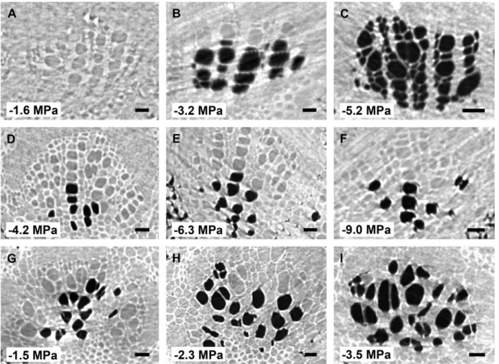
A total of 99 microCT scans were taken within the 72 h of beamtime, with leaves of the three species experiencing progres-sive levels of drought stress. Embolized conduits could easily be distinguished from functional, water-filled vessels in microCT scans due to the excellent phase contrast obtained: the lumina of functional vessels showed a grey appearance, while gas-filled vessels were considerably dark (Fig. 1). Secondary and third-order veins were frequently found near the midrib (Fig. 2). Observation of these veins along the 1.7 mm axial scan area was useful to find a connection between the vein and midrib, which frequently confirmed the status of the secondary vein order. In various cases, however, the scan area was too small to identify the vein connection, which made it impossible to distinguish secondary veins from third-order veins. As such, both second- and third-order veins were grouped together in our analyses.
Cutting the veins within the scan area induced embolism artificially (Supplementary Fig. S1), and enabled us to quantify the amount of embolized conduits at a particular water poten-tial before cutting (Figs 1, 2). The P50 values of leaf midribs were −2.01 ± 0.09 MPa for L. tulipifera, −2.87 ± 0.14 MPa for B. pendula, and −6.46 ± 0.24 MPa for L. nobilis (Fig. 1;
Table 1; Supplementary Table S1). The vulnerability to embol-ism in midribs versus second- and third-order veins differed across species (Fig. 3; Table 1). For L. tulipifera, the difference of 0.13 MPa observed between the midrib and second or third vein order was not significant according to visual inspection of the 95% confidence intervals, but larger differences were found for B. pendula (0.41 MPa) and L. nobilis (1.37 MPa), with the midrib showing lower embolism resistance than the second and third vein orders (Fig. 3; Table 1). The slope of the vulnerability curve fitted for midribs and second- or third-order veins was rather similar for B. pendula and L. tulipifera, showing strong overlapping of the confidence bands (Fig. 3;
Supplementary Table S1). However, the slope of the vulner-ability curve for the second- and third-order veins of L. nobilis was less steep (7.75 ± 2.38) than the midrib (14.76 ± 2.24) (Fig. 3; Supplementary Table S1).
Branch embolism resistance based on the centrifuge flow method showed P50 values of −1.8 ± 0.01 MPa for B. pendula and −2.1 ± 0.02 MPa for L. tulipifera (Fig. 3; Table 1).
Embolism resistance between leaves and branches was more or less within the same range, although differences were observed across the three species studied. In L.
tulipif-era, vulnerability curves of the midrib, second- and third-order
veins, and branch xylem overlapped in their confidence inter-vals, with their P50 values within a narrow range of −2.01 to −2.14 MPa (Fig. 3C). In B. pendula, confidence intervals of vulnerability curves of leaf and stem xylem did not overlap, with the leaf midrib P50 (−2.87 MPa) being 1 MPa more nega-tive than stem P50 (−1.8 MPa) (Fig. 3A). The opposite situation was found for L. nobilis, with P50 values being more negative in branch xylem (−9.2 MPa) than second and third vein orders (−7.83 MPa), and the least negative P50 values were for the leaf midrib (−6.46 MPa).
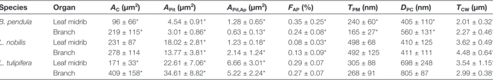
Xylem anatomy in relation to embolism resistance
Conduit area (AC) was wider in branches than in midribs of B. pendula and L. tulipifera (P=0.03 and P=0.01, respectively),
whereas no significant difference was observed for L. nobilis (P=0.81; Table 2). In B. pendula and L. nobilis, the conduit wall thickness (TCW) was significantly thicker in branches than mid-ribs (Table 2). Conduit wall thickness was 2.01 ± 0.32 µm in
leaf xylem of B. pendula and 2.27 ± 0.46 µm in stem xylem (P=0.04). For L. nobilis, conduit walls were 3.62 ± 0.49 µm thick in leaf midribs and 4.48 ± 0.64 µm thick in stem xylem (P<0.01). No significant difference was observed in conduit wall thickness between stems and midribs for L. tulipifera (P=0.70).
Significant differences were found in various pit characters between branch xylem and the xylem of leaf midribs (Table 2). The pit border surface area (APit) was larger in xylem of leaf midribs than branches for B. pendula and L. nobilis, but the opposite was found for L. tulipifera. Pit aperture surface areas (APit,Ap) were larger in the midrib than in the branches for B. pendula and L. tulipifera. Laurus nobilis, however, showed a
larger mean pit aperture area in its branches than in the leaf midrib. The ratio between the pit aperture surface area and pit membrane surface area (FAP) was slightly higher in the leaf midrib than in branches for B. pendula and L. tulipifera, with values between 0.24 ± 0.08 and 0.35 ± 0.25. This indicates that the pit aperture area represents about 30% of the total pit border area for both species. FAP values for L. nobilis were lower, with pit apertures occupying 8% of the pit border area in the midrib and 13% in branch xylem.
Pit chamber depth (DPC) was largest in L. tulipifera (698 and 805 nm for the midrib and branch xylem, respectively),
Fig. 1. MicroCT slices showing cross sections of xylem tissue in leaf midribs of Betula pendula (A, B, C), Laurus nobilis (D, E, F), and Liriodendron
tulipifera (G, H, I). Water-filled xylem conduits appear grey, while embolized conduits appear dark. The dark cells in the pith tissue of (G) do not represent
embolized conduits. The corresponding water potential for each image is shown in the bottom left corner of each image. The adaxial side of all leaves is at the top of each image. X, xylem tissue. Scale bar: 50 µm for all images.
followed by B. pendula (405 and 560 nm), and most shallow in
L. nobilis (410 and 411 nm). These data suggest that pit borders
in the leaf midrib were shallower than in branch xylem in the species studied, although this difference was only statistically significant for B. pendula (Table 2).
The pit membranes between neighbouring conduits showed a general similarity in ultrastructure between leaves and branches (Fig. 4). In all species and organs, pit membranes included dark (i.e. highly electron dense) particles, which were typically largest near the outermost layers of the pit mem-brane (Fig. 4F). Pit membranes of L. nobilis had an equally dark appearance in xylem of the branch and midrib (Fig. 4C,
D), while pit membranes were considerably darker in midribs than in branches of B. pendula (Fig. 4A, B). Pit membranes in L. tulipifera (Fig. 4E, F) were most transparent, showing a smaller amount of dark nanoparticles after treatment with OsO4 than B. pendula and L. nobilis.
Values of mean pit membrane thickness (TPM) were highest in L. nobilis, where they were close to 500 nm in the midrib and stem xylem, while thinner interconduit pit membranes occurred in L. tulipifera (305 and 268 nm for midribs and branches, respectively) and B. pendula (Table 2; Figs 4, 5). The thinnest pit membranes were observed in B. pendula, which was the only species showing significant difference in pit
membrane thickness between the leaf midribs (240 ± 60 nm) and branches (165 ± 27 nm) (Fig. 4A, B). This significant dif-ference in TPM for B. pendula was linked to a 1 MPa differ-ence in P50 values for both organs: the thickest pit membranes (240 ± 60 nm) in the midrib corresponded to a more negative
P50 value (−2.87 ± 0.14 MPa), while the thinner pit mem-branes (165 ± 27 nm) corresponded to a branch xylem P50 of −1.8 ± 0.01 MPa. When analysing TPM and P50 values for the three species, including branches and midribs, a significant negative correlation was found (r=−0.92, n=6, P<0.01).
Discussion
The hydraulic vulnerability segmentation hypothesis
Our microCT observations show that leaf xylem of the three angiosperm species studied is fairly resistant to embolism, with
P50 values around −2 MPa or less negative (Fig. 3; Table 1). High embolism resistance was especially found in leaf veins of L. nobilis, despite the fact that all microCT scans were per-formed on cut branches, which may underestimate embolism resistance due to artificial air entry. The relatively high resistance to xylem embolism observed agrees with recent studies based on direct, non-invasive methods, such as the optical method
Fig. 2. MicroCT slices showing cross sections of xylem conduits in second or third vein orders of Betula pendula (A, B, C), Laurus nobilis (D, E, F), and Liriodendron tulipifera (G, H, I), with reference to the corresponding xylem water potential measured. The exact vein order (second or third) could frequently not be determined based on microCT scans. The adaxial side of all leaves is at the top of each image. Scale bar: 20 µm for all images.
(Brodribb et al., 2016a, b; Rodriguez-Dominguez et al., 2018;
Lamarque et al., 2018) and earlier microCT observations of leaves (Bouche et al., 2016a; Skelton et al., 2017; Scoffoni et al., 2017a). Moreover, minor variation in xylem embolism resist-ance within a leaf may occur, because second- and third-order veins were found to show an equal or even higher embolism resistance than the midrib, as suggested previously (Brodribb et al., 2007; Scoffoni et al., 2017b).
Comparing xylem embolism resistance of leaves with stems was feasible because non-xylem leaf tissue was excluded in our microCT analyses. While the major advantage of microCT observations is direct, visual observation of embolism without artefacts, which may be introduced by manipulating a hydraulic system under negative pressure, the drawback of this method is that hydraulic conductance cannot be measured directly (Jansen et al., 2015). Instead, vulnerability curves based on microCT rely either on the assumption that all water-filled conduits are functional, or on the difference in filled versus embolized con-duits before and after cutting the scanned tissue (Supplementary Fig. S1). There is, however, convincing evidence that microCT observations agree well with hydraulic measurements for vari-ous species (Choat et al., 2010, 2016; Bouche et al., 2016b;
Nolf et al., 2017; Torres-Ruiz et al., 2017; Charrier et al., 2018;
Lamarque et al., 2018). Alternative, indirect methods to deter-mine xylem vulnerability to embolism in leaves should be interpreted with caution, because no direct comparison can be made between microCT observations (excluding the non-xylem tissue) and methods that estimate whole-leaf responses (i.e. including xylem and non-xylem tissue) to the decline in leaf hydraulic conductance (Bouche et al., 2016a; Trifiló et al., 2016; Scoffoni et al., 2017b; Scoffoni and Sack, 2017). Leaves of
L. tulipifera, for instance, were found to have a 50% reduction
of the whole-leaf hydraulic conductance at −1.2 MPa based on the rehydration kinetics method (Johnson et al., 2012; 2016). Therefore, the xylem tissue seems to be more resistant to dys-function than the outer xylem tissue, which provides the most important bottleneck causing a decline of leaf hydraulic con-ductance (Scoffoni et al., 2017b; Scoffoni and Sack, 2017).
A major result of this paper is that xylem embolism resist-ance is more or less similar between leaves and stems. This
finding was also supported by similarity in bordered pit mem-brane thickness, which is a useful determinant of drought-induced embolism resistance (Li et al., 2016). In particular, we found similar embolism resistance of xylem between leaves and branches for L. tulipifera. A previously published P50 value of −2.99 MPa for stems of L. tulipifera would indicate higher embolism resistance of stems based on the air-injection method (Johnson et al., 2011), but could also reflect intraspe-cific variation and/or methodological artefacts (Yin and Cai, 2018). A branch P50 value for seedlings of L. tulipifera based on microCT was −1.61 MPa (data not shown), which is slightly less negative than the P50 values for leaves and stems reported here (Table 3), suggesting that leaf xylem of this species is not more vulnerable to embolism than are branches.
Fig. 3. Vulnerability to drought-induced embolism for xylem tissue of different organs in three woody angiosperm species: Betula pendula (A), Laurus nobilis (B), and Liriodendron tulipifera (C). Percentage loss of conductance (PLC; leaves) or conductivity (branches) is plotted against stem water potential (MPa). Theoretical PLC values were determined based on microCT for leaf midribs and second or third vein orders, while hydraulic PLC values were based on a flow-centrifuge method for branches of B. pendula and L. tulipifera. Branch vulnerability curves were based on a flow-centrifuge method; stem data of L. nobilis were from Lamarque et al. (2018) and based on branches from different specimens than the one used for leaf sampling. (This figure is available in colour at
JXB online.) Table 1. Hydraulic vulnerability to embolism (P50, MPa) in leaf and
stem xylem of Betula pendula, Laurus nobilis, and Liriodendron tulipifera based on X-ray computed tomography (leaf midrib, second and third vein order) and a flow-centrifuge method (branch)
Species P50 (MPa)
X-ray microtomography Centrifuge method Leaf midrib Second and
third vein orders
Branch Betula pendula −2.87 ± 0.14 −3.28 ± 0.31 −1.80 ± 0.01
Laurus nobilis −6.46 ± 0.24 −7.83 ± 0.74 −9.21 ± 0.23a
Liriodendron tulipifera −2.01 ± 0.09 −2.14 ± 0.17 −2.10 ± 0.02
All measurements were based on branches and leaves from a single specimen per species. Fitted value ±SE.
An even higher embolism resistance for leaves than branches was found for B. pendula, which is the opposite of what would be predicted based on the hydraulic vulnerability segmenta-tion hypothesis. Previously published P50 values for branches of B. pendula were in good agreement with our centrifuge-flow
measurements, with values between −1.8 MPa and −2.3 MPa (Cochard et al., 2005; González-Muñoz et al., 2018). Moreover, the thicker pit membranes in leaves than in branches (Fig. 5) are in line with their high leaf embolism resistance.
Lower xylem embolism resistance in leaves than branches was found for Vitis vinifera (Charrier et al., 2016; Hochberg et al., 2016) and olive (Rodriguez-Dominguez et al., 2018), and could be suggested for L. nobilis, although our data should be interpreted with caution because of the use of different specimens. Stem P50 values of L. nobilis reported in literature are highly variable, ranging from −1.5 to −3.5 MPa (Salleo and Lo Gullo, 1993; Salleo et al., 2009; Nardini et al., 2017), but these values may underestimate embolism resistance due to
Fig. 4. Transmission electron microscopy images of interconduit pit borders and pit membranes in Betula pendula (A, B), Laurus nobilis (C, D), and
Liriodendron tulipifera (E, F) from branch xylem (A, C, E) and leaf midribs (B, D, F). Pit apertures are (partly) visible when the section was cut through the
centre of the pit border (A, C, D, E). Scale bars: 500 nm.
Table 2. Wood anatomical features related to conduit and pit characteristics for xylem of branches and leaf midribs in Betula pendula,
Laurus nobilis, and Liriodendron tulipifera
Species Organ AC (µm2) APit (µm2) APit,Ap (µm2) FAP (%) TPM (nm) DPC (nm) TCW (µm)
B. pendula Leaf midrib 96 ± 66* 4.54 ± 0.91* 1.28 ± 0.65* 0.35 ± 0.25* 240 ± 60* 405 ± 110* 2.01 ± 0.32* Branch 219 ± 115* 3.01 ± 0.86* 0.63 ± 0.13* 0.24 ± 0.08* 165 ± 27* 560 ± 131* 2.27 ± 0.46*
L. nobilis Leaf midrib 231 ± 87 18.02 ± 2.81* 1.23 ± 0.18* 0.08 ± 0.03* 498 ± 68 410 ± 125 3.62 ± 0.49* Branch 278 ± 114 13.77 ± 3.81* 2.14 ± 1.24* 0.13 ± 0.09* 492 ± 125 411 ± 111 4.48 ± 0.64*
L. tulipifera Leaf midrib 171 ± 33* 22.61 ± 7.06* 6.66 ± 3.01* 0.29 ± 0.07 305 ± 88 698 ± 248 3.54 ± 1.15* Branch 409 ± 158* 34.61 ± 8.82* 5.22 ± 2.24* 0.27 ± 0.07 268 ± 91 805 ± 87 2.99 ± 0.38*
AC, conduit area; APit, bordered pit area; APit,Ap, pit aperture area; DPC, pit chamber depth; FAP, pit aperture fraction; TCW, interconduit wall thickness; TPM, interconduit pit membrane thickness. Values are given as mean ±SD. *Significant differences between branches and midribs.
artefacts (Cochard et al., 2015). The leaf P50 values obtained in this study for L. nobilis (−6.46 and −7.83 MPa for the midrib and second-/third-order veins, respectively) are much more negative than these earlier records, but within the same order of magnitude as the −7.94 to −9.21 MPa values based on three different methods: (i) microCT of stems from intact plants, (ii) the optical method of leaf veins, and (iii) the centrifuge-flow method on 1 m-long stem segments (Lamarque et al., 2018). While the latter authors showed that embolism resistance was not different between stem and leaf veins of L. nobilis when using samples from the same plant, embolism resistance of leaves based on microCT could be interpreted as being equally vulnerable to that of stems.
The relatively high embolism resistance observed in leaves does not imply that hydraulic segmentation may not occur without any difference in xylem embolism resistance. First, even if the stem and leaf xylem show equal resistance to embolism, or if the leaves show a higher resistance than the stems, leaves can still provide hydraulic segmentation because the outer xylem tissue (i.e. the non-vascular leaf tissue) can act as a hydraulic bottleneck in the system, protecting the xylem in the vascular bundles (Scoffoni et al., 2017a). In this way, leaf xylem would be less expendable as outside xylem, which may recover more easily from drought-induced decline due to aquaporin activ-ity (Maurel et al., 2016; Groszmann et al., 2017). Recovery of embolized xylem conduits in leaves, on the other hand, shows physical constraints and is unlikely to represent a dynamic and fast process on a daily basis (e.g. Mayr et al., 2014; Bouche et al., 2016a). Second, leaves can be at more negative water poten-tials than stems via transpiration and/or cuticular conductance.
Therefore, leaves may still experience xylem embolism before stems, creating a hydraulic fuse for stem xylem (Tyree and Ewers, 1991; Wolfe et al., 2016). Stem and leaf xylem water potential measurements would be required to estimate hydraulic safety margins (defined as the most minimum xylem water poten-tial experienced by a plant in the field minus the P50 value of a tissue; Meinzer et al., 2009; Choat et al., 2012) and to put the findings of this paper in an ecological context. Based on the minimum xylem water potentials reported in literature, which are −1.19 for L. tulipifera (Johnson et al., 2012), −2.64 for
B. pendula (Cochard et al., 2005), and −4.2 MPa for L. nobilis (Lamarque et al., 2018), hydraulic safety margins would be posi-tive for L. nobilis and L. tulipifera, except for branches of
B. pen-dula. A positive hydraulic safety margin would indicate that
embolism levels corresponding to P50 are unlikely for branches and leaves in the field, while a negative hydraulic safety mar-gin for B. pendula would suggest that embolism in the field is slightly higher for this species. The lab-based data presented in this paper, however, do not allow us to make any firm statement about embolism occurrence in the field.
Moreover, future work should focus on whole plant model-ling that takes into account the vulnerability of each organ to embolism to better understand the degree of ‘true’ segmentation across species. While it is clear that xylem conduits form a mor-phological and developmental continuum over long distances, from the roots to the minor leaf veins, anatomical bottlenecks are well known to occur at nodes, bifurcations, and stem–petiole abscission zones, making conduit connections non-randomly distributed and potentially segmented (Salleo et al., 1984; André et al., 1999; André, 2005; Rančić et al., 2010; Wolfe et al., 2016).
Embolism resistance in relation to xylem anatomy
Additional support for relatively high embolism resistance in leaves is provided by the interconduit pit membrane thick-ness, which was similar between both organs in L. nobilis and
L. tulipifera, but significantly higher in leaves than in branches
for B. pendula. Thick pit membranes could provide higher hydraulic safety due to narrow pore volumes and/or long pore pathways, which would increase the number of constrictions within this pathway and therefore bubble snap-off (Roof, 1970;
Kovscek et al., 2007; Schenk et al., 2017). The difference in pit membrane thickness between leaves and stems of B. pendula may explain the 1 MPa difference in P50 between branches and midribs of this species. While intervessel pit membrane thick-ness was found to be a determinant of P50 (Li et al., 2016), these findings also suggest that variation in pit membrane thickness does not always correspond to considerable changes in P50, at least not among organs within a plant. Given the differences in conduit size between leaves and stems, especially in
L. tulip-ifera and B. pendula, our findings also suggest that pit
mem-brane thickness and the associated resistance to air-seeding is not affected by conduit size, which would be expected based on the assumption that wide conduits are more vulnerable to xylem embolism than narrow ones (Hargrave et al., 1994;
Sperry et al., 1994; Tyree and Zimmermann, 2002).
To our knowledge, our TEM observations of the midrib xylem are the first to show that post-fixation with OsO4 results Fig. 5. Comparison of interconduit pit membrane thickness (nm) for xylem
of leaf midribs (dark grey bars) and branches (light grey bars) in Betula
pendula, Laurus nobilis, and Liriodendron tulipifera. The bars show mean
values, and the error bars the standard errors. Different letters indicate intraspecific and interspecific statistical significance.
in a dark staining of interconduit pit membranes in leaves, simi-lar to what has been found for stems and branches (Schmid and Machado, 1968; Jansen et al., 2009; Schenk et al., 2017, 2018;
Zhang et al., 2017). This staining reaction is most likely caused by binding of osmium particles to double carbon bonds in unsatur-ated fatty acid chains of lipids (Riemersma, 1968; Schenk et al., 2017, 2018). More research of xylem tissue across different organs is needed to address the functional role of lipid surfactants in pit membranes and their potential effect on air-seeding (Jansen and Schenk, 2015; Schenk et al., 2015, 2017, 2018; Jansen et al., 2018).
Despite the limited number of species studied, some anatomical features could explain the high embolism resistance in
L. nobi-lis. Compared with B. pendula and L. tulipifera, L. nobilis showed
the thickest interconduit walls (TCW), thickest interconduit pit membranes (TPM), most shallow pit chambers (DPC), and a low pit aperture fraction (FAP, i.e. the ratio between the pit aperture surface area to the pit border surface area). Thick intervessel walls can be suggested to increase hydraulic safety by avoiding wall col-lapse under negative pressure (Hacke et al., 2001), while shallow pit chambers and a low pit aperture fraction could provide mech-anical support for aspirated pit membranes, resulting in increased air-seeding pressures (Choat et al., 2008; Lens et al., 2011). The relatively low (8–13%) pit aperture fraction of L. nobilis had a simi-lar order of magnitude to the pit aperture fractions reported in
Acer (Lens et al., 2011), but more data on additional species would be needed to provide a functional explanation of this feature.
Conclusions
In summary, the findings of this paper raise questions about the assumption that leaf xylem is the main driving mechanism behind the hydraulic vulnerability segmentation hypothesis, because embolism resistance in leaves and stems can be similar (L. tulipifera), possibly higher in leaves than stems (B. pendula), or possibly lower in leaves than stems (L. nobilis). Further work is required to test whether the underlying mechanism behind hydraulic segmentation between plant organs is species-spe-cific across a wide range of angiosperms, and to what extent there is intraspecific variation. We recommend that sufficient care should be taken in distinguishing xylem embolism resist-ance in leaf veins from whole-leaf hydraulic measurements. Finally, anatomical features such as interconduit pit membranes may provide additional evidence for embolism resistance and could improve our poor understanding of the mechanisms behind air-seeding. More detailed anatomical observations are also needed to investigate whether anatomical segmentation or compartmentalization occurs at the root to the stem tran-sition, at the stem–petiole tissue level, and/or along different vein orders or vein connections (Cochard et al., 1992; Hacke and Sauter, 1996).
Supplementary data
Supplementary data are available at JXB online.
Fig. S1. MicroCT slices showing cross sections of xylem tissue in leaf midribs of Betula pendula, Laurus nobilis, and
Liriodendron tulipifera.
Fig. S2. Three dimensional reconstruction based on microCT showing a cross section through a leaf of Liriodendron
tulipifera at −2.3 MPa.
Table S1. Statistical comparison of the theoretical hydraulic loss of conductance between different organs and species.
Acknowledgements
We acknowledge the Paul Scherrer Institut, Villigen, Switzerland for provision of synchrotron radiation beamtime at the TOMCAT beam-line X02DA of the SLS and would like to thank Dr Iwan Jerjen and Dr Anne Bonnin for assistance. The Botanical Garden of Ulm University is acknowledged for providing plant material. The Electron Microscopy Section of Ulm University is acknowledged for technical support with electron microscopy. YZ acknowledges financial support from the Chinese Scholarship Council (CSC). LJL was supported by an IdEx Bordeaux International Post-doctoral program fellowship (UB101 CR1024-R s/CR1024-6M), and CS hold a Humboldt Research Fellowship for Postdoctoral Researchers. MN and BC acknowledge travel funding from the International Synchrotron Access Program managed by the Australian Synchrotron.
References
André J-P. 2005. Vascular organization of angiosperms. A new vision. Enfield, NH, USA: Science Publishers.
André J-P, Catesson AM, Liberman M. 1999. Characters and origin of vessels with heterogenous structure in leaf and flower abscission zones. Canadian Journal of Botany 77, 253–261.
Böhme J. 1893. Capillarität und Saftsteigen. Berichte der Deutschen botanischen Gesellschaft 11, 203–212.
Bouche PS, Delzon S, Choat B, et al. 2016a. Are needles of Pinus
pinaster more vulnerable to xylem embolism than branches? New insights
from X-ray computed tomography. Plant, Cell & Environment 39, 860–870. Bouche PS, Jansen S, Sabalera JC, Cochard H, Burlett R, Delzon S. 2016b. Low intra-tree variability in resistance to embolism in four Pinaceae species. Annals of Forest Science 73, 681–689.
Brodersen CR, McElrone AJ, Choat B, Lee EF, Shackel KA, Matthews MA. 2013. In vivo visualizations of drought-induced embolism spread in
Vitis vinifera. Plant Physiology 161, 1820–1829.
Brodribb TJ, Bienaimé D, Marmottant P. 2016a. Revealing catastrophic failure of leaf networks under stress. Proceedings of the National Academy of Sciences, USA 113, 4865–4869.
Brodribb TJ, Feild TS, Jordan GJ. 2007. Leaf maximum photosynthetic rate and venation are linked by hydraulics. Plant Physiology 144, 1890–1898. Brodribb TJ, Skelton RP, McAdam SA, Bienaimé D, Lucani CJ, Marmottant P. 2016b. Visual quantification of embolism reveals leaf vulnerability to hydraulic failure. New Phytologist 209, 1403–1409.
Buckley TN, John GP, Scoffoni C, Sack L. 2017. The sites of evaporation within leaves. Plant Physiology 173, 1763–1782.
Charrier G, Delzon S, Domec JC, et al. 2018. Drought will not leave your glass empty: low risk of hydraulic failure revealed by long-term drought observations in world’s top wine regions. Science Advances 4, eaao6969.
Charrier G, Torres-Ruiz JM, Badel E, et al. 2016. Evidence for hydraulic vulnerability segmentation and lack of xylem refilling under tension. Plant Physiology 172, 1657–1668.
Choat B, Badel E, Burlett R, Delzon S, Cochard H, Jansen S. 2016. Noninvasive measurement of vulnerability to drought-induced embolism by X-ray microtomography. Plant Physiology 170, 273–282.
Choat B, Brodersen CR, McElrone AJ. 2015. Synchrotron X-ray microtomography of xylem embolism in Sequoia sempervirens saplings during cycles of drought and recovery. New Phytologist 205, 1095–1105. Choat B, Cobb AR, Jansen S. 2008. Structure and function of bordered pits: new discoveries and impacts on whole-plant hydraulic function. New Phytologist 177, 608–625.
Choat B, Drayton WM, Brodersen C, Matthews MA, Shackel KA, Wada H, McElrone AJ. 2010. Measurement of vulnerability to water stress-induced cavitation in grapevine: a comparison of four techniques applied to a long-vesseled species. Plant, Cell & Environment 33, 1502–1512. Choat B, Jansen S, Brodribb TJ, et al. 2012. Global convergence in the vulnerability of forests to drought. Nature 491, 752–755.
Cochard H, Bréda N, Granier A, Aussenac G. 1992. Vulnerability to air embolism of three European oak species (Quercus petraea (Matt) Liebl, Q
pubescens Willd, Q robur L). Annales des Sciences Forestières 49, 225–233. Cochard H, Damour G, Bodet C, Tharwat I, Poirier M, Améglio T. 2005. Evaluation of a new centrifuge technique for rapid generation of xylem vulnerability curves. Physiologia Plantarum 124, 410–418.
Cochard H, Delzon S, Badel E. 2015. X-ray microtomography (micro-CT): a reference technology for high-resolution quantification of xylem embolism in trees. Plant, Cell & Environment 38, 201–206.
Dixon HH. 1914. Transpiration and the ascent of sap in plants. London: Macmillan and Co., Ltd.
Dixon MA, Tyree MT. 1984. A new stem hygrometer, corrected for temperature gradients and calibrated against the pressure bomb. Plant, Cell & Environment 7, 693–697.
Domec J-C, Warren JM, Meinzer FC, Lachenbruch B. 2009. Safety factors for xylem failure by implosion and air-seeding within roots, trunks and branches of young and old conifer trees. International Association of Wood Anatomists Journal 30, 101–120.
Duursma R, Choat B. 2017. Fitplc – an R package to fit hydraulic vulnerability curves. Journal of Plant Hydraulics 4, 2.
González-Muñoz N, Sterck F, Torres-Ruiz JM, et al. 2018. Quantifying
in situ phenotypic variability in the hydraulic traits of four tree species across
their distribution range in Europe. PLoS One 13, e0196075.
Groszmann M, Osborn HL, Evans JR. 2017. Carbon dioxide and water transport through plant aquaporins. Plant, Cell & Environment 40, 938–961. Hacke U, Sauter JJ. 1996. Drought-induced xylem dysfunction in petioles, branches, and roots of Populus balsamifera L. and Alnus glutinosa (L.) Gaertn. Plant Physiology 111, 413–417.
Hacke UG. 2014. Irradiance-induced changes in hydraulic architecture 1. Botany 92, 437–442.
Hacke UG, Sperry JS, Pockman WT, Davis SD, McCulloh KA. 2001. Trends in wood density and structure are linked to prevention of xylem implosion by negative pressure. Oecologia 126, 457–461.
Hales S. 1727. Vegetable staticks, or, An account of some statical experiments on the sap in vegetables: being an essay towards a natural history of vegetation: also, a specimen of an attempt to analyse the air, by a great variety of chymio-statical experiments; which were read at several meetings before the Royal Society. London: W. & J. Innys and T. Woodward. Hargrave KR, Kolb KJ, Ewers FW, Davis SD. 1994. Conduit diameter and drought-induced embolism in Salvia mellifera Greene (Labiatae). New Phytologist 126, 695–705.
Hochberg U, Albuquerque C, Rachmilevitch S, Cochard H, David-Schwartz R, Brodersen CR, McElrone A, Windt CW. 2016. Grapevine petioles are more sensitive to drought induced embolism than stems: evidence from in vivo MRI and microcomputed tomography observations of hydraulic vulnerability segmentation. Plant, Cell & Environment 39, 1886–1894.
Holbrook NM, Burns MJ, Field CB. 1995. Negative xylem pressures in plants: a test of the balancing pressure technique. Science 270, 1193–1194. Jansen S, Choat B, Pletsers A. 2009. Morphological variation of intervessel pit membranes and implications to xylem function in angiosperms. American Journal of Botany 96, 409–419.
Jansen S, Klepsch MM, Li S, Kotowska MM, Schiele S, Zhang Y, Schenk HJ. 2018. Challenges in understanding air-seeding in angiosperm xylem. Acta Horticulturae (in press).
Jansen S, Nardini A. 2014. From systematic to ecological wood anatomy and finally plant hydraulics: are we making progress in understanding xylem evolution? New Phytologist 203, 12–15.
Jansen S, Schenk HJ. 2015. On the ascent of sap in the presence of bubbles. American Journal of Botany 102, 1561–1563.
Jansen S, Schuldt B, Choat B. 2015. Current controversies and challenges in applying plant hydraulic techniques. New Phytologist 205, 961–964.
Jinagool W, Lamarque L, Delmas M, Delzon S, Cochard H, Herbette S. 2018. Is there variability for xylem vulnerability to cavitation in Walnut tree cultivars and species (Juglans spp.)? HortScience 53, 132–137.
Johnson DM, McCulloh KA, Meinzer FC, Woodruff DR, Eissenstat DM. 2011. Hydraulic patterns and safety margins, from stem to stomata, in three eastern U.S. tree species. Tree Physiology 31, 659–668.
Johnson DM, McCulloh KA, Woodruff DR, Meinzer FC. 2012. Hydraulic safety margins and embolism reversal in stems and leaves: why are conifers and angiosperms so different? Plant Science 195, 48–53.
Johnson DM, Wortemann R, McCulloh KA, Jordan-Meille L, Ward E, Warren JM, Palmroth S, Domec JC. 2016. A test of the hydraulic vulnerability segmentation hypothesis in angiosperm and conifer tree species. Tree Physiology 36, 983–993.
Kovscek AR, Tang G-Q, Radke CJ. 2007. Verification of roof snap off as a foam-generation mechanism in porous media at steady state. Colloids and Surfaces A: Physicochemical and Engineering Aspects 302, 251–260. Lamarque LL, Corso D, Torres-Ruiz JM, et al. 2018. An inconvenient truth about xylem resistance to embolism in the model species for refilling Laurus
nobilis L. Annals of Forest Science, doi: 10.1007/s13595-018-0768-9.
Lamy JB, Delzon S, Bouche PS, Alia R, Vendramin GG, Cochard H, Plomion C. 2014. Limited genetic variability and phenotypic plasticity detected for cavitation resistance in a Mediterranean pine. New Phytologist 201, 874–886.
Lens F, Sperry JS, Christman MA, Choat B, Rabaey D, Jansen S. 2011. Testing hypotheses that link wood anatomy to cavitation resistance and hydraulic conductivity in the genus Acer. New Phytologist 190, 709–723. Li S, Klepsch M, Jansen S, Schmitt M, Lens F, Karimi Z, Schuldt B, Espino S, Schenk HJ. 2016. Intervessel pit membrane thickness as a key determinant of embolism resistance in angiosperm xylem. IAWA Journal 37, 152–171.
Losso A, Anfodillo T, Ganthaler A, Kofler W, Markl Y, Nardini A, Oberhuber W, Purin G, Mayr S. 2018. Robustness of xylem properties in conifers: analyses of tracheid and pit dimensions along elevational transects. Tree Physiology 38, 212–222.
Maurel C, Verdoucq L, Rodrigues O. 2016. Aquaporins and plant transpiration. Plant, Cell & Environment 39, 2580–2587.
Mayr S, Schmid P, Laur J, Rosner S, Charra-Vaskou K, Dämon B, Hacke UG. 2014. Uptake of water via branches helps timberline conifers refill embolized xylem in late winter. Plant Physiology 164, 1731–1740. Meinzer FC, Johnson DM, Lachenbruch B, McCulloh KA, Woodruff DR. 2009. Xylem hydraulic safety margins in woody plants: coordination of stomatal control of xylem tension with hydraulic capacitance. Functional Ecology 23, 922–930.
Nardini A, Savi T, Losso A, Petit G, Pacilè S, Tromba G, Mayr S, Trifilò P, Lo Gullo MA, Salleo S. 2017. X-ray microtomography observations of xylem embolism in stems of Laurus nobilis are consistent with hydraulic measurements of percentage loss of conductance. New Phytologist 213, 1068–1075.
Nolf M, Lopez R, Peters JM, Flavel RJ, Koloadin LS, Young IM, Choat B. 2017. Visualization of xylem embolism by X-ray microtomography: a direct test against hydraulic measurements. New Phytologist 214, 890–898. Paganin D, Mayo SC, Gureyev TE, Miller PR, Wilkins SW. 2002. Simultaneous phase and amplitude extraction from a single defocused image of a homogeneous object. Journal of Microscopy 206, 33–40. Pammenter NW, Van der Willigen C. 1998. A mathematical and statistical analysis of the curves illustrating vulnerability of xylem to cavitation. Tree Physiology 18, 589–593.
Pivovaroff AL, Sack L, Santiago LS. 2014. Coordination of stem and leaf hydraulic conductance in southern California shrubs: a test of the hydraulic segmentation hypothesis. New Phytologist 203, 842–850.
Pockman WT, Sperry JS, O’Leary JW. 1995. Sustained and significant negative water pressure in xylem. Nature 378, 715–716.
Rančić D, Quarrie SP, Radosević R, Terzić M, Pećinar I, Stikić R, Jansen S. 2010. The application of various anatomical techniques for studying the hydraulic network in tomato fruit pedicels. Protoplasma 246, 25–31.
Riemersma JC. 1968. Osmium tetroxide fixation of lipids for electron microscopy. A possible reaction mechanism. Biochimica et Biophysica Acta 152, 718–727.