Hesperidin contributes to the vascular protective effects of orange
juice: a randomized crossover study in healthy volunteers
1–3Christine Morand, Claude Dubray, Dragan Milenkovic, Delphine Lioger, Jean Franc¸ois Martin, Augustin Scalbert, and Andrzej Mazur
ABSTRACT
Background: Although numerous human studies have shown con-sistent effects of some polyphenol-rich foods on several intermedi-ate markers for cardiovascular diseases, it is still unknown whether their action could be specifically related to polyphenols.
Objective:We investigated the effect of orange juice and its major
flavonoid, hesperidin, on microvascular reactivity, blood pressure, and cardiovascular risk biomarkers through both postprandial and chronic intervention studies.
Design:Twenty-four healthy, overweight men (age 50–65 y) were
included in a randomized, controlled, crossover study. Throughout the three 4-wk periods, volunteers daily consumed 500 mL orange juice, 500 mL control drink plus hesperidin (CDH), or 500 mL control drink plus placebo (CDP). All measurements and blood col-lections were performed in overnight-fasted subjects before and after the 4-wk treatment periods. The postprandial study was con-ducted at the beginning of each experimental period.
Results: Diastolic blood pressure (DBP) was significantly lower after 4 wk consumption of orange juice or CDH than after consump-tion of CDP (P = 0.02), whereas microvascular endothelium-related reactivity was not significantly affected when measured after an overnight fast. However, both orange juice and CDH ingestion sig-nificantly improved postprandial microvascular endothelial
reactiv-ity compared with CDP (P, 0.05) when measured at the peak of
plasma hesperetin concentration.
Conclusions: In healthy, middle-aged, moderately overweight men, orange juice decreases DBP when regularly consumed and post-prandially increases endothelium-dependent microvascular reactiv-ity. Our study suggests that hesperidin could be causally linked to the beneficial effect of orange juice. This trial is registered at
clin-icaltrials.gov as NCT00983086. Am J Clin Nutr 2011;93:73–80.
INTRODUCTION
A growing number of epidemiologic studies have consistently shown a protective effect of polyphenol-rich foods (fruit, tea, wine, and cocoa or chocolate) against cardiovascular diseases (CVDs) (1–4). This evidence is supported by results from nu-merous studies conducted in animal models, with nutritionally realistic levels of isolated flavonoids (5, 6), and in humans with flavonoid-rich foods. The most convincing clinical data are available for only a few flavonoid-rich products, probably be-cause they have been more extensively studied than others (7). Thus, the consumption of chocolate and cocoa, green tea, and soy
protein isolate has been reported to exert beneficial effects on some intermediate risk factors for CVD, such as LDL cholesterol, blood pressure (BP), and endothelial function (7). Only a few clinical trials have dealt with the oral administration of chemi-cally pure flavonoids, making it difficult to dissociate the specific effect of flavonoid compounds from that of the entire food. However, the ingestion of pure dietary epicatechin and epi-gallocatechin gallate (EGCG) has been causally linked to the beneficial effect of catechin-rich cocoa and tea on vascular function (8–10).
Clinical data that examine the potential efficacy of flavonoids on CVD risk factors are lacking for some flavonoid subclasses commonly consumed as part of a normal diet, such as antho-cyanins and flavanones (11). A focus on flavanones is particularly relevant because of their high content in citrus and because of high citrus fruit consumption, particularly orange juice, world-wide. According to the Phenol-Explorer database (12), a single glass of orange juice (150 mL) may contain’90 mg flavanone glycosides. Hesperidin (hesperetin-7-O-rutinoside) represents 90% of total flavanones in the orange; the remaining flavanones comprise narirutin (naringenin-7-O-rutinoside). Moreover, fla-vanones are among the flavonoid compounds displaying the highest bioavailability (13).
The consumption of citrus fruit has been associated with a lower risk of acute coronary events and stroke (14, 15). From clinical data, citrus juice consumption reduces oxidative DNA damage in blood cells (16) and improves plasma concentrations of markers of inflammation and oxidative stress (17–19). In addition, the consumption of citrus juices improves lipemia in men with previous coronary bypass surgery (20). Furthermore, in hypertensive subjects, the consumption of flavanone-rich grapefruit juice exerts a significant beneficial effect on BP (21).
1
From INRA, UMR 1019, UNH, CRNH Auvergne, Clermont-Ferrand, France (CM, DM, DL, JFM, AS, and AM); Clermont Universite´, Universite´ d’Auvergne, Unite´ de Nutrition Humaine, Clermont-Ferrand (CM, DM, JFM, AS, and AM); and Inserm, CIC 501, Clermont-Ferrand (CD).
2
Supported by a grant from the Florida Department of Citrus (to CM). The funders had no role in study design, data collection and analysis, de-cision to publish, or preparation of the manuscript.
3
Address correspondence to C Morand, UMR1019, Unite´ de Nutrition Humaine, INRA, Centre Clermont-Ferrand–Theix, 63122 St Gene`s Cham-panelle, France. E-mail: christine.morand@clermont.inra.fr.
Received July 13, 2010. Accepted for publication October 26, 2010. First published online November 10, 2010; doi: 10.3945/ajcn.110.004945.
Am J Clin Nutr 2011;93:73–80. Printed in USA.Ó 2011 American Society for Nutrition 73
Further data are needed to determine the specific role of fla-vanones in the health benefit induced by the consumption of citrus foods. Therefore, the aim of the present study was to compare the effects of orange juice with those of pure hesperidin on endothelial microvascular reactivity, BP, and systemic markers linked to CVD risk in healthy men to determine whether hesperidin contributes to the protective effects of orange juice.
SUBJECTS AND METHODS Subjects
Twenty-four overweight male volunteers, 50–65 y of age, were recruited by newspaper advertisements. All subjects were healthy and had no evidence of chronic disease. Exclusion criteria were as follows: use of medications, antioxidants, or vitamin supple-ments; smoking; alcohol consumption (.20 g alcohol/d); intense physical activity (.5 h/wk); intestinal disorders; or vegetarian-ism. Subjects presenting with a high consumption of flavonoid-rich beverages, such as tea, herb tea, coffee, wine, cocoa, and fruit juice, were also excluded when the daily consumption of one or more of these products exceeded 500 mL (estimated from a food-frequency questionnaire).The study was approved by and performed under the guidelines of the French Human Ethics Committee of the South East VI. Written informed consent was obtained from each of the subjects before commencement of the study. All subjects completed the study except for one who was excluded because of underlying disease.
Test drinks and products
Orange juice from concentrate and the analysis of its con-stituents (Table 1) were provided by the Florida Department of Citrus (Lake Alfred, FL). Subjects were instructed to keep the orange juice in their home refrigerator until consumption. As shown in Table 1, the flavanone content in 500 mL orange juice (corresponding to the daily administered dose) was 292 mg
hesperidin and 47.5 mg narirutin. The control drink (CD) had a sugar composition similar to that of orange juice, and 500 mL of each drink contained 180 and 194 kcal, respectively (Table 1). Volunteers were asked to reconstitute the control drink from a sachet of caster sugar, distributed at the beginning of the experimental period. Each sachet contained 22.5 g of the sugar mix, which had to be dissolved in 250 mL tap water just before drinking. Hesperidin capsules were filled with the orange bi-oflavonoid complex (OBC; 90%), containing 99.2% hesperidin (Nutrafur, Murcia, Spain). Each capsule contained 146 mg OBC, corresponding to the amount of hesperidin present in 250 mL orange juice. The placebo capsules consisted of starch (146 mg/capsule) and were visually identical to those con-taining hesperidin.
Study design
The main goal of the present study was to examine the acute and chronic effects of orange juice consumption on functional and systemic markers associated with cardiovascular risk and to determine the specific role of hesperidin, its major flavonoid, in the observed effects. For that purpose, the study was a controlled, randomized, crossover 4-wk dietary intervention trial with 3 treatment groups: control drink + placebo (CDP), control drink + hesperidin (CDH), and orange juice. For the orange juice in-tervention, the trial was open, but it was double-blinded for CDP and CDH. There was a 3-wk washout period between each treatment. The order of administration was determined by using computer-generated random numbers.
Each subject was assigned successively to 3 periods of 4-wk dietary treatments with daily consumption of either 1) 500 mL of a control drink plus 2 placebo capsules (2· 146 mg starch), 2) 500 mL of the control drink plus 2 capsules of pure hesperidin (2 · 146 mg), or 3) 500 mL orange juice, which naturally provided 292 mg hesperidin. At home, subjects were instructed to divide the total daily dose into 2 equal intakes (250 mL beverage plus one capsule, according to experimental group), one with the breakfast and the other one at lunchtime.
During the protocol, volunteers made 6 visits to the clinical research unit on the first (visits 1, 3, and 5) and last (visits 2, 4, and 6) day of each of experimental period for cardiovascular measurements and biological sample collection. In practice, for the 4-wk chronic study, overnight-fasted subjects arrived at the clinical research unit between 0800 and 0900. After a 10-min rest while supine, BP and microvascular reactivity were measured, and a blood sample was collected. For each intervention period, volunteers were asked to collect urine over the 24-h period before their last visit. On the first day of each experimental period (visits 1, 3, and 5), acute response to the corresponding dietary treatment was examined by measuring microvascular reactivity after 6 h, corresponding to the reported maximal flavanone plasma concentration (22). Briefly, after measurements and blood sampling were performed, volunteers had to consume the complete daily dose of the treatment together with a breakfast consisting of bread, butter, and ham (280 kcal) within 15 min. Subjects were then allowed to consume only tap water. Six hours after the consumption of breakfast, blood was collected and microvascular reactivity was immediately measured. Vol-unteers received a light meal before leaving the clinical center.
TABLE 1
Main constituents and phytochemical contents of the test drinks (500 mL) used in the study1
Orange juice Control drink
Total carbohydrates (g)2 45 45
Total organic acids (g)3 4.72 —
Total minerals (g)4 1.31 — Total pectins (mg) 568 — Total vitamin C (mg) 180 — a-Tocopherol (mg) 1.05 — Vitamin B-9 (mg) 0.085 — Total carotenoids (mg)5 0.135 — Flavonoids (mg) 341.9 — Hesperidin 292 — Narirutin 47.5 — Others6 2.4 — Energy (kcal) 194 180 1
All values are means.
2
Includes sucrose (50%), glucose (25%), and fructose (25%).
3
Includes citric acid, isocitric acid, and malic acid.
4
Includes Ca, Mg, Cu, Fe, Na, P, K, and Zn.
5
Includeb-carotene, b-cryptoxanthin, and lutein.
6
Includes sinensitin, nobiletin, and tangeretin.
During the entire study period, subjects were asked to maintain their lifestyle and their normal dietary habits with the exception that they were instructed to completely refrain from consuming citrus-containing foods and to limit their total intake of flavonoid-rich beverages (tea, coffee, cocoa, wine, fruit juice) to,200 mL/d. To assess whether the volunteers maintained their dietary habits throughout the study period, a 3-d food record was performed during the last week of each intervention period. To assess dietary compliance, subjects kept a diary in which consumption of the study products (drinks and capsules) was recorded daily, and they were asked to return the unconsumed products at the end of each period. Subjects used the same diary to record occurrences and use of medications. The diaries were collected and checked at the end of each experimental period.
Processing and analysis of biological samples
Fasting blood samples and 24-h urine specimens were collected before and after each intervention period (CDP, CDH, and orange juice). Venous blood was collected into evacuated tubes con-taining EDTA or Na-heparin. Plasma was immediately isolated, processed, and stored at280°C until analysis. Plasma samples for vitamin C analysis were immediately transferred into dark Eppendorf tubes containing 2 volumes of meta-phosphoric acid (5%) and stored at280°C. The laboratory technicians in charge of analysis were not aware of the interventions.
Plasma total cholesterol, HDL cholesterol, triglycerides, and glucose were measured according to standard laboratory pro-cedures; LDL cholesterol was calculated with the Friedewald formula. Plasma insulin was assessed by radioimmunoassay (Human Insulin Radioimmunoassay kit; DiaSorin srl, Saluggia, Italy). Uric acid was measured by using a commercial colori-metric assay (Biomerieux, Marcy l’Etoile, France). The ferric-reducing ability of plasma (FRAP) was measured with the method of Benzie and Strain (23). Inflammatory and endothelial activation biomarkers were assayed by ELISA using kits from Assaypro (St Charles, MO) for C-reactive protein (CRP) and von Willebrand factor (vWF), eBioscience (San Diego, CA) for in-terleukin-6 (IL-6), and Abcys (Paris, France) for soluble intercellular adhesion molecule 1 (sICAM-1) and soluble vascular cellular ad-hesion molecule 1 (sVCAM-1). The nitric oxide metabolite (NOx) assessment in plasma was based on the conversion of nitrate to nitrite in the presence of nitrate reductase, followed by the colorimetric determination of nitrite by using Griess reagent (24).
Vitamin C was quantified in deproteinized plasma by HPLC with a fluorescent detector (excitation wavelength, 360 nm; emission wavelength, 440 nm) as previously described (25). Plasma a-tocopherol and b-cryptoxanthin were extracted with hexane and then quantified by HPLC with diode array detection, as detailed previously (26). Quantification of plasma hesperetin was performed after enzymatic hydrolysis of its metabolites by using a b-glucuronidase/sulfatase mixture from Helix pomatia (Sigma, L’Isle d’Abeau, Chesnes, France), followed by an extraction procedure with acidic methanol. Quantification of plasma hesperetin was then carried out by HPLC by using a gradient elution procedure coupled to electrospray ionization– mass spectrometry/mass spectrometry (API 2000; Perkin Elmer, Courtaboeuf, France), as previously described (27).
Urinary 8-isoprostane (8-iso-prostaglandin 2a), as a systemic measure of lipid peroxidation, was assayed by immunoassay
(Euromedex; Oxford Biomedical Research, Oxford, United Kingdom) with correction for urinary creatinine concentration. Urinary creatinine was determined by using a commercial kit (Biome´rieux, Marcy l’Etoile, France).
BP measurements
BP was measured on the nondominant upper arm by a trained, certified staff member, who was blinded to the study protocol, in a quiet, temperature-controlled (22°C 6 1°C) room, with a vali-dated oscillometric device and appropriately sized cuffs (Omron 705 CP; Omron Matsusaka Co Ltd, Matsusaka City, Japan). Before BP recordings were made, participants rested 15 min in a seated position. At each assessment, 3 consecutive BP readings were recorded at 5-min intervals. The average of these measures was considered for statistical analysis.
Assessment of microvascular reactivity by using combined laser Doppler flowmetry and iontophoresis
The same physician, who was not aware of the study design, performed all measurements. Laser Doppler measurements were carried out in a quiet, temperature-controlled room, with subjects in a supine position, after a 10-min acclimatization period and before blood sampling. Before starting, the flexor surface of the forearm skin was gently cleaned with alcohol. A laser Doppler instrument (Periflux System 5001; Perimed AB, Stockholm, Sweden) (wavelength 780 nm) and 2 ION6 perspex iontophoresis chambers with an internal platinum wire electrode (Moor Instruments Ltd, Axminster, United Kingdom) were used for noninvasive and continuous measurement of perfusion changes during skin microvessel stimulation. This system uses a dispos-able gel sponge soaked with 2% acetylcholine (Sigma) and 2% sodium nitroprusside (Nipride; Roche Laboratory, Meylan, France) solutions before being put into a self-contained chamber with the Doppler probe. Acetylcholine and sodium nitroprusside were introduced in the anodal and cathodal chambers, re-spectively. Acetylcholine is the standard test drug for assessing endothelial function because it mediates vasodilation via medi-ators secreted by the endothelium. Sodium nitroprusside is an NO donor that reacts with tissue sulfhydryl groups under physiologic conditions to produce NO directly and to thereby stimulate vascular relaxation; thus, sodium nitroprusside is used as an endothelium-independent control.
The iontophoresis chambers were carefully attached to the forearm, and the reference electrode connected to the ionto-phoresis controller was attached to the cheek to complete the circuit. Drug delivery was achieved by using a constant-current iontophoresis controller (PF 382b Pe´rilont Power Supply; Per-imed AB), with the current increasing incrementally from 5 to 20 microAmperes, providing a total charge of 8 milliCoulombs. Voltage across the chambers was measured to enable calculation of the electrical resistance of the skin. Measurement of skin perfusion was carried out by using the laser Doppler fluximeter. The laser Doppler signal is proportional to the number and ve-locity of moving blood cells in illuminated superficial skin microvessels. The laser beam penetrates the skin and is partially backscattered by moving blood cells. According to the Doppler principle, a frequency shift occurs, generating a signal that is linearly related to red blood cell flow, as predicted by theoretical
and experimental models. The laser Doppler output is semi-quantitative and expressed in perfusion units (PU) of output voltage (1 PU = 10 mV) in accordance with general consensus (European Laser Doppler Users Groups, London, 1992). The laser Doppler outputs were recorded continuously by an inter-faced computer with acquisition software dedicated to Periflux instruments (Perisoft; Perimed AB). For calibration, we used a device composed of colloidal latex particles; the Brownian motion provides the standard value. Because the output cannot be easily translated into absolute values of blood flow, the magnitude of the changes in skin perfusion was calculated as the ratio between peak and mean baseline perfusions. The reproducibility of laser Doppler flowmetry has been studied in stable emulsions with an intraassay CV of’6%. In humans, the technique shows a CV of 16–21% (28), which was in agreement with the day-to-day variability determined in our laboratory in 6 subjects on 4 different occasions.
Statistical analysis
Statistical analyses were made by using SAS statistical soft-ware (version 9.2; SAS Institute Inc, Cary, NC). Data at the end of treatment were analyzed by analysis of covariance, with baseline measurement as the covariate, by using a mixed linear model. In this model, subjects were treated as the random effect, and dietary treatment, period, and interaction between treatment and period were treated as the fixed effects. The PROC MIXED (SAS In-stitute) procedure was used to analyze the fixed effects of dietary treatment on all the measured variables. A significant main effect of dietary treatment was indicated when the P value of the F test for the main effect was ,0.05. When analysis of covariance showed a statistically different main effect, least-squares-means comparisons (adjusted for multiple comparisons by the Tukey-Kramer method) were carried out to identify differences be-tween pairs of treatment means. Statistical significance was set at P, 0.05. The PROC MIXED procedure was used to analyze carryover effects from one diet period to the next. No carryover effect (order of treatment, interaction between period, and treatment) was observed for any of the outcomes measured. Unless otherwise specified, presented values are least-squares means (6SEM) adjusted according to the baseline at the be-ginning of each of the treatment periods. Pearson’s correlation analyses were conducted to assess the association between hesperetin concentration in plasma and change in microvascular endothelial reactivity.
RESULTS
Baseline characteristics of subjects and intervention compliance
The baseline characteristics of subjects are summarized in Table 2. Subjects enrolled in the study were healthy men (age 51– 63 y) who were slightly overweight [body mass index (in kg/m2) ranging from 25.2 to 30.5]. Subjects ranged from normal to mildly hyperlipidemic, as calculated from the baseline value for plasma cholesterol and triglyceride concentrations (Table 2). Among the recruited subjects, two-thirds were normotensive, and one-third was hypertensive.
The dietary survey, performed at the end of each experimental period, did not reveal any significant changes in food habits
throughout the intervention study. Mean (6SEM) total daily energy intakes were similar between groups (CDP: 2283 6 112 kcal; CDH: 2271 6 123 kcal; and orange juice: 2404 6 120 kcal; P = 0.24). No significant change in anthropometric variables was observed between dietary treatments; least-squares mean (6SEM) weights were as follows: CDP, 84.8 6 0.36 kg; CDH, 85.66 0.36 kg; and orange juice, 85.2 6 0.36 (P = 0.209). According to participants’ self-reports and the monthly monitoring of the returned unconsumed products (juice, capsules, bags), none of the subjects were classified as non-compliant. Compliance for the orange juice group is shown by the plasma concentrations of b-cryptoxanthin and vitamin C (Table 3).
Microvascular reactivity and biological variables
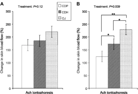
Endothelial function was assessed by measuring changes in endothelium-dependent microvascular relaxation in the overnight-fasted subjects who alternately received CDP, CDH, and orange juice for 4 wk. As shown in Figure 1A, no significant differ-ences in microvascular relaxation were observed between di-etary treatments in response to acetylcholine (P = 0.120), even though orange juice tended to increase vasodilation when com-pared with CDP. Similarly, there was no significant effect (P = 0.080) of the treatment on fasted plasma NOx, but orange juice tended to increase its concentration when compared with CDP or CDH (Table 3).
When acute changes in endothelium-dependent vasodilation were measured 6 h after treatments, significant differences between the groups were observed (P = 0.009; Figure 1B). The higher acetylcholine-induced vasodilatory response was observed after orange juice consumption, with a difference reaching +105.306 25.48% when compared with CDP (P, 0.01). Consumption of CDH also acutely increased the endothelium-dependent vasodi-lation when compared with CDP consumption (+48.656 25.54%, P, 0.05). The difference in endothelium-dependent vasodilation between CDH and orange juice treatments (256.64 6 25.54%) was also significant (P, 0.05), suggesting that orange juice was more potent than CDP to acutely stimulate the microvascular endothelial reactivity. The positive changes in microvascular re-activity are coincident with the detection of hesperetin in the plasma of volunteers sampled 6 h after orange juice or CDH
TABLE 2
Baseline characteristics of the entire study population on the screening day (n = 24)
Mean6 SEM Range
Age (y) 566 1 51–63
BMI (kg/m2) 27.46 0.3 25.2–30.5
Systolic blood pressure (mm Hg) 1346 3 108–169
Diastolic blood pressure (mm Hg) 826 2 64–109
Triglycerides (mmol/L) 1.426 0.14 0.50–2.60
Total cholesterol (mmol/L) 5.656 0.17 4.00–6.70
HDL cholesterol (mmol/L) 1.496 0.05 1.02–1.97
LDL cholesterol (mmol/L) 3.846 0.16 2.36–5.02
Fasting glucose (mmol/L) 5.486 0.17 4.60–8.80
Insulin (lU/mL) 15.016 0.75 7.26–19.74
C-reactive protein (mg/L) 1.596 0.13 0.61–3.36
intake (means6 SEMs: 0.86 6 0.10 lM and 0.77 6 0.16 lM, respectively, P = 0.636), whereas this flavanone was not detected after CDP intake. Pearson’s correlation was used to examine as-sociations between plasma hesperetin concentrations and changes in microvascular endothelial reactivity in this postprandial study. These variables were significantly correlated in both the CDH (r = 0.698, P = 0.0001) and orange juice (r = 0.434, P = 0.039) groups. Plasma vitamin C concentration was significantly higher (P , 0.001) 6 h after orange juice intake (70.5 6 4.6 lM) when compared with CDP (52.86 4.6 lM) or CDH (50.8 6 4.6 lM), whereas plasma NOx concentrations were not significantly dif-ferent between groups (data not shown).
Endothelium-independent microvascular relaxation (in re-sponse to sodium nitroprusside) did not differ between CDP, CDH, and orange juice in the acute 6-h postintervention study as well as when measured in the fasted subjects at the end of the 4-wk intervention period. The changes in skin blood flows mea-sured in response to sodium nitroprusside iontophoresis after CDP, CDH, and orange juice were 120.66 13.9%, 118.7 6 14.0%, and 111.06 13.9%, respectively (P = 0.866).
BP
BP was measured in the overnight fasted subjects at the be-ginning and end of each experimental period (Table 3). The 4-wk consumption of orange juice as well as of CDH resulted in a significantly lower diastolic BP (DBP) compared with that measured after consumption of CDP (P = 0.023). The differences
in DBP between CDP and CDH, as well as that between CDP and orange juice, were significantly different (P , 0.05). Re-gardless of the experimental dietary group, systolic BP (SBP) was similar after the 4-wk supplementation period. There was no significant effect of dietary interventions on pulse pressure (Table 3).
CVD risk factors and markers of inflammation and endothelial dysfunction
Fasting glucose, insulinemia, total cholesterol, LDL choles-terol, HDL cholescholes-terol, and triglycerides were not significantly different between groups (Table 3). Similarly, none of the plasma concentrations of CRP, IL-6, vWF, VCAM-1, and ICAM-1 differed significantly at the end of the 3 dietary interventions. However, sVCAM-1 concentration tended to be lower after the consumption of orange juice or CDH than after consumption of CDP (P = 0.089).
Plasma antioxidant status and biomarkers of oxidative stress
After 4 wk consumption of orange juice, concentrations of vitamin C and b-cryptoxanthin were significantly higher in fasted plasma compared with those measured after CDP and CDH (Table 3). After 4 wk consumption of orange juice, con-centrations of uric acid in plasma were significantly lower than after consumption of CDP (Table 3). These changes in plasma
TABLE 3
Blood pressure and systemic biomarkers for control drink + placebo (CDP) and differences between dietary interventions after 4 wk (n = 23)1
Means6 SEMs Least-squares means6 SEMs
CDP CDP2 CDH CDP2 OJ CDP CDP2 CDH CDP2 OJ P value2 Blood pressure (mm Hg) Systolic 134.36 3.6 20.6 6 3.1 3.06 2.7 133.76 2.1 22.0 6 2.9 2.06 2.8 0.362 Diastolic 84.66 2.9 3.26 1.5 5.56 1.8 84.96 2.1 5.36 2.0* 4.56 2.0* 0.023 Pulse pressure 49.76 2.4 22.5 6 2.3 23.8 6 2.8 49.26 2.0 26.1 6 2.7 22.2 6 2.7 0.092 Plasma concentrations Glucose (mmol/L) 6.16 0.2 0.26 0.2 0.16 0.2 6.16 0.2 0.26 0.2 06 0.2 0.489 Insulin (lU/mL) 17.16 1.9 1.16 1.6 1.96 1.5 16.96 1.2 1.36 1.5 1.26 1.5 0.642 Triglycerides (mmol/L) 1.36 0.2 06 0.1 06 0.1 1.36 0.1 20.1 6 0.1 20.1 6 0.1 0.642
Total cholesterol (mmol/L) 5.56 0.2 20.3 6 0.2 20.2 6 0.2 5.46 0.2 20.4 6 0.2 20.3 6 0.2 0.100
LDL cholesterol (mmol/L) 3.56 0.2 20.3 6 0.2 20.3 6 0.2 3.56 0.2 20.4 6 0.2 20.3 6 0.2 0.091 HDL cholesterol (mmol/L) 1.46 0.1 06 0.1 06 0.1 1.46 0.1 06 0.1 06 0.1 0.388 CRP (mg/L) 1.646 0.23 0.076 0.19 20,34 6 0.21 1.606 0.17 0.376 0.20 0.206 0.20 0.209 IL-6 (pg/mL) 2.006 0.26 20.21 6 0.42 0.246 0.24 1.986 0.25 0.196 0.29 20.11 6 0.29 0.586 vWF (U/mL) 2.756 0.28 20.10 6 0.25 0.066 0.35 2.626 0.21 20.28 6 0.30 20.17 6 0.31 0.648 sICAM-1 (ng/mL) 355.06 17.0 4.96 29.3 228.1 6 23.2 3606 19 216 6 27 228 6 26 0.243 sVCAM-1 (ng/mL) 11936 161 2426 161 3236 150 11996 119 2836 157 3026 154 0.089 NOx (lmol/L) 39.06 5.6 3.26 6.0 212.4 6 5.6 37.96 5.6 0.86 7.0 213.5 6 6.9 0.080 FRAP (lmol Fe2+/mL) 10236 41 22 6 42 96 42 10286 40 126 44 206 44 0.905
Uric acid (lmol/L) 64.16 3.3 2.46 2.1 6.06 2.0 64.36 3.0 3.16 2.1 6.36 2.1* 0.017
a-Tocopherol (lmol/L) 26.86 1.1 20.5 6 0.9 21.0 6 1.0 27.36 0.9 06 1.1 0.26 1.1 0.978
b-Cryptoxanthin (lmol/L) 0.106 0.01 20.01 6 0.01 20.24 6 0.02 0.106 0.01 06 0.01 20.22 6 0.01* 0.0001
Vitamin C (lmol/L) 35.06 3.3 25.4 6 4.9 242.1 6 4.2 36.46 3.5 23.5 6 4.7 239.8 6 4.7* 0.0001
1
CDH, control drink + hesperidin; OJ, orange juice; CRP, C-reactive protein; IL-6, interleukin-6; vWF, von Willebrand factor; sICAM-1, soluble intercellular adhesion molecule 1; sVCAM-1, soluble vascular cell adhesion molecule 1; NOx, nitric oxide metabolites; FRAP, ferric-reducing ability of plasma. Statistical analyses were performed on least-squares means. *Differences between dietary interventions least-squares means were significantly different from zero, P, 0.05 (Tukey-Kramer adjustment for multiple comparisons).
2
P value for dietary treatment effect; ANCOVA with baseline measurement at the beginning of each dietary intervention as the covariate was used (PROC MIXED; SAS Institute, Cary, NC).
concentrations of antioxidant molecules were not accompanied by changes in total plasma antioxidant capacity, as reflected by similar FRAP values (Table 3). Total urinary 8-isoprostane con-centrations, a reliable and specific biomarker of oxidative stress, measured at the end of each experimental period were not sig-nificantly different between treatments: mean (6SEM) CDP, 1.566 0.17 ng/mg creatinine; CDH, 1.47 6 0.11 ng/mg creati-nine; and orange juice, 1.606 0.17 ng/mg creatinine (P = 0.78).
DISCUSSION
The study’s main finding is that 4 wk consumption of orange juice or purified hesperidin, the major orange phenolic, signifi-cantly decreased DBP in healthy subjects. A significant im-provement in endothelium-dependent microvascular reactivity was also observed postprandially after both orange juice and hesperidin ingestion, at a time point coincident with maximal hesperidin bioavailability.
The DBP-lowering effect after 4 wk of orange juice or CDH consumption is of particular interest with regard to their potential health benefits, because DBP is an indicator of peripheral vessel resistance. This effect on DBP was not associated with significant modification of either SBP or pulse pressure. Our present results are consistent with data reported by Reshef et al (21), who showed that consumption of high-naringin sweetie fruit (a hybrid between grapefruit and pummelo) juice for 5 wk reduced DBP in hypertensive subjects when compared with the low-naringin juice. Even if the observed BP-lowering effect was moderate, in persons aged 50–69 y with an SBP of 150 mm Hg and a DBP of 90 mm Hg, a 3–4 mm Hg reduction in DBP would reduce the incidence of coronary artery disease by 20% (29). Previous clinical trials in which healthy or hypertensive volunteers con-sumed dark chocolate, black tea, or pure quercetin as part of their usual diet also supported the beneficial effects of these supple-ments on BP (30–32). The possible mechanisms by which these flavonoid-rich foods lowered BP may involve a chronic increase
in the production of NO by vascular endothelium (33). In the present study, we showed that only orange juice consumption tended to increase NO plasma concentration. Other mechanisms, such as an inhibitory effect on angiotensin-converting enzyme, could also be responsible for the BP-lowering effects of flava-nones (34).
Endothelial function was assessed in the present study on microvessels by using laser Doppler flowmetry, which measures cutaneous perfusion accompanied by iontophoresis of acetyl-choline and sodium nitroprusside (35). Acetylacetyl-choline mediates vasodilatation via the endothelium-dependent production of NO or prostanoids, whereas sodium nitroprusside, an NO donor that stimulates smooth muscle cell relaxation, is used as an endothelium-independent control. Cutaneous microvascular circu-lation was shown to be associated with high BP (36) and coronary artery disease (37). In our postprandial study, we observed a significant improvement of acetylcholine-mediated vasodila-tion after intake of orange juice or CDH, without significant change in the sodium nitroprusside-mediated dilation. This result indicates that the effect of interventions on microvascular function was focused on endothelium-dependent vasorelaxation. Similarly, in several previous postprandial studies, other flavo-noid-rich beverages have also favorably affected endothelial function in healthy subjects (38–40). In agreement with our results obtained with purified hesperidin, the few studies that used supplementation with isolated flavonoids (epicatechin, EGCG) reported improved postprandial endothelium-dependent vasodi-lation (9, 10). In these studies, and ours, the changes in vascular function paralleled plasma concentrations of the flavonoids. Furthermore, the observed changes in microvascular endothelial reactivity were positively correlated with the plasma concen-trations of hesperetin after intake of CDH or orange juice, suggesting that hesperetin could at least partially explain the effect of orange juice.
The ability of flavonoids to activate endothelial NO synthase is likely the mechanism underlying improved endothelial function
FIGURE 1.Least-squares mean (6SEM) changes in endothelium-dependent microvascular reactivity in response to each dietary treatment in the chronic (A) and acute (B) studies. For each experimental group, the changes in skin blood flow in response to iontophoresis of acetylcholine (Ach) were determined in fasted subjects in the chronic study and 6 h after intake in the postprandial acute study. n = 23. The PROC MIXED procedure with Tukey adjustment in SAS (SAS Institute, Cary, NC) was used. *,**Differences between groups are shown when the P value for treatment was significant: *P, 0.05, **P , 0.01. CDP, control drink plus placebo; CDH, control drink plus hesperidin; OJ, orange juice.
(40, 41). However, from our data, the postprandial acetylcholine-mediated dilation improved after ingestion of orange juice or pure hesperidin without significant changes in plasma NO concen-tration, suggesting that other mediators also contribute. Prosta-glandins can play an important role in acetylcholine-mediated vasodilation (35); therefore, one can speculate that orange juice and hesperidin positively affect endothelial microvascular function by acting on the cyclooxygenase pathway. This hy-pothesis is supported by studies showing the ability of dietary flavonoids to increase prostacyclin production (42, 43).
Because the effect of orange juice on postprandial endothelial microvascular reactivity seems to be greater than that of pure hesperidin, the contribution of other specific components of orange juice cannot be ruled out. In particular, orange juice is rich in vitamin C, which may contribute to the maintenance of a healthy vasculature through the regulation of prostacyclin pro-duction and NO bioactivity (44). In the present study, the con-sumption of orange juice induced a +40-lmol/L increase in fasted plasma vitamin C concentration after 4 wk. This result could be particularly relevant considering that a 20-lmol/L rise in plasma vitamin C concentration has been associated with an approximate 20% reduction in risk of total mortality and CVD mortality in the general population (45).
Putative changes in markers of endothelial activation after di-etary supplementation have also been assessed in our study. A tendency toward plasma sVCAM-1 reduction (P , 0.08) was observed only after 4 wk consumption of orange juice or pure hesperidin. In patients with peripheral arterial disease, 4 wk daily consumption of 500 mL orange plus black currant juice compared with a reference sugar drink reduced markers of inflammation (CRP, fibrinogen) (46). However, in our study in healthy volun-teers, orange juice consumption did not affect inflammatory markers. This result was not surprising because the control sugar beverage did not display any adverse proinflammatory effect.
In dyslipidemic subjects, the consumption of hesperidin, provided as a pure compound or in orange juice, beneficially modified the blood lipid profile only when administered at a high dose (.400 mg) (20, 47). Our study, carried out in healthy subjects who consumed a lower amount of juice or of hesperi-din, showed no significant effect on plasma lipid concentrations. Interestingly, we observed a significant reduction in the con-centration of uric acid after 4 wk orange juice consumption. This result is in agreement with a previous study that reported an inverse association between high plasma concentrations of vi-tamin C and lower concentrations of uric acid in subjects after orange juice consumption (19). This lowering effect of orange juice consumption on uric acid concentration is particularly interesting because various observations suggest an association between the concentration of serum uric acid and cardiovascular morbidity and mortality (48), and serum uric acid is a de-terminant of metabolic syndrome (49).
In our study, the intake of’300 mg hesperidin administered as orange juice (500 mL) or as pure compound led to similar plasma concentrations of hesperetin and induced positive effects on vascular protection. Further clinical research should de-termine the minimal and maximal ranges of hesperidin in orange juice that provide the benefits described in this study. Estab-lishment of effective amounts of hesperidin in orange juice may lead to recommendations for servings of orange juice on a daily basis to maximize such benefits.
The following limitations of our study should be noted. Al-though care was taken for optimal BP measurements, we ac-knowledge that 24-h ambulatory BP monitoring would have been more accurate. Second, a possible association between the ob-served effects on BP and on skin microvascular reactivity should be treated with caution. The relevance of skin microvessels in BP control remains uncertain because the skin microcirculation might not be the site of BP regulation. Furthermore, we observed a trend for a chronic effect of the interventions on microvascular endothelial reactivity. It could be suggested that our study was underpowered; therefore, a larger study might have positive findings. In conclusion, we observed favorable changes in BP and endothelial function after the consumption of orange juice in healthy subjects and showed that the flavanone hesperidin could be responsible for the observed effects. On the basis of these results, it would be interesting to encourage the consumption of citrus foods, which are the unique dietary sources of flavanones.
We are indebted to all of the subjects who volunteered in the clinical trial. We also thank Dominique Bayle and Se´verine Thien for performing biochem-ical analyses, Mathieu Rambeau for assistance in HPLC analyses, and the medical team of the clinical research unit for their technical assistance in conducting the clinical aspects of this study.
The authors’ responsibilities were as follows—CM: study concept and design, data interpretation, and manuscript preparation; DB: study design, volunteer recruitment, clinical study management, and revision of the man-uscript; DM: study design, data interpretation, and manuscript preparation; DL: protocol implementation, sample acquisition, and data analysis and col-lection; JFM: statistical analysis; AS: study design and revision of the man-uscript; and AM: study design, data interpretation, and writing and revision of the manuscript. None of the authors had any conflicts of interests with regard to the research described in this article.
REFERENCES
1. Peters U, Poole C, Arab L. Does tea affect cardiovascular disease? A meta-analysis. Am J Epidemiol 2001;154:495–503.
2. Di Castelnuovo A, Rotondo S, Iacoviello L, Donati MB, De Gaetano G. Meta-analysis of wine and beer consumption in relation to vascular risk. Circulation 2002;105:2836–44.
3. Mink PJ, Scrafford CG, Barraj LM, et al. Flavonoid intake and car-diovascular disease mortality: a prospective study in postmenopausal women. Am J Clin Nutr 2007;85:895–909.
4. Buijsse B, Weikert C, Drogan D, Bergmann M, Boeing H. Chocolate consumption in relation to blood pressure and risk of cardiovascular disease in German adults. Eur Heart J 2010;31:1616–23.
5. Auclair S, Milenkovic D, Besson C, et al. Catechin reduces athero-sclerotic lesion development in apo E-deficient mice: a transcriptomic study. Atherosclerosis 2009;204:e21–7.
6. Norata GD, Marchesi P, Passamonti S, Pirillo A, Violi F, Catapano AL. Anti-inflammatory and anti-atherogenic effects of cathechin, caffeic acid and trans-resveratrol in apolipoprotein E deficient mice. Athero-sclerosis 2007;191:265–71.
7. Hooper L, Kroon PA, Rimm EB, et al. Flavonoids, flavonoid-rich foods, and cardiovascular risk: a meta-analysis of randomized controlled trials. Am J Clin Nutr 2008;88:38–50.
8. Loke WM, Hodgson JM, Proudfoot JM, McKinley AJ, Puddey IB, Croft KD. Pure dietary flavonoids quercetin and (–)-epicatechin augment ni-tric oxide products and reduce endothelin-1 acutely in healthy men. Am J Clin Nutr 2008;88:1018–25.
9. Schroeter H, Heiss C, Balzer J, et al. (–)-Epicatechin mediates beneficial effects of flavanol-rich cocoa on vascular function in humans. Proc Natl Acad Sci USA 2006;103:1024–9.
10. Widlansky ME, Hamburg NM, Anter E, et al. Acute EGCG supple-mentation reverses endothelial dysfunction in patients with coronary artery disease. J Am Coll Nutr 2007;26:95–102.
11. Erdman JW Jr, Balentine D, Arab L, et al. Flavonoids and heart health. Proceedings of the ILSI North America Flavonoids Workshop, May 31– June 1, 2005, Washington, DC. J Nutr 2007;137:718S–37S.
12. Neveu V, Perez-Jimenez J, Vos F, et al. Phenol-Explorer: an online comprehensive database on polyphenol contents in foods. Available from: http://www.phenol-explorer.eu (cited June 2010).
13. Manach C, Williamson G, Morand C, Scalbert A, Re´me´sy C. Bio-availability and bioefficacy of polyphenols in humans: I- A review of 97 bioavailability studies. Am J Clin Nutr 2005;81(suppl):230S–42S. 14. Johnsen SP, Overvad K, Stripp C, Tjonneland A, Husted SE, Sorensen
HT. Intake of fruit and vegetables and the risk of ischemic stroke in a cohort of Danish men and women. Am J Clin Nutr 2003;78:57–64. 15. Dauchet L, Ferrieres J, Arveiler D, et al. Frequency of fruit and
vege-table consumption and coronary heart disease in France and Northern Ireland: the PRIME study. Br J Nutr 2004;92:963–72.
16. Guarnieri S, Riso P, Porrini M. Orange juice vs vitamin C: effect on hydrogen peroxide-induced DNA damage in mononuclear blood cells. Br J Nutr 2007;97:639–43.
17. Johnston CS, Dancho CL, Strong GM. Orange juice ingestion and supplemental vitamin C are equally effective at reducing plasma lipid peroxidation in healthy adult women. J Am Coll Nutr 2003;22:519–23. 18. Ghanim H, Sia CL, Upadhyay M, et al. Orange juice neutralizes the proinflammatory effect of a high-fat, high-carbohydrate meal and pre-vents endotoxin increase and Toll-like receptor expression. Am J Clin Nutr 2010;91:940–9.
19. Sanchez-Moreno C, Cano MP, de Ancos B, et al. Effect of orange juice intake on vitamin C concentrations and biomarkers of antioxidant status in humans. Am J Clin Nutr 2003;78:454–60.
20. Kurowska EM, Spence JD, Jordan J, et al. HDL-cholesterol-raising ef-fect of orange juice in subjects with hypercholesterolemia. Am J Clin Nutr 2000;72:1095–100.
21. Reshef N, Hayari Y, Goren C, Boaz M, Madar Z, Knobler H. Antihy-pertensive effect of sweetie fruit in patients with stage I hypertension. Am J Hypertens 2005;18:1360–3.
22. Manach C, Morand C, Gil-Izquierdo A, Bouteloup-Demange C, Remesy C. Bioavailability in humans of the flavanones hesperidin and narirutin after the ingestion of two doses of orange juice. Eur J Clin Nutr 2003;57:235–42. 23. Benzie IF, Strain JJ. The ferric reducing ability of plasma (FRAP) as a measure of “antioxidant power”: the FRAP assay. Anal Biochem 1996; 239:70–6.
24. Moshage H, Kok B, Huizenga JR, Jansen PL. Nitrite and nitrate deter-minations in plasma: a critical evaluation. Clin Chem 1995;41:892–6. 25. Tessier F, Birlouez-Aragon I, Tjani C, Guilland JC. Validation of a
mi-cromethod for determining oxidized and reduced vitamin C in plasma by HPLC-fluorescence. Int J Vitam Nutr Res 1996;66:166–70.
26. Lyan B, Azais-Braesco V, Cardinault N, et al. Simple method for clinical determination of 13 carotenoids in human plasma using an isocratic high-performance liquid chromatographic method. J Chromatogr B Bi-omed Sci Appl 2001;751:297–303.
27. Ito H, Gonthier MP, Manach C, et al. Polyphenol levels in human urine after intake of six different polyphenol-rich beverages. Br J Nutr 2005; 94:500–9.
28. Farkas K, Kolossvary E, Jarai Z, Nemcsik J, Farsang C. Non-invasive assessment of microvascular endothelial function by laser Doppler flowmetry in patients with essential hypertension. Atherosclerosis 2004; 173:97–102.
29. Law MR, Morris JK, Wald NJ. Use of blood pressure lowering drugs in the prevention of cardiovascular disease: meta-analysis of 147 rando-mised trials in the context of expectations from prospective epidemio-logical studies. BMJ 2009;338:b1665.
30. Edwards RL, Lyon T, Litwin SE, Rabovsky A, Symons JD, Jalili T. Quercetin reduces blood pressure in hypertensive subjects. J Nutr 2007; 137:2405–11.
31. Grassi D, Desideri G, Necozione S, et al. Blood pressure is reduced and insulin sensitivity increased in glucose-intolerant, hypertensive subjects after 15 days of consuming high-polyphenol dark chocolate. J Nutr 2008;138:1671–6.
32. Taubert D, Roesen R, Lehmann C, Jung N, Schomig E. Effects of low habitual cocoa intake on blood pressure and bioactive nitric oxide: a randomized controlled trial. JAMA 2007;298:49–60.
33. Grassi D, Desideri G, Croce G, Tiberti S, Aggio A, Ferri C. Flavonoids, vascular function and cardiovascular protection. Curr Pharm Des 2009; 15:1072–84.
34. Actis-Goretta L, Ottaviani JI, Fraga CG. Inhibition of angiotensin converting enzyme activity by flavanol-rich foods. J Agric Food Chem 2006;54:229–34.
35. Turner J, Belch JJ, Khan F. Current concepts in assessment of micro-vascular endothelial function using laser Doppler imaging and ionto-phoresis. Trends Cardiovasc Med 2008;18:109–16.
36. Serne EH, Stehouwer CD, ter Maaten JC, et al. Microvascular function relates to insulin sensitivity and blood pressure in normal subjects. Circulation 1999;99:896–902.
37. Shamim-Uzzaman QA, Pfenninger D, Kehrer C, et al. Altered cutaneous microvascular responses to reactive hyperaemia in coronary artery dis-ease: a comparative study with conduit vessel responses. Clin Sci (Lond) 2002;103:267–73.
38. Agewall S, Wright S, Doughty RN, Whalley GA, Duxbury M, Sharpe N. Does a glass of red wine improve endothelial function? Eur Heart J 2000;21:74–8.
39. Heiss C, Dejam A, Kleinbongard P, Schewe T, Sies H, Kelm M. Vascular effects of cocoa rich in flavan-3-ols. JAMA 2003;290: 1030–1.
40. Lorenz M, Jochmann N, von Krosigk A, et al. Addition of milk prevents vascular protective effects of tea. Eur Heart J 2007;28:219–23. 41. Madeira SV, Auger C, Anselm E, et al. eNOS activation induced by
a polyphenol-rich grape skin extract in porcine coronary arteries. J Vasc Res 2009;46:406–16.
42. Schramm DD, Wang JF, Holt RR, et al. Chocolate procyanidins decrease the leukotriene-prostacyclin ratio in humans and human aortic endo-thelial cells. Am J Clin Nutr 2001;73:36–40.
43. Hermenegildo C, Oviedo PJ, Garcia-Perez MA, Tarin JJ, Cano A. Ef-fects of phytoestrogens genistein and daidzein on prostacyclin pro-duction by human endothelial cells. J Pharmacol Exp Ther 2005;315: 722–8.
44. Hornig B. Vitamins, antioxidants and endothelial function in coronary artery disease. Cardiovasc Drugs Ther 2002;16:401–9.
45. Khaw KT, Bingham S, Welch A, et al. Relation between plasma ascorbic acid and mortality in men and women in EPIC-Norfolk prospective study: a prospective population study. European Prospective Investigation into Cancer and Nutrition. Lancet 2001;357:657–63.
46. Dalga˚rd C, Nielsen F, Morrow JD, et al. Supplementation with orange and blackcurrant juice, but not vitamin E, improves inflammatory markers in patients with peripheral arterial disease. Br J Nutr 2009;101: 1–7.
47. Miwa Y, Yamada M, Sunayama T, et al. Effects of glucosyl hesperidin on serum lipids in hyperlipidemic subjects: preferential reduction in elevated serum triglyceride level. J Nutr Sci Vitaminol (Tokyo) 2004;50: 211–8.
48. Alderman MH. Uric acid and cardiovascular risk. Curr Opin Pharmacol 2002;2:126–30.
49. Onat A, Uyarel H, Hergenc G, et al. Serum uric acid is a determinant of metabolic syndrome in a population-based study. Am J Hypertens 2006; 19:1055–62.