Nutritional regulation of glucokinase: a cross-species story
Ste´phane Panserat
1*, Nicole Rideau
2and Sergio Polakof
31INRA, UR1067 Nutrition Metabolism Aquaculture (NUMEA), French National Institute for Agricultural Research (INRA), Aquapoˆle, F-64310 Saint-Pe´e-sur-Nivelle, France
2INRA, UR83 Recherches Avicoles, French National Institute for Agricultural Research (INRA), F-37380 Nouzilly, France 3INRA, Human Nutrition Unit (UNH), French National Institute for Agricultural Research (INRA), Clermont-Ferrand/Theix Research Centre, F-63122 Saint-Gene`s-Champanelle, France
Abstract
The glucokinase (GK) enzyme (EC 2.7.1.1.) is essential for the use of dietary glucose because it is the first enzyme to phosphorylate glucose in excess in different key tissues such as the pancreas and liver. The objective of the present review is not to fully describe the biochemical characteristics and the genetics of this enzyme but to detail its nutritional regulation in different vertebrates from fish to human. Indeed, the present review will describe the existence of the GK enzyme in different animal species that have naturally different levels of carbohydrate in their diets. Thus, some studies have been performed to analyse the nutritional regulation of the GK enzyme in humans and rodents (having high levels of dietary carbohydrates in their diets), in the chicken (moderate level of carbohydrates in its diet) and rainbow trout (no carbohydrate intake in its diet). All these data illustrate the nutritional importance of the GK enzyme irrespective of feeding habits, even in animals known to poorly use dietary carbohydrates (carnivorous species).
Key words:Glucokinase: Humans: Fish: Birds: Carbohydrates
Introduction
The glucokinase (GK) enzyme, also known as hexokinase (HK) IV (or D), is one of the four glucose-phosphorylating isoenzymes described initially in the vertebrate liver, characterised by a low affinity for glucose, sigmoidal kin-etics and lack of inhibition by glucose-6-phosphate. The others, being called HK I (or A), II (or B) and III (or C), are more widely distributed and do not meet the character-istics cited above(1,2). This enzyme has been the object of numerous studies mainly because of its role in hepatic metabolism, but also in glucose homeostasis, given its involvement in metabolism-dependent insulin secretion. In the latter case, research has been focused on the role of GK in type 2 diabetes mellitus. Very good reviews about the biochemical and molecular regulation of GK can be found in the literature and the reader is encouraged to consult those of Printz et al.(3), Iynedjian(4), Ca´rdenas(5) and Matschinsky(6,7).
Most of the reviews published so far have dealt with the role and regulation of GK in the rodent model applied to human pathophysiological issues(7). In the present review we will not concentrate on the pharmacological
or molecular regulation of the mammalian GK, as this topic has been approached on numerous occasions (see above). Rather, we will focus our attention on the nutri-tional regulation of the enzyme. In this sense, comparative studies of glucose-phosphorylating activity in the liver have shown that in regular conditions (standard diet) the number of isoenzymes may vary as a function of the species and that GK appears to be absent in several of them(8) (see Fig. 1). Moreover, other questions remain unresolved, including a different nutritional regulation of hepatic and pancreatic GK (due to different promoters) and the possibility that in some animal species GK has not been detected due to the lack of nutritional induction or evolutionary reasons (such as loss of the gene or con-version to a pseudogene). Beyond the well-known rodent model for the study of GK, in the present review we explore the knowledge accumulated concerning the nutritional regulation of GK in other animal species with different nutritional habits and then alternative mechan-isms of glucose regulation.
Thus, our objective is twofold: on one hand, to offer a glance over the knowledge about GK in other vertebrate groups, such as fish and birds, but also in rarely explored
* Corresponding author: Dr Stephane Panserat, fax þ 33 5 59 54 51 52, email panserat@st-pee.inra.fr
Abbreviations: GK, glucokinase; GKA, GK activator; GKR, glucokinase regulator; GKRP, glucokinase regulatory protein; HC, high carbohydrate; HF, high fat; HK, hexokinase; Km, Michaelis constant; mRNA, messenger RNA.
qThe Authors 2014
mammalian species, such as cats and dogs. On the other hand, we aim to make a comparative analysis of the nutri-tional regulation of GK between the classical mammalian models and the other groups in which GK is differentially
regulated based on different evolutionary pressures and nutritional habits. Finally, several outputs will be discussed from a perspective point of view, including: (i) other nutri-tional roles for GK (other than those related to hepatic glucose metabolism and insulin secretion); (ii) implications in human nutrition from data obtained in genome-wide association studies; and (iii) impact on the context of dele-terious diets (rich in fat or carbohydrates) for some species and not for others.
Glucokinase in mammals
Among vertebrates, the mammals are without discussion the best-represented group concerning the study of GK. Actually, the enzyme was discovered in rats, which became very quickly the best model for studying human GK: 37 % of the papers published on GK are focused on the rat GK, while 42 % are studies in human subjects, a number importantly boosted by pharmacological and mutation studies. After mice (16 %), the rest of mammalian species (all of them below 2 %) seem not to have attracted the attention of scientists (see Fig. 2). This may be due to the fact that GK activity has been found in twenty-two mammalian species, twelve of them rodents (see Table 1). In contrast, at the protein level a similar number of species express GK, but most of them are primates (Fig. 3). In this sense, the biggest cluster includes the primates and rodents, while the rest of the species are classified in more or less diverse groups. It is worth mentioning that nutritional habits do not seem to have an impact on this classification.
GK in mammals has been mainly studied in rodent species, such as the rat (Rattus norvegicus) and the mouse (Mus musculus), and, when possible, in human biopsy samples. More recently, some research teams have been also put some attention to the regulation of GK in other species of interest, such as domestic cats and dogs, although the level of knowledge remains quite limited.
Glucokinase function and regulation in mammals
The GK enzyme is a glucose-phosphorylating enzyme initially discovered in rat liver in the early 1960s. The rapid characterisation of its biochemical properties led to the idea that GK was actually the key regulator enzyme of hepatic glucose metabolism. As a matter of fact, GK does not work as any other HK found in eukaryotic cells, given that it is not inhibited by the product of the reaction that it catalyses (glucose-6-phosphate), has a low affinity for glucose (S0.5about 7·5 mM) and a characteristic sigmoi-dal kinetics (Hill coefficient about 1·6). Thanks to these characteristics, GK is able to cope and handle the postpran-dial glucose increase observed after a meal. Given its importance in the postprandial regulation of glucose metabolism, it is not surprising that GK is tightly regulated by insulin. Cyprinus carpio Ctenopharyngodon idella Lateolabrax japonicus Sparus aurata Culter ilishaeformis Pelteobagrus vachellii Salmo marmoratus Carassius auratus Xenopus tropicalis Xenopus laevis Danio rerio Oreochromis niloticus* Oryzias latipes* Meleagris gallopavo* Anas platyrhynchos* Chelonia mydas Taeniopygia guttata* Gallus gallus Columba livia Ornithorhynchus anatinus* Ailuropoda melanoleuca* Mustela putorius furo*
Cavia porcellus* Mesocricetus auratus* Nomascus leucogenys* Tursiops truncatus*
Loxodonta africana*
Saimiri boliviensis boliviensis* Trichechus manatus latirostris*
Echinops telfairi* Pteropus alecto Mus musculus Rattus norvegicus Octodon degus* Heterocephalus glaber** Sorex araneus* Jaculus jaculus* Callithrix jacchus* Pan paniscus* Pongo abelii‡ Homo sapiens Orcinus orca* Equus caballus§ Sus scrofa|| Ovis aries¶ Felis catus¶ Otolemur garnettii* Gorilla gorilla gorilla* Bos grunniens mutus Condylura cristata* Cricetulus griseus†
Sarcophilus harrisii* Monodelphis domestica* Ceratotherium simum simum*
Bos taurus† Papio anubis* Ochotona princeps* Pan troglodytes‡
Myotis davidii
Canis lupus familiaris* Dasypus novemcinctus* Myotis brandtii Odobenus rosmarus divergens* Oryctolagus cuniculus* Macaca mulatta‡ Equus grevyi Tupaia chinensis Ficedula albicollis* Melopsittacus* undulatus Anolis carolensis* Takifugu rubripes* Maylandia zebra* Oncorhynchus mykiss HK4 HK2 HK1 HK1 HK2 HK4 HK4 HK4 HK2 HK1 HK3 HK3 Fish and amphibians
Reptiles and birds
Mammals
Fig. 1. Venn diagrams representing the putative presence of the four hexo-kinase (HK) isoforms (HK1, HK2, HK3, HK4) in fish and amphibians, reptiles and birds, and mammals. The represented data were obtained from the National Center for Biotechnology Information (NCBI) protein database. * Predicted sequences. † Predicted HK2. ‡ Predicted sequences except HK1. § Predicted sequences except HK2. [capsverbar] Predicted sequences except HK1 and HK2. { Predicted sequences except HK4. ** Predicted sequence HK1. (A colour version of this figure can be found online at http://www. journals.cambridge.org/nrr).
Very soon GK was also reported to be present in labo-ratory animals(9) and human pancreatic tissue(10), and then its role in glucose homeostasis as a glucosensor was proposed(11). This was further supported by the fact that GK is encoded by a single gene, but controlled by two specific promoters: one hepatic and one pancreatic(12,13). More recently, the GK network has been largely enriched as GK has been found in several other cellular types, including neurons (glucose excited and glucose inhibited), enteroendocrine cells (K and L)(14), a-cells(15)and pituitary gonadotropes(16).
The presence of GK in rodent pancreatic tissue was discovered more than 20 years ago(17), and given that the activity of pancreatic GK is glucose dependent, this enzyme is today inseparable from the glucose sensor con-cept(18,19). The GK glucose sensor paradigm is integrated with the threshold concept for glucose-stimulated insulin release, given that GK constitutes the rate-limiting step in the generation of a trigger metabolite of a constellation of metabolic signals initiating the secretory process. Since its discovery, pancreatic GK has been extensively studied, given that more than 150 mutations have been discovered in the pancreatic GK gene that alters this glucosensing capacity. However, while most of the energy of the scientists has been directed to this aspect, very little has been done in the nutritional field, and the behaviour of GK in pancreatic tissue under different real nutritional conditions is still only partially known.
GK is known to be tightly regulated at numerous levels, including modifications of conformational status, physical interaction with other proteins, hormonal control and a tissue-dependent molecular expression.
The most rapid regulation of GK takes place at the con-formational level given the cooperativity of its kinetics with regard toD-glucose. GK has two conformations, an active
and an inactive form with high and low glucose affinity, respectively, which allow optimal substrate sensitivity at the fasting level of blood glucose in humans and many lab-oratory animals(20). The second level of regulation in terms of rapidity would be its interaction with the bifunctional
enzyme 6-phosphofructo-2-kinase/fructose-2,6-biphospha-tase (6PF2K/F26P2ase)(21) and the GK regulatory protein (GKRP). While in the first case very little is known, more information is available concerning the GKRP regulation of GK. The actual ligands for this protein are fructose-1-phosphate and fructose-6-fructose-1-phosphate, which have antagon-ist effects on the GK – GKRP binding tandem: while GKRP binds to GK and inhibits the enzyme activity competitively with respect to glucose, fructose-6-phosphate reinforces the inhibitory effect of GKRP and fructose-1-phosphate abolishes binding and subsequent inhibition(22,23). Later, other authors showed that this was only a partial descrip-tion of the reguladescrip-tion mechanism and that the main feature of GKRP was that this protein sequesters GK in the nucleus in the absence of a high concentration of glucose or fruc-tose(24,25). In the opposite conditions (i.e. high glucose or fructose concentration), GK is released by GKRP and translocates into the cytoplasm where it exerts its action. Long-term GK control is mainly exerted by hormones and GK expression control. Hormonal stimulation is able to induce (insulin) or repress (glucagon) GK expression, protein and activity. Maybe the major feature of the regulation of GK expression is the dual control of this enzyme by insulin in the liver and by glucose in the pancreatic b-cells, which is based on the existence of a downstream and an upstream promoter within the GK gene – the ‘one gene, two promoters’ concept for control of GK expression(4). Finally, other levels of regulation include the epigenetic hypermethylation of the GK gene (age-related, in a way that increased methylation is nega-tively associated with hepatic GK expression(26)), and GK nitration, that leads to pancreatic(27) and hepatic inacti-vation of the enzyme(28).
Nutritional regulation of glucokinase in mammals
Very few studies have focused on the macro- and micronu-trient interactions and regulation of this enzyme. Given that the micronutrient effects are only known in some mamma-lians species(29 – 31), in the present review we will focus on
60 (a) (b) (c) 50 40 30 20 10 0 Trout Sea bream Car p Sea bassZebr
afish
SalmonGoldfishPerchHalib ut Omniv orous Car nivorous Herbiv orous Omniv orous Car nivorous Herbiv orous Omniv orous Car nivorous Herbiv orous Chic ken Duck Quail Pigeon Owl Human Rat
MouseDogRab bit Big Guinea-pig Cat SheepMonk ey Cow Number of pub lications 25 3800 3700 3600 1500 1000 500 0 20 15 10 5 0
Fig. 2. Representation of the number of publications concerning the enzyme glucokinase in three groups of vertebrates: fish (a), birds (b) and mammals (c). Amphibians and reptiles were omitted given the low number of publications (, 5). For each group, the number of publications (. 1) per species was included, as well as the total number of publication depending on nutritional and feeding habits. Data were obtained from Scopus (Copyrightq2013 Elsevier B.V.) using the
key words ‘glucokinase’ and the searched species (i.e. ‘rat’ or ‘chicken’).
Table 1. Glucokinase activity in the liver of forty-two vertebrate species, including twenty-two mammals, seven birds, three reptiles, two amphibians and eight fish*
Species Activity (U/g liver) Activity (mU/mg protein) Tissue preparations
and assay conditions Reference Mammals
Degu (Octodondegus) 1·1 About 4·3 – 251
Field mouse (Akodonolivaceus) 2·1 About 11 – 251
Long-haired mouse (Akodon longipilis) 1·9 About 9·6 – 251
Leaf-eared mouse (Phyllotis darwini) 3 About 13·3 – 251
Hamster (Mesocricetus auratus) 0·5 About 2·4 251
– 12·2 105 000 g 252
Guinea-pig (Carla porcellus) 1 About 5·6 251
– 22 105 000 g 252
Coipo (Myocastor coypu) 1·25 About 5·8 – 251
Cururo (Spalacopus cyanus) 2 About 11 – 251
Squirrel monkey (Saimiri scuirea) 2·5 – 251
– 79·1 105 000 g 252
Marsupial yaca (Marmosa elegans) 1·5 10 251
Sheep (Ovis aries) 0·02 – 228C; 25 000 g 253
Camel (Camelus dromedaries) 0·05 – 228C; 25 000 g 253
Possum (Trichosurus vulpecula) 6·5 – 25 000 g 254
Rabbit (Oryctolagus cuniculus) 1·3 – 25 000 – 100 000 g 254
2·25 10 251
41
Dog (Canis familiaris) 1·8 – 25 000 g 254
Pig (Sus scrofa) 3·2 – 25 000 g 254
– 12·9 105 000 g 252
Man (Homo sapiens) 1·01 1.2 – 6·0 10 000 – 100 000 g 63
Fasted and fed activities 41
Mouse (Mus musculus) 3·8 4 – 21 12 000 – 100 000 g 41, 255
Fed and fasted
Rat (Rattus norvergicus) 3·2 – 5·7 6 – 33 27 000 – 100 000 g 41, 44
Sand rat (Merianes hurrianae) – 40 105 000 g 252
Gerbil (Cricetidae gerbillus) – 140 105 000 g 252
Cat (Felis domesticus) – 5 105 000 g 252
Birds
Finch (Serinus canaries) – 8·8 105 000 g 252
Chicken (Gallus gallus) 0 – 0·1 0·3 – 1·5 600 – 900 g 160, 164, 200, 256
0·1 – 0·3 – 27 000 g 158
0·40 47 100 000 g 41
Pigeon (Columba livia) 0·60 – 100 000 g 41
Mallard (Pekin) (Ana splatyrhynchos) 0·45 2·2 – 4·1 100 000 g 41
– 900 g 183
Mule duck (male Cairina moschata £ female Anas plathyrhynchos) – 1·8 – 3·6 900 g 183
0·2 – 1·8 7 – 96 27 000 g 151
Muscovy (Cairina moschata) – 1·8 900 g 183
Hinny (male Anas plathyrhynchos £ female Cairina moschata) – 1·9 – 4·4 900 g 183
Reptiles
Spotted turtle (Cleramys guttala) – 11·2 105 000 g 252
Argentine tortoise (Geochelone chilensis) 0·15 1·7 40 000 rpm 257
Argentine snake-necked turtle (Hydromedusa tectifera) 0·13 1·0 40 000 rpm 257
Amphibians
American bullfrog tadpole (Rana catesbeiana)† – 13·4 105 000 g 252
Salamander (Ambystom amacu) – 8·6 105 000 g 252
Fish
Goldfish (Carassius auratus) – 1·6 900 g 47
2·1 105 000 g 252
Trout (Oncorhynchus mykiss) – 3·3 – 55·2 900 g 165
Gilthead sea bream (Sparus auratus) – 1·0 – 29·9 900 g 165
Common carp (Cyprinus carpio) – 1·1 – 9·7 900 g 165
Atlantic salmon (Salmo salar) – 6·0 – 9·4 900 – 100 000 g 154, 198
5·2 – 13·5 0 – 30 % starch
Atlantic halibut (Hippoglossu shipoglossus) – 27·1 – 98·4 900 – 100 000 g 242
Brockmann bodies
European sea bass (Dicentrarchus labrax) – 0·6 – 9·6 900 g 224
Blackspot sea bream (Pagellus bogaraveo) 0·1 0·7 – 0·9 900 g 258
* When possible, activities were expressed in both U/g liver and mU/mg protein. When available, different extraction procedures were included. † Renamed Lithobates catesbeianus.
the nutritional impact of macronutrients and more in gen-eral the nutritional status of the animals on GK regulation. Despite the lack of human data on GK we do know, thanks to studies developed in rodents, that nutrition plays a key role in GK regulation and that all the macro-nutrients seem to have an important impact on this enzyme. In rodents, high fat (HF) feeding results in reduced hepatic and pancreatic GK activity and impaired glucose tolerance. In contrast, short-term fructose feeding in human subjects seems to have a beneficial effect on glycaemia control, given that it would be able to release GK from the GKRP binding, allowing increased hepatic glucose uptake(32,33). However, longer studies in human subjects do not confirm these results and chronic fructose feeding in animals clearly leads to hepatic steatosis and insulin resistance(34). In this sense, it is worth mentioning that the hepatic metabolism of fructose is not completely
equivalent to that of glucose. While glucose metabolism is regulated by insulin and controlled by GK and phospho-fructokinase (regulated by the level of ATP), fructose con-version to triose phosphate is an insulin-independent process and very fast (given the low Michaelis constant (Km) of the fructokinase enzyme)(35). Moreover, this fruc-tose metabolism is not regulated by ATP or citrate levels, leading to a transient depletion of free phosphate and a decrease in ATP in liver cells in response to fructose.
Given the increasing consumption of cafeteria-like diets (very rich in fat) and fructose syrup-based soft drinks in Western countries, the potential impact of fats and fructose feeding on human GK should not be ignored.
Studies on the nutritional regulation of GK in non-rodent animals are much less abundant. The interest in GK could be divided into two groups: on the one hand GK could be important to species of commercial interest such as live-stock. However, most of them (ruminants) do not rely of glucose as primary fuel and then the role of GK remains to be elucidated. In single-stomached species (such as pigs, horse, rabbits) GK has been barely approached and then its importance is still unknown. On the other hand, in recent years the presence of GK in domestic species such as cats and dogs has been extensively studied, given that these species (with important protein require-ments) are currently fed high-carbohydrate (HC)/HF diets with a low cost when comparing with protein. As a result, an important part of the canine and feline popu-lation is obese and diabetic and a possible role of GK in this new animal epidemic has been suggested.
Given the key role of GK on glucose metabolism and homeostasis, we will focus the present review on two groups of mammals depending on their metabolic and nutritional orientation: species relying mainly on glucose as a primary energy fuel (omnivorous and some herbivor-ous) and those that utilise other sources of nutrients, such as protein or fat (mainly carnivorous).
Carbohydrate-dependent mammalian species
Most of the available information on GK nutritional regulation came from fasting – refeeding cycle experiments, and the role of GK under different feeding regimens and diets is almost unknown. In the next sections of the review we will use as reference the data published on rodent species, stressing the data coming from other species when they do exist.
Glucokinase regulation by nutritional status: focus on fed – fasted – refeeding cycles and postprandial changes – liver. The early studies that focused on GK showed that, in the liver, GK activity is highly affected by nutritional status, being inhibited by a classical 48 h food-deprivation protocol(36 – 38), and practically undetectable after 72 h of fasting(39,40)(Table 2 and Fig. 4). Given the key role of insulin in controlling GK activity, the reduction in GK activity during fasting has been traditionally related to the Xenopus laevis
Xenopus silurana tropicalis Rhabdosargus sarba Carassius auratus Cyprinus carpio Danio rerio Erythroculter ilishaeformis Ctenopharyngodon idella Pelteobagrus vachellii Oreochromis niloticus Lateolabrax japonicus Sparus aurata Oncorhynchus mykiss Salmo marmoratus Gallus gallus Heterocephalus glaber Felis catus Bos taurus Ovis aries Cricetulus griseus Homo sapiens Pteropus alecto Mus musculus 0·1 Rattus norvegicus Carassius auratus Pengze
Fig. 3. Glucokinase protein multiple alignment between sequences of forty vertebrate species, including thirteen fish, two amphibians, one bird and nine mammals available at the National Center for Biotechnology Information (NCBI) protein database. Multiple progressive alignment was done using COBALT (Constraint-based Multiple Protein Alignment Tool)(249). The picture
was created using TreeViewX from the Nexus file obtained after COBALT analysis. Protein accession numbers are available upon request.
very low insulin levels during the food deprivation periods. Interestingly, GK activity in the liver of fasted human sub-jects (overnight) is quite low (1 – 3·5 mU/mg)(41) when compared with 3-d fasted rodents (10 mU/mg), suggesting that the capacity of the human liver to handle dietary glucose is not as high as in rodents. This is in line with the fact that GK is detected in human liver biopsies but only when patients are well nourished, while in poorly nourished individuals the activity is undetectable(42).
The molecular studies on GK showed that messenger RNA (mRNA) levels are strongly reduced in the fasted liver, while GK expression increases quickly when glucose is administrated(43)(Table 2 and Fig. 4). The concentration of the GK protein in liver increases during the transition from the fasted to the refed state, similarly to the mRNA levels, which are undetectable in the liver of fasted rats, but which are strongly induced by a glucose load(17). A very complete study by Iritani et al.(44) showed in detail how GK activity and expression are regulated by feeding in the rat liver. Only 2 h after feeding a control diet (67 % carbohydrate) the mRNA levels of GK increased dramati-cally up to 20-fold when compared with the fasted state. At 8 h after feeding, this level of expression was already reduced 50 %, and 16 h after the meal the fasting level was achieved. In contrast, activity levels increased differen-tially, given that the first significant changes are observed 8 h after the meal, increasing the levels even 24 h after feeding.
The information available concerning the nutritional regulation of the GK – GKRP tandem is very scarce. GKRP quantity decreases with fasting and increases with refeed-ing(45). Thus, disruption of the GK – GKRP complex and translocation of active GK to the cytosol has been reported during transition from the fasting (24 h) to refed (1 and 2 h refeeding) conditions in rats(46,47).
Glucokinase regulation by nutritional status: focus on fed – fasted – refeeding cycles and postprandial changes – pancreas. The earliest studies reported that the mRNA levels of pancreatic GK remained unaltered during the fed – fasting – refeeding cycle(17). However, more recent papers showed that there exists a dependence on the nutri-tional state given that after 2 d of fasting the mRNA levels of GK were reduced by 50 % when comparing with the fed control, while after 4 h of refeeding the expression was normalised(48). Results obtained at the protein level are contradictory, given that in vivo studies did not detect any nutrient-dependent increase in freshly isolated islets from starved and refed rats(17) or after infusion of glu-cose(49). However, GK protein did increase in vitro when pancreatic islets were incubated in the presence of high glucose concentrations, but for a long period (3 – 7 d)(50). Contrary to the data obtained at the mRNA and protein levels, but consistent with the induction of pancreatic GK by insulin, GK activity in cytoplasmic fractions of pan-creatic islets decreases with the duration of fasting and is increased by refeeding(51 – 53). GK activity contributed
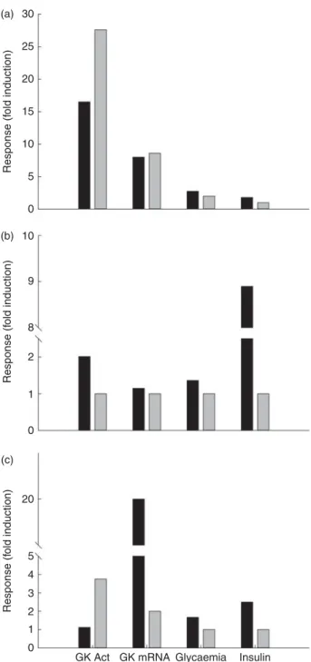
Table 2 . Glucokinase a ctivities in the liver o f omnivorous a nd carnivorous piscine, avian and m ammalian s pecies submitted to several n utritional treatment s* Omnivorous Carnivorous Fish (common c arp) Birds (chicken) Mammals (rat) Fish (rainbow trout) Birds (owl) M ammals (dog ) Fed 100 (14 % CHO) (211) 100 (15 9 ) 100 (4 h ) (25 8 ) 141 (6 % CHO) (20 7) ND 100 (43 % CHO) (13 6) Fasted ND 46 (24 h ) (15 9 )† 3 7 (72 h) (25 8 ) 100 (259) ND ND High CHO 129 (21 % CHO) (211) 100 (38 % CHO) (16 2 ) 105 (100 % CHO) (62 ) 1200 (20 % CHO) (259) 108 (38 % CHO) (16 2 ) ND High fat N D N D 4 8 (6 % fat, 20 d) (82) 93 (26 % fat) (224) ‡ N D 4 1 (52 % fat) (13 6 ) High protein 2 0 (0 % CHO) (21 1 ) 94 (14 % CHO) (162) † 6 7 (90 % p rotein, 20 d) (82 ) 100 (0 % CHO) (25 9) 100 (14 % CHO) (16 2 ) ND Refed N D 5 6 (4 h ), 100 (24 h) (62 ) 388 (6 % CHO, 1 week) (207) ND ND CHO tolerance test ND 201 (oral saccharose 3 g/kg) (163) 86 (oral glucose 3 ·6 g/kg) (40 ) 179 (glucose IP 250 m g p er kg) (13 )§N D N D CHO, carbohydrate; ND, no data a vailable in the lit erature; IP, intraperiton eally. * Data a re expressed as fold-induction when comp ared with a nutritional condition consid e red a s s tabl e for the enzyme activity (va lue 100) from a nutr ition a l point of view and depending on the spe cies. For the omnivorous speci es data a re comp ared with the fed status on a regular diet (14, 37 and 60 % o f c a rbohydrates for carp, chicken a n d rat, respectively). For the carnivorous sp ecies, the diet used as reference was in gen eral high in protein (57, 55 and 90 % o f p rotein for trout, owl and dog, respectively). Other comparisons are explained in the table. N o data have been found concernin g herb ivorous spe cies. † Compared with HC (about 38 % CHO). ‡ Compared with low fat (11 % fat/16 % CHO). § Compared with fasted.
about 75 % to the total glucose phosphorylation capacity in cytoplasmic fractions of normal pancreatic islets. This has been later confirmed at the histological level, showing that the spatial pattern of intracellular GK distribution, rather than major changes in the absolute amounts of the enzyme protein(54), parallels changes in the nutrient status of the animals(55). The importance of pancreatic GK in human glucose homeostasis came from evidence obtained in GK mutants(56 – 58). Here again, very little is known about how these mutants handle glucose homeo-stasis under a nutritional challenge. A very interesting study has been carried out by Klupa et al.(59) in which patients having ten different GK-inactivating mutations were fed either a low-carbohydrate diet (25 % of daily energy intake as carbohydrates) or a HC diet (60 % of daily energy intake as carbohydrates) for 2 d. In all the cases, glycaemia was lower when feeding the low-carbohydrate diet, which seems to compensate for the non-functional GK and then promises a potential clinical impact when handling glucose homeostasis in these patients.
Regulation of glucokinase by dietary carbohydrates: liver. Data concerning the regulation of GK activity under different diets are essential in order to understand the nutritional role of this key enzyme. Unfortunately, studies focused in this topic are not abundant and only a few reports explored the combination of macronutrients as GK regulators. The early studies combining diets showed that GK worked properly only when rats were fed with a diet containing enough carbohydrates (Table 2). GK activity increased rapidly in rats in response to the
feeding of both the high-glucose and high-fructose diets(60). The response to glucose is consistently greater than that to fructose. No change from fasting levels was observed in the groups fed diets containing no utilisable carbohydrate. In rats fed the high-glucose diet (60 % glu-cose), GK activity increased only 4 h after the start of feeding, while in the fructose-fed rats, the first change was noted at 12 h. GK levels continued to increase over a 24 h period in the glucose-fed rats but not in the fruc-tose-fed rats. Similarly, the expression of GK increased up to 6 h after feeding a HC diet (57 % of energy supplied by carbohydrates), with basal levels 18 h after the intake(61). In another similar approach, Perez et al.(62) fasted rats for 48 h, obtaining a 50 % reduction in GK activity. At 6 h after being refed with a 100 %-carbohydrate diet, rats recovered their normal GK levels. Activity remained stable up to 72 h after the last meal. After 2 d on a HF diet (80 %), activity was reduced by more than 50 %. In human subjects hepatic GK has been shown to be inducible by dietary carbohydrates (400 g/d)(63). Thus, the feeding of normal voluntary subjects with a carbo-hydrate-poor diet (5 g/d) for 8 d caused a considerable decrease in GK activity measured in hepatic biopsy samples, while a 2 d feeding with a HC diet was enough to restore the normal values. After fasting, both glucose infusion (either intragastric or intraperitoneal) and HC feeding (refeeding) were able to restore the normal hepatic GK activities in rats(38,39). Thus, intragastric glucose infu-sion is able to restore basal GK activities in only 4 h(40).
In the absence of specific nutritional studies, maybe the most interesting article published so far concerning the nutritional regulation of GKRP is that of Chu et al.(64). In this study, rats were intraduodenally infused with glucose (initial bolus of 500 mg/kg and then 28 mg/kg per min). The authors showed that the translocation of GK from the nucleus to the cytoplasm is quite fast (20 – 30 min), explaining the switch in net hepatic glucose balance from output to uptake in response to glucose ingestion. Similarly, GK translocation in the liver in response to hyperglycaemia and hyperinsulinaemia was impaired in Zucker rats(65). Interestingly and unlike GK, GKRP mRNA is induced by high glucose concentrations but not by insulin(66). Toyoda et al.(67) also showed that the GK – GKRP complex was disrupted after the oral administration of either glucose (solution 20 %), fructose (solution 2·5 %) or glucose plus fructose to 24 h fasted rats, with a conco-mitant increase in glucose phosphorylation. This is in accordance with the data of Watford(68) showing that small amounts of fructose-1-phosphate, in the presence of relatively high glucose levels, markedly stimulate GK through a novel mechanism of regulation involving dis-sociation from a regulatory protein and translocation from the nucleus into the cytosol. Nevertheless, this beneficial effect of fructose on liver glucose uptake does not persist during chronic nutrition (as in total parenteral nutrition)(69). 1200 400 GK activity (relativ e units v . f asted state) 200 0 Fasted Fed HP HF ND ND ND HC Refed CHO tol test
Fig. 4. Glucokinase (GK) activity in the liver of rainbow trout (B), chicken (A) and rat (B) submitted to different nutritional conditions, including fasting (24 – 72 h), regularly feeding, refeeding after fasting, response to a carbo-hydrate tolerance test (CHO tol test) and to diets rich in carbocarbo-hydrates (high carbohydrate; HC), protein (high protein; HP) and fat (high fat; HF). The fed status was based on the regularly used diet for each species, with a pro-portion of carbohydrates of 6, 37 and 65 % for trout, chicken and rat, respect-ively. The HF diet contained 15 and 65 % of fat for trout and rat, and the HP diet contained 57, 55 and 90 % of protein for trout, chicken and rat. The toler-ance test was made orally for chicken (saccharose 3 g/kg) and rat (glucose 3.6 g/kg), and intraperitoneally for trout (250 mg glucose/kg). The refeeding period was also dependent on the species: 1 week for trout and 24 h for rats. Data are presented as fold-induction when compared with the fasted group (value ¼ 100). ND, no data found in the literature. For more details and refer-ences, see Table 2.
However, this effect is not common to all mammalian species. For example, pigs fed a high-fructose diet (40 % sucrose) exhibited lower GK activities than in the control group (1.23 v. 2·3 mmol hexose phosphorylated/g tissue) 4 h after the last meal, suggesting that GK would be less induced in the presence of fructose, showing in that case a high specificity for dietary glucose(70).
A few studies dealt with the developmental regulation of hepatic GK. In general, evidence shows that activity in newborns is lower than in adults(71). In the rat, GK activity is detected for the first time 16 d after birth, with activities equivalent to those of the adult 10 d later(72). Although both insulin and glucose are needed for the normal devel-opment of GK, their infusion before the sixteenth day has no effect. In contrast, if a HC diet is given only 2 d after the detection of GK, then the activity is inducible(73), demon-strating that the neonatal rat liver is sensitive to nutrients present in the HC diet. In a more complete approach, Walker & Eaton(74) showed that normal GK development results from the nutritional transition between a HF diet (rat milk) and a HC solid diet (regular laboratory chow). In this set of experiments, GK activity was only inducible with a high-glucose (60 %) or high-dextrin (60 %) diet at day 18, only 2 d after the appearance of the enzyme activity in the liver. However, this ability to be induced can be abolished or severely retarded by diets without carbohydrates. The mRNA levels of the hepatic GK are not detected before the tenth day after birth, being detected from the fourteenth day, with an activity increas-ing up to 30-fold in the next 2 weeks(61). Moreover, if fasting – refeeding cycles are applied during develop-ment, then GK is resynthesised more rapidly than in normal pups. However, GK appearance can also be delayed if a carbohydrate-free diet is given at that stage of development.
Regulation of glucokinase by dietary carbohydrates: pancreas. GK activity in the pancreas is mainly controlled by glucose(50,75), at least when cells are isolated in vitro or the organ maintained in culture, even if other studies did not detect any changes in GK activity after infusion of glu-cose and isolation of fresh islets(49). However, GK protein levels did increase in vitro when pancreatic islets were incubated in the presence of high glucose concentrations (30 mM), but for a long period (3 – 7 d)(50). At the molecular level, GK mRNA levels increased 3-fold in rats receiving an oral glucose load (4 g/kg) only 1 h after the treatment(76). It is worth mentioning that in the context of glucose regulation the role of GK interaction with the enzyme 6-phosphofructokinase-2-kinase/fructose-2,6-biphosphatase seems to be major in pancreatic tissue, given that such interaction increases in parallel with the glucose concen-tration in the culture medium(77).
Concerning HC feeding, most of the information comes from the rodent model fed with simple sugars, like the high-fructose diet. Thus, quantities of dietary fruc-tose between 10 and 60 % for 3 weeks are able to enhance
pancreatic GK activity and protein levels in rodents as part of the adaptative process of the islet glucose metabolism and glucose-induced insulin secretion(78). In contrast, longer periods of feeding resulted in a blunted response due to the lipoglucotoxicity associated with the insulin resistance and dyslipidaemia in this nutritional model(79). Finally, while GKRP is also expressed in the pancreas, no nutritional information is available(80). GK has been also detected in pig islets at the protein level(81), but unfortu-nately no information about its nutritional regulation is available.
Regulation of glucokinase by dietary proteins: liver. Consistent with their natural requirements, GK activity is maximal in rats fed a 65 % dextrin diet. However, if carbo-hydrates are artificially substituted by protein (90 % protein instead of 20 %), GK activity is abnormally reduced(82) (Table 2 and Fig. 4). This reduction is not visible after 4 d of feeding, but significant after 40 d of feeding(83). Consistently, GK gene expression is also lower in HP-fed rats (50 % milk protein)(84). GK activity is lower in HP-fed rats than in HC-fed ones(38).
The effects of individual amino acids have also been tested in a few studies. In rodents, branched-chain amino acids dose-dependently enhanced the mRNA levels of GK in rat liver and strongly increased GK mRNA expression and protein levels in HepG2 cells in a glucose-dependent manner(85), suggesting an improved hepatic glucose metabolism through an enhanced glucosensing system. In growing piglets fed with a carnitine-supplemented diet (0·5 %) GK expression was induced 27-fold as compared with controls. This indicates that carnitine has a dramatic effect on glucose metabolism underlying one of the mech-anisms involved in the health-related effects of carnitine, such as protection against neurodegeneration, mitochon-drial decay, and oxidative stress as well as improvement in glucose tolerance and insulin sensitivity(86). Weanling rats from dams maintained on a low-protein/HC diet dis-played impaired insulin secretion associated with a lower Km and protein levels for GK(87). Similarly, the ability of hepatic GK to be induced during development (about 18 d after birth) can be abolished or severely retarded by diets high in protein (75 %)(74).
Regulation of glucokinase by dietary proteins: pancreas. Animal models of protein malnutrition have provided important insights into the adaptive mechanisms involved in insulin secretion in malnutrition(88). Mice supplemented with taurine (2 %) have higher insulin content, and insulin secretion from isolated islets accompanied by higher expression of genes required for glucose-stimulated insulin secretion including GK(89,90).
Regulation of glucokinase by dietary fat: liver. In rats fed a HF (65 % fat) diet, hepatic GK activity is strongly reduced(82) or even not different from that observed in food-deprived animals (75 % energy as fat)(83) (Fig. 4 and Table 2). However, while in the early stages of continuous overnutrition in mice with a HF diet (32 % safflower-seed
oil) hepatic GK is up-regulated(91), chronic studies showed also that in the long term a fatty-acid-rich diet (cafeteria diet, 59 % fat) reduces GK activity ( – 29 %) and protein levels in rats(92,93). Additionally, higher GK activities than in the control group were reported in rats fed on a HF (25 % coconut oil) – high cholesterol (1 %) diet, which has been associated with the needs of the liver to syn-thesise other substrates when glucose provided by the diet is low(94).
In this sense, it is known that NEFA inhibit GK activity through allosteric binding(95) and impair GK translocation in hepatocytes(96). The effect of fatty acids depends on their nature. In the rat, GK mRNA increases when MUFA (10 % of triolein) is added to a carbohydrate-enriched diet. However, if this fatty acid is replaced by PUFA (men-haden oil), the induction of GK mRNA is decreased by 60 to 70 %(97). In vivo NEFA induce hepatic insulin resistance, probably due to an impairment of the ability of insulin to increase glucose cycling (through GK) and in vivo GK activity (for a review, see Lam et al.(98)). In this sense, in the HF diet-induced obese mice, decreased levels of the hepatic sirtuin (silent mating type information regulation 2 homologue 1; SIRT1), AMP-protein kinase a (AMPKa) and GK-3b proteins were described in comparison with the lean controls and were associated with steatosis, oxidative stress and inflammation(93).
The ability of hepatic GK to be induced during deve-lopment (about 18 d after birth) can be abolished or severely retarded by diets high in fat (25 %)(74).
In human subjects, variability at the GKRP gene locus (LIPGENE study) showed that n-3 PUFA levels were asso-ciated with the degree of insulin resistance and plasma concentrations of C-reactive protein(99). This suggests that a recommendation to increase n-3 PUFA could have an even more beneficial effect on insulin resistance and inflammatory markers only among metabolic syndrome patients carrying the C/C genotype.
Regulation of glucokinase by dietary fat: pancreas. Kim et al.(100)demonstrated that pancreatic GK is nutrition-ally regulated by dietary lipids. When feeding rats with a HF diet (40 % fat) they observed a 50 % reduction in GK mRNA levels and insulin secretion in comparison with the group fed the control diet (HC/low fat). This suggests that the deleterious effect of a HF diet on glucose homeo-stasis could be based on a direct impact on pancreatic GK. Further support for this deleterious effect of dietary lipids is found in the study of Gremlich et al.(101) in which elevated fatty acid levels decrease the mRNA and protein levels of GK and insulin secretion in an in vitro rat pancreatic model.
The powerful effect of early nutrition on pancreatic GK and later b-cell functionality has been further confirmed using HF diets. Thus, weanlings from dams fed on a HF diet throughout both gestation and lactation have reduced GK mRNA and protein expression(102 – 104).
Regulation of glucokinase by feeding-related hormones. Glucokinase in the diabetic state and other metabolic-related disorders: liver. The first evidence of insulin regu-lating GK activity came from experiments in alloxan-dia-betic rats, where insulin levels are almost undetectable. Thus, hepatic GK activity in diabetic rats is much lower than in normal rats(36,37)and if insulin is administrated to diabetic-induced alloxan rats then GK activity is recovered and normal levels are achieved between 16 and 24 h of infusion(40). Consistent with this, insulin administration has been shown to increase GK activity in rats, either acutely(39)or chronically (up to 14 d)(105).
In contrast to this, although GK mRNA levels recover from the diabetic state when insulin is administrated(43), the kinetics of the mRNA levels was not equivalent, as higher GK expression has been found only 1 h after stimu-lation, with a maximum level 8 h after. In comparison, in in vitro studies, the mRNA levels of GK were rapidly increased, with the maximum levels observed between 4 and 8 h after insulin stimulation, which was independent of the glucose concentration in the medium(106). Further-more, the effect of insulin on GK was prevented by the addition of glucagon.
Hepatic GKRP quantity increases with insulin and decreases in the diabetic state(45), which is further sup-ported by the fact that glucagon inhibits in vitro the GK translocation to the cytoplasm(25).
A recent study also suggests that the impaired inhibition of hepatic glucose production and increased glucose uptake in the liver of hyperglycaemic Zucker rats could be due to a failure of GK to be released from the GK – GKRP complex(107).
The relationship between human GK and insulin was first evident from the study of hepatic activity in diabetic patients, in which GK can be depressed 50 % when compared with a healthy volunteer(63). Further data con-firm this, as the hyperinsulinaemia observed at certain stages of the diabetic condition is also able to actually increase GK activity in human subjects(108). The confir-mation of an insulin-sensitive GK in the human liver came from the studies by Iynedjian et al.(109)using hepato-cytes isolated from fasted human subjects, where GK activity and expression are strongly up-regulated, with a peak of induction 8 h after stimulation by insulin. Consist-ent with this, the addition of glucagon to the medium abolishes the action of insulin, highlighting the inhibitory effect of this hormone on human GK.
A very recent paper has shown that liver GK gene expression (in the fasting state) is associated with de novo lipogenesis markers and hepatic TAG content in the human liver, suggesting that GK activity may induce fatty liver and its metabolic and hepatic consequences in humans(110).
Regulation of glucokinase by feeding-related hormones. Glucokinase in the diabetic state and other metabolic-related disorders: pancreas. At least two characteristics
differentiate hepatic and pancreatic GK, including that fact that the b-cell-specific promoter does not show a dramatic nutritional regulation by insulin and glucagon, and that glucose rather than insulin (as occurs in the liver) acts as its major regulator.
Among the diabetic and obesity animal models it is worth mentioning that in the sand rat (Psammomys obesus) after 1 week feeding with a high-energy diet, the pancreatic b-cell volume was reduced by one-third in hyperglyaemic animals. Insulin and GK immunostaining in the cytoplasm of the pancreatic b-cells were reduced by more than 50 %. After 3 weeks of high-energy diet feed-ing, all changes observed after 1 week were even more pronounced, with reductions in the range of 70 – 95 %. For all changes observed, there was a significant corre-lation with the increase in blood glucose concentration. Thus, increasing glycaemia appears to be the factor responsible for the deterioration of the pancreatic b-cell function and the resulting loss of the insulin-secretory capacity in Psammomys. The final result of this develop-ment is an irreversible diabetic state due to the feeding of the high-energy diet characterised by muscle insulin resistance and the inability of insulin to activate insulin signalling(111).
Another model of GK-related diabetes developed in recent years is the double (liver and pancreas) mutants for gkdel/wt that show a reduced insulin secretion in response to glucose. The impaired glucose tolerance, and compensatory hyperinsulinaemia observed in response to a HF diet, seen in the gkdel/wtmice is similar to the pheno-type recently reported in a HF-fed b-cell-specific gkdel/wt mouse strain(112), suggesting that b-cells and not hepatic GK status may be the driver in the development of diabetes in these mice.
In humans, the impact of GK on metabolic-related diseases is significant, given that even minor changes in this enzyme would result in major changes in glucose homeostasis. Spontaneous mutations of the GK gene are manifested in a wide range of pathologies of glucose homeostasis, including hypoglycaemias, milder forms of persistent hyperinsulinaemic hypoglycaemia of infancy, borderline and mild hyperglycaemias of maturity-onset diabetes of the young type 2 (MODY2), and life-threatening permanent neonatal diabetes mellitus requiring intensive and lifelong insulin therapy (for a review, see Matschinsky(6)). Similarly, several mutations in the GKRP gene have been associated with metabolic disorders, including liver fat accumulation in Hispanics on a high-sugar diet(113) and postprandial TAG on whole-grain intake(114). GK mRNA levels in human diabetic pancreatic tissue is 50 % lower than in healthy controls, which could be associated with the defect in insulin secretion in these patients(115). The low activities of hepatic GK of untreated diabetic patients were restored to normal values by treatment with insulin or tolbutamide.
A direct effect of leucine on pancreatic GK has recently been reported in rat islets and type 2 diabetic human islets. Leucine culture for 2 d increased glucose-induced cytosolic Ca2þ elevation, ATP level, and insulin secretion as well as GK mRNA and protein levels. The increase in GK mRNA levels occurred as early as day 1 and lasted through to 1 week(116).
As stated before, GK is also expressed in the pancreatic a-cell, where it works as the limiting step for further glu-cose metabolism(117). Given that glucagon production is inhibited by glucose, the fact that a-cells express this protein could suggest that in the postprandial period GK may mediate glucose-inhibited glucagon production. However, this hypothesis needs to be explored further.
Carbohydrate-independent mammalian species
Glucokinase in carnivorous species: the example of the cat. During its evolutionary development the cat has adapted to a diet high in protein (about 54 % of DM) and low in carbohydrates (about 8 % of DM), directly linked to its natural diet consisting of animal prey(118). In response to these dietary habits, when compared with omnivorous species (such as rodents or humans), cats have lower activities of carbohydrate digestive enzymes in the gastrointestinal tract, slower glucose incorporation rate into glycogen and elongated glucose elimination time in the glucose tolerance test(119). These facts imply that the cat as a carnivorous animal is not well adapted to readily metabolise large glucose loads(120). However, on the basis of all these facts, no dietary requirement for carbohydrates has been established for cats(121). On the other hand, commercial cat foods often contain consi-derable amounts of carbohydrates, mainly as starch(122). According to the carnivore connection theory of Brand Miller and Colagiuri(123,124), an unnaturally high intake of carbohydrates in carnivores – especially carbohydrates with a high glycaemic index like simple sugars – may con-tribute to the development of diabetes mellitus. This could certainly be the case with domestic cats(119,122). Cats have a very low functional GK with no capacity to be inducible by dietary carbohydrates, which associated with a minimal activity of hepatic glycogen synthase makes felines unable to rapidly minimise blood glucose after a HC meal. Thus, additional starch in the diet that is not stored as muscle glycogen or used for energy is stored as fat with the associ-ated risk of obesity and insulin resistance. As stassoci-ated above, cats are strict carnivores and considered to be deficient in hepatic GK based on their low GK expression and activity(125 – 127), which has been proposed as the cause of feline diabetes(126). Paradoxically, the presence of GKRP in the liver of cats has not been detected, which discards the hypothesis of a low GK activity caused by its seques-tration by the regulatory protein(45,128). However, the existence of high HK activities and gluconeogenic enzymes in the feline liver seems to point to a different strategy,
where low GK activity would be compensated by HK and the liver would be oriented to glucose production and export rather than glucose use, which is coherent with the nutritional profile of cats(129). Given the nature of strict carnivores, the lack of GK in the feline liver has been traditionally considered as a natural feature of their nutritional condition rather than a metabolic defect(128). Could it be then that the lack of hepatic GK acts as a ‘carnivorous marker’? The answer is not clear given that other carnivorous species do have GK activity in the liver, including several fish species (see the ‘Glucokinase in fish’ section). In this sense, the GK observed in fish is often inducible by the presence of carbohydrates in the diet, even if they are not natural ingredients in carnivorous diets. As a matter of fact, most of the studies with cats have been made with fasted cats, even if they were fed with high proportions of carbohydrates in the diet. Given that the ability of GK to be induced by dietary carbo-hydrates is still unknown in the cat liver, then the perform-ance of postprandial studies would be valuable. The main consequence of this putative lack of adaptative GK in the cat liver is that cats have a very poor control of glycaemia following a glucose load. This would result in a more persistent hyperglycaemia and slower glucose clearance than in other comparable mammals, like dogs(130). Given that the glycogen-storage pathway is also very low in the cat(131), then carbohydrates will be stored as lipids through de novo lipogenesis with the consistent associated pathophysiological complications, diabetes included(122).
In contrast to the situation observed in the liver, nutrient-sensing machinery seems to be present in feline pancreatic tissue, where the activity and expression of GK are comparable with those of other mammalian species(127).
Glucokinase in carnivorous species: the example of the dog. Very little information about dog (Canis familiaris) GK is available in the literature. For instance, most of the studies have been conducted using fasted animals, in which GK activity and expression have been character-ised(126). The earlier studies showed that GK could play a role from a nutritional point of view, given that as in other species, dogs have an adaptative GK(132)that seems to be regulated by insulin. This conclusion came from studies with diabetic dogs, which have 50 % less GK activity in the liver than normal dogs(133). More similarities are also shared with other mammalian species including the possible interaction with a regulatory protein, given that infused fructose (1·7 – 6·7 mmol/kg per min) is able to release GK from the binding with its regulatory protein, showing that the translocation of GK is a major deter-minant of hepatic glucose metabolism in dogs(134). GK is also expressed in the dog pancreas. Finally, it is worth mentioning that more work is still needed regarding the characterisation of GK activity in dogs, given that some inconsistencies remain visible. Thus, the earliest
studies reported quite high activity of hepatic GK, about 45 mU/mg protein in the fasted state. However, more recent studies do not agree with these results, and situate the GK activity in two different ranges: either about 10 mU/mg protein(126) or 10 – 15 mU/mg protein(135). While these differences are probably related to the experimental conditions rather than to the activity assessing method, further studies should be developed in order to better characterise this enzyme in the dog. At the nutritional level, very little is known in dogs. Recently, Coate et al.(136)showed that in normal dogs GK activity increases in response to a meal, while in HF/high-fructose-fed dogs the GK protein content and activity are significantly reduced, resulting also in impaired net hepatic glucose uptake and glycogen storage(129). Similarly, 4 weeks of a HF/high-fructose-diet feeding, a diet inducing glucose intolerance, markedly reduced GK protein content and GK activity in vivo in dogs, leading to impaired hepatic glucose uptake and postprandial hyperglycaemia(129,137).
Glucokinase in ruminants. In contrast to single-stomached animals, in ruminants only small amounts of glucose are absorbed and transported from the gastro-intestinal tract to the liver. Micro-organisms in the rumen ferment carbohydrates to the volatile fatty acids acetate, propionate and butyrate and these largely replace glucose as the main metabolic fuel of the tissues in the fed condition. As a result, blood glucose con-centrations in cattle are significantly lower than in dogs, ranging between 2·4 – 4·5 mM in sheep and 2·3 – 4·2 mM
in cattle(138,139). The insulin-secretory response in rumi-nants also differs from that in non-rumirumi-nants, plasma propionate being a more potent stimulus for insulin release in ruminants(140). Since no or very little glucose passes through the liver, the activity of the enzyme necessary for glucose phosphorylation (hepatic GK) is very low(141,142). The very little free glucose that is nor-mally absorbed by the ruminant intestinal tract does not allow the induction of this hepatic enzyme. On the other hand, the reliance of ruminants on fatty acids, especially volatile fatty acids, makes glucose an unattrac-tive candidate as a primary fuel, also taking into account that the lack of hepatic GK may represent a metabolic advantage considering the continual need for gluconeo-genesis in ruminants.
GK has been described to be present in several other mammalian species (revised by Cardenas(8)) (Table 1). Among those species of special interest, it is worth mentioning that GK has not been detected in the liver of ruminants like dairy cows(142). However, once more, the experimental design could be critical in order to detect this enzyme activity. Thus, while on this occasion cows were unfasted, no information about the exact post-prandial period is offered. Moreover, cows were fed on a low-carbohydrate diet, which certainly needs to be explored more deeply. GK gene expression has been reported in the liver of offspring sheep at 1 year of age.
It was increased in obese sheep as compared with lean sheep, but maternal diet restriction had no influence on the stimulatory effect of obesity on the GK gene(143). Finally, the possibility of the expression of a fetal GK on ruminants has not been explored yet. Given that possible episodes of hyperglycaemia can exist during the nutritional transitions associated with animal development, further studies on this subject could be of interest.
Glucokinase in birds
Concerning birds, very little is known in comparison with mammals, and only a few species have been approached so far (Table 1). Among them, 72 % of the published papers have focused on the chicken GK, the only species for which a partially exploitable GK sequence is available. The rest of the species are anecdotally represented, yet, very interestingly, species from different nutritional habits have been studied.
GK studies related in the present paper concern princi-pally two granivorous domesticated species (chicken and duck) and a carnivorous wild species (owl) (Table 1).
Peculiarities of glucose metabolism in avian species
Birds develop adaptive mechanisms ensuring them an active energy metabolism characterised by a high basal temperature and glycaemia (428C and 11 mM). In spite of
the high plasma glucose level, less evidence (see Scanes & Braun(144)) supports close consistency in the control of metabolism in wild and domesticated birds, some species being more tolerant than others as revealed by glucose tolerance tests. The ability of birds to maintain and tolerate comparatively high plasma glucose concentrations appears to have evolved independently of other vertebrate groups(145). The basis for the relatively high glucose levels in birds is still unknown. All carbohydrate metabolic pathways studied in mammals appear to operate in birds; they are merely observed in different proportions. For instance, there is a quick transition to gluconeogenesis in granivorous birds because of their small glycogen stores and high metabolic rates. Carnivorous birds demonstrate continuous gluconeogenesis from amino acids in both the fed and the fasted state, enabling meat-eating species to use a meal-eating schedule(146). Despite significant research focus on glucose metabolism in poultry and to a smaller extent in wild birds (for recent reviews, see Braun & Sweazea(147)and Scanes(148)), certain fields have long been debated such as the existence of GK in avian species(1), or remain still enigmatic including the still-undefined nature of GLUT in avian muscle and fat tissue(149). These fields covering two limiting steps in peripheral glucose utilisation may, however, be a key to further understanding of the peculiarities of glucose homeostasis in avian species. Data concerning avian GK (mainly domestic birds) are further developed.
Characterisation of glucokinase at the biochemical and molecular level in avian species
Liver glucokinase activity. The presence of GK has long been controversial in avian species. Studies involved only liver GK activity and led to divergent results, mostly because of methodological difficulties in differentiating GK activity from other HK activities (Table 1). The methods currently used to measure GK activity in the 20 000 to 100 000 g supernatant fraction from liver extracts accor-ding to methods used in mammals(37)have failed to detect a GK-like activity in avian species and its induction by a carbohydrate- or glucose-enriched meal (for a review, see Berradi et al.(150)). This has long precluded establishing the presence of a typical GK in avian carbohydrate metab-olism(1,151,152). From a comparative physiological point of view this was a relevant and interesting question because GK plays an important role in mammalian glucose homeo-stasis and because avian species present several specific characteristics regarding glucose metabolism (see above). Two different extraction procedures(153,154) made it poss-ible to measure higher glucose phosphorylation at 100 mM-glucose (H) and at 0·5 mM-glucose (L), with in both cases a H/L value higher than 1·5 accepted as a preliminary suggestion of GK-like activity(153,155). This is not sufficient, however, to prove the presence of GK, as N-acetylglucosa-mine kinase (EC 2.7.1.59) can also phosphorylate glucose with low affinity. Precaution is thus needed for avoiding confusion between GK and N-acetylglucosamine kinase, such as verifying that the enzyme is competitively inhibited by N-acetylglucosamine(156) or using additional criteria. Wals & Katz(154) identified GK-like activities in chicken (Gallus gallus) and Japanese quail (Coturnix japonica) liver extracts in fractions (1000 g and 27 000 g pellets) that were discarded by previous investigators. Klandorf et al.(157) further confirmed the presence of a GK-like enzyme in chicken liver extracts and showed equal distri-bution in the soluble and particulate fractions. A GK activity detected in 1000 g pellets suggested that avian GK was present in the nuclear fraction. Berradi et al.(150) also reported GK-like activity in soluble and particulate fractions of duck liver extracts. It was, on the other hand, possible to measure a soluble GK-like activity in the natant fraction of crude liver homogenates (900 g super-natant fraction) from the chicken and duck as described by Panserat et al.(158). This method first reported for Atlan-tic salmon (Salmo salar)(153)is commonly used to measure GK activity in fish species. GK-like activity measured in crude liver homogenates from fed chickens presented a high apparent half-saturating concentration of glucose of 8.6 mM(159). In addition, N-acetyl-D-glucosamine, a known inhibitor of GK activity in mammals, significantly inhibited chicken GK activity by increasing the apparent Km for glucose(160), which confirmed specificity of GK activity measurement. In addition, changes in GK kinetic para-meters in response to GK activator (GKA) provide
unequivocal evidence for the presence of a functional GK in chicken liver, most probably very similar in its structure and activity to those of mammalian GK(161). Immunological and physiological studies further support that GK activities can be measured.
Comparison of glucokinase-like activity levels. It is dif-ficult to compare GK activities of various species measured under different experimental conditions, media and tech-niques. However, some comparative studies carried out by the same author or using the same technique are avail-able (Tavail-able 1). These studies suggest that GK activity is lower in the liver of avian species than in that of various mammals or fish species. Using a radiochemical method in the presence of glucose-6-phosphate as an inhibitor of low-Km HK, Stanley et al.(41) reported three to ten times lower GK activities (U/g liver; nutritional state not defined) in the 100 000 g supernatant fraction of various avian species (chickens, pigeons and ducks) than in mammals (rats, mice and human subjects). GK activities reported by Myers & Klasing (U/mg protein) were 100 times weaker in the liver of the chicken or owl (Tyto alba) than in the rat in this same 100 000 g supernatant frac-tion(162). When compared with fish species in the fed state, liver GK activity (U/mg protein) measured in the 10 000 g supernatant fraction was weaker in the chicken (standard diet, 35 % carbohydrates) than in the rat (stan-dard rodent chow, 60 % carbohydrate) or the fish (10 % carbohydrate)(47). GK activity (U/mg protein) measured in the 900 g supernatant fraction from chicken liver(163) was similar to those of common carp (Cyprinus carpio) and gilthead sea bream (Sparus aurata) but were three to six times lower than that of rainbow trout (24 h after meal, . 20 % starch)(164) (see Table 1). At the moment, there is no hypothesis for the low GK activities in the liver of chickens and ducks as well as in those of some fish. Chicken – duck comparison. GK-like activity was reported in the liver of 15-week-old overfasted mule ducks and 5-week-old overfasted broiler chickens(160). In this study, liver GK activities were higher in ducks than in broilers (chicken diet 37 % carbohydrates/duck diet 45 % carbohydrates). The difference may be linked to age or body weight. However, a species-related difference is possible and may be a characteristic of avian species susceptible to hepatic steatosis, such as Muscovy ducks (Cairina moschata) or Landes geese (Anser anser)(165,166). Moreover, in the absence of overfeeding, fatty acid synthase activity of duckling livers was 10-fold higher than that of chicken livers(167). The molecular basis for the high incorporation rate of glucose into fatty acids and the high level of lipogenic enzymes may involve GK, as suggested by the high level of GK (1·76 v. 0·88 mU/mg protein) and by the GK:HK ratio (GK:HK ¼ 3·2 v. 1·01), respectively, observed in fasted ducks compared with fasted chickens(160).
Chicken – owl comparison. In the owl, a carnivorous bird species that consumes a diet consisting almost
exclusively of animal prey, GK-like activity is six times lower than in the chicken(162). Chickens and barn owls have large differences in the shapes of their glucose toler-ance curves after either an oral (22·2 mmol glucose/kg body weight in 10 ml by oral administration) or intra-venous glucose (5 ml/kg body weight of 1·1M) challenge.
Very low liver GK activity in barn owls may be one possible explanation for these large differences in rates of glucose clearance(162). Similarly, the lack of liver GK activity reported in cats (strict carnivorous mammal species) is believed to contribute to the prolonged glucose elevation observed following a glucose challenge(130).
Pancreas glucokinase. Preliminary tests performed in our laboratory (N Rideau, unpublished results) showed the presence of GK-like activity in the particulate fraction of chicken pancreas obtained according to Wals & Katz(154)(the soluble fraction was not studied). A GK-like activity in the 900 g supernatant fraction from chicken pan-creas was, according to Tranulis et al.(153), however, not detectable. It is possible that the enzyme links with some particulate fraction that protects it from degradation (in particular by pancreatic enzymes). However, isolated islets of Langerhans constitute a better medium of study than whole pancreas homogenates. In mammals indeed, GK activity is studied using isolated islets of Langerhans.
Liver and pancreatic glucokinase messenger RNA and protein. Developments in molecular biology techniques have confirmed the presence of the GK gene in chickens and ducks. A gene coding for GK has not yet been found in the chicken genome database (http://www. ensembl.org/Gallus_gallus/). It is, however, noteworthy that the chicken genome has not been fully sequenced to date. A chicken complementary DNA fragment exhibiting high homology with human GK (. 80 %) was, however, sequenced(163) from chicken liver and pancreas (gi 148743499 (1326 bp mRNA) and gi 44888569 (751 bp mRNA), respectively). A physical radiation hybrid mapping in the chicken suggests that the GK gene is most probably located on GGA22, a not yet sequenced microchromosome (N Rideau and M Morisson, personal communication cited in Nadaf et al.(168)). A predicted Gallus gallus GK (HK 4) (GK) mRNA sequence present on chromosome 22 was recently reported in Genbank databases (gi: 363745396, 1263 bp mRNA). A single fragment from mule duck liver (600 bp) was also obtained, purified, cloned and sequenced(160). Comparison of the deduced amino acid (a.a.) sequence of chicken (Q8JHA6_CHICK; a.a. 1 – 442) and human liver GK (a.a. 1 – 465) shows 83.9 % identity and 90.1 % similarity(161,169). The deduced ‘avian GK pro-tein’ presents no amino acid modification at the glucose or ATP binding sites(170), at the allosteric activator site and hydrophobic surface for GKA(171), at the GK regulator (GKR) binding site except N166(K) and L355(F) that participate in the binding of GKRP(172,173). Looking for hyperglycaemic mutations (avian species have a high basal glucose level and they are relatively insensitive to