CASE REPORT
Isolated radial nerve palsy in newborns
—case report
of a bilateral manifestation and literature review
Elisabeth Böhringer&Peter Weber
Received: 7 February 2013 / Accepted: 1 May 2013 / Published online: 18 May 2013 # Springer-Verlag Berlin Heidelberg 2013
Abstract Uni- or bilateral radial nerve palsy in newborn is a rare symptom. We report about a case of unusual bilateral radial nerve palsy in a term-born girl who recovered completely after 10 months and review the English-speaking literature about this condition. Review of the literature shows less than 60 reported cases of radial nerve palsy, most of them unilateral. Conclusion: Besides the clinical examination, in most cases, no further diagnostic investigation is necessary. An incomplete restitution is rare as recovery mostly occurs within 3–6 months.
Keywords Isolated radial nerve palsy . Newborn . Bilateral
Introduction
In contrast to neonatal brachial plexus palsy, an isolated peripheral nerve lesion at the upper limb is a rare occurrence in newborns. Until now, in a small number of publications, less than 60 cases of newborns with an isolated radial nerve palsy are reported and only four cases with a bilateral lesion. With respect to the clinical features, sometimes this con-dition could be misdiagnosed as a variant of brachial plexus palsy. However, these conditions must be distinguished, because management and prognosis are different.
Case report
We report about a girl, who was born after 38 weeks and 3 days. The prenatal diagnostic screening, as well as the whole pregnancy, was normal. The delivery took place in a regional hospital and was documented as normal without
any special problems such as using instruments, or pressing or pulling at the arms. The duration of delivery was less than 12 h. It was the mother’s second pregnancy. The birth weight was 2,630 g, APGAR values was 9/10/10.
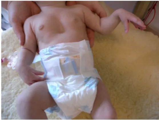
During the initial hospitalisation at the birth clinic, no abnormalities were documented; the child seemed to be healthy, she drank well, and had no local or generalised signs of infection. At the time of discharge, no special features were documented, especially no plexus brachialis palsy. The par-ents noted the special hand position in the second week of life, but they were not worried because, in general, the child was well and they planned to ask the paediatrician at the next screening examination. The parents did not observe any ab-normalities on the skin of the arms, nor redness, nor retraction. At the age of 4 weeks, the girl was admitted to our hospital with a cough and a stridor during inspiration caused by a coronavirus infection. The neurological examination showed a severe degree of wrist drop of both upper limbs associated with an impaired abduction of the thumbs (Fig.1). The grasp reflex was normal and no other muscular or neurological impairment was detectable, so that a bilateral isolated radial nerve palsy diagnosis was made. A careful exam showed no skin lesions of the arms. With respect to the clinical features of isolated radial nerve palsy, we decided to renounce further technical or laboratory investigations. Both occupational and physiotherapy were initiated and the wrists were splinted.
After 8 weeks of therapy, only marginal improvement of the palsy was seen. The 3-month-old girl started with hand-to-hand contact, although she did not play with her fingers. She ex-plored both hands only together with the mouth; no single-hand-to-mouth contact was observed. She started to hold things in her hands sporadically. The wrists were still dropped and the impaired abduction of the thumbs persisted. After stimulation, she was able to bring the right hand in the 0-grade position, but not the left hand. There were no contractures or spasticity. Bicep reflexes were positive; brachioradialis reflexes were not elicited clearly.
E. Böhringer
:
P. Weber (*)Division of Neuropediatrics and Developmental Medicine, University Children`s Hospital Basel, University of Basel, Spitalstr. 33CH 4056 Basel, Switzerland
e-mail: Peter.Weber@ukbb.ch Eur J Pediatr (2014) 173:537–539 DOI 10.1007/s00431-013-2033-4
At the last follow-up, the girl was aged 10 months. In the meantime, the parents had stopped the therapies due to organisational problems. There was no wrist drop anymore but the finger was not flexed at rest. She showed normal, active finger movements and played equally with both hands. There were normal hand-to-hand movements and no weakness in clinching the fingers into a fist. Tendon reflexes were normal.
Discussion
Bilateral isolated radial nerve palsy in newborns is uncom-mon. Former case reports were identified by an electronic search in the database PubMed, without time limitations, using the terms “radial nerve palsy” and “newborn”. We
reviewed the identified articles as well as “related citations in PubMed”. We excluded all cases in which the radial nerve palsy was:
(a) caused by a trauma, fracture, or a congenital deforma-tion (i.e. angioleiomyoma)
(b) associated with abnormalities such as hypo- or hyper-plasia of the radius
(c) not isolated, meaning, i.e. combined with nerve palsies of other peripheral nerves
Reviewing the English-speaking literature, we found 55 reported isolated radial nerve palsies in newborns (Table1), most of them unilateral (93 %) [1–7,9,10,12–15]; in only five cases, including our report, bilateral manifestations were described [4, 11, 13]. Other than in our case, in all newborns with bilateral radial nerve palsies, a difficult de-livery and skin lesions on the dorsolateral side of the upper arms were described.
In general, in 77 % of these patients, a skin lesion like local fat necrosis [1,2,4–7,10–13,15], ecchymosis [4,12] or a “circumferential” or “constriction” band [13,14] of the distal upper arm were remarked. Prolonged labour (>18 h) was present in about 85 % [1–4,7,11–13]. This leads most authors to the conclusion that intrauterine constriction/amnion bands or pressure on the arm either in utero or during delivery causes local fat necrosis, irritating the radial nerve when leaving the radial groove which therefore could be responsible for the radial nerve palsy.
However, our reported child did not have any history of prolonged or complicated delivery and no skin marks were
Fig. 1 Bilateral wrist drop at the age of 4 weeks. Besides this feature, the infant showed a funnel chest, without additional clinical problems
Table 1 Summary of the reported cases in the literature, including our own case
Authors No of cases Uni-/bilateral Skin lesions Complications
at birth
Outcome
Alsubhi et al. [1] 25 Unilateral +17/25 +24/25 Complete recovery, 1 week–2 months
Coppotelli et al. [2] 1 Unilateral + + Improvement, 3 weeks
Deshmukh et al. [3] 2 Unilateral − +2/2 Complete recovery
Feldman [4] 8 Uni- (6/8) and bilateral (2/8) +8/8 +8/8 Complete recovery, 1 week–3 months
Ghinescu et al. [5] 1 Unilateral + − Complete recovery, 3 months
Haider [6] 1 Unilateral + − Complete recovery, 10 weeks
Hayman et al. [7] 4 Unilateral +3/4 +3/4 Complete recovery, 4 days–4 months
Lenn et al. [9] 1 Unilateral − − Improvement, 1 month
Lightwood [10] 2 Unilateral +2/2 +1/2 Complete recovery, 1–2 months
Lundy et al. [11] 1 Bilateral + + Complete recovery, 2 weeks
Monica et al. [12] 4 Unilateral + 4/4 + 4/4 Complete recovery, 3 months–2 years
Morgan [13] 2 Uni- (1/2) and bilateral (1/2) +2/2 +2/2 Complete recovery, few weeks–3 months
Richardson et al. [14] 1 Unilateral + − Incomplete recovery, 6 months after surgery
Ross et al. [15] 2 Unilateral +2/2 − Complete recovery, 4 months
Our case 1 Bilateral − − Complete recovery, 10 months
Total 56 Uni- (51) and bilateral [5] +43/56 +46/56 Complete recovery, 53/56;
4 days–2 years
detected. The literature query revealed 22 % of newborns without skin lesions [1,3,7,9] and 15 % without complications at birth [1,6,9,14,15] although suffering from isolated radial nerve palsy. Most authors assume that pressure due to abnormal intrauterine position accounts for radial nerve palsy, too.
In general, the outcome of this lesion is good: complete recovery was seen in 52 (94 %) children during 4 days to 2 years, treated with wrist splinting and physiotherapy [1,
3–7,10–14]. In two children, there was improvement at the last follow-up (3 weeks, 1 month) [2,9], and only one child underwent surgery [14]. All children with a bilateral mani-festation completely recovered.
In some cases, further investigations were done, such as ultrasound [7, 11], radiographs [7, 8], electromyography (EMG) [7, 8,11,14, 15], and biopsy [7,10]. In an ultra-sound, one author detected a thickening of the radial nerve [13]. In other cases, histology fat necrosis [7, 10] and a granulomatous reaction [10] were reported in histological examinations and an EMG showed either normal results [11], absent sensory and motor response [7,8] or fibrillation [15]. In most cases, no further investigation has been done. In a recently published case [5], a subcutaneous fat necrosis and a deep-seated hematoma in the region of the radial nerve are documented. In our case, we decided to renounce further investigation due to the typical clinical features.
In a differential diagnosis, a local bacterial infection such as arthritis, osteomyelitis, or abscess at the shoulder region [8] or humerus fracture has to be considered. The presence of the grasp reflex is the main differential clinical feature between radial nerve palsy of the newborn and distal symp-toms of brachial plexus palsy.
Conclusion
Our case describes the first case of bilateral radial nerve palsy without a complicated delivery and without skin le-sions on the upper arms. Isolated radial nerve palsy in
newborns, with or without skin lesions, has predominantly a favourable outcome with conservative treatment like wrist splinting, occupational, and physiotherapy and has to be clearly distinguished from brachial plexus palsy with a highly variable outcome.
References
1. Alsubhi FS, Althunyan AM, Curtis CG, Clarke HM (2011) Radial nerve palsy in the newborn: a case series. CMAJ 183:1367–1370
2. Coppotelli BA, Lonsdale JD, Kass E (1979) Sclerema neonatorum complicated by radial nerve palsy following nontraumatic delivery. Mt Sinai J Med 46:143–144
3. Deshmukh NV, Phillips GE (2002) Isolated radial nerve palsy in a newborn: report of two cases. Hand Surg 7:293–294
4. Feldman GV (1957) Radial nerve palsies in the newborn. Arch Dis Child 32:469–471
5. Ghinescu CE, Kamalanathan AN, Morgan C (2012) Unilateral radial nerve palsy in a newborn. Arch Dis Child Fetal Neonatal Ed 97:F153 6. Haider S (2012) Images in paediatrics: subcutaneous fat necrosis caus-ing radial nerve palsy BMJ Case Reports.http://casereports.bmj.com/ content/2012/bcr.10.2011.4904.full
7. Hayman M, Roland EH, Hill A (1999) Newborn radial nerve palsy: report of four cases and review of published reports. Pediatr Neurol 21:648–651
8. Lejman T, Strong M, Michno P (1995) Radial-nerve palsy associ-ated with septic shoulder in neonates. J Pediatr Orthop 15:169–171 9. Lenn NJ, Hamill JS (1983) Congenital radial nerve pressure palsy.
Clin Pediatr (Phila) 22:388–389
10. Lightwood R (1951) Radial nerve palsy associated with localized subcutaneous fat necrosis in the newborn. Arch Dis Child 26:436–437 11. Lundy CT, Goyal S, Lee S, Hedderly T (2009) Bilateral radial
nerve palsy in a newborn. Neurology 72:576
12. Monica JT, Waters PM, Bae DS (2008) Radial nerve palsy in the newborn. A report of four cases and literature review. J Pediatr Orthop 28:460–462
13. Morgan L (1948) Radial nerve paralysis in the newborn. Arch Dis Child 23:137–139
14. Richardson GA, Humphrey MS (1989) Congenital compression of the radial nerve. J Hand Surg (Am) 14:901–903
15. Ross D, Jones R Jr, Fisher J, Konkol RJ (1983) Isolated radial nerve lesion in the newborn. Neurology 33:1354–1356