The Rockefeller University Press $30.00
Rheumatoid arthritis (RA) affects 0.5–1% of
the population in most studied communities
(Neovius et al., 2011). Today, the detection of
prototypic autoantibodies, so-called ACPAs
(anti-citrullinated protein antibodies; Schellekens
et al., 1998), is part of the diagnostic criteria for
RA (Aletaha et al., 2010), and approximately
two thirds of patients are seropositive (Klareskog
et al., 2008). Typically, sera from ACPA
+RA
pa-tients contain antibodies toward several
differ-ent citrullinated autoantigens (Verpoort et al.,
2007; Snir et al., 2010). Anticitrulline antibodies
often emerge before onset of disease
(Rantapää-Dahlqvist et al., 2003; Nielen et al., 2004; van de
Stadt et al., 2011), and we have recently
demon-strated their accumulation in synovial fluid (i.e.,
active rheumatic joints) as compared with sera,
suggesting that they are at least partly produced
in the inflamed lesions (Snir et al., 2010).
Col-lectively, the anticitrulline immunity in RA
pro-vides an interesting and multifaceted case of
potentially pathogenic humoral autoimmunity.
To gain a more thorough understanding of the
humoral aspect of this autoimmunity, we
inves-tigated the cellular and molecular basis of the
production of antibodies to various citrullinated
autoantigens in RA patients.
CORRESPONDENCE Vivianne Malmström: [email protected] OR Khaled Amara: [email protected] Abbreviations used: ACPA, anticitrullinated protein antibody; CCP, cyclic citrullinated peptide; CDR, complementarity-determining region; FWR, framework region; PEI, polyethylenimine; RA, rheumatoid arthritis; SHM, somatic hypermutation; SPR, surface plasmon resonance.J. Steen and F. Murray contributed equally to this paper.
Monoclonal IgG antibodies generated
from joint-derived B cells of RA patients
have a strong bias toward citrullinated
autoantigen recognition
Khaled Amara,
1Johanna Steen,
1Fiona Murray,
1Henner Morbach,
2Blanca M. Fernandez-Rodriguez,
3Vijay Joshua,
1Marianne Engström,
1Omri Snir,
1Lena Israelsson,
1Anca I. Catrina,
1Hedda Wardemann,
4Davide Corti,
3Eric Meffre,
2Lars Klareskog,
1and Vivianne Malmström
11Rheumatology Unit, Department of Medicine, Karolinska University Hospital, Karolinska Institutet, SE-171 76 Solna, Stockholm, Sweden
2Department of Immunobiology, Yale University School of Medicine, New Haven, CT 06520 3Institute for Research in Biomedicine, CH-6500 Bellinzona, Switzerland
4Max Planck Research Group Molecular Immunology, Max Planck Institute for Infection Biology, D-10117 Berlin, Germany
Antibodies targeting citrullinated proteins (ACPAs [anticitrullinated protein antibodies])
are commonly found in patients with rheumatoid arthritis (RA), strongly associate with
distinct HLA-DR alleles, and predict a more aggressive disease course as compared with
seronegative patients. Still, many features of these antibodies, including their site of
production and the extent of MHC class II–driven T cell help, remain unclarified. To
address these questions, we have used a single B cell–based cloning technology to isolate
and express immunoglobulin (Ig) genes from joint-derived B cells of active RA patients.
We found 25% of synovial IgG-expressing B cells to be specific for citrullinated
auto-antigens in the investigated ACPA
+RA patients, whereas such antibodies were not found
in ACPA
patients. The citrulline-reactive monoclonal antibodies did not react with the
unmodified arginine peptides, yet several reacted with more than one citrullinated
anti-gen. A role for active antigen selection of the citrulline-reactive synovial B cells was
supported by the strong bias toward amino acid replacement mutations in ACPA
+antibodies and by their loss of reactivity to citrullinated autoantigens when somatic
mutations were reverted to the corresponding germline sequences.
© 2013 Amara et al. This article is distributed under the terms of an Attribution– Noncommercial–Share Alike–No Mirror Sites license for the first six months after the publication date (see http://www.rupress.org/terms). After six months it is available under a Creative Commons License (Attribution–Noncommercial–Share Alike 3.0 Unported license, as described at http://creativecommons.org/licenses/ by-nc-sa/3.0/).
The Journal of Experimental Medicine
on March 16, 2013
jem.rupress.org
Downloaded from
most commonly rearranged genes for both ACPA
+and ACPA
patients (Fig. 1 a and Table S3). The distribution of IgG
sub-classes of synovial B cells was similar to that of normal human
serum, dominated by IgG1 and IgG2 and with low numbers
of IgG3 (Fig. 1, e and f ). When analyzing the CDR3
(com-plementarity-determining region 3) features, there were no
significant differences between ACPA
+and ACPA
samples
and only subtle differences in the CDR3 lengths (Fig. 1 b). In
terms of light chain gene usage, V1, V3, and J3 were most
commonly used among kappa clones and V1, V2, and J3
for lambda (Fig. 1, c and d; and Table S3). Collectively, these
results indicate that the Ig gene usage in synovial B cells from
the inflamed joints of ACPA
+and ACPA
patients display a
similarly broad Ig gene diversity.
Citrulline-specific B cells are common in ACPA
+but not in ACPA
RA synovial fluid
Out of the 258 sequenced antibodies, we expressed 160, 93
from ACPA
+and 67 from ACPA
patient samples, using an
Ig1 construct as previously published (Wardemann et al.,
2003; Tiller et al., 2008). Hereby, monoclonal recombinant
human antibodies were generated and could then be assessed
for their antigen specificity. We first studied ACPA fine
specific-ity by screening for reactivspecific-ity toward the citrullinated candidate
RESULTS AND DISCUSSION
Synovial fluid IgG
+B cells display extensive clonal diversity
Single cell sorting and subsequent recombinant expression of
antibodies from synovial IgG
+CD19
+B cells from six RA
patients, three ACPA
+and three ACPA
, was performed.
Patient demographics are displayed in
Table S1
. Synovial fluid
was shown to contain significantly lower numbers of CD19
+B cells as compared with peripheral blood, although many
B cells were IgG switched and 5–15% of these cells displayed
an early plasmablast phenotype (CD19
dim/CD27
high; not
de-picted). Serologically, we have previously demonstrated an
enrichment of citrulline-specific IgG antibodies in the joints
of ACPA
+RA patients (Snir et al., 2010) and thus postulated
the presence of ACPA-producing B cells/plasma cells in the
joints of such patients. After our single B cell approach, we
could analyze the synovial B cell repertoire from the analysis
of sequences of the variable parts of the Ig genes. Our data
demonstrate a wide variation among the patients as well as
among individual clones in terms of the gene usage, and
over-all, the majority of the functional Ig genes were represented
among these IgG-expressing B cells (Fig. 1 and
Table S3
).
In total, 258 IgH () and corresponding IgL gene sequences
were generated from ACPA
+(n = 132) and ACPA
(n = 126)
patients. For the Ig heavy chain, IGHV3 and IGHJ4 were the
Figure 1. Similar IgG gene characteristic in synovial B cells from ACPA+ and ACPA RA patients. (a) Summary of VH and JH family gene usage in seropositive (ACPA+) and negative (ACPA) patient samples. (b) Overview of IgH () CDR3 amino acid characteristics: length (left) and neg-atively (middle) and positively (right) charged amino acids in ACPA+ and ACPA patients. (c and d) Light chains data depicting V/J (c) and V/J (d) gene family usage. (e) Distribution of IgG subclasses displayed per patient with the total number of sequences analyzed represented in the mid-dle of the pie charts. (f) IgG subclass distribution in ACPA+ and ACPA patient samples. Differences between individual fractions were not statisti-cally significant (P > 0.5), as determined by Fisher’s exact test.
on March 16, 2013
jem.rupress.org
positive clones for one or several of the different citrullinated
peptides. Notably, such clones were generated only from
ACPA
+and not from ACPA
patient samples (Fig. 2 a). All
clones reacting toward citrullinated peptides failed to recognize
RA autoantigens -enolase (CEP-1; amino acids 5–21),
vimen-tin (amino acids 60–75), and fibrinogen (amino acids 36–52).
Overall, CEP-1 was the dominant B cell epitope recognized by
these citrulline-specific antibodies, and we identified several
Figure 2. Citrulline-reactive antibodies could be generated from ACPA+ but not from ACPA RA patients. (a) Pie charts summarizing the frequency of the citrulline-reactive clones (to any of the tested citrulline peptides) in individual patient samples. The top panel represents the three ACPA+ patient samples and the three in the bottom panel, the ACPA samples. The absolute number of tested antibodies is indicated in the center of each pie chart. (b) Frequency comparison of antigen reactivity in the clones generated from the different ACPA+ and ACPA patients (one symbol summarizes all generated antibodies from each individual; range 15–37 clones per patient sample; in total 93 antibodies were tested from ACPA+ and 67 from ACPA patients). CCP denotes general citrulline reactivity; polyreactivity is based on positivity of two or more of the following: insulin, LPS, and double-stranded DNA; and lastly tetanus toxoid was used as a proxy for recall response. Horizontal lines represent the mean values of reac-tivity for all RA patients. (c) ELISA graphs of the various ACPA fine specificities: CEP-1 (citrullinated -enolase, amino acids 5–21), cit-fib (citrullinated fibrinogen, amino acids 36–52), and cit-vim (citrullinated vimentin, amino acids 60–75). Black lines represent individual IgG+ memory B cells antibod-ies. (c) Green lines represent the high positive control (serum pool of ACPA+ antibodies). (c and e) Red lines represent the negative control antibody (serum pool of ACPA antibodies). Each vertical row represents one ACPA+ patient sample. (d) ELISA graphs of the corresponding arginine versions of the peptides. (c–e) Dashed horizontal lines show a cutoff of OD450 for positive reactivity. (e) Sequences from four selected antibodies with high reac-tivity to two or more of the citrullinated peptides that were subsequently reverted into their predicted germline sequence (in both the FWRs and CDRs) and then expressed as recombinant antibodies. ELISA graphs illustrate the citrulline reactivity of the original antibodies (i.e., mutated; left) compared with those encoded by the predicted germline sequences (right). Data are representative of three to six independent experiments.
on March 16, 2013
jem.rupress.org
Table 1.
Ig gene usage, CDR3 amino acid features, and reactivity of recombinant c
itrulline-specific IgG antibodies generated from synovial memory B cells of RA patients
Clone Heavy-chain Light-chain Reactivity V D J IgG SC V-Mut. CDR3 (aa) (+) Length Family V J V-Mut. CDR3 (aa) (+) Length CEP-1 cit-fib cit-vim CCP Polyreactivity Tetanus 1276SF-B01 3-49 3-3 3 IgG1 4 (3R/1S) EYVDFWSDPSRARFDI 2 16 1-44 3 2 (2R) AAWDDSLNGWV 0 11 + + + neg neg neg 1276SF-B07 3-23 4-4 4 IgG1 8 (7R/1S) DWRHNNYGPPHSFDY 3 15 2-14 3 4 (3R/1S) SSYTSSSTWV 0 10 + neg neg neg 1276SF-C07 3-7 3-10 4 IgG1 6 (4R/2S) RGKCFFDC 2 8 7-43 2 6 (3R/3S) VLYMGSGISV 0 10 ++ + pos neg neg 1276SF-D10 3-30 6-13 6 IgG1 6 (4R/2S) VRGAAATGYYYGMDV 1 15 3-1 2 6 (6R) QAWDSSTVV 0 9 +++ ++ ++ pos pos neg 1276SF-F12 4-59 4-17 4 IgG1 3 (2R/1S) RLLGDYIFDY 1 10 3-1 2 4 (4R) VRRGTAAVV 2 9 + + pos pos neg 1276SF-G08 4-39 3-10 4 IgG1 1 (1R) VRGYFDY 1 7 3-15 1 3 (2R/1S) QHYNNWPPWT 1 10 ++ + + neg neg neg 1276SF-H10 4-39 3-22 4 IgG1 4 (3R/1S) LPNYYDSSGYARGGFDY 1 17 3-15 3 1 (1R) QQYNNWPRGGFT 1 12 + pos neg neg 1103SF-A03 4-39 5-18 4 IgG1 7 (2R/5S) RRGYSYGYSRARGTTFDY 5 20 1-51 3 9 (6R/3S) GTWDSSLSAGV 0 11 +++ ++ neg neg neg 1103SF-A04 1-2 3-3 3 IgG1 16 (14R/2S) DRSPIDYDFWSGSTFYSYGMDV 2 24 1-47 2 6 (4R/2S) AAWDDSLSGVV 0 11 +++ + ++ pos pos neg 1103SF-B02 1-69 3-9 4 IgG1 8 (5R/3S) INTSPILTGYYFPGVHDY 1 18 1-12 3 6 (4R/2S) QQANSFPFT 0 9 ++ + + pos pos neg 1103SF-B03 4-31 3-22 4 IgG1 3 (2R/1S) GAREGLIVVDTFDY 1 14 1-39 3 4 (4R) HQSYSTPQT 1 9 + neg neg neg 1103SF-B04 3-30-3 2-2 5 IgG1 4 (3R/1S) DLGDTSCYT 0 10 1-39 1 5 (4R/1S) QQSYSTPQT 0 9 +++ neg neg neg 1103SF-H05 3-30-3 6-6 4 IgG1 2 (1R/1S) DPYRGKATQDY 2 11 1-39 3 7 (5R/2S) QQSYSTPFT 0 9 + + pos pos neg 1325SF-A04 3-23 2-21 4 IgG1 34 (27R/7S) VKAWEIIAS 1 9 3-21 3 29 (21R/8S) QVWDSSADHPV 1 11 +++ + pos neg neg 1325SF-B05 1-8 4-23 6 IgG1 25 (18R/7S) GGRPYYYYYGMDV 2 17 2-8 2 26 (19R/7S) SSYAGGNVVV 0 10 ++ pos neg neg 1325SF-B07 3-9 3-22 4 IgG1 2 (1R/1S) GARASDSSGSNYFDY 1 15 1-5 5 4 (3R/1S) QQHNHYSPIT 2 10 + neg neg neg 1325SF-C02 1-2 3-10 4 IgG1 2 (1R/1S) SGASITMIRGALEN 1 14 1-47 3 7 (5R/2S) AAWDDSLRWV 1 10 + neg neg neg 1325SF-C04 3-21 4-23 4 IgG1 8 (3R/5S) GITVITPGSY 0 11 3-21 2 10 (7R/3S) QVWDTSSDHHVV 2 12 ++ pos pos neg 1325SF-C05 1-2 4-17 5 IgG1 8 (8R) VMDPRPPYGDYAISH 2 15 1-47 2 9 (8R/1S) AAWDDSLR 1 8 ++ pos neg neg 1325SF-D06 3-74 5-24 2 IgG1 25 (15R/10S) DAGNSGHDWYFDL 1 13 2-23 3 19 (15R/4S) CSYAGRGLGV 1 10 +++ +++ + pos neg neg 1325SF-G12 4-39 5-24 4 IgG3 5 (4R/1S) HRNPPIAIVIFVFSPMDY 1 18 1-51 1 11 (7R/4S) GTWDTSLNVPYV 0 12 + pos neg neg 1325SF-H07 3-23 3-3 4 IgG1 3 (2R/1S) GSLYDFWSGYPDSFDY 0 16 1-5 1 8 (6R/2S) QQYNTYLWT 0 9 + + pos pos neg 1325SF-B109 3-49 3-3 3 IgG1 4 (4R) TRDEYYDFWSGPSRAFDI 2 18 1-44 3 2 (2R) AAWDDSLNGWV 0 11 ++ + + neg neg neg 1325SF-B117 4-59 5-12 4 IgG2 13 (6R/7S) LDVEYSGFDLAYYFDS 1 18 2-23 3 19 (16R/3S) CSHARSYSLV 2 10 +++ neg neg neg 1325SF-C127 3-15 2-15 6 IgG1 5 (4R/1S) TTDPGYCSGGRCYHHFYYGMDV 3 22 1-51 3 5 (2R/3S) GTWDSSLSPWV 0 11 ++ ++ neg pos neg
For reactivity, increasing signal strength is denoted as + (negative control signal < OD 450 nm < 1.0), ++
(1.0 < OD 450 nm < 2.0), and +++ (OD 450 nm > 2.0).
on March 16, 2013
jem.rupress.org
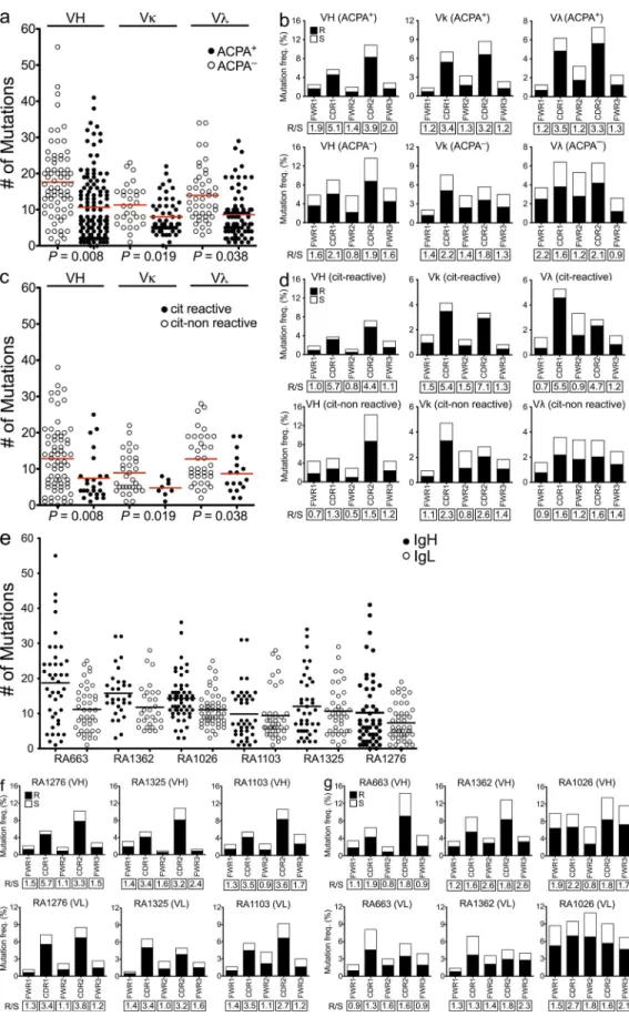
random mutations (ratio > 2.9; Shlomchik et al., 1987). R/S
mutation ratios in the framework regions (FWRs) are,
how-ever, usually lower than in the CDRs but may have a role in
the antibody structure (Berek et al., 1991). In our study, the
sequences generated from cells of ACPA
patients contained
significantly more somatic mutations than those from ACPA
+patients (Fig. 4, a and e). The overrepresentation of mutations
in ACPA
patients could be a consequence of recycling of
nonreactive or low-affinity B cells within the germinal centers
(Allen et al., 2007). Interestingly, sequences generated from
ACPA
+patient samples displayed a bias toward replacement
mutations as compared with sequences isolated from ACPA
patients, resulting in a striking difference in the R/S mutation
ratios, especially in the CDR1 and CDR2 regions, which
nor-mally interact with antigens (Fig. 4, b, f, and g). These results
suggest that antigen-driven B cell activation and somatic
hy-permutations (SHMs) are characteristic of antibodies isolated
from ACPA
+patients. Thus, although these data provide
sup-portive evidence for an antigen-driven response, we cannot
exclude the possibility of a concomitant component of
poly-clonal B cell stimulation in these synovial samples (Shlomchik
et al., 1987; Randen et al., 1992; Bridges et al., 1993).
Next we performed reanalyses of the mutation patterns
of the monoclonal antibodies generated from ACPA
+pa-tient samples based on citrulline reactivity. As seen in Fig. 4 c,
the citrulline-specific antibodies displayed fewer overall
mutations but higher R/S ratios in their CDRs as compared
the native nonmodified arginine version of the peptide (Fig. 2,
c and d). Several clones displayed cross-reactivity to one or
more citrulline antigens, warranting more detailed affinity
analysis in addition to the ELISA. For this purpose, surface
plasmon resonance (SPR) measurements were performed to
study the kinetics of antibody–peptide interactions, and the
analyzed citrulline-specific monoclonal antibodies were shown
to display cross-reactivity to various citrullinated peptides with
variable affinities. Overall, the K
dvalues were in micro- to
nanomolar range for all analyzed antibodies: from 1.36 × 10
6to 5.94 × 10
10M for CEP-1, from 3.84 × 10
5to 3.01 ×
10
9M for cit-fib, and from 1.56 × 10
6to 1.06 × 10
10M
for cit-vim (
Table S2
). The antibody 1276SF-D10 showed the
highest binding affinity for CEP-1 with a K
d= 5.94 × 10
10M
but exhibited 20-fold and almost 400-fold lower binding
af-finities to cit-fib (K
d= 3.10 × 10
9M) and cit-vim (K
d=
1.55 × 10
8M), respectively. The antibody 1103SF-B02
showed the highest binding affinity for cit-vim with a K
d=
1.06 × 10
10M but exhibited 3- and 100-fold lower binding
affinities to CEP-1 (K
d= 3.03 × 10
9M) and cit-fib (K
d=
1.01 × 10
8M), respectively. Many antibodies displayed low
to medium overall binding affinity and cross-reacted to
sev-eral citrullinated antigens, whereas the antibodies with high
binding affinity for one antigen only displayed low cross-
reactivity to other citrullinated antigens. These observations
suggest that several different target antigens may initially drive
the autoimmune responses before being refined and focused
on one particular epitope.
Next, the generated recombinant antibodies were tested
for alternative antigen specificities, first with the RA
diagnos-tic andiagnos-ticyclic citrullinated peptide (anti-CCP) assay (Snir et al.,
2010) and subsequently also for polyreactivity, using a
pre-viously validated panel of ubiquitous antigens toward which
RA and systemic lupus erythematosus patient–derived B cells
often show reactivity (Wardemann et al., 2003; Tiller et al.,
2008), as well as for the recall antigen tetanus toxoid.
Reac-tivity to CCP was only found for antibodies derived from
ACPA
+patients (P < 0.001), whereas polyreactivity and
tet-anus reactivity were found in both groups at similar
fre-quencies (Fig. 2 b and Table S3). However, none of the
citrulline-reactive antibodies were cross-reactive to tetanus,
whereas 8 out of 25 displayed polyreactivity (Table 1). Two of
the citrulline-reactive monoclonal antibodies (1325SF-B109
and 1276SF-D10) were used for the staining of synovial tissue
biopsies from three RA patients to analyze the presence of
the target antigens in the rheumatic joint. Both antibodies
showed distinct staining of inflamed synovial tissues from RA
patients, as exemplified in in Fig. 3, whereas staining with the
isotype control was negative in all experiments (Fig. 3 c).
Citrulline-specific Igs display a different mutational pattern
as compared with citrulline-negative Ig derived
from the same inflamed joint
A signature of antigen-driven selection is represented by an
increase in the replacement to silent (R/S) mutation ratios in
the CDRs as compared with what would be expected for
Figure 3. Immunohistochemical localization of citrullinated proteins in synovial tissues of RA patients. (a and b) Immunohisto-chemistry using two of the recombinant citrulline-specific antibodies, 1276SF-D10 (a) and 1325SF-B109 (b), brown staining of both the lining (black arrows) and sublining (white arrows) layers in an inflamed syno-vial biopsy, obtained at the time of joint arthroplasty from a RA patient. (c) Staining of a matched irrelevant IgG2a-negative control used at similar concentration. Insets show the same samples at higher magnifi-cation. Similar results were observed in two other RA synovial tissues (not depicted). Data presented in this figure are representative of three independent experiments from four patients. Bars, 68 µm.
on March 16, 2013
jem.rupress.org
Figure 4. Citrulline-reactive antibodies display a bias toward nonsynonymous mutations in the CDRs. (a) Comparison of the absolute numbers of somatic mutations in individual VH, V, and V genes of the antibodies generated from ACPA+ and ACPA patients. (b) Frequencies of replacement (R) and silent (S) mutations in the CDRs and FWRs of VH, V, and V genes of synovial IgG+ memory B cells from ACPA+ and ACPA RA patients. (c) Comparison of the absolute numbers of somatic mutations between citrulline-positive versus -negative antibodies generated
on March 16, 2013
jem.rupress.org
carriage of the PTPN22 risk allele (two out of three ACPA
+and one out of three ACPA
). ACPA
+patients were also
selected for having high titers in the CCP test (>300, cut off
for positivity 25). Still, our data demonstrate a considerable
heterogeneity in the fine specificity of the citrulline-reactive
B cells. None of the antibodies generated appeared to be
clonally related based on the dissimilarity of the VH-DH-JH
joints, a finding which could be explained by the many
dif-ferent candidate autoantigens that have been found to be
targeted by ACPA in RA patients (Lundberg et al., 2012).
Clearly, understanding the role of ACPAs in the
pathogene-sis of RA will require careful dissection of several different
epitopes, a notion well-compatible with observations in the
mouse, where different anticitrulline antibodies are able to
trigger or enhance development of arthritis (Kuhn et al.,
2006; Hill et al., 2008; Uysal et al., 2009) or specific features of
arthritis such as bone erosions (Harre et al., 2012). Extended
studies on synovial tissue and possible differences to synovial
fluid would also be needed to fully dissect the local
contribu-tion of B cells within the rheumatic joint.
Our study gives rise to several downstream questions
relating to the mechanisms that determine accumulation of
ACPA-producing B cells and plasma cells in joints and how
and where these cells differentiate and undergo SHM. Key
factors for driving plasma cell differentiation such as IL-6,
IL-21, APRIL, and CXCL13 are present (and elevated) in the
rheumatic joint (McInnes and Schett, 2007; Humby et al.,
2009), and plasma cell differentiation (without assessment of
antigen specificity) has been demonstrated to occur in the
rheumatic joint by extensive V gene analysis (Scheel et al.,
2011). Indeed, a subset of the IgG
+B cells we analyzed
dis-played an early plasmablast phenotype, which is indicative
that at least some of the antibodies we have generated are also
secreted in vivo.
Besides the role of antibodies as effector molecules in RA
pathology, it is also tempting to speculate that the synovial
IgG-expressing B cells specific for citrullinated autoantigens
may function as local antigen-presenting cells important for
T cell reactivation. Here the B and T cell epitopes may be
ex-pected to overlap, as has been shown to be the case for
citrul-linated vimentin (Verpoort et al., 2007; Snir et al., 2011).
A similar scenario has been discussed in the context of celiac
disease, another condition under which high numbers of
autoantigen-specific B cells are found in the target organ
(Di Niro et al., 2012).
In conclusion, the present findings have demonstrated the
utility of a single cell approach for generation of monoclonal
antibodies from the target organ of a classical autoimmune
disease, with resulting new insights on the specificity as well
as mutational features of the resulting IgGs. Notably, the
with the citrulline-negative clones (Fig. 4 d). Traditionally,
selection of high-affinity B cells during affinity maturation is
primarily dependent on the presentation of antigens to T cells
and takes place in germinal centers (Liu et al., 1997).
How-ever, it is known that such affinity maturation may also take
place at extrafollicular sites (William et al., 2002). To address
whether the citrulline-specific antibodies had undergone
affinity-driven maturation, we reverted four antibodies with
high citrulline reactivity to their predicted germline sequences.
Reversion of mutations in these four citrulline-specific
mono-clonal antibodies led to a loss of reactivity to all the autoantigens
(Fig. 2 e). The above observations suggest that the
genera-tion of citrulline-specific antibodies may result from T cell–
dependent B cell immune responses (Shlomchik et al., 1987;
Weiss and Rajewsky, 1990; Berek et al., 1991). Moreover,
SHM was likely required for development of high-affinity
citrulline-binding antibodies because reversion of these four
representative citrulline-specific antibodies to the
corre-sponding germline led to complete loss of binding to the
citrullinated peptides.
We and others have previously postulated that
auto-immunity against citrullinated autoantigens may initially be
triggered at sites distant from the joints, e.g., in lungs during
smoking (Klareskog et al., 2008) or in gums during infection
with Porphyromonas gingivalis or other bacteria (Lundberg
et al., 2008; Berthelot and Le Goff, 2010), whereas the factors
behind the transition to arthritis are still unclear. Our data
provide the first evidence that specificity of IgGs of B cells
from joints of ACPA
+RA patients can be drastically biased
toward reactivity with the candidate citrullinated
autoanti-gens. More extensive investigations of B cell/plasma cell
spec-ificities in the blood or other lymphoid organs would be
required to determine the degree to which this bias is caused
by local immune reactions driven by T cells present in
in-flamed joints or whether immune activation against
citrulli-nated autoantigens also happens outside the joint. The analysis
of SHM patterns suggests that at least antibodies generated
from synovial fluid B cells are driven by T cell immunity.
In this context, it is of interest that T cell responses to the
same sets of RA-associated citrullinated autoantigens have been
described and associate to the same set of HLA-DR alleles
(Sebbag et al., 2006; James et al., 2010; Snir et al., 2011; Pieper
et al., 2012).
RA patients are known to display a B cell repertoire
gen-erally biased toward autoimmunity (Samuels et al., 2005;
Menard et al., 2011), and some underlying factors have been
identified such as increased frequency of the C1858T PTPN22
risk allele in ACPA
+RA patients (Menard et al., 2011). Our
patient samples were selected and characterized concerning
the presence of the RA-associated HLA-DRB1 alleles and
from ACPA+ patients. (d) R/S mutation ratios in the CDRs and FWRs of the VH, V, and V genes in the citrulline-positive and -negative clones gener-ated from ACPA+ patients. (e) Absolute numbers of somatic mutations in individual VH and IgL genes of the antibodies generated from the individual patient samples. (a, c, and e) Horizontal bars indicate mean. (f and g) Frequencies of R and S mutations in the CDRs and FWRs of VH and VL of syno-vial IgG+ memory B cell from the individual ACPA+ (f) and ACPA (g) RA patients. The R/S ratios are indicated below the respective graphs.
on March 16, 2013
jem.rupress.org
for each region based on the absolute number of nucleotides in all analyzed sequences as defined by IgBLAST. IgG isotype subclasses were determined using the international ImMunoGeneTics database (Table S3).
Cloning and sequencing
Restriction sites for expression vector cloning were introduced by using gene-specific primers and first PCR products as template as previously described (Wardemann et al., 2003; Tiller et al., 2008). The digested PCR products from each single cell were cloned into expression vectors con-taining human Ig1, Ig, or Ig constant regions as previously described (Wardemann et al., 2003; Tiller et al., 2008). Ligation reactions were per-formed using the quick ligase kit (New England Biolabs, Inc.) according to the manufacturer’s instructions. Expression vectors containing IgH and the corresponding IgL genes were transformed into DH5 bacteria (Gibco Invitrogen) and sequentially isolated using NucleoSpin plasmid DNA purification kits (Macherey-Nagel) according to the manufacturer’s instructions. To confirm identity with the original PCR products, the isolated IgH and IgL plasmids from each clone were sequenced.
Antibody production and purification
Antibodies were produced by transient cotransfection of exponentially growing 293 human embryonic kidney fibroblasts (Gibco Invitrogen) using the polyethylenimine (PEI)-precipitation method as described previously with some modifications (Boussif et al., 1995; Mouquet et al., 2011). Adherent (293A) as well as suspension (Freestyle 293F) cells were used to generate recombinant proteins (Gibco Invitrogen). 293A cells growing in DMEM + GlutaMAX (Gibco Invitrogen) supplemented with 10% ultra-low IgG FBS (Gibco Invitrogen) and antibiotics/antimycotics (Gibco Invi-trogen) were washed at 70% cell confluency using serum-free DMEM for 5 min. The medium was then replaced with serum-free DMEM + Gluta-MAX supplemented with 1% Nutridoma-SP (Roche) and antibiotics/ antimycotics (Gibco Invitrogen) before PEI-mediated transfection with equal amounts (10 µg) of IgH and corresponding IgL chain vector DNA. Anti-bodies were alternatively produced by transient transfection of suspension cultured 293 Freestyle cells (Gibco Invitrogen) with PEI-Max (Polysciences) as transfection agent and a total of 20 µg of vector DNA, as previously de-scribed (Corti et al., 2011). Supernatants were collected after 6 d of culture, and antibodies were purified by binding to protein G–Sepharose (Sigma-Aldrich) and eluted with 0.1 M glycine buffer, pH 3, into storage buffer (1 M Tris-HCl, pH 8). Antibody concentrations were determined by anti– human IgG1 ELISA using human monoclonal IgG1 as standard (Sigma-Aldrich) as previously described (Wardemann et al., 2003; Tiller et al., 2008). Expression of antibodies consisting of both heavy and light chain, as well as the protein purity, was verified by PAGE.
Reversion of hypermutated sequences to germline
Antibodies for reversion experiments were chosen according to their level of reactivity to the citrullinated autoantigens (Table 1). Germline sequences were determined by reverting mutations to the germline sequence while retaining the original CDR3 junctions and terminal deoxynucleotidyl transferase (TdT) N nucleotides. Germlined VH and VL nucleotide sequences were codon op-timized and synthesized by Genscript and their accuracies were confirmed by sequencing. Recombinant mutated and germline-reverted antibodies were tested for citrulline reactivity by ELISA as described above in comparison.
Antibody specificities
Assessment of IgG antibodies reactivity against -enolase. Anti–-enolase reactivity was determined by ELISA as described previously with some modifications (Snir et al., 2010). In brief, 96-well Nunc plates were coated with 2.5 µg/ml of the -enolase peptide 1 in its native (REP-1) or citrullinated (CEP-1) form (Kinloch et al., 2005; Lundberg et al., 2008). Purified antibodies, diluted in blocking buffer, were used at concentrations of 5, 1, 0.2, and 0.04 µg/ml to generate dilution curves. Positive and negative controls included sera from patients and healthy individuals, respectively. In all ELISAs, HRP-conjugated goat anti–human IgG (Jackson ImmunoResearch
generated antibodies will also provide the immunological/
rheumatological community the opportunity to investigate
the role of specific immunity to citrullinated autoantigens in
arthritis and to identify ways to counteract the emergence, as
well as the effects, of such immunity.
MATERIALS AND METHODS Patients
RA patients attending the Rheumatology Clinic at Karolinska University Hospital and fulfilling the American College of Rheumatology criteria for the diagnosis of RA (Arnett et al., 1988) were included in the study. In-formed consent was obtained from all patients in accordance with a protocol approved by the Ethical Review Committee North (KI forskningsetikkom-mitte Nord) of Karolinska University Hospital.
Synovial fluid samples from six HLA-DR shared epitope–positive RA patients (five females and one male) with a median age of 39 yr (range 35–47 yr) and median disease duration of 10 yr (range 3–28 yr) were included. The joint fluid was taken at a time point of local disease activity that required arthrocentesis and subsequent injection of local corticosteroids.
The demographic and clinical features of all six patients are summarized in Table S1. In brief, three of the patients were ACPA+ based on a CCP test, whereas three were negative. The ACPA+ patients showed variable positivity for citrullinated -enolase (CEP-1; Kinloch et al., 2005; Lundberg et al., 2008), vimentin (amino acids 60–75), and fibrinogen (Table S1; Verpoort et al., 2007).
Flow cytometric analysis
Mononuclear cells were prepared from synovial fluid samples by Ficoll-Paque Plus preparation (GE Healthcare). Immunofluorescence labeling for flow cytometry was performed by incubating cell suspensions with anti– human CD19 conjugated to PE-Cy7 (BioLegend), anti–human IgG conju-gated to APC (Miltenyi Biotec), anti–human CD3 (BioLegend) and anti–human CD14 (BioLegend) both conjugated to PB, anti–human CD27 conjugated to APC-Cy7 (BioLegend), anti–human IgD conjugated to FITC (BD), and anti–human IgM conjugated to PE (BioLegend). Cell incubation with anti-bodies was performed at 4°C for 30 min in PBS/1% human serum. Flow cyto-metric analysis was performed using a Cyan flow cytometer (Dako) and FlowJo software version 8.8.7 (Tree Star).
Single B cell sorting
Cryopreserved synovial mononuclear cells were thawed, and the cell suspen-sions were stained with anti–human CD19 conjugated to PE (Miltenyi Bio-tec), anti–human IgG conjugated to APC (Miltenyi BioBio-tec), and anti–human CD3 (BD) and anti–human CD14 (BD) both conjugated to FITC. Single synovial B cells expressing CD19 and IgG, while lacking CD3 and CD14, were sorted by flow cytometry using a Cytomation MoFlo (Dako) into 96-well PCR plates containing 5 µl/well of 0.5× PBS containing 10 mM DTT and 8 U RNAsin inhibitor (Promega) as previously described (Warde-mann et al., 2003; Tiller et al., 2008).
cDNA synthesis and PCR amplification
Single cell cDNA was synthesized in a total volume of 15 µl in the origi-nal 96-well PCR plate using the SuperScript III RT (Gibco Invitrogen). Individual IgH () and IgL chain ( or ) gene rearrangements were am-plified independently, using the cDNA as template, by two successive rounds of PCR (50 cycles each) using primers as previously described (Tiller et al., 2008).
Ig gene sequence analysis
From each 96-well PCR plate, wells from which matching IgH () and Ig/ Ig amplicons were obtained were sequenced (Eurofins MWG Operon) and analyzed for Ig gene usage, CDR3 features, and number of V gene SHMs by IgBLAST comparison (Table S3). The Ig CDR3 length was determined as indicated previously (Kabat et al., 1983; Kabat and Wu, 1991). Replacement (R) and silent (S) mutation frequencies in FWRs and CDRs were calculated
on March 16, 2013
jem.rupress.org
Fc. Parallel sections were stained with irrelevant origin–, mouse monoclonal IgG2a isotype–, and concentration-matched antibody as negative control (Sigma-Aldrich). The next day, sections were first blocked with 1% normal goat serum and then incubated for 30 min with biotin-conjugated goat anti– mouse secondary antibody (Invitrogen). Staining was performed using the VECTASTAIN Elite ABC kit (Vector Laboratories) and visualized with 3,3-diaminobenzidine (DAB). Sections were counterstained with Mayer’s hema-toxylin, permanently mounted, and viewed by a light microscope (Reichert Polyvar 2 type 302001; Leica).
SPR analysis of antibody affinities
To analyze the interactions between the citrullinated autoantigens and the citrulline-specific monoclonal antibodies, we performed an SPR analysis on a Biacore T200 (GE Healthcare) using a streptavidin capture (CAP) sensor chip according to the manufacturer’s instructions. Initially, Biotin CAPture reagent, which is a modified form of streptavidin, was immobilized on the CAP sensor chip for 5 min at a flow rate of 2 µl/min. Next, to immobilize the biotinylated citrullinated peptides on the streptavidin surface of the CAP-chip, the CEP-1 cit-fib and cit-vim (50 nM concentrations in 0.3 M sodium phosphate buffer, pH 7.4) were injected for 3 min at a flow rate of 10 µl/min. Once the citrulli-nated peptide surface on the CAP-chip was prepared, five different concentra-tions of each of the citrulline-specific monoclonal antibodies (ranging from 5 nM to 1.5 µM) were injected into the flow cells at a flow rate of 30 µl/min. For each concentration used, cycles of injection for 3 min and dissociation period were performed. All SPR analyses were performed at 25°C. Binding datasets from five different concentrations of monoclonal antibodies were collected using a single-cycle kinetics mode (Karlsson et al., 2006). The bind-ing data were analyzed usbind-ing the Biacore T200 Evaluation software version 1.0 (GE Healthcare) and were fitted with a 1:1 binding model.
Statistics
Ig gene repertoire analyses, analysis of positive and negative charges in IgH CDR3, and antibody reactivity were calculated by Fisher’s exact test or 2 test. Differences in CDR3 length and V gene mutations were calculated by paired two-tailed Student’s t test (with Wilcoxon’s signed rank test). Ratios of V gene SHM were calculated by one-way nonparametric Kruskal-Wallis test followed by Dunn’s multiple comparison tests. Differences were consid-ered to be statistically significant at values of P ≤ 0.05. All statistical analyses were performed by Prism software version 5.0c (GraphPad Software).
Online supplemental material
Table S1 shows patient characteristics. Table S2 shows kinetic rates and af-finity of synovial IgG antibodies to CEP-1, cit-fib, and cit-vim measured by SPR. Table S3, included as a separate Excel file, shows sequence data and reactivity of IgG antibodies from synovial memory B cells of RA patients. Online supplemental material is available at http://www.jem.org/cgi/ content/full/jem.20121486/DC1.
We would like to thank Annika van Vollenhoven (Centre for Molecular Medicine, Stockholm, Sweden) for her excellent assistance in single cell sorting as well as Cornelia Kreschel (Max Planck Institute for Infection Biology, Berlin, Germany) and Jason Bannock (Yale University School of Medicine, New Haven, CT) for their valuable technical assistance. We would like to thank Stephen Rapecki, Emily Barry, Ruth Morris, Hannah Hailu, and Kerry Tyson (UCB Celltech, Slough, England, UK) for their valuable help in the murinization of the human monoclonal antibodies used for immunohistochemistry. We would like to acknowledge GE Healthcare for the use of Biacore T200 at their Demolab at Science for Life Laboratories, Stockholm. This work was supported by the European Research Council (grant number 250167), the Innovative Medicines Initiative Be The Cure Joint Undertaking program (grant number 115142-2), and the Swedish Combine program, as well as grants from the Swedish Association Against Rheumatism, the King Gustaf the V’s 80-Year Foundation, the Swedish Research Council (project grants to V. Malmström and L. Klareskog and a Linneaus center grant with L. Klareskog as co-investigator), National Institutes of Health National Institute of Allergy and Infectious Diseases (grant number AI071087 to E. Meffre), and the Deutsche Forschungsgemeinschaft (DFG; MO 2160/2-1 to H. Morbach).
Laboratories, Inc.) was used as the detecting antibody and visualized using the chromogenic substrate 3,3,5,5-tetramethylbenzidine (Bio-Rad Labo-ratories). Reactivity was detected at 450 nm with a reference of 650 nm, and the minimum OD450 at which antibodies were considered reactive is indi-cated in each graph by the red line (Fig. 2, c and e). To be considered reac-tive, the results for any given antibody had to be confirmed in at least two independent experiments.
Assessment of IgG antibody reactivity against fibrinogen and vimentin. Anti-vimentin and -fibrinogen reactivity was determined by ELISA as described previously with some modifications (Snir et al., 2010). In brief, streptavidin-coated high binding–capacity 96-well plates (Thermo Fisher Scientific) were coated with 1 µg/ml of biotinylated vimentin (amino acids 60–75) or fibrinogen (amino acids 36–52) peptides in their native and citrullinated forms (Verpoort et al., 2007). All other stages of the vimentin and fibrinogen ELISAs were performed exactly as for the -enolase ELISA described above.
Anti-CCP assay. Anti-CCP reactivity was determined using a commercial anti-CCPlus ELISA kit according to the manufacturer’s instructions (Euro-Diagnostica). Purified antibodies were used at concentrations of 5, 1, 0.2, and 0.04 µg/ml in blocking buffer. Positive and negative controls included sera from patients and healthy individuals, respectively. Reactivity was de-tected at 450 nm with a reference of 650 nm, and the minimum OD450 at which antibodies were considered reactive is indicated in each graph. To be considered reactive, the results for any given antibody had to be confirmed in at least two independent experiments.
Assessment of IgG antibodies polyreactivity. IgG antibodies were screened for reactivity against specific antigens as previously described (Wardemann et al., 2003). Three different control antibodies were used in all ELISAs: mGO53 (nonreactive), JB40 (weak reactive), and ED38 (high reactive; Wardemann et al., 2003; Meffre et al., 2004). Antibodies were considered polyreactive when they recognized at least two antigens out of the three analyzed antigens that include double-stranded DNA, insulin, and LPS. In all ELISAs, HRP-conjugated goat anti–human IgG (Jackson ImmunoResearch Laboratories, Inc.) was used as detecting antibody and revealed using the chromogenic substrate 3,3,5,5-tetramethylbenzidine (Bio-Rad Laboratories). Reactivity was detected at 450 nm with a refer-ence of 650 nm and the minimum OD450. To be considered reactive, the results for any given antibody had to be confirmed in at least two inde-pendent experiments.
Assessment of IgG antibody reactivity against human tetanus. Anti-tetanus antibodies were detected using a commercial anti–human Anti-tetanus ELISA kit according to the manufacturer’s instructions (MyBioSource). Purified antibodies were used at a concentration of 0.5 µg/ml. Samples were added to the appropriate microtiter plate wells and incubated for 20 min at 37°C. ELISAs were developed with HRP-conjugated tetanus antigen and revealed using the chromogenic substrate 3,3,5,5-tetramethylbenzidine. Reactivity was detected at 450 nm with a reference of 650 nm and the mini-mum OD450. To be considered reactive, the results for any given antibody had to be confirmed in at least two independent experiments.
Immunohistochemical analysis
Immunohistochemical staining was performed on synovial tissue sections to investigate the presence of citrullinated proteins. Biopsy specimens were ob-tained from three RA patients at the time of joint replacement. Serial cryo-sections (7 µm) were fixed for 20 min with 2% (vol/vol) formaldehyde (Sigma-Aldrich) and stored at 70°C until use. For the immunostaining, synovial tissue sections were blocked with 1% H2O2 and 20% AB human serum (Akademiska pharmacy) for 20 min and incubated overnight in a moist chamber at 4°C with the purified recombinant “murinized” antibodies (range 3–10 µg/ml). The murinization of the human monoclonal antibodies was performed by replacing the full human IgG1 Fc by the murine IgG2a
on March 16, 2013
jem.rupress.org
posttranslationally modified (citrullinated) fibrinogen in DR4-IE trans-genic mice. J. Exp. Med. 205:967–979. http://dx.doi.org/10.1084/jem .20072051
Humby, F., M. Bombardieri, A. Manzo, S. Kelly, M.C. Blades, B. Kirkham, J. Spencer, and C. Pitzalis. 2009. Ectopic lymphoid structures support ongoing production of class-switched autoantibodies in rheumatoid synovium. PLoS Med. 6:e1. http://dx.doi.org/10.1371/journal.pmed .0060001
James, E.A., A.K. Moustakas, J. Bui, G.K. Papadopoulos, G. Bondinas, J.H. Buckner, and W.W. Kwok. 2010. HLA-DR1001 presents “altered- self” peptides derived from joint-associated proteins by accepting citrulline in three of its binding pockets. Arthritis Rheum. 62:2909–2918. http://dx.doi.org/10.1002/art.27594
Kabat, E.A., and T.T. Wu. 1991. Identical V region amino acid sequences and segments of sequences in antibodies of different specificities. Relative contributions of VH and VL genes, minigenes, and complementarity-determining regions to binding of antibody-combining sites. J. Immunol. 147:1709–1719.
Kabat, E.A., T.T. Wu, H.M. Perry, K.S. Gotteman, and C. Foeller. 1983. Sequences of Proteins of Immunological Interest. U.S. Dept. of Health and Human Services, Public Health Service, National Institutes of Health, Bethesda, MD. 323 pp.
Karlsson, R., P.S. Katsamba, H. Nordin, E. Pol, and D.G. Myszka. 2006. Analyzing a kinetic titration series using affinity biosensors. Anal. Biochem. 349:136–147. http://dx.doi.org/10.1016/j.ab.2005.09.034
Kinloch, A., V. Tatzer, R. Wait, D. Peston, K. Lundberg, P. Donatien, D. Moyes, P.C. Taylor, and P.J. Venables. 2005. Identification of citrulli-nated alpha-enolase as a candidate autoantigen in rheumatoid arthri-tis. Arthritis Res. Ther. 7:R1421–R1429. http://dx.doi.org/10.1186/ ar1845
Klareskog, L., J. Rönnelid, K. Lundberg, L. Padyukov, and L. Alfredsson. 2008. Immunity to citrullinated proteins in rheumatoid arthritis. Annu. Rev.
Immunol. 26:651–675. http://dx.doi.org/10.1146/annurev.immunol
.26.021607.090244
Kuhn, K.A., L. Kulik, B. Tomooka, K.J. Braschler, W.P. Arend, W.H. Robinson, and V.M. Holers. 2006. Antibodies against citrullinated proteins enhance tis-sue injury in experimental autoimmune arthritis. J. Clin. Invest. 116:961– 973. http://dx.doi.org/10.1172/JCI25422
Liu, Y.J., O. de Bouteiller, and I. Fugier-Vivier. 1997. Mechanisms of selection and differentiation in germinal centers. Curr. Opin. Immunol. 9:256–262. http://dx.doi.org/10.1016/S0952-7915(97)80145-8
Lundberg, K., A. Kinloch, B.A. Fisher, N. Wegner, R. Wait, P. Charles, T.R. Mikuls, and P.J. Venables. 2008. Antibodies to citrullinated alpha-enolase peptide 1 are specific for rheumatoid arthritis and cross-react with bacterial enolase. Arthritis Rheum. 58:3009–3019. http://dx.doi.org/ 10.1002/art.23936
Lundberg, K., C. Bengtsson, N. Kharlamova, E. Reed, X. Jiang, H. Källberg, I. Pollak-Dorocic, L. Israelsson, C. Kessel, L. Padyukov, et al. 2012. Genetic and environmental determinants for disease risk in subsets of rheumatoid arthritis defined by the anticitrullinated protein/peptide antibody fine specificity profile. Ann. Rheum. Dis. http://dx.doi.org/ 10.1136/annrheumdis-2012-201484.
McInnes, I.B., and G. Schett. 2007. Cytokines in the pathogenesis of rheu-matoid arthritis. Nat. Rev. Immunol. 7:429–442. http://dx.doi.org/ 10.1038/nri2094
Meffre, E., A. Schaefer, H. Wardemann, P. Wilson, E. Davis, and M.C. Nussenzweig. 2004. Surrogate light chain expressing human periph-eral B cells produce self-reactive antibodies. J. Exp. Med. 199:145–150. http://dx.doi.org/10.1084/jem.20031550
Menard, L., D. Saadoun, I. Isnardi, Y.S. Ng, G. Meyers, C. Massad, C. Price, C. Abraham, R. Motaghedi, J.H. Buckner, et al. 2011. The PTPN22 allele encoding an R620W variant interferes with the removal of devel-oping autoreactive B cells in humans. J. Clin. Invest. 121:3635–3644. http://dx.doi.org/10.1172/JCI45790
Mouquet, H., F. Klein, J.F. Scheid, M. Warncke, J. Pietzsch, T.Y. Oliveira, K. Velinzon, M.S. Seaman, and M.C. Nussenzweig. 2011. Memory B cell antibodies to HIV-1 gp140 cloned from individuals infected with clade A and B viruses. PLoS ONE. 6:e24078. http://dx.doi.org/ 10.1371/journal.pone.0024078
The authors have no conflicting financial interests. However, a patent application covering the citrulline-specific monoclonal antibodies has been submitted by K. Amara, L. Klareskog, and V. Malmström.
Author contributions: K. Amara conceived the study, performed experiments, analyzed data, prepared figures, and contributed to the writing of the manuscript. J. Steen, F. Murray, and H. Morbach performed experiments and revised the manuscript. B.M. Fernandez-Rodriguez and L. Israelsson performed experiments and revised the manuscript. V. Joshua and M. Engström performed immunohistochemistry experiments. O. Snir recruited study subjects and performed experiments. A.I. Catrina provided the associated clinical data from the subjects. D. Corti performed the germline sequence reversion, revised the manuscript, and provided valuable suggestions. H. Wardemann and E. Meffre revised the manuscript and provided valuable suggestions. L. Klareskog conceived and supervised the study and revised the manuscript. V. Malmström conceived and supervised the study, analyzed data, and prepared the manuscript. All authors discussed the results, commented on the manuscript at all stages, and approved the final manuscript.
Submitted: 7 July 2012 Accepted: 23 January 2013
REFERENCES
Aletaha, D., T. Neogi, A.J. Silman, J. Funovits, D.T. Felson, C.O. Bingham III, N.S. Birnbaum, G.R. Burmester, V.P. Bykerk, M.D. Cohen, et al. 2010. 2010 rheumatoid arthritis classification criteria: an American College of Rheumatology/European League Against Rheumatism col-laborative initiative. Ann. Rheum. Dis. 69:1580–1588. http://dx.doi.org/ 10.1136/ard.2010.138461
Allen, C.D., T. Okada, H.L. Tang, and J.G. Cyster. 2007. Imaging of ger-minal center selection events during affinity maturation. Science. 315: 528–531. http://dx.doi.org/10.1126/science.1136736
Arnett, F.C., S.M. Edworthy, D.A. Bloch, D.J. McShane, J.F. Fries, N.S. Cooper, L.A. Healey, S.R. Kaplan, M.H. Liang, H.S. Luthra, et al. 1988. The American Rheumatism Association 1987 revised criteria for the classification of rheumatoid arthritis. Arthritis Rheum. 31:315– 324. http://dx.doi.org/10.1002/art.1780310302
Berek, C., A. Berger, and M. Apel. 1991. Maturation of the immune response in germinal centers. Cell. 67:1121–1129. http://dx.doi.org/10.1016/ 0092-8674(91)90289-B
Berthelot, J.M., and B. Le Goff. 2010. Rheumatoid arthritis and periodontal disease. Joint Bone Spine. 77:537–541. http://dx.doi.org/10.1016/j.jbspin .2010.04.015
Boussif, O., F. Lezoualc’h, M.A. Zanta, M.D. Mergny, D. Scherman, B. Demeneix, and J.P. Behr. 1995. A versatile vector for gene and oligonu-cleotide transfer into cells in culture and in vivo: polyethylenimine. Proc.
Natl. Acad. Sci. USA. 92:7297–7301. http://dx.doi.org/10.1073/pnas
.92.16.7297
Bridges, S.L. Jr., S.K. Lee, W.J. Koopman, and H.W. Schroeder Jr. 1993. Analysis of immunoglobulin gamma heavy chain expression in synovial tissue of a patient with rheumatoid arthritis. Arthritis Rheum. 36:631–641. http:// dx.doi.org/10.1002/art.1780360509
Corti, D., J. Voss, S.J. Gamblin, G. Codoni, A. Macagno, D. Jarrossay, S.G. Vachieri, D. Pinna, A. Minola, F. Vanzetta, et al. 2011. A neutralizing antibody selected from plasma cells that binds to group 1 and group 2 influenza A hemagglutinins. Science. 333:850–856. http://dx.doi.org/ 10.1126/science.1205669
Di Niro, R., L. Mesin, N.Y. Zheng, J. Stamnaes, M. Morrissey, J.H. Lee, M. Huang, R. Iversen, M.F. du Pré, S.W. Qiao, et al. 2012. High abundance of plasma cells secreting transglutaminase 2-specific IgA autoantibodies with limited somatic hypermutation in celiac disease intestinal lesions. Nat.
Med. 18:441–445. http://dx.doi.org/10.1038/nm.2656
Harre, U., D. Georgess, H. Bang, A. Bozec, R. Axmann, E. Ossipova, P.J. Jakobsson, W. Baum, F. Nimmerjahn, E. Szarka, et al. 2012. Induction of osteoclastogenesis and bone loss by human autoantibodies against citrul-linated vimentin. J. Clin. Invest. 122:1791–1802. http://dx.doi.org/10 .1172/JCI60975
Hill, J.A., D.A. Bell, W. Brintnell, D. Yue, B. Wehrli, A.M. Jevnikar, D.M. Lee, W. Hueber, W.H. Robinson, and E. Cairns. 2008. Arthritis induced by
on March 16, 2013
jem.rupress.org
Snir, O., M. Widhe, M. Hermansson, C. von Spee, J. Lindberg, S. Hensen, K. Lundberg, A. Engström, P.J. Venables, R.E. Toes, et al. 2010. Antibodies to several citrullinated antigens are enriched in the joints of rheumatoid arthritis patients. Arthritis Rheum. 62:44–52. http://dx.doi.org/10.1002/ art.25036
Snir, O., M. Rieck, J.A. Gebe, B.B. Yue, C.A. Rawlings, G. Nepom, V. Malmström, and J.H. Buckner. 2011. Identification and functional characterization of T cells reactive to citrullinated vimentin in HLA-DRB1*0401-positive humanized mice and rheumatoid arthritis pa-tients. Arthritis Rheum. 63:2873–2883. http://dx.doi.org/10.1002/art .30445
Tiller, T., E. Meffre, S. Yurasov, M. Tsuiji, M.C. Nussenzweig, and H. Wardemann. 2008. Efficient generation of monoclonal antibodies from single human B cells by single cell RT-PCR and expression vector cloning. J. Immunol. Methods. 329:112–124. http://dx.doi.org/10.1016/ j.jim.2007.09.017
Uysal, H., R. Bockermann, K.S. Nandakumar, B. Sehnert, E. Bajtner, A. Engström, G. Serre, H. Burkhardt, M.M. Thunnissen, and R. Holmdahl. 2009. Structure and pathogenicity of antibodies specific for citrullinated collagen type II in experimental arthritis. J. Exp. Med. 206:449–462. http://dx.doi.org/10.1084/jem.20081862
van de Stadt, L.A., M.H. de Koning, R.J. van de Stadt, G. Wolbink, B.A. Dijkmans, D. Hamann, and D. van Schaardenburg. 2011. Development of the anti-citrullinated protein antibody repertoire prior to the onset of rheumatoid arthritis. Arthritis Rheum. 63:3226–3233. http://dx.doi .org/10.1002/art.30537
Verpoort, K.N., K. Cheung, A. Ioan-Facsinay, A.H. van der Helm-van Mil, J.K. de Vries-Bouwstra, C.F. Allaart, J.W. Drijfhout, R.R. de Vries, F.C. Breedveld, T.W. Huizinga, et al. 2007. Fine specificity of the anti-citrullinated protein antibody response is influenced by the shared epitope alleles. Arthritis Rheum. 56:3949–3952. http://dx.doi.org/10 .1002/art.23127
Wardemann, H., S. Yurasov, A. Schaefer, J.W. Young, E. Meffre, and M.C. Nussenzweig. 2003. Predominant autoantibody production by early human B cell precursors. Science. 301:1374–1377. http://dx.doi.org/10 .1126/science.1086907
Weiss, U., and K. Rajewsky. 1990. The repertoire of somatic antibody mutants accumulating in the memory compartment after primary immunization is restricted through affinity maturation and mirrors that expressed in the secondary response. J. Exp. Med. 172:1681– 1689. http://dx.doi.org/10.1084/jem.172.6.1681
William, J., C. Euler, S. Christensen, and M.J. Shlomchik. 2002. Evolution of autoantibody responses via somatic hypermutation outside of germi-nal centers. Science. 297:2066–2070. http://dx.doi.org/10.1126/science .1073924
Neovius, M., J.F. Simard, and J. Askling; ARTIS study group. 2011. Nationwide prevalence of rheumatoid arthritis and penetration of disease-modifying drugs in Sweden. Ann. Rheum. Dis. 70:624–629. http://dx.doi.org/10.1136/ard.2010.133371
Nielen, M.M., D. van Schaardenburg, H.W. Reesink, R.J. van de Stadt, I.E. van der Horst-Bruinsma, M.H. de Koning, M.R. Habibuw, J.P. Vandenbroucke, and B.A. Dijkmans. 2004. Specific autoantibodies pre-cede the symptoms of rheumatoid arthritis: a study of serial measure-ments in blood donors. Arthritis Rheum. 50:380–386. http://dx.doi.org/ 10.1002/art.20018
Pieper, J., M. Rieck, E.A. James, C. Sandin, L. Klareskog, J. Buckner, and V. Malmström. 2012. -enolase specific T cells in rheumatoid arthritis – a MHC class II tetramer approach. Ann. Rheum. Dis. 71:A33–A34. http:// dx.doi.org/10.1136/annrheumdis-2011-201234.5
Randen, I., D. Brown, K.M. Thompson, N. Hughes-Jones, V. Pascual, K. Victor, J.D. Capra, O. Førre, and J.B. Natvig. 1992. Clonally related IgM rheumatoid factors undergo affinity maturation in the rheumatoid syno-vial tissue. J. Immunol. 148:3296–3301.
Rantapää-Dahlqvist, S., B.A. de Jong, E. Berglin, G. Hallmans, G. Wadell, H. Stenlund, U. Sundin, and W.J. van Venrooij. 2003. Antibodies against cyclic citrullinated peptide and IgA rheumatoid factor predict the de-velopment of rheumatoid arthritis. Arthritis Rheum. 48:2741–2749. http://dx.doi.org/10.1002/art.11223
Samuels, J., Y.S. Ng, C. Coupillaud, D. Paget, and E. Meffre. 2005. Impaired early B cell tolerance in patients with rheumatoid arthritis. J. Exp. Med. 201:1659–1667. http://dx.doi.org/10.1084/jem.20042321
Scheel, T., A. Gursche, J. Zacher, T. Häupl, and C. Berek. 2011. V-region gene analysis of locally defined synovial B and plasma cells reveals selected B cell expansion and accumulation of plasma cell clones in rheumatoid arthritis. Arthritis Rheum. 63:63–72. http://dx.doi.org/ 10.1002/art.27767
Schellekens, G.A., B.A. de Jong, F.H. van den Hoogen, L.B. van de Putte, and W.J. van Venrooij. 1998. Citrulline is an essential constituent of antigenic determinants recognized by rheumatoid arthritis-specific autoantibodies. J. Clin. Invest. 101:273–281. http://dx.doi.org/10.1172/ JCI1316
Sebbag, M., N. Moinard, I. Auger, C. Clavel, J. Arnaud, L. Nogueira, J. Roudier, and G. Serre. 2006. Epitopes of human fibrin recognized by the rheumatoid arthritis-specific autoantibodies to citrullinated pro-teins. Eur. J. Immunol. 36:2250–2263. http://dx.doi.org/10.1002/eji .200535790
Shlomchik, M.J., A. Marshak-Rothstein, C.B. Wolfowicz, T.L. Rothstein, and M.G. Weigert. 1987. The role of clonal selection and somatic muta-tion in autoimmunity. Nature. 328:805–811. http://dx.doi.org/10.1038/ 328805a0