DOI 10.1007/s00249-014-0943-3 BIOPHYSICS LETTER
On the use of one‑step perturbation to investigate the dependence
of NOE‑derived atom–atom distance bound violations of peptides
upon a variation of force‑field parameters
Zhixiong Lin · Chris Oostenbrink · Wilfred F. van Gunsteren
Received: 23 October 2013 / Revised: 13 January 2014 / Accepted: 17 January 2014 / Published online: 7 February 2014 © European Biophysical Societies' Association 2014
Keywords Molecular dynamics (MD) simulation ·
One-step perturbation · Reweighting · NOE distance bound violations · Peptide
Molecular dynamics (MD) simulation represents a use-ful tool for investigating the properties of molecular sys-tems relevant in physics, chemistry, and biology. However, the quality of the results obtained from MD simulations depends on the extent of sampling of the conformational space accessible to a molecular system and on the accuracy of the underlying force field (van Gunsteren and Berendsen 1990). A major effort in the realm of molecular simulation is thus directed towards the parametrization and validation of force fields against available experimental data (Cornell et al. 1995; Jorgensen et al. 1996; MacKerell et al. 1998; Reif et al. 2012).
The parametrization philosophy of the GROMOS force field (van Gunsteren et al. 2006; van Gunsteren et al. http://www.gromos.net) is to determine as many values of the parameters as possible by fitting particular properties of small molecules in the condensed phase as simulated to corresponding experimental values (Horta et al. 2011), and to subsequently validate the force field against experimen-tal data of biomolecular systems such as peptides, proteins, nucleic acids, carbohydrates, and lipids. To avoid jeopard-izing the transferability of parameters between groups of atoms in different molecules, data on large molecules are not used in the parametrization phase. If one were to use such data from molecules such as proteins, e.g., structures from the Protein Data Bank, properties of groups of atoms may well be dependent on their particular environment in the folded molecule. Furthermore, the Protein Data Bank contains structures derived from measurements at different thermodynamic conditions of pH, ionic strength, etc. Using
Abstract The method of one-step perturbation can be
used to predict from a single molecular dynamics simu-lation the values of observable quantities as functions of variations in the parameters of the Hamiltonian or bio-molecular force field used in the simulation. The method is used to predict violations of nuclear overhauser effect (NOE) distance bounds measured in nuclear magnetic resonance (NMR) experiments by atom–atom distances of the NOE atom pairs when varying force-field parameters. Predictions of NOE distance bound violations between different versions of the GROMOS force field for a
hexa-β-peptide in solution show that the technique works for
rather large force-field parameter changes as well as for very different NOE bound violation patterns. The effect of changing individual force-field parameters on the NOE distance bound violations of the β-peptide and an
α-peptide was investigated too. One-step perturbation,
which in this case is equivalent to reweighting configu-rations, constitutes an efficient technique to predict many values of different quantities from a single conformational ensemble for a particular system, which makes it a power-ful force-field development technique that easily reduces the number of required separate simulations by an order of magnitude.
Z. Lin · W. F. van Gunsteren (*)
Laboratory of Physical Chemistry, Swiss Federal Institute of Technology, ETH, 8093 Zurich, Switzerland
e-mail: [email protected] C. Oostenbrink
Institute for Molecular Modeling and Simulation, University of Natural Resources and Life Sciences (BOKU), Vienna, Austria
the GROMOS force field, generally, good agreement with experimental data of biomolecules was obtained; see, for example, a recent validation of the new GROMOS 54A8 force field (Reif et al. 2013). However, in case experimen-tal data for test molecules are not reproduced, e.g., the con-formational preference of a hexa-β-peptide in solution was shown to be wrongly described by the GROMOS 54A7 force field (Lin and van Gunsteren 2013), the responsi-ble force-field parameter is to be identified. This could be done by running additional simulations with different val-ues of force-field parameters and different combinations thereof, which is, however, computationally very costly. In this communication, we illustrate a technique that can give an indication of which force-field parameters should be changed without the need of repeating simulations.
One-step perturbation (Liu et al. 1996) is an efficient method to calculate many free energy differences from a single simulation (Oostenbrink 2012). It can, however, also be applied to predict structural properties from a sin-gle simulation (Oostenbrink and van Gunsteren 2005; Hritz and Oostenbrink 2009; Li and Brüschweiler 2010; Bach-mann et al. 2013). Examples are the occurrence of hydro-gen bonds (Oostenbrink and van Gunsteren 2005), NMR
3J-coupling constants (Hritz and Oostenbrink 2009), and
properties of liquid water (Bachmann et al. 2013). The same idea was also applied to optimize the Amber ff99SB force field with respect to chemical shifts of proteins (Li and Brüschweiler 2010).
Using one-step perturbation, structural properties can be predicted as follows. For a quantity Q( rN) that depends
on the coordinates �rN = (�r
1,�r2, . . . , �rN) of a system of N
particles, the average Q A of the quantity Q( rN) calculated
over the ensemble of configurations rN generated using the
Hamiltonian HA can be predicted from a reference
simula-tion of the reference Hamiltonian HR,
in which 〈···〉R denotes the ensemble average over the
configurations sampled in the reference simulation. In this way, the quantity Q A for many different
Ham-iltonians HA can be predicted from a single simula-tion using the reference Hamiltonian HR. This applica-tion of one-step perturbaapplica-tion consists of reweighting the configurations rN of the ensemble R with the factor e−(HA(�rN)−HR(�rN))/kBT/ e−(HA(�rN)−HR(�rN))/kBT and is
sim-ilar to the unbiasing step in umbrella sampling (Torrie and Valleau 1977).
The accuracy of Eq. (1) in predicting structural proper-ties depends on the overlap of the configurations belong-ing to HA and HR, and on the accuracy of the function
Q( rN) that connects the observable Q to a configuration rN.
(1) �Q�A= Qe−(HA−HR)/kBT R e−(HA−HR)/kBT R ,
Regarding the latter, the relation between a chemical shift and a molecular structure is not completely understood, and the Karplus relation 3J(θ) relating a 3J-coupling
con-stant to a torsional angle θ has a large statistical uncertainty (Steiner et al. 2012). On the other hand, a NOE intensity is related to the atom–atom distance rij of the atoms i and j involved in the NOE,
So, this quantity Q will be used in this study. For a pro-tein, the set of NOE atom–atom distances may not be very sensitive to changes in force-field parameters as long as the particular fold of the protein is maintained (Eichenberger et al. 2010). For small flexible peptides though, different conformations may be present in the ensemble and may contribute to Q A, and the relative occurrence of the
dif-ferent conformations is generally sensitive to force-field parameter changes.
Based on the above considerations, we use one-step per-turbation or reweighting to predict the NOE distance bound violations for a hexa-β-peptide (Seebach et al. 1998; Daura et al. 1999) (H2+-β2-HVal-β3-HAla-β2-HLeu-β3-HVal-β2
-HAla-β3-HLeu-OH) and a hepta-α-peptide (Seebach et al.
2005; Wang et al. 2010) (Val-Ala-Leu-Aib-Ile-Met-Phe). The accuracy of the technique is first tested through per-turbations between different GROMOS force fields for the
β-peptide. Next, the method is used to predict the effect of
changing individual force-field parameters on the NOE dis-tance bound violations of both peptides.
The simulations of the two peptides were carried out in explicit methanol solvent using the GROMOS11 simula-tion package (van Gunsteren et al. http://www.gromos.net; Eichenberger et al. 2011; Riniker et al. 2011; Schmid et al. 2011a, 2012). The simulation temperatures and lengths are listed in Table 1. The details of the simulation setup are described by Lin and van Gunsteren (2013) for the
β-peptide and Wang et al. (2010) for the α-peptide.
Inter-proton distance bounds derived from the NOE cross-peak (2)
Q rij ∝ rij−6.
Table 1 Overview of the MD simulations
a GROMOS force fields used for the peptides: 45A3 (Schuler et al. 2001), 53A6 (Oostenbrink et al. 2004), 54A7 (Schmid et al. 2011b)
b The 53A6 force field was used for the β-peptide, and 45A3 for the
methanol solvent Peptide Simulation
name
Force
fielda No. solvent molecules T (K) Simulation time (ns)
Pβ Pβ_45A3 45A3 1,123 340 500
Pβ_53A6*b – 1,123 340 500
Pβ_53A6 53A6 1,123 340 500 Pβ_54A7 54A7 1,123 340 500
intensities (Seebach et al. 1998, 2005) were compared with the corresponding average effective interproton distances in the simulations, calculated as 〈r−6〉−1/6. The hydrogen–
hydrogen distances involving aliphatic hydrogen atoms were calculated by defining virtual (CH1), prochiral (ste-reospecific CH2), and pseudo (CH3 and nonstereospecific
CH2) atomic positions, and the distance bounds for the lat-ter were modified to include pseudo-atom distance bound corrections (0.10 nm for nonstereospecific CH2, 0.15 nm for CH3, and 0.29 nm for nonstereospecific rotating
meth-yls) (Wüthrich et al. 1983).
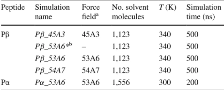
The NOE distance bound violations out of the 32 NOE distance bounds (Seebach et al. 1998) of the β-peptide using different GROMOS force fields are shown in Fig. 1a, d, and g for GROMOS 45A3, 53A6, and 54A7, respec-tively. The 45A3 (Schuler et al. 2001) and 53A6 (Oos-tenbrink et al. 2004) force fields have sizable differences in their parameter values of the backbone atom partial charges (up to 20 %), terminal group partial charges (up to 50 %), as well as the van der Waals parameters of the polar backbone atoms (up to 20 %), yet the simulations
of the β-peptide using these two force fields show a very similar picture of NOE bound violations; i.e., for both force fields, there are six positive violations <0.1 nm for the same hydrogen–hydrogen distances (Lin and van Gun-steren 2013). In contrast, the 54A7 force field (Schmid et al. 2011b) gives a completely different violation pattern with five positive NOE bound violations larger than 0.1 nm (Lin and van Gunsteren 2013), although it only differs from 53A6 (Oostenbrink et al. 2004) in the backbone ϕ and ψ torsional angle energy terms and the backbone N and O van der Waals interactions for this β-peptide.
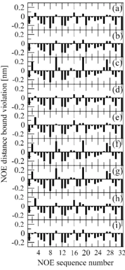
Figure 2 shows a comparison of NOE bound violations of the β-peptide predicted by one-step perturbation and evaluated from direct MD simulations. Since 45A3 has a different solvent methanol model, an additional simulation was carried out with 53A6 as the solute force field, and 45A3 as the methanol solvent force field (Table 1). This allows for determination of the effect of changing only the solute force-field parameters. According to Fig. 2, the pre-diction by one-step perturbation is very accurate; i.e., the deviations between the predicted violations and the simu-lated ones are mostly within 0.02 nm. The accurate pre-diction when perturbing from 45A3 to 53A6 shows that the technique works for rather large force-field parameter changes, and the perturbation between 53A6 and 54A7 illustrates that the technique works for very different NOE bound violation patterns. We note in passing that the 53A6 force field (blue and green symbols in Fig. 2) yields much
Fig. 1 Comparison of r−6 averaged NOE distances as obtained from simulations or predicted by one-step perturbation and experimen-tal data for Pβ. a Pβ_45A3; b 53A6 predicted by Pβ_45A3; c 54A7 predicted by Pβ_45A3; d Pβ_53A6; e 45A3 predicted by Pβ_53A6;
f 54A7 predicted by Pβ_53A6; g Pβ_54A7; h 45A3 predicted by
Pβ_54A7; i 53A6 predicted by Pβ_54A7
Fig. 2 Comparison of NOE atom–atom distance bound violations of
the β-peptide predicted by one-step perturbation and evaluated from MD simulation. Blue plus sign NOE violations predicted by Pβ_45A3 compared with the ones evaluated from Pβ_53A6*; red cross NOE violations predicted by Pβ_53A6 compared with the ones evaluated from Pβ_54A7; green circle NOE violations predicted by Pβ_54A7 compared with the ones evaluated from Pβ_53A6. The dashed lines indicate a deviation of 0.02 nm. The hydrogen atom pairs with viola-tions larger than 0.1 nm are listed. H*-Cβ refers to nonstereospecific protons bound to Cβ
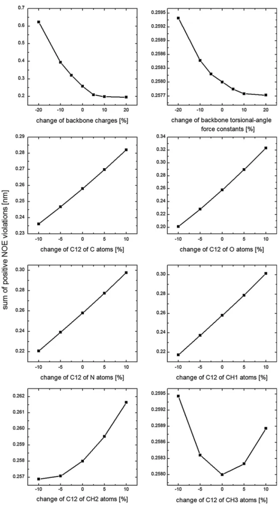
Fig. 3 Sum of the positive
NOE distance bound violations for the β-peptide predicted by one-step perturbation using the Pβ_53A6 simulation as the reference state and varying dif-ferent force-field parameters
smaller violations than the 54A7 one (red symbols in Fig. 2).
In a previous study, Bachmann et al. (2013) concluded that one-step perturbation can only be used to qualitatively predict different properties of liquid water. In contrast, the NOE distance bound violations of the β-peptide can be pre-dicted using one-step perturbation not only qualitatively, but also quantitatively. The less accurate prediction in the previous study is related to the larger size of the pertur-bation considered in that study, i.e., changing force-field parameters by 5 % for up to 5,000 water molecules (Bach-mann et al. 2013).
Next, one-step perturbation was used to investigate the effect of changing individual parameters on the NOE dis-tance bound violations of the β-peptide. The following solute parameters were separately varied: backbone par-tial charges, force constants of backbone torsional-angle energy terms (changes up to ±20 %), and C12 repulsive van der Waals parameters of GROMOS atom types (Oost-enbrink et al. 2004) C, O, N, CH1, CH2, and CH3 (changes up to ±10 %). The results for changes from the 53A6 force field are shown in Fig. 3. The biggest effect comes from the change in backbone atomic partial charges. Increasing them improves the agreement with the experimental NOE
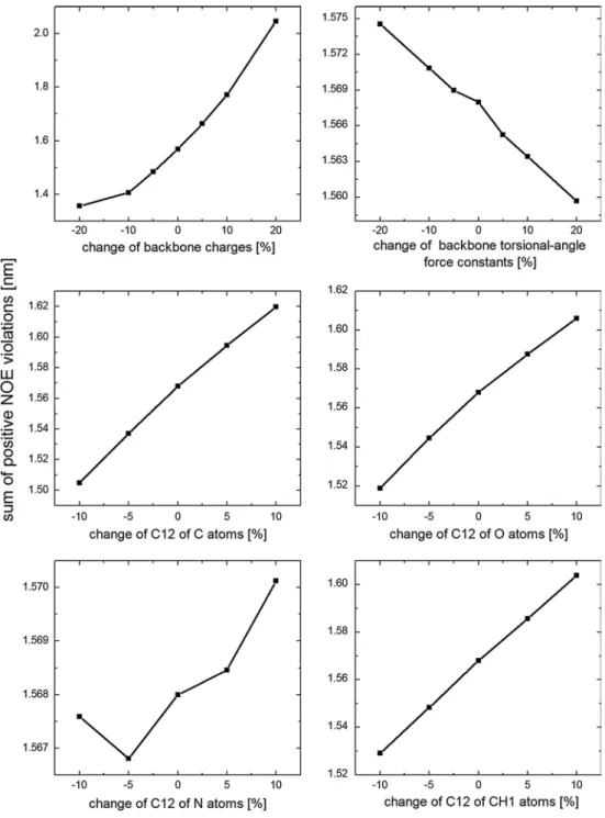
Fig. 4 Sum of the positive
NOE distance bound violations for the α-peptide predicted by one-step perturbation using the Pα_53A6 simulation as the reference state and varying dif-ferent force-field parameters
data, and vice versa. This may be due to stabilization of the helical conformation of the β-peptide over the nonhelical ones by the increase of backbone partial charges. Increas-ing or decreasIncreas-ing the force constants of backbone torsional-angle energy terms has only a very small effect on improv-ing or impairimprov-ing the agreement with the experimental data. Decreasing the repulsive C12 van der Waals parameters improves the agreement except for the atom type CH3.
For the hepta-α-peptide, similar conclusions were obtained for the changes in backbone torsional-angle force constants and C12 parameters of the backbone atoms (Fig. 4). However, an opposite relation between force-field parameters and NOE violations was observed for the change of backbone partial charges; i.e., reducing the backbone par-tial charges improves the agreement with the experimental NOE data for the α-peptide. This may be caused by the
α-peptide adopting predominantly unfolded conformations
(Wang et al. 2010). Increasing the backbone partial charges destabilizes unfolded conformations over helical ones, and thus impairs the agreement with experimental data for the
α-peptide while it improves the agreement for the β-peptide.
This observation nicely demonstrates that force-field param-eters should not be obtained based on single simulations of macromolecules. A quantity Q( rN) may pose conflicting
requirements on force-field parameters for different sys-tems, and different quantities Q( rN) may show conflicting
requirements for a single system. This illustrates the chal-lenge of transferable force-field development.
In summary, one-step perturbation, which in this case is equivalent to reweighting configurations, constitutes an efficient technique to predict violations of NOE distance bounds of peptides upon changing force-field parameters. However, a necessary condition is that conformations rel-evant to both Hamiltonians be present in the conformational ensemble used to obtain 〈Q〉 values. Reweighting can also
be used to predict quantities 〈Q〉 other than NOE distance
bound violations such as X-ray diffraction intensities, resi-dential dipolar couplings, or circular dichroism (CD) spectra for a particular system, which makes it a powerful technique for force-field development that easily reduces the number of required separate simulations by an order of magnitude.
Acknowledgments This work was financially supported by the
National Center of Competence in Research (NCCR) in Structural Biology and by grant number 200020–137827 of the Swiss National Science Foundation, and by grant number 228076 of the European Research Council (ERC), which is gratefully acknowledged.
References
Bachmann S, Dolenc J, van Gunsteren WF (2013) On the use of one-step perturbation to investigate the dependence of different prop-erties of liquid water upon a variation of model parameters from a single simulation. Mol Phys 111(14–15):2334
Cornell WD, Cieplak P, Bayly CI, Gould IR, Merz KM, Ferguson DM, Spellmeyer DC, Fox T, Caldwell JW, Kollman PA (1995) A second generation force field for the simulation of pro-teins, nucleic acids, and organic molecules. J Am Chem Soc 117(19):5179–5197
Daura X, Gademann K, Jaun B, Seebach D, van Gunsteren WF, Mark AE (1999) Peptide folding: when simulation meets experiment. Angew Chem Int Ed 38(1–2):236–240
Eichenberger AP, Gattin Z, Yalak G, van Gunsteren WF (2010) Molec-ular dynamics simulation of ester-linked hen egg white lysozyme reveals the effect of missing backbone hydrogen bond donors on the protein structure. Helv Chim Acta 93(10):1857–1869 Eichenberger AP, Allison JR, Dolenc J, Geerke DP, Horta BAC, Meier
K, Oostenbrink C, Schmid N, Steiner D, Wang DQ, van Gun-steren WF (2011) GROMOS plus plus software for the analysis of biomolecular simulation trajectories. J Chem Theory Comput 7(10):3379–3390
Horta BAC, Fuchs PFJ, van Gunsteren WF, Hünenberger PH (2011) New interaction parameters for oxygen compounds in the GRO-MOS force field: improved pure-liquid and solvation properties for alcohols, ethers, aldehydes, ketones, carboxylic acids, and esters. J Chem Theory Comput 7(4):1016–1031
Hritz J, Oostenbrink C (2009) Efficient free energy calculations for compounds with multiple stable conformations separated by high energy barriers. J Phys Chem B 113(38):12711–12720
Jorgensen WL, Maxwell DS, TiradoRives J (1996) Development and testing of the OPLS all-atom force field on conformational energetics and properties of organic liquids. J Am Chem Soc 118(45):11225–11236
Li DW, Brüschweiler R (2010) NMR-based protein potentials. Angew Chem Int Ed 49(38):6778–6780
Lin ZX, van Gunsteren WF (2013) Refinement of the application of the GROMOS 54A7 force field to β-peptides. J Comput Chem 34(32):2796–2805
Liu HY, Mark AE, van Gunsteren WF (1996) Estimating the relative free energy of different molecular states with respect to a single reference state. J Phys Chem 100(22):9485–9494
MacKerell AD, Bashford D, Bellott M, Dunbrack RL, Evanseck JD, Field MJ, Fischer S, Gao J, Guo H, Ha S, Joseph-McCarthy D, Kuchnir L, Kuczera K, Lau FTK, Mattos C, Michnick S, Ngo T, Nguyen DT, Prodhom B, Reiher WE, Roux B, Schlenkrich M, Smith JC, Stote R, Straub J, Watanabe M, Wiorkiewicz-Kucz-era J, Yin D, Karplus M (1998) All-atom empirical potential for molecular modeling and dynamics studies of proteins. J Phys Chem B 102(18):3586–3616
Oostenbrink C (2012) Free energy calculations from one-step per-turbations. In: Baron R (ed) Computational drug discovery and design, methods in molecular biology, vol 819. Humana, New York, pp 487–499
Oostenbrink C, van Gunsteren WF (2005) Efficient calculation of many stacking and pairing free energies in DNA from a few molecular dynamics simulations. Chem Eur J 11(15):4340–4348 Oostenbrink C, Villa A, Mark AE, van Gunsteren WF (2004) A
bio-molecular force field based on the free enthalpy of hydration and solvation: the GROMOS force-field parameter sets 53A5 and 53A6. J Comput Chem 25(13):1656–1676
Reif MM, Hünenberger PH, Oostenbrink C (2012) New interaction parameters for charged amino acid side chains in the GROMOS force field. J Chem Theory Comput 8(10):3705–3723
Reif MM, Winger M, Oostenbrink C (2013) Testing of the GROMOS force-field parameter set 54A8: structural properties of electro-lyte solutions, lipid bilayers, and proteins. J Chem Theory Com-put 9(2):1247–1264
Riniker S, Christ CD, Hansen HS, Hünenberger PH, Oostenbrink C, Steiner D, van Gunsteren WF (2011) Calculation of rela-tive free energies for ligand-protein binding, solvation, and
conformational transitions using the GROMOS software. J Phys Chem B 115(46):13570–13577
Schmid N, Allison JR, Dolenc J, Eichenberger AP, Kunz APE, van Gunsteren WF (2011a) Biomolecular structure refine-ment using the GROMOS simulation software. J Biomol NMR 51(3):265–281
Schmid N, Eichenberger AP, Choutko A, Riniker S, Winger M, Mark AE, van Gunsteren WF (2011b) Definition and testing of the GROMOS force-field versions 54A7 and 54B7. Eur Biophys J 40(7):843–856
Schmid N, Christ CD, Christen M, Eichenberger AP, van Gunsteren WF (2012) Architecture, implementation and parallelisation of the GROMOS software for biomolecular simulation. Comput Phys Commun 183(4):890–903
Schuler LD, Daura X, van Gunsteren WF (2001) An improved GRO-MOS96 force field for aliphatic hydrocarbons in the condensed phase. J Comput Chem 22(11):1205–1218
Seebach D, Abele S, Gademann K, Guichard G, Hintermann T, Jaun B, Matthews JL, Schreiber JV (1998) β2- and β3-peptides with proteinaceous side chains: synthesis and solution structures of constitutional isomers, a novel helical secondary structure and the influence of solvation and hydrophobic interactions on folding. Helv Chim Acta 81(5):932–982
Seebach D, Mathad RI, Kimmerlin T, Mahajan YR, Bindschadler P, Rueping M, Jaun B, Hilty C, Etezady-Esfarjani T (2005) NMR-solution structures in methanol of an α-heptapeptide, of a
β3/β2-nonapeptide, and of an all-β3-icosapeptide carrying the 20 proteinogenic side chains. Helv Chim Acta 88(7):1969–1982 Steiner D, Allison JR, Eichenberger AP, van Gunsteren WF (2012) On
the calculation of 3Jαβ-coupling constants for side chains in
pro-teins. J Biomol NMR 53(3):223–246
Torrie GM, Valleau JP (1977) Nonphysical sampling distributions in Monte Carlo free-energy estimation: umbrella sampling. J Com-put Phys 23(2):187–199
van Gunsteren WF, Berendsen HJC (1990) Computer-simulation of molecular-dynamics: methodology, applications, and perspec-tives in chemistry. Angew Chem Int Ed 29(9):992–1023 van Gunsteren WF, Bakowies D, Baron R, Chandrasekhar I,
Chris-ten M, Daura X, Gee P, Geerke DP, Glättli A, Hünenberger PH, Kastenholz MA, Oostenbrink C, Schenk M, Trzesniak D, van der Vegt NFA, Yu HB (2006) Biomolecular modeling: goals, prob-lems, perspectives. Angew Chem Int Ed 45(25):4064–4092 van Gunsteren WF et al. http://www.gromos.net
Wang DQ, Friedmann M, Gattin Z, Jaun B, van Gunsteren WF (2010) The propensity of α-aminoisobutyric acid (=2-methylalanine; Aib) to induce helical secondary structure in an α-heptapeptide: a computational study. Helv Chim Acta 93(8):1513–1531
Wüthrich K, Billeter M, Braun W (1983) Pseudo-structures for the 20 common amino acids for use in studies of protein confor-mations by measurements of intramolecular proton-proton dis-tance constraints with nuclear magnetic resonance. J Mol Biol 169(4):949–961