CLINICAL ARTICLE - VASCULAR
Posterior auricular artery as an alternative donor vessel
for extracranial-intracranial bypass surgery
Menno R. Germans&Luca Regli
Received: 10 July 2014 / Accepted: 11 August 2014 / Published online: 27 August 2014 # Springer-Verlag Wien 2014
Abstract
Background Sometimes the superficial temporal artery (STA) is not available for an extracranial-intracranial (EC-IC) bypass procedure. An alternative vessel for an EC-IC bypass is the posterior auricular artery (PAA) if it extends to the temporoparietal area with a diameter large enough. We assessed the prevalence of an appropriate PAA as an alterna-tive donor vessel and report three illustraalterna-tive cases in which the PAA was used for EC-IC bypass surgery.
Methods A literature search was performed on the use of the PAA as a donor vessel for bypass surgery. Secondly, a pro-spective database of bypass surgeries was reviewed to calcu-late the prevalence of a PAA with a diameter of at least 1 mm in the parietotemporal area. Finally, three illustrative cases are reported that describe various indications for the revascularisation procedures with their clinical, surgical and imaging features.
Results Two articles have previously described the use of the PAA for bypass surgery and their results are summarised. The prevalence of a PAA that would be appropriate for an EC-IC bypass in patients with intracranial vascular pathology is 5.7 %. The presented cases demonstrate that the PAA can be successfully used for EC-IC bypass surgery with good flow velocities and patency.
Conclusions The PAA is a rarely described as an appropriate donor vessel for an EC-IC bypass. Its prevalence is 5.7 % and it can successfully be used as an alternative donor vessel. The awareness among cerebrovascular surgeons about the pres-ence of a PAA and knowledge about its anatomy may be valuable.
Keywords Cerebral revascularisation . Extracranial-intracranial arterial bypass . Posterior auricular artery . Middle cerebral artery . Case reports . Prevalence studies
Introduction
An extracranial-intracranial (EC-IC) bypass is an impor-tant revascularisation technique for treatment of complex cerebrovascular cases, such as intracranial aneurysm sur-gery with planned parent artery sacrifice or steno-occlusive disease, like moyamoya disease [1]. Recent literature showed that EC-IC bypass for symptomatic atherosclerotic internal carotid artery occlusion and hae-modynamic cerebral ischaemia does not reduce ischae-mic stroke [5, 14]. However, there is still a subset of patients who benefit from flow augmentation surgery, although those patients have to be selected carefully [2]. The most common used technique for EC-IC bypass sur-gery is the superficial temporal artery (STA) to middle cere-bral artery (MCA) anastomosis [11]. An alternative method is connecting the external carotid artery (ECA) with an interpo-sition graft (saphenous vein or radial artery) to the intracranial internal carotid artery (ICA) or MCA, which is mostly used for high-flow bypasses [3]. Other, rarely used, donor arteries are the occipital artery [7,12] or the posterior auricular artery (PAA) [8,16].
The STA cannot be used as a donor artery for bypass surgery due to various causes. In these cases an interpo-sition graft can be used as an alternative, but this tech-nique has its risks and drawbacks [3]. Recently, the PAA was used as an additional bypass in a refractory case of moyamoya disease [8] and for a revascularisation proce-dure [16], indicating that the PAA might be a potential candidate for a EC-IC bypass.
M. R. Germans (*)
:
L. RegliDepartment of Neurosurgery, University Hospital Zürich, Frauenklinikstrasse 10, 8091 Zürich, Switzerland e-mail: mrgermans@hotmail.com
This article summarises an overview of the current litera-ture regarding bypass surgery that incorporates the PAA and additionally assesses the prevalence of a PAA that would be appropriate for an EC-IC bypass in patients with intracranial vascular pathology. Finally, clinical, operative and radiologi-cal features are presented of three patients in whom the PAA was used for an EC-IC bypass to the MCA.
Methods
This study was performed with approval of the Institutional Research Board of the University of Zürich.
Literature search
We performed a literature search in PubMed and EMBASE with the search words “posterior auricular artery” and “bypass” in May 2014 to get an indication about the current knowledge regarding bypass surgery with a PAA.
Database search
We retrospectively reviewed prospectively collected data of consecutive patients who received bypass surgery in a period of 7 years. All preoperative radiological investi-gations visualising the extracranial arteries (computed tomographic angiography [CTA], magnetic resonance an-giography [MRA] and digital subtraction anan-giography [DSA]) were reviewed by the first author. The patients with an extracranial artery running posterior to the ex-ternal acoustic meatus (EAM) until at least the temporoparietal area (i.e. the vascular anatomy of the PAA [9]) with a diameter of at least 1 mm were selected and the confidence interval (CI) was calculated using the modified Wald interval with an alpha of 0.05.
Case reports
We describe the clinical and radiological features, intraopera-tive flow measurements in the bypass of three patients in
Table 1 Overview of current literature describing posterior auricular artery to middle cerebral artery bypass
Reference Indication Side Reason to use PAA Intraoperative flow (ml/min) Horiuchi et al. [8] Refractory moyamoya Right STA already used for revascularisation n.a.
Tokugawa et al. [16] ICA stenosis Left STA already used for revascularisation n.a. Current study, case 1 Fusiform MCA aneurysm Right More favourable location for bypass 29 Current study, case 2 Moyamoya Left STA already used for revascularisation
(double barrel bypass)
25 Current study, case 3 ICA dissection Left More favourable location for bypass 25–35 PAA posterior auricular artery, STA superficial temporal artery, ICA internal carotid artery, MCA middle cerebral artery, n.a. not applicable
Fig. 1 Case 1. DSA of the right extracranial arteries with posterior auricular artery (a, arrow), which was used for an EC-IC bypass (b). Note that the craniotomy was extended posteriorly (closer to the Sylvian point) to find M4 recipient branches more easily
whom the PAA was actually used for the EC-IC bypass. In one patient we report the long-term follow-up.
Results
Literature search
Our literature search resulted in three hits, of which one article was not relevant to the topic. The selected articles have both recently been published in neurosurgical journals and de-scribed a bypass of the PAA to the MCA [8,16]. An overview of the reported data, added with the data of the current report, is presented in Table1.
Database search
The search in our database of 175 patients revealed 10 patients (5.7 %; 95 % CI 3.0–10.3) with a PAA that would be
appropriate for EC-IC bypass surgery. Three of these patients actually received a bypass incorporating the PAA.
Case reports
Case 1
This man had a headache for several days before the devel-opment of a sudden left-sided hemiparesis. A cerebral infarct of the right parietal MCA territory was diagnosed with an additional mycotic aneurysm of the M2 branch. Several days after the diagnosis, he developed an acute myocardial infarc-tion, which was treated with a cardiac stent, and mitral valve replacement with aortic valve reconstruction 2 weeks later. The diagnosis of endocarditis was confirmed by the presence of Streptococcus salivarius on the mitral valve. Follow-up imaging showed an increase in aneurysm size despite antibi-otic therapy and, after discussion in our multidisciplinary neurovascular team, we decided to explore the aneurysm surgically. The preoperative DSA showed a large right-sided PAA, which was planned to be used as a donor artery because
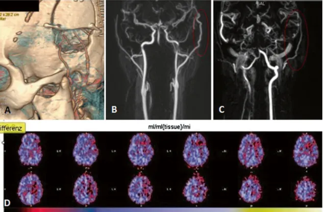
Fig. 2 Case 2. a DSA of the left internal carotid artery showing moyamoya disease. b DSA of the left external carotid artery showing parietal branch of superficial temporal artery (black arrow) and posterior auricular artery (grey arrow). c Preoperative PET imaging with acetazolamide (Diamox)
its location was more favourable than the MCA in this partic-ular case (see Fig.1a). A straight incision directly over the PAA exposed the artery and a bypass with 10–0 monofilament interrupted sutures was made to the MCA (M4 branch) with-out any significant difficulties. The initial flow through the bypass was 11 ml/min. The mycotic aneurysm was treated by clipping the parent M2 branch proximal to the aneurysm (inflow occlusion). A complete trapping was not performed, as the adhesions around the aneurysm did not allow for a safe microsurgical dissection of the outflow region. The flow increased to 29 ml/min after M2 clipping (see Table1). There were no perioperative complications and a patent bypass was seen on a CTA the next day (see Fig.1b).
Case 2
A 26-year old woman was referred to our clinic because of hypesthesia in the right side of her face and right hand. She recurrently developed these symptoms after standing up rap-idly, indicating an orthostatic component. The additional in-vestigations, consisting of CTA, MRA and DSA confirmed the diagnosis moyamoya disease (see Fig.2a) and showed a patent STA and a large bore PAA at left ECA injection (see Fig.2b). The positron emission tomography (PET) showed reduced cerebrovascular reactivity to acetazolamide in the left MCA territory, consistent with her clinical symptoms (see Fig.2c).
Because of the extensive involvement of the MCA territory and also reduced cerebrovascular reserve of the left anterior cerebral artery, a double-barrel bypass to the infrasylvian and suprasylvian M4 branches of the MCA was planned. Both the parietal branch of the STA and the PAA were exposed by a question mark incision and the bypasses were performed with 10–0 monofilament sutures. The flow directly after establish-ment of the PAA-STA bypass was 25 ml/min. No perioperative complications occurred with patent bypasses on the postoper-ative CTA (see Fig.3a). The 6-month follow-up investigations, consisting of MRA and PET imaging, showed widely patent bypasses and a normal cerebrovascular reactivity in a patient without neurological deficits (see Fig.3b-d).
Case 3
This 44-year old woman presented to our hospital because of pain in her neck after scuba diving with associated symptoms of word finding disturbances and right-sided hemihypesthesia and hemihypalgesia. Her neurological exam showed a left-sided Horner syndrome and confirmed her semiology. The CTA/CT perfusion (CTP) showed a left-sided ICA occlusion, most probably due to dissection, and a thrombus in a left M2 branch with a reduced mean transit time (MTT) in the parietal MCA territory. Despite a start of intravenous thrombolysis at 225 min after the onset of symptoms, followed by
intra-Fig. 3 Postoperative three-dimensional CTA reconstruction of the bypasses performed in case 2 (a). A patent superficial temporal artery (b) and posterior auricular artery (c) with normal cerebrovascular reactivity (d) after 6 months
arterial treatment, her symptoms continued to fluctuate. A follow-up MRI showed similar results as the CT, with an unchanged area of restricted perfusion, but only a small area
of cerebral infarction (see Fig.4aandb). Because of the MRI findings and continuing symptoms, we proceeded with an emergency EC-IC bypass. The preoperative DSA confirmed
Fig. 5 a DSA of common carotid artery with dissection of internal carotid artery and large superficial temporal (black arrow) and posterior auricular arteries (arrowhead). b Postoperative CTA with patent bypass (grey arrow). Postoperative CT perfusion with small infarct Fig. 4 Case 3. a Preoperative MRI with perfusion restriction in MCA territory. b Preoperative diffusion-weighted MRI with evidence for small infarct in parietal area
the ICA dissection and showed large STA and PAA arteries (see Fig.5a). The angiographic anatomy and area of hypoper-fusion determined that the PAA was the best donor artery for the EC-IC bypass. With a retroauricular incision directly over the PAA, a bypass was performed with interrupted 10–0 monofilament sutures to the temporal branch of the MCA without any difficulties. In comparison to the“free flow” in the PAA of 15 ml/min, the flow through the bypass after reperfusion was between 25 and 35 ml/min. Directly postop-eratively, she had a right-sided hemiparesis which resolved after a few hours. The postoperative CTA/CTP showed a patent bypass with normalised perfusion of the MCA territory and an unchanged small cortical infarct (see Fig.5bandc).
Discussion
This report adds three additional patients with a PAA to MCA bypass to the scarce literature. The intraoperative flow mea-surements and follow-up imaging showed highly patent by-passes. The calculated incidence of a PAA that would be appropriate for an EC-IC bypass in patients with intracranial vascular pathology is 5.7 % and higher than expected from the current literature.
Low-flow EC-IC bypass surgery is the primary treatment for moyamoya disease and indicated in highly selected pa-tients with symptomatic steno-occlusive disease and exhausted cerebrovascular reserve capacity. Additionally, flow replacement surgery is a good option in a selected group of patients with complex or fusiform middle cerebral artery aneurysms which are difficult to treat with direct clipping or endovascular treatment options [1].
Sometimes the STA is not available for bypass surgery due to hypoplasia of the artery, division of the artery at previous craniotomy, damage of the artery during dissection, or it is already used for an intracranial bypass. In such cases, the PAA can be used as an alternative donor when it ascends to the temporoparietal area with a large enough diameter [8,16].
Most of the time the PAA leaves the ECA just superior to the occipital artery, but in 10–15 % it arises from the occipital artery after an occipitoauricular trunk [17]. It then courses between the mastoid tip and auricle, and runs almost vertically towards the vertex until a mean distance of 7.5 cm from the mastoid tip [9]. In 33 %, the PAA ascends to the temporoparietal area and therefore the length is sufficient to use it as a donor artery for an intracranial bypass, provided that the vessel diameter is large enough [13]. As it courses approximately 1.2 cm posterior to the EAM [9], it is located ideally in the posterior margin of a standard craniotomy around the Sylvian point (see Fig. 1) [11]. The PAA is probably the dominant artery for the auricle [13], where it mainly vascularises the medial part through three to five branches and extends anteriorly, where
it anatomises with the STA [13, 15]. Myocutaneous and myofascial flaps based on the PAA have been used in recon-structive [4] and ENT [6] surgery. Recognising the PAA as a potential donor artery preoperatively is important to plan the correct skin incision and avoid injury along its retroauricular ascending trajectory.
The use of a PAA for bypass surgery has rarely been described. Recently, the PAA was used as a donor artery for additional revascularisation in a patient with a refractory case of moyamoya disease despite direct STA and occipital artery bypasses [8]. Another case report [16] describes the use of the PAA for a double-barrel revascularisation procedure, which is comparable to our second case. Our report adds three addi-tional PAA to MCA bypasses, and by describing perioperative flow dynamics and long-term postoperative imaging in one patient, we show that the PAA can be as patent as the STA [10] and therefore could be a reliable alternative.
Although the PAA is not a standard donor artery for EC-IC bypass surgery, its availability is higher than expected and increasing awareness among cerebrovascular surgeons about its anatomy may be valuable. In 5.7 %, it ascends to the temporoparietal area with a sufficient large diameter, where it can be used as an alternative donor vessel when the STA is not available.
Conflicts of interest None.
References
1. Amin-Hanjani S (2011) Cerebral revascularization: extracranial-intracranial bypass. J Neurosurg Sci 55:107–116
2. Amin-Hanjani S, Barker FG, Charbel FT, Connolly ES Jr, Morcos JJ, Thompson BG (2012) EC-IC bypass for stroke—is this the end of the line or a bump in the road? Neurosurgery 71:557-561
3. Bisson EF, Visioni AJ, Tranmer B, Horgan MA (2008) External carotid artery to middle cerebral artery bypass with the saphenous vein graft. Neurosurgery 62:1419–1424
4. Choung PH (1996) The auriculomastoid fasciocutaneous island flap: a new flap for orofacial reconstruction. J Oral Maxillofac Surg 54: 559–567
5. Fluri F, Engelter S, Lyrer P (2010) Extracranial-intracranial arterial bypass surgery for occlusive carotid artery disease. Cochrane Database Syst Rev
CD005953-6. Gibb AG, Tan KK, Sim RS (1997) The Singapore swing. J Laryngol Otol 111:527–530
7. Hayashi T, Shirane R, Tominaga T (2009) Additional surgery for postoperative ischemic symptoms in patients with moyamoya dis-ease: the effectiveness of occipital artery-posterior cerebral artery bypass with an indirect procedure: technical case report. Neurosurgery 64:E195–E196
8. Horiuchi T, Kusano Y, Asanuma M, Hongo K (2012) Posterior auricular artery-middle cerebral artery bypass for additional surgery of moyamoya disease. Acta Neurochir (Wien) 154:455–456 9. McKinnon BJ, Wall MP, Karakla DW (1999) The vascular anatomy
and angiosome of the posterior auricular artery. A cadaver study. Arch Facial Plast Surg 1:101–104
10. Nakayama N, Kuroda S, Houkin K, Takikawa S, Abe H (2001) Intraoperative measurement of arterial blood flow using a transit time flowmeter: monitoring of hemodynamic changes during cerebrovas-cular surgery. Acta Neurochir (Wien) 143:17–24
11. Newell DW, Vilela MD (2004) Superficial temporal artery to middle cerebral artery bypass. Neurosurgery 54:1441–1448
12. Pandey P, Steinberg GK (2011) Neurosurgical advances in the treat-ment of moyamoya disease. Stroke 42:3304–3310
13. Pinar YA, Ikiz ZA, Bilge O (2003) Arterial anatomy of the auricle: its importance for reconstructive surgery. Surg Radiol Anat 25:175–179
14. Powers WJ, Clarke WR, Grubb RL Jr, Videen TO, Adams HP Jr, Derdeyn CP (2011) Extracranial-intracranial bypass surgery for stroke prevention in hemodynamic cerebral ischemia: the Carotid Occlusion Surgery Study randomized trial. JAMA 306:1983–1992 15. Tilotta F, Lazaroo B, Laujac MH, Gaudy JF (2009) A study of the
vascularization of the auricle by dissection and diaphanization. Surg Radiol Anat 31:259–265
16. Tokugawa J, Nakao Y, Kudo K, Iimura K, Esaki T, Yamamoto T, Mori K (2013) Posterior auricular artery-middle cerebral artery
bypass: a rare superficial temporal artery variant with well-developed posterior auricular artery: case report. Neurol Med Chir (Tokyo). doi:10.2176/nmc.cr2012-0233
17. Zumre O, Salbacak A, Cicekcibasi AE, Tuncer I, Seker M (2005) Investigation of the bifurcation level of the common carotid artery and variations of the branches of the external carotid artery in human fetuses. Ann Anat 187:361–369
Comment
This manuscript nicely describes another potential donor for rare but existing circumstances of low flow bypasses where STA is not available. Although PAA is seldomly of appropriate size and orientation for routine bypasses, but vascular neurosurgeons should consider it as a potential alternative for low flow revascularization procedures.
Amir Dehdashti NY, USA