HAL Id: hal-01099081
https://hal.inria.fr/hal-01099081
Submitted on 6 Jan 2015
HAL is a multi-disciplinary open access
archive for the deposit and dissemination of
sci-entific research documents, whether they are
pub-lished or not. The documents may come from
teaching and research institutions in France or
abroad, or from public or private research centers.
L’archive ouverte pluridisciplinaire HAL, est
destinée au dépôt et à la diffusion de documents
scientifiques de niveau recherche, publiés ou non,
émanant des établissements d’enseignement et de
recherche français ou étrangers, des laboratoires
publics ou privés.
Evaluation of morphometric descriptors of deep brain
structures for the automatic classification of patients
with Alzheimer’s disease, mild cognitive impairment and
elderly controls
Alexandre Routier, Pietro Gori, Ana B. Graciano Fouquier, Sophie Lecomte,
Olivier Colliot, Stanley Durrleman
To cite this version:
Alexandre Routier, Pietro Gori, Ana B. Graciano Fouquier, Sophie Lecomte, Olivier Colliot, et al..
Evaluation of morphometric descriptors of deep brain structures for the automatic classification of
patients with Alzheimer’s disease, mild cognitive impairment and elderly controls. MICCAI Workshop,
Sep 2014, Cambridge, United States. pp.8. �hal-01099081�
Evaluation of morphometric descriptors of deep
brain structures for the automatic classification
of patients with Alzheimer’s disease, mild
cognitive impairment and elderly controls
Alexandre Routier1,2,3,4,5,6, Pietro Gori5,1,2,3,4, Ana B. Graciano
Fouquier1,2,3,4,5, Sophie Lecomte1,2,3,4,5,6, Olivier Colliot1,2,3,4,5,6, Stanley
Durrleman5,1,2,3,4,6 and the Alzheimer’s Disease Neuroimaging Initiative
1 Sorbonne Universit´es, UPMC Univ Paris 06, UMR S 1127, ICM, F-75013, Paris 2
Inserm, U1127, 75013, Paris, France
3
CNRS, UMR 7225, 75013, Paris, France
4
Institut du Cerveau et de la Moelle ´epini`ere, ICM, 75013, Paris, France
5
Inria Paris-Rocquencourt, 75013, Paris, France
6 Centre d’Acquisition et de Traitement des Images (CATI), Paris, France
1
Introduction
Our participation in the MICCAI 2014 CADDementia challenge aims at evalu-ating the performance of morphometric descriptors in multi-class classification tasks for the prediction of Alzheimer’s disease and Mild Cognitive Impairment from structural Magnetic Resonance Images (MRIs).
We used the method for the construction of population-specific atlases that is described in [6, 5]. The method takes as input a set of segmented brain structures, which take the form of the union of labelled 3D surface meshes, called shape complexes. The method estimates an anatomical model, called template, which is representative of the shape complexes within a group of subjects. The variability in shape within the group is captured by 3D space deformations of the ambient space, which warps the anatomical model to the anatomical shape complex of each subject. The method estimates the anatomical model together with the deformation parameters.
The method requires to use the same set of homologous structures for all subjects. We choose a subset of 12 deep brain structures that were segmented from MRIs: caudate nucleus, putamen, pallidum, thalamus, hippocampus and amygdala of each hemisphere. We do not include the lateral ventricles because of a large variability in the segmentation of the horns of the ventricles, which could have masked other patterns of shape variability in the statistical analysis. We do not include the cortical surface because of the subject-specific gyrification.
Deformation parameters are seen as a multi-variate descriptor, which encodes the differences in shape between each subject’s anatomical configuration and the anatomical model. This descriptor encodes different patterns such as the shift of the caudate nucleus due to the ventricular enlargement and the hippocampal atrophy, for instance. The residual shape, namely the difference between the
deformed template and the subject’s shape complex, is considered as noise. The combination of the two terms gives the likelihood of a given anatomical shape complex, which will be used in classification.
We use a sub-set of the Alzheimer’s Disease Neuroimaging Initiative (ADNI) database to build the anatomical models. We build three anatomical models considering a group of Cognitively Normal (CN) subjects, subjects with Mild Cognitive Impairment (MCI) and patients with Alzheimer’s disease (AD). Once the models are built, we test any new subject by registering each model to the shape complex of this subject and computing its likelihood. We then classify according to the maximum likelihood. We test our classifiers on another sub-set of the ADNI database and the CADDementia database.
The method is fully automatic. The atlas construction method uses the con-cept of varifolds [3] for mesh comparison and therefore does not require specific mesh pre-processing. The method is indeed robust with respect to changes in topology between meshes, small holes, spikes, irregular sampling and inconsis-tency in normal orientations. We do not perform quality control of the segmen-tations as small errors in the position of the boundaries are likely to be averaged out in this kind of shape analysis. The use of smooth 3D deformations also acts as low-pass filter which smoothes out irregularities in the boundaries of the structures. Few important failures in segmentations are likely to be considered as outliers in the statistical analysis. We use the implementation of the method in the software Deformetrica, which is freely available at www.deformetrica.org. Building the anatomical models took 3 days, 15 hours on average (with a parallelization on 40 threads). Registering the anatomical models to test subjects took 10 hours and 20 minutes on average, with a standard deviation of about 1 hour and 30 minutes. The computations were made on a computer cluster wich is composed of two types of machines. The first one (with 32 computing nodes) is running on an Intel XeonR Processor X5650 (2x6 Cores, 2.66 GHz)R
and 12x4GB 1333MHz DDR3 Memory and the second one (with 2 computing nodes) is running on an Octo-processor Intel XeonR Processor X7550 (8x2x8R
Cores, 2 GHz) and 128x2GB 1066MHz DDR3 Memory.
2
Material and Methods
2.1 Data sets
We use the baseline images from the ADNI database to build the statistical models. We choose the same set of 509 subjects as the ones selected in [4], de-composed into 162 cognitively normal controls (CN), 210 patients with Mild Cognitive Impairment (MCI) and 137 patients diagnosed with Alzheimer’s dis-ease (AD) at baseline. We split the data set into a training set of 50 CN, 50 MCI and 50 AD, the rest being our test set.
We perform the same pre-processing to all ADNI and CADDementia data. The atlas construction is performed only on the training sub-set of the ADNI data. Classification are performed on the test set of the ADNI data and the CADDementia data.
2.2 Data pre-processing
The data pre-processing consists of the following steps:
– We run FreeSurfer1 on the T1 MRI data [7] with default parameters. The
output is volumetric segmentation of various structures. At this stage, we exclude from the ADNI dataset, 2 subjects for which the FreeSurfer pipeline failed.
– We run a marching cube algorithm (as implemented in FreeSurfer) to re-construct 3D triangular meshes from the volumetric segmentation of the 12 selected structures on the RAS coordinate system (Right, Anterior, Supe-rior). We do not perform any other processing on the meshes, although they have holes, spikes and irregular meshing.
– We register all images to the image of a control young adult from the ADNI training data set (126 S 0405 S14635 I38828) using FSL software2 [9]. We
use rigid and scaling transformation with 7 degrees of freedom. The trans-formations are then applied to the meshes. The transformed meshes are the inputs given to the software Deformetrica.
Additionally, we build a naive prototype initialization for the anatomical models to give as input of Deformetrica. We build this prototype by mapping a sphere to each structure of the reference subject with very smooth parameters. The corresponding initial anatomical model is shown in Fig. 1-left.
2.3 Atlas construction on ADNI training data
We use the Deformetrica software to build the anatomical models and esti-mate the deformation parameters. The method minimizes the following criterion (see [5] for details):
E(X0, c, α0, . . . , αN) = N X i=1 (12 X k=1 1 2σ2 k φαi(X0,k) − Ski 2 W + α T iKVαi ) (1) where
– X0= {X0,k}k=1,...,12 denotes the position of the vertices of the anatomical
model with 12 components, one for each anatomical structure,
– c denotes a set of control points which are supposed to move to the most variable parts of the anatomical model,
– {αi}i=1,...,Ndenotes momentum vectors attached to the control points which
parameterize the deformations of the anatomical model to each subject’s anatomical configuration (among N the number of subjects),
– Si
k denotes the mesh of the k-th structure of the i-th subject,
1
http://surfer.nmr.mgh.harvard.edu
2
– {φαi}
i=1,...,Ndenotes the smooth 3D deformation from the anatomical model
to the i-th subject,
– k.kW denotes the varifold norm,
– σk2 denotes the variance of the noise of the k-th structure in the space of varifolds,
– KV is the deformation kernel matrix, so that αTiKVαimeasures the squared
norm of the initial velocity of the deformation
We choose the following parameters, using the rationale detailed in [5]: – deformation kernel width: σV = 10 mm,
– varifold kernel width: σW = 5 mm,
– variance of noise: σ2
k = 16 for all structures,
– template kernel width 0.5σV,
other parameters being the ones by default in Deformetrica.
2.4 Classification of ADNI test data and CADDementia data Any test image is transformed into a set of sub-cortical structures after the pre-processing steps explained in 2.2. We then register each atlas to this subject’s shape complex. The registration is performed by minimizing the following crite-rion, which is essentially (1) for N = 1 and keeping fixed the atlas parameters: the template shape X0 and the control points c:
E(α) = 12 X k=1 1 2σ2 k kφα(X0,k) − Skk 2 W + α T KVα, (2)
where the Sk’s denotes the test subject shapes.
The value of the criterion E at convergence is an approximation of the log-likelihood of the test data [1, 2]. In order to take into account the covariance of the deformation parameters, we replace the matrix KV by the inverse of
the regularized empirical covariance matrix of the momentum vectors αi. This
corrected value of the criterion is used in classification.
3
Results
3.1 Results on the ADNI data
In Fig. 2, the 3 estimated template shape complexes are shown. The template of the MCI class falls in-between the template of the CN and AD classes. These shapes show the shift of the caudate nucleus toward the lateral parts of the brain due to a larger and larger ventricular enlargement. We notice also a greater and greater atrophy of the hippocampus.
The confusion matrix of the classification performed on the test sub-set of the ADNI database is shown in Table 1. The accuracy, assuming the probability of 1/3 for each class, is 51% (i.e. 13P3
k=1nk,k/nk where nk =
P3
total number of samples of the class k). We notice that our classifier tends to empty the MCI class, and to classify MCI subjects as either CN or AD with equal probability. This may be explained by the fact that our descriptors of MCI subjects overlap the descriptors of CN and AD classes, as if there is a continuum between the three classes. In other words, our classifier does not detect shape patterns that are specific to MCI subjects. This conclusion is corroborated by the visualization of the 3 template shapes complexes in Fig. 2.
The ROC curves of pairwise classification are shown in Fig. 3. As expected, the AD versus CN classification has overall better performance than classification of AD or CN against MCI.
Fig. 1. Initial prototype given as input of Deformetrica (left) and an instance of esti-mated atlas given as output (right): template shapes are representative of the group and momenta arrows parameterize template-to-subject deformations.
Fig. 2. Superimposition of the 3 template shapes for the CN, MCI and AD classes in green, blue and red respectively. Anterior view (left) and posterior view (right)
True class
AD MCI CN
Hyp othesized classAD
66
83
28
MCI 6
13
5
CN
14
64
78
Table 1. Confusion matrix on ADNI test data set.
0 0.1 0.2 0.3 0.4 0.5 0.6 0.7 0.8 0.9 1 0 0.1 0.2 0.3 0.4 0.5 0.6 0.7 0.8 0.9 1
False positive rate
True positive rate
EC vs AD (AUC = 0.818) MCI vs EC (AUC = 0.651) MCI vs AD (AUC = 0.615)
Fig. 3. ROC curves of pairwise classification on the ADNI database.
3.2 Results on the CADDementia training data
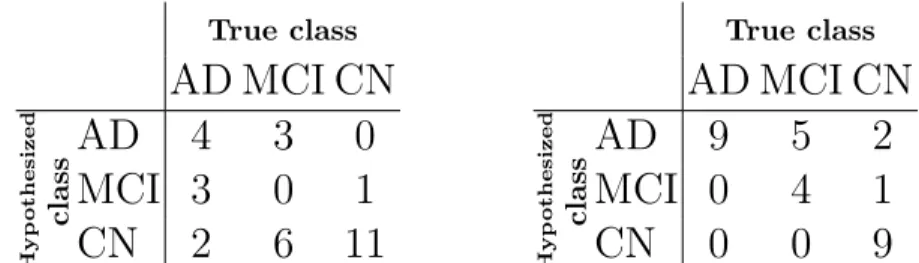
We test our classifier on the 30 subjects of the training database of CADDe-mentia. Table 2 shows the confusion matrix using the thresholds that maximize the accuracy of the classifier on the ADNI data set, for which the accuracy is 50%. These two thresholds determine the position of the boundaries between the three classes. The optimization of these two thresholds on the given 30 subjects of the CADDementia database yields the confusion matrix in Table 3 and an accuracy of 73%. Differences in optimum thresholds between the two databases may come from differences in patients, differences in age distribution, differences in clinical practice for the diagnosis of mild cognitive impairment and dementia. Optimizing the thresholds on only 30 subjects is also not ideal, as they might not generalize well to the rest of the data set. For these reasons, we decided to submit two predictions for each subject: one using the thresholds estimated from
the ADNI data set and the other one using the thresholds estimated from the CADDementia training data set.
True class
AD MCI CN
Hyp oth esized classAD
4
3
0
MCI 3
0
1
CN
2
6
11
Table 2. Confusion matrix for the classi-fication of the CADDementia training set, using the thresholds that are optimum for the ADNI data set.
True class
AD MCI CN
Hyp ot hesized classAD
9
5
2
MCI 0
4
1
CN
0
0
9
Table 3. Confusion matrix for the clas-sification of the CADDementia training set after optimizing the thresholds for this data set.
4
Discussion and conclusion
This work evaluates the performance of the Deformetrica software in classi-fication tasks. The software computes shape descriptors for anatomical shape complex of sub-cortical structures that are known to be markers of disease pro-gression. The approach is essentially multi-variate and combine different shape patterns such as the effect of hippocampal atrophy and ventricular enlargement on the shape of the sub-cortical structures. Our results suggest that the method does not find shape features that are characteristic of MCI subjects. The method tends to position the anatomy of MCI subjects, as an intermediate stage of dis-ease progression. This fact may come from the method itself, which does not capture characteristics of such non-demented subjects. It may also come from the heterogeneity of the MCI group.
Our goal was to use the software Deformetrica “out of the box” as a test case, whereas several improvements could be made such as the estimation of the covariance of deformation parameters and noise variance during the training phase along the lines of [1, 8]. We could have determined also the best thresholds using cross-validation on the ADNI database. Correction for age and sex could also have improved classification performance.
Acknowledgements
This work has been supported by the “Centre d’Acquisition et de Traitement des Images” (CATI) and the program “Investissements d’Avenir” ANR-10-IAIHU-06. Data used in the preparation of this article were obtained from the Alzheimer’s Disease Neuroimaging Initiative (ADNI) database3. As such, the investigators
3
within the ADNI contributed to the design and implementation of ADNI and/or provided data but did not participate in analysis or writing of this report. ADNI investigators include (complete listing available at http://www.loni.ucla.edu/ ADNI/Collaboration/ADNI Author ship list.pdf).
References
1. Allassonni`ere, S., Amit, Y., Trouv´e, A.: Towards a coherent statistical framework for dense deformable template estimation. Journal of the Royal Statistical Society Series B 69(1), 3–29 (2007)
2. Allassonni`ere, S., Kuhn, E., Trouv´e, A.: Construction of bayesian deformable models via a stochastic approximation algorithm: A convergence study. Bernoulli Journal 16(3), 641–678 (2010)
3. Charon, N., Trouv´e, A.: The varifold representation of non-oriented shapes for dif-feomorphic registration. SIAM J. Imaging Sci. 6(4), 25472580 (2013), accepted for publication
4. Cuingnet, R., Gerardin, E., Tessieras, J., Auzias, G., Lehricy, S., Habert, M., Chupin, M., Benali, H., Colliot, O., ADNI: Automatic classification of patients with alzheimer’s disease from structural mri: a comparison of ten methods using the adni database. Neuroimage 56(2), 766–81 (2011)
5. Durrleman, S., Prastawa, M., Charon, N., Korenberg, J., Joshi, S., Gerig, G., Trouv´e, A.: Morphometry of anatomical shape complexes with dense deformations and sparse parameters. Neuroimage (2014), under minor revision
6. Durrleman, S., Prastawa, M., Korenberg, J.R., Joshi, S., Trouv´e, A., Gerig, G.: Topology preserving atlas construction from shape data without correspondence using sparse parameters. In: Ayache, N., Delingette, H., Golland, P., Mori, K. (eds.) Med Image Comput Comput Assist Interv. Med Image Comput Comput Assist Interv., vol. LNCS 7512, pp. 223–230. Springer (2012)
7. Fischl, B., Salat, D., Busa, E., Albert, M., Dieterich, M., Haselgrove, C., van der Kouwe, A., Killiany, R., Kennedy, D., Klaveness, S., Montillo, A., Makris, N., Rosen, B., Dale, A.: Whole brain segmentation: automated labeling of neuroanatomical structures in the human brain. Neuron 33, 341–355 (2002)
8. Gori, P., Colliot, O., Worbe, Y., Marrakchi-Kacem, L., Lecomte, S., Poupon, C., Hartmann, A., Ayache, N., Durrleman, S.: Bayesian atlas estimation for the vari-ability analysis of shape complexes. In: Proc. Med Image Comput Comput Assist Interv. vol. LNCS 8149, pp. 267–274 (2013)
9. Smith, S.M., Jenkinson, M., Woolrich, M.W., Beckmann, C.F., Behrens, T.E., Johansen-Berg, H., Bannister, P.R., Luca, M.D., Drobnjak, I., Flitney, D.E., Niazy, R.K., Saunders, J., Vickers, J., Zhang, Y., Stefano, N.D., Brady, J.M., Matthews, P.M.: Advances in functional and structural {MR} image analysis and implementa-tion as {FSL}. NeuroImage 23, Supplement 1(0), S208 – S219 (2004)