HAL Id: inserm-02454988
https://www.hal.inserm.fr/inserm-02454988
Submitted on 19 May 2020HAL is a multi-disciplinary open access
archive for the deposit and dissemination of sci-entific research documents, whether they are pub-lished or not. The documents may come from teaching and research institutions in France or abroad, or from public or private research centers.
L’archive ouverte pluridisciplinaire HAL, est destinée au dépôt et à la diffusion de documents scientifiques de niveau recherche, publiés ou non, émanant des établissements d’enseignement et de recherche français ou étrangers, des laboratoires publics ou privés.
Multiple roles of macrophage migration inhibitory factor
in pulmonary hypertension
Gael Jalce, Christophe Guignabert
To cite this version:
Gael Jalce, Christophe Guignabert. Multiple roles of macrophage migration inhibitory factor in pul-monary hypertension. American Journal of Physiology - Lung Cellular and Molecular Physiology, American Physiological Society, 2020, 318 (1), pp.L1-L9. �10.1152/ajplung.00234.2019�. �inserm-02454988�
The Multiple Roles of Macrophage Migration Inhibitory Factor
in Pulmonary Hypertension
Gael Jalce
1and Christophe Guignabert
2, 3Short title: MIF in PH/PAH
1APAXEN, Gosselies, Belgium; gael.jalce@apaxen.com
2INSERM UMR_S 999, Hôpital Marie Lannelongue, Le Plessis-Robinson, France
3Faculté de Médecine, Université Paris-Sud, Université Paris-Saclay, Le Kremlin-Bicêtre, France.
Contents:
1. Introduction ... 2
2. Targeting the vicious cycle between the dysfunctional pulmonary endothelium and the inflammatory component ... 3
3. Macrophage migration inhibitory factor (MIF) and its signaling in PH/PAH ... 5
3.1. MIF circulating levels: a new emerging PAH marker? ... 6
3.2. Elevated levels of MIF in other types of PH ... 8
3.3. Dysregulation of the MIF signaling pathway and its role in PAH ... 10
4. Therapeutic potential of MIF inhibition in preclinical models ... 11
5. Conclusions ... 13
6. References ... 16
Conflict of interest: G.J. and C.G. are the inventors on patent WO2015173433 and have no conflict
of interest to disclose.
Manuscript word count: 3,209
Corresponding author: Christophe Guignabert, PhD INSERM UMR_S 999 133, Avenue de la Résistance, 92350 Le Plessis-Robinson, France. Tel: +33-1-40948833; Fax: +33-1-40942522 Email: christophe.guignabert@inserm.fr
Abstract
Pulmonary hypertension (PH) is a life-threatening condition arising from the loss and obstructive remodeling of the pulmonary arteries, leading to the sustained elevation of pulmonary arterial pressure (PAP) and pulmonary vascular resistance (PVR) and subsequently right ventricular (RV) failure and death. PH encompasses a group of multifactorial diseases, such as pulmonary arterial hypertension (PAH) and chronic thromboembolic PH (CTEPH), for which there is no treatment that can stop or reverse the progression of remodeling of the pulmonary vasculature. The identification of new molecular targets for the development of more effective drugs is thus urgently needed. In this context, macrophage migration inhibitory factor (MIF), a pleiotropic upstream pro-inflammatory mediator, is emerging as a promising molecular target, as it contributes to perivascular inflammation and pulmonary arterial remodeling, two key hallmarks of PAH that are not specifically targeted by currently approved therapies. The objective of this review is to summarize the scientific evidence on the pathogenic roles of MIF and its potential as a biomarker and therapeutic target in PH/PAH.
Keywords: macrophage migration inhibitory factor, vascular remodeling, inflammation, endothelial dysfunction, target, therapeutics
1. Introduction
Pulmonary hypertension (PH) is a hemodynamic and pathophysiological condition defined as a mean pulmonary arterial pressure (mPAP) of greater than 20 mmHg at rest, measured during right heart catheterization (RHC), which frequently culminates in right ventricular failure and death (17, 53). PH can result from pre-capillary (arterial) or post-capillary (venous) pathomechanisms. The new classification of PH proposed by the 6th World Symposium (2019) gathers the various PH entities into five groups based on their similar clinical presentation, pathological findings, hemodynamic characteristics, and treatment strategy: Group 1, pulmonary arterial hypertension (PAH); Group 2, PH due to left heart disease; Group 3, PH due to lung diseases and/or hypoxia; Group 4, PH due to pulmonary artery obstructions; Group 5, PH with unclear and/or multifactorial mechanisms (53).
PAH, defined as an increased mPAP > 20 mmHg and pulmonary vascular resistance (PVR ≥ 3 Wood units) with normal left heart filling pressures (pulmonary artery wedge pressure ≤ 15 mmHg) measured during resting RHC, includes the most serious and lethal forms of PH but is relatively rare (17, 53). In Europe, the prevalence and incidence of PAH are in the range of 15 to 60 subjects per million of the population and 5 to 10 cases per million per year, respectively. Although mortality rates have improved in the last few decades with the current available therapeutic options, most patients still die from PAH or fail to adequately respond to medical therapy, with a five-year survival rate of 59% (7). Pulmonary vascular remodeling in PAH, which is the cause of increased mPAP and PVR in PAH patients, is a complex and multifactorial process that involves multiple mechanisms, including, among others: dysfunction of the pulmonary endothelium, sustained vasoconstriction, chronic lung inflammation and dysimmunity, altered communication between the various types of vascular cells within the pulmonary arterial wall (pulmonary endothelial (ECs) and vascular smooth muscle cells (PA-SMCs), fibroblasts, myofibroblasts, and pericytes) and between the pulmonary vascular cells and the various types of immune cells, as well as excessive activation of signaling pathways and transcription factors by various key growth factors, hormones, chemokines, and cytokines (21, 22, 29, 32). In addition, several genetic and environmental factors are known to play a predisposing or
Figure 1: Components of the pulmonary vascular remodeling associated with pulmonary arterial hypertension (PAH).
Currently approved therapies mainly focus on the imbalance between vasoconstriction and vasodilation. Although such therapies have improved patient outcomes over the last 20 years, they all consist of symptomatic vasodilators, with little impact on the progression of aberrant pulmonary arterial remodeling. Median survival is thus still unsatisfactory (7) and new drugs that could halt or even reverse the progression of the disease by targeting other causal mechanisms that drive pulmonary vascular remodeling are urgently needed.
This review provides insights concerning the anti-inflammatory strategy in PH/PAH that aims to target key immune system mediators, with a focus on the pro-inflammatory mediator macrophage migration inhibitory factor (MIF).
2. Targeting the vicious cycle between the dysfunctional pulmonary
endothelium and the immune/inflammatory component
The structural and functional alterations of the endothelium in pulmonary arteries are now well established to be central in the pathogenesis of PAH (22, 29, 31). Briefly, the main characteristics of
Environment & Genetic predisposition Altered intercellular communications between resident and immune cells Pulmonary endothelial dysfunction Chronic lung inflammation & Dysimmunity
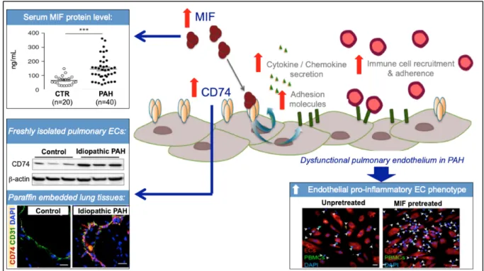
pulmonary EC alterations or dysfunction in PAH include, among others, a reduction in the secretion of vasodilator molecules, such as prostacyclin (PGI2) and nitric oxide (NO), and an increase in the potent vasoconstrictor endothelin (ET)-1. However, the dysfunctional pulmonary endothelial cells in PAH also exhibit a decrease in tube formation capacity, metabolic alterations, and a pro-inflammatory phenotype (32). Indeed, the dysfunctional pulmonary endothelium in PAH overexpresses E-selectin, intercellular adhesion molecule (ICAM)-1, and vascular cell adhesion molecule (VCAM)-1 and secretes locally excessive amounts of numerous inflammatory mediators, such as interleukin (IL)-1 a, IL-6, IL-8, IL-12, CCL2 (MCP-1), and MIF (39). As previously reviewed (48), this pro-inflammatory EC phenotype, along with other abnormalities affecting the immune/inflammatory component of PAH, facilitates the infiltration of inflammatory cells in the perivascular area. The pro-inflammatory environment within the pulmonary arterial walls plays a central role in pulmonary vascular remodeling through the promotion of cell survival and migration (39, 58, 61).
In accordance with these features, a link between circulating levels of various pro-inflammatory mediators and clinical outcomes and/or survival has been described in PH/PAH, suggesting a possible relationship between chronic lung inflammation and disease progression (33, 54, 58). Recently, four PAH clusters with distinct proteomic immune profiles have been found in cohorts of PAH patients by unsupervised analysis of blood proteomic profiles, highlighting the potential existence of distinct immune phenotypes (57). The authors found tumor necrosis factor (TNF)-related apoptosis-inducing ligand (TRAIL), C-C motif chemokine ligand (CCL)-5, CCL7, CCL4, TNF-β, and MIF to be among the panel of up-regulated circulating pro-inflammatory mediators central to immune network-cluster 1, which was associated with the most unfavorable five-year transplant-free survival rates (57). These authors also identify two other different panels of circulating mediators (composed of specific inflammatory molecules and growth factors) that were able to distinguish between groups of patients with low- (cluster 3) and intermediate-risk (cluster 4). Using this specific multiplex approach, they also found a subgroup of patients with intermediate-risk (cluster 2) who had low levels of these circulating mediators similar to healthy controls (56). These observations highlight the need to confirm these findings in large, well-characterized populations with different technologies and arise
the question on how these events detected in the circulation can be used to guide the management of these patients, and how they reflect what is occurring in the PAH lung.
The interplay between alterations of the pulmonary endothelium and the inflammatory component is also illustrated by the presence of circulating autoantibodies directed against antinuclear antigens, ECs, and fibroblasts in several forms of PH/PAH. Maladaptation of the immune response is also underscored by the presence of lymphoid neogenesis, T helper 17 (Th17) cell immune polarization, and dendritic-cell recruitment in the lungs of PH/PAH patients. As a result, the clinical evaluation of anti-inflammatory drugs has become a recent focus in PAH clinical trials. The anti-IL6 and anti-CD20 monoclonal antibodies tocilizumab and rituximab are examples of anti-inflammatory agents under clinical investigation in PAH (ClinicalTrials.gov identifiers NCT02676947 and NCT01086540). Patients with rheumatoid arthritis (RA) that responded to tocilizumab treatment were those that showed significantly lower MIF serum levels, indicating a potential interplay between these two cytokines in RA (34).
Despite its considerable therapeutic potential, targeting inflammation or the vicious cycle between the dysfunctional pulmonary endothelium and the inflammatory component is still highly challenging and highlights the importance of understanding the mechanistic diversity of inflammatory pathways that contribute to PH/PAH (30). Because the excessive activity of the MIF/CD74 axis in pulmonary ECs has been shown to be partly involved in the acquisition and maintenance of the pro-inflammatory endothelial phenotype in PAH (39), MIF and its signaling are emerging as promising therapeutic targets.
3. Macrophage migration inhibitory factor (MIF) and its signaling in
PH/PAH
MIF is a pro-inflammatory mediator with multiple biological functions and pleiotropic effects that is expressed by a variety of immune cells, including macrophages and lymphocytes, as well as endothelial and epithelial cells under certain conditions. MIF occupies an upstream position in the inflammatory cascade and promotes the production of various other pro-inflammatory mediators, such
as TNF-alpha, IL-6, IL-1 beta, etc. Because MIF is associated with acute and chronic inflammatory disorders, as well as glucocorticoid resistance, it represents a promising therapeutic target in several diseases, such as rheumatoid arthritis (RA), irritable bowel disease (IBD), systemic lupus erythematous (SLE), multiple sclerosis (MS), type 1 diabetes mellitus, and PH/PAH (25).
3.1.
Circulating MIF levels: a new emerging PAH marker?
Even if concentration levels vary greatly in the existing literature, circulating MIF levels have been found to be elevated in several forms of PH/PAH (Table 1). Importantly, these results support the notion that circulating MIF levels could serve, alone or in conjunction with other inflammatory mediators, as a potential biomarker of PAH severity (14, 20, 35, 44, 57). Of note, no significant difference in blood levels of the MIF homolog d-dopachrome tautomerase (DDT or MIF-2) between patients with PAH and control subjects without established cardiovascular disease (7.4 ± 2.2 versus 4.6 ± 1.0 ng/mL) (39).
Table 1. Circulating levels of MIF in patients with PAH:
The variability around the reported results, probably due to the type of samples, technology or to the small numbers of patients studied, emphasizes the need to conduct a replicative study with a
Ref. Sample type
& Technology PAH subgroups
Number of patients
Idiopathic and heritable PAH 40
Control subjects 20
PAH (both systemic sclerosis-associated and idiopathic) 101
Healthy controls 35
PAH associated with connective tissue disease 87
Idiopathic PAH 84
Drugs and toxin-induced PAH 49
PAH associated with congenital heart disease 38
Portopulmoanry hypertension 7
Idiopathic PAH 53
PAH associated with connective tissue disease 37 PAH associated with congenital heart disease 7 Portopulmoanry hypertension 7
Healthy controls 88
Idiopathic PAH 13
Systemic sclerosis-associated PAH 15 Systemic sclerosis without PAH 14 Portopulmonary hypertension 21 Controls with liver disease without both pulmonary
hypertension and hepatopulmonary syndrome 31 Plasma & ELISA [14] [56] Plasma & Bio-Plex® multiplex immunoassay
PAH external validation cohort (n=104):
PAH discovery cohort (n=281): Serum & ELISA [38] [54] Serum & ELISA Serum & ELISA [34] 31.19 ng/mL 147 ± 14 ng/mL 66 ± 7 ng/mL 1170 pg/mL 175 pg/mL 504 pg/mL (496-555) Cluster 1 (n=26): high-risk Cluster 2 (n=33): intermediate-risk Cluster 3 (n=36): low-risk Cluster 4 (n=9): intermediate-risk 640 pg/mL (531-816) 467 pg/mL (354-622) 732 pg/mL (575-832) 733 pg/mL (620-931) Cluster 1 (n=58): high-risk Cluster 2 (n=109): intermediate-risk Cluster 3 (n=77): low-risk Cluster 4 (n=37): intermediate-risk 715 pg/mL (598-917) 534 pg/mL (451-644) 565 pg/mL (517-687) 599 pg/mL (501-714)
Circulating MIF concentration
270 ± 636.2 pg/mL 333 ± 520 pg/mL 175 ± 68.68 pg/mL 46.68 ng/mL
sufficiently large sample size and a reproducible technology to measure MIF levels. Indeed, a positive correlation between MIF serum levels and pulmonary vascular resistance (PVR), which is used as a primary endpoint in phase II PAH clinical trials, has been found in both idiopathic and systemic sclerosis-associated PAH (SSc-PAH patients) (35). Consistent with this notion, MIF levels in patients with porto-pulmonary hypertension (PoPH) were found to positively correlate with PVR and inversely with cardiac output (CO), two hemodynamic parameters of disease severity in PAH patients (14). Stefanantoni et al. (55) also reported increased MIF levels in idiopathic PAH and SSc-PAH patients with higher NYHA functional classes. More recently, Marshall et al. (44) reported significant positive correlations between MIF levels and those of seven angiogenic factors (angiopoietin-1, vascular endothelial growth factor (VEGF), platelet-derived growth factor (PDGF)-BB, fibroblast growth factor (FGF)-2, epidermal growth factor (EGF), placental growth factor (PLGF), and Dickkopf (DKK)-1) in patients with idiopathic PAH and those with connective tissue disease-associated PAH (CTD-PAH) in a cohort of patients enrolled in the FREEDOM C-2 trial. However, the authors did not find any correlation between MIF levels and prognostic markers, such as six-minute walk distance (6MWD) or the composite clinical outcome, in this cohort (44). The potential of MIF as a PAH severity biomarker is also supported by the recent work of Sweat et al. (57), who used unsupervised machine learning to categorize PAH patients into four proteomic immune clusters with distinct circulating cytokine signatures that were independent of PAH etiology and associated with various clinical-risk parameters and the long-term prognosis (outcome). Cluster 1, which contains a set of selective upregulated cytokines, including MIF, was associated with high risk. Cluster 1 was comprised of a high proportion of PAH patients with functional class IV symptoms (22.4%), NT-proBNP > 1500 pg/mL (39.7%), high-risk composite REVEAL, elevated mPAP (52 mmHg [45-60]) and right atrial pressure (11 mmHg [5-14]), impaired right ventricular function, and low five-year survival rates (47.6%, CI 35.4-64.1%) (57). This study provides support for the use of immune phenotyping to potentially help in selecting PAH patients who are the most likely to respond to cytokine and growth-factor antagonists, such as MIF antagonists. Interestingly, T-cell lymphocytes were shown to be a source of MIF overabundance in patients with PAH, with no significant difference
3.2.
Elevated levels of MIF in other types of PH
a) PH associated with heart failure (HF) with preserved ejection fraction (PH-HFpEF)
Heart failure (HF) with preserved ejection fraction (HFpEF) is currently the dominant form of HF and a common disease among older adults. Up to 80% of these patients develop PH, which is associated with worse symptoms and higher mortality. The prevalence of PH in HFpEF, based on echocardiographic diagnosis, is 83% and five-year overall mortality is more than 50% from the time of diagnosis (27, 37). In HFpEF patients, plasma MIF levels above the median value (51.58 ng/mL) were found to be associated with higher pulmonary artery systolic pressure (PASP), a surrogate parameter of pulmonary artery pressure (PAP). In addition, MIF levels were also found to correlate with markers of heart failure, the level of natriuretic peptides BNP and NT-proBNP, and a combined endpoint of death or hospitalization after 180 days. These results suggest that MIF could be a useful biomarker, as it is associated with clinical outcomes and could be involved in the pathophysiology of patients with HFpEF and PH (41, 42).
b) PH associated with congenital cardiac shunts
PH is a relatively common complication of congenital heart disease (CHD), with an adult prevalence that ranges from 5 to 10% (15, 19, 60). Serum MIF levels are higher in pediatric patients with PH associated with congenital cardiac shunt, a type of CHD, than those of controls. In these patients, high circulating MIF levels were found to be associated with medial hypertrophy of the small pulmonary arteries and elevated PVR (43) and inversely associated with pulmonary blood flow, estimated noninvasively by transthoracic echocardiography (VTIPV) (67). A common single nucleotide polymorphism (SNP) in the Carbamyl-Phosphate Synthetase I (CPSI) gene is known to cause the substitution of asparagine (Asn) for threonine (Thr) at position 1405 (T1405N) in the critical cofactor-binding domain of the enzyme (56). The CPSI T1405N functional polymorphism has been shown to be associated with the development of PH in the pediatric population (9). Remarkably, patients with a high PVR and the AC-CPSI T1405N genotype profile were those with the highest MIF levels.
c) PH associated with congenital diaphragmatic hernia (CDH)
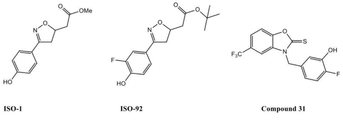
Congenital diaphragmatic hernia (CDH) is a rare congenital anomaly and life-threatening disease characterized by a diaphragmatic defect. PH is a common complication in newborn infants with CDH, in addition to lung hypoplasia. The vascular defects associated with PH, which are characterized by increased muscularization of the arterioles and capillaries, start to develop early in gestation (67). MIF expression has been shown to be 10-fold higher in newborns than in children and adults (50). In the rat neonate model of CHD induced by nitrofen, which mimics the pulmonary abnormalities described for human CDH, including lung hypoplasia and pulmonary vascular remodeling, treatment with the MIF inhibitor ISO-92 led to decreased pulmonary cellularity, vascular wall thickness, and right ventricular systolic pressure (RVSP). However, in contrast to CD74 expression in fetuses of CHD rats, ISO-92 did not affect MIF expression or secretion relative to the nitrofen group (47).
d) PH caused by chronic lung diseases
PH is a common and severe complication of interstitial lung diseases (ILDs), such as idiopathic pulmonary fibrosis (IPF), and can adversely affect symptoms, functional capacity, and survival. PH affects nearly 8 to 15% of patients with IPF at diagnosis and 30 to 50% at the time of evaluation for lung transplantation. Drugs approved for the treatment of PAH are ineffective in IPF-PH and there are currently no approved treatments for PH in IPF (IPF-PH) (11).
Zhang et al. (66) reported that median plasma MIF levels were higher in patients with interstitial lung disease-associated PH (ILD-PH) (median 1,424 pg/mL; range 519-4,396; p = 0.007) than in those with ILD without PAH (median 803 pg/mL; p = 0.076) or a randomly recruited population-based control group (median 365 pg/mL; range 142-4,707).
MIF has also been found to be particularly abundant in the bronchoalveolar lavage (BAL) of patients with IPF and expressed in actively fibrosing areas, such as fibroblast foci and lung remodeling zones (2). Interestingly, higher numbers of cells positive for MIF or its two main receptors, CD74 and CXCR4, have been found in the perivascular area and vascular wall in the lungs of IPF-PH patients (23). In contrast, fibroblast foci of IPF-PH patients have been found to not express CD74, but low
levels of MIF and CXCR4 (23). MIF levels have also been shown to be higher in the sputum and BAL of patients with chronic obstructive pulmonary disease (COPD) than those of non-smokers and healthy smokers (51).
3.3.
Dysregulation of the MIF signaling pathway and its role in PAH
Engagement of MIF with CD74, CXC chemokine receptors (CXCR)-2, CXCR4 and CXCR7 is partly responsible for the multiple biological activities associated with MIF (1, 24, 49). Interestingly, several of these receptors exhibit a more intense immunoreactivity in lungs from patients with PAH and from rodents with established PH (5, 10, 16, 18, 23, 40, 45, 52, 65).
Consistent with these observations, emerging evidence support the notion that MIF and its signaling could contribute to the progression of the pulmonary vascular remodeling associated with PAH. Activation of the MIF/CXCR2 and MIF/CXCR4 axes is known to facilitate leukocyte recruitment (3), induce PASMCs proliferation (64, 66), and promote fibroblast migration in scratch-wounded monolayers in vitro (13). In addition, up-regulation of the MIF/CD74 axis in human pulmonary ECs induces the expression of specific adhesion molecules (i.e. ICAM-1, VCAM-1, and E-selectin), facilitating the adhesion and recruitment of inflammatory cells to the pulmonary vascular wall (Figure 2). Although CXCR2 and CXCR4 mRNA levels in pulmonary ECs derived from patients with idiopathic PAH are unaltered (3), it cannot be excluded that MIF could act through these endothelial receptors to interfere with particular endothelial functions. Interestingly, Burton et al. have reported that the loss of signaling through the bone morphogenetic protein receptor type II (BMPR-II) increased CXCR2 protein levels in human pulmonary artery endothelial cells (8). Furthermore, the excessive secretion of MIF by dysfunctional pulmonary ECs has been shown to increase FoxM1 expression and activity in PA-SMCs, mediating SMC proliferation and pulmonary vascular remodeling (12).
Despite this knowledge, there are still many challenges and open questions. It is necessary to define more precisely the role of MIF on the maintenance of cardiac homeostasis under stress and during the right ventricular hypertrophy that occurs in PAH (59). Although several studies support that
cardiac-detrimental effect of MIF in myocardial ischemia under certain circumstances. Plasma MIF levels also have been reported to be associated with infarct size and the extent of post-infarct cardiac remodeling (62). Inhibition of MIF in mice with myocardial ischemia reduced inflammatory cell infiltration, expression of CCL2 and decreased the incidence of post-myocardial ischemia cardiac rupture (62). Thus, a better understanding of the temporal MIF expression during cardiac ischemia and repair is needed. Given that MIF has both intracellular and extracellular activity, further studies are also required to precise their functions and to address the role of MIF polymorphisms in PAH. Bossini-Castillo et al. (6) found an association of the MIF rs755622*C allele with a higher risk of developing PAH in patients with the diffuse cutaneous form of SSc (dcSSc).
Figure 2: The MIF/CD74 axis contributes to the endothelial pro-inflammatory phenotype and leukocyte recruitment in pulmonary arterial hypertension (PAH).
4. Therapeutic potential of MIF inhibition in preclinical models
Although animal models do not recapitulate the full spectrum of the human disease (4), several preclinical studies have demonstrated that the inhibition of MIF may provide a novel efficient anti-inflammatory approach in the treatment of PH/PAH. Phenotypically, MIF-deficient (KO Mif -/-) mice
appear to live longer and develop normally on multiple genetic backgrounds (26, 28), and are protected against chronic hypoxia-induced PH (36, 66).
In addition, chronic treatment with the prototypical MIF inhibitor ISO-1 or its analogue ISO-92 (Figure 3) have been reported to reduce pulmonary vascular remodeling, cardiac hypertrophy, and right ventricular systolic pressure in the mouse model of PH induced by chronic hypoxia (63, 64, 66). It has been shown that MIF inhibition with ISO-92 enhances pulmonary angiogenesis and lung development in a model of congenital diaphragmatic hernia by increasing the activity of p-eNOS and VEGF and reducing that of arginase 1, arginase 2, and Sflt-1 (47). These observations are consistent with the findings obtained by Le Hiress et al., who demonstrated that curative treatment with the MIF antagonists ISO-1 or N-(3-hydroxy-4-fluorobenzyl)-5 trifluoromethylbenzoxazol-2-thione 31 or anti-CD74 antibodies reversed established PH in monocrotaline-injected rats, reducing total PVR and lung remodeling (Figure 3) (38). MIF inhibition was shown to also attenuate the increase in IL-6 and CCL2 (MCP-1) serum levels in monocrotaline-injected rats, two key pro-inflammatory cytokines known to contribute to the pathogenesis of PAH. However, treatment of monocrotaline-injected rats with ISO-1 has been shown to promote cardiac fibrosis relative to chronic treatment with anti-CD74 antibodies or compound 31 (38, 39). In accordance with these results, Günther et al. reported that chronic treatment of injected mice with ISO-1 or compound 31 attenuated bleomycin-induced inflammation and lung fibrosis and prevented the development of PH in mice (23).
5. Conclusions
Although the exact pathophysiology of PH/PAH is still unknown, there is increasing evidence that inflammation plays an important role in remodeling of the pulmonary arteries, leading to the development and progression of PH/PAH (Figure 4). MIF is an upstream pleiotropic proinflammatory mediator associated with acute and chronic inflammatory disorders and glucocorticoid resistance. Elevated MIF levels in the blood of patients with PH/PAH have been shown to correlate with disease severity, suggesting that serum MIF levels may be a potential biomarker. Pharmacological inhibition of MIF with the small molecules ISO-1, ISO-92, or compound 31, as well neutralizing CD-74 antibodies, significantly improves PH in various animal models of the disease. These pharmacological efficacy studies suggest that targeting MIF may be an effective approach to treat pulmonary vascular remodeling, particularly in PH/PAH patients with high circulating levels of MIF. Efforts are currently underway to design small molecule MIF inhibitors with optimized drug properties for clinical trials in immune, inflammatory, and cardiological diseases and cancer. To date, the most advanced anti-MIF product is Imalumab, a monoclonal anti-oxMIF antibody that is currently under clinical investigation for cancer treatment (NCT01765790). The importance of understanding the pathogenic role of inflammation and the vicious cycle between the dysfunctional pulmonary endothelium and the inflammatory component that contributes to PH/PAH is essential for the design of new therapies for targeting relevant inflammatory pathways in PH/PAH.
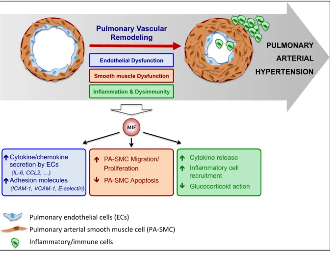
Figure 4: Schematic diagram summarizing the contribution of MIF to the 3 main components of the pulmonary vascular remodeling associated to pulmonary arterial hypertension (PAH).
é Cytokine release é Inflammatory cell recruitment ê Glucocorticoid action é Cytokine/chemokine secretion by ECs (IL-6, CCL2, …) é Adhesion molecules
(ICAM-1, VCAM-1, E-selectin)
é PA-SMC Migration/ Proliferation ê PA-SMC Apoptosis PULMONARY ARTERIAL HYPERTENSION Pulmonary Vascular Remodeling Endothelial Dysfunction
Smooth muscle Dysfunction
Inflammation & Dysimmunity
MIF
Inflammatory/immune cells
Pulmonary arterial smooth muscle cell (PA-SMC) Pulmonary endothelial cells (ECs)
Acknowledgments:
This research was supported by grants from the French National Institute for Health and Medical Research (INSERM), the University of Paris-Sud and the Université Paris-Saclay, the Marie Lannelongue Hospital, the French National Agency for Research (ANR) grant no. ANR-16-CE17-0014 (TAMIRAH), the Fondation pour la Recherche Médicale (FRM) grant no. DEQ20150331712 (Equipe FRM 2015), and in part by the Département Hospitalo-Universitaire (DHU) Thorax Innovation (TORINO), the Assistance Publique-Hôpitaux de Paris (AP-HP), Service de Pneumologie, Centre de Référence de l’Hypertension Pulmonaire Sévère, LabEx LERMIT (grant no. ANR-10-LABX-0033), the French PAH patient association (HTAP France), and the french Fonds de Dotation "Recherche en Santé Respiratoire" - (FRSR) - Fondation du Souffle (FdS).
6. References
1. Alampour-Rajabi S, El Bounkari O, Rot A, Muller-Newen G, Bachelerie F, Gawaz M,
Weber C, Schober A, and Bernhagen J. MIF interacts with CXCR7 to promote receptor
internalization, ERK1/2 and ZAP-70 signaling, and lymphocyte chemotaxis. FASEB J 29: 4497-4511, 2015.
2. Bargagli E, Olivieri C, Nikiforakis N, Cintorino M, Magi B, Perari MG, Vagaggini C, Spina
D, Prasse A, and Rottoli P. Analysis of macrophage migration inhibitory factor (MIF) in
patients with idiopathic pulmonary fibrosis. Respir Physiol Neurobiol 167: 261-267, 2009. 3. Bernhagen J, Krohn R, Lue H, Gregory JL, Zernecke A, Koenen RR, Dewor M, Georgiev
I, Schober A, Leng L, Kooistra T, Fingerle-Rowson G, Ghezzi P, Kleemann R, McColl SR, Bucala R, Hickey MJ, and Weber C. MIF is a noncognate ligand of CXC chemokine receptors
in inflammatory and atherogenic cell recruitment. Nat Med 13: 587-596, 2007.
4. Bonniaud P, Fabre A, Frossard N, Guignabert C, Inman M, Kuebler WM, Maes T, Shi W,
Stampfli M, Uhlig S, White E, Witzenrath M, Bellaye PS, Crestani B, Eickelberg O, Fehrenbach H, Guenther A, Jenkins G, Joos G, Magnan A, Maitre B, Maus UA, Reinhold P, Vernooy JHJ, Richeldi L, and Kolb M. Optimising experimental research in respiratory
diseases: an ERS statement. Eur Respir J 51: 2018.
5. Bordenave J, Thuillet R, Tu L, Phan C, Cumont A, Marsol C, Huertas A, Savale L, Hibert
M, Galzi JL, Bonnet D, Humbert M, Frossard N, and Guignabert C. Neutralization of
CXCL12 attenuates established pulmonary hypertension in rats. Cardiovasc Res 2019.
6. Bossini-Castillo L, Campillo-Davo D, Lopez-Isac E, Carmona FD, Simeon CP, Carreira P,
Callejas-Rubio JL, Castellvi I, Fernandez-Nebro A, Rodriguez-Rodriguez L, Rubio-Rivas M, Garcia-Hernandez FJ, Madronero AB, Beretta L, Santaniello A, Lunardi C, Airo P, Hoffmann-Vold AM, Kreuter A, Riemekasten G, Witte T, Hunzelmann N, Vonk MC, Voskuyl AE, de Vries-Bouwstra J, Shiels P, Herrick A, Worthington J, Radstake T, Martin J, and Spanish Scleroderma G. An MIF Promoter Polymorphism Is Associated with
Susceptibility to Pulmonary Arterial Hypertension in Diffuse Cutaneous Systemic Sclerosis. J Rheumatol 44: 1453-1457, 2017.
7. Boucly A, Weatherald J, Savale L, Jais X, Cottin V, Prevot G, Picard F, de Groote P,
Jevnikar M, Bergot E, Chaouat A, Chabanne C, Bourdin A, Parent F, Montani D, Simonneau G, Humbert M, and Sitbon O. Risk assessment, prognosis and guideline
implementation in pulmonary arterial hypertension. Eur Respir J 50: 2017.
8. Burton VJ, Holmes AM, Ciuclan LI, Robinson A, Roger JS, Jarai G, Pearce AC, and Budd
DC. Attenuation of leukocyte recruitment via CXCR1/2 inhibition stops the progression of PAH
in mice with genetic ablation of endothelial BMPR-II. Blood 118: 4750-4758, 2011.
9. Canter JA, Summar ML, Smith HB, Rice GD, Hall LD, Ritchie MD, Motsinger AA,
Christian KG, Drinkwater DC, Jr., Scholl FG, Dyer KL, Kavanaugh-McHugh AL, and Barr FE. Genetic variation in the mitochondrial enzyme carbamyl-phosphate synthetase I
predisposes children to increased pulmonary artery pressure following surgical repair of congenital heart defects: a validated genetic association study. Mitochondrion 7: 204-210, 2007. 10. Costello CM, McCullagh B, Howell K, Sands M, Belperio JA, Keane MP, Gaine S, and
McLoughlin P. A role for the CXCL12 receptor, CXCR7, in the pathogenesis of human
11. Cottin V, Price LC, and Valenzuela C. The unmet medical need of pulmonary hypertension in idiopathic pulmonary fibrosis. Eur Respir J 51: 2018.
12. Dai Z, Zhu MM, Peng Y, Jin H, Machireddy N, Qian Z, Zhang X, and Zhao YY. Endothelial and Smooth Muscle Cell Interaction via FoxM1 Signaling Mediates Vascular Remodeling and Pulmonary Hypertension. American journal of respiratory and critical care medicine 198: 788-802, 2018.
13. Dewor M, Steffens G, Krohn R, Weber C, Baron J, and Bernhagen J. Macrophage migration inhibitory factor (MIF) promotes fibroblast migration in scratch-wounded monolayers in vitro. FEBS Lett 581: 4734-4742, 2007.
14. DuBrock HM, Rodriguez-Lopez JM, LeVarge BL, Curry MP, VanderLaan PA, Zsengeller
ZK, Pernicone E, Preston IR, Yu PB, Nikolic I, Xu D, Thadhani RI, Channick RN, and Ananth Karumanchi S. Macrophage migration inhibitory factor as a novel biomarker of
portopulmonary hypertension. Pulm Circ 6: 498-507, 2016.
15. Engelfriet PM, Duffels MG, Moller T, Boersma E, Tijssen JG, Thaulow E, Gatzoulis MA,
and Mulder BJ. Pulmonary arterial hypertension in adults born with a heart septal defect: the
Euro Heart Survey on adult congenital heart disease. Heart 93: 682-687, 2007.
16. Farkas D, Kraskauskas D, Drake JI, Alhussaini AA, Kraskauskiene V, Bogaard HJ, Cool
CD, Voelkel NF, and Farkas L. CXCR4 inhibition ameliorates severe obliterative pulmonary
hypertension and accumulation of C-kit(+) cells in rats. PLoS One 9: e89810, 2014.
17. Galie N, Humbert M, Vachiery JL, Gibbs S, Lang I, Torbicki A, Simonneau G, Peacock A,
Vonk Noordegraaf A, Beghetti M, Ghofrani A, Gomez Sanchez MA, Hansmann G, Klepetko W, Lancellotti P, Matucci M, McDonagh T, Pierard LA, Trindade PT, Zompatori M, and Hoeper M. 2015 ESC/ERS Guidelines for the diagnosis and treatment of pulmonary
hypertension: The Joint Task Force for the Diagnosis and Treatment of Pulmonary Hypertension of the European Society of Cardiology (ESC) and the European Respiratory Society (ERS): Endorsed by: Association for European Paediatric and Congenital Cardiology (AEPC), International Society for Heart and Lung Transplantation (ISHLT). Eur Respir J 46: 903-975, 2015.
18. Gambaryan N, Perros F, Montani D, Cohen-Kaminsky S, Mazmanian M, Renaud JF,
Simonneau G, Lombet A, and Humbert M. Targeting of c-kit+ haematopoietic progenitor cells
prevents hypoxic pulmonary hypertension. Eur Respir J 37: 1392-1399, 2011.
19. Gatzoulis MA, Beghetti M, Landzberg MJ, and Galie N. Pulmonary arterial hypertension associated with congenital heart disease: recent advances and future directions. Int J Cardiol 177: 340-347, 2014.
20. Grieb G, Kim BS, Simons D, Bernhagen J, and Pallua N. MIF and CD74 - suitability as clinical biomarkers. Mini Rev Med Chem 14: 1125-1131, 2014.
21. Guignabert C, and Dorfmuller P. Pathology and Pathobiology of Pulmonary Hypertension. Seminars in respiratory and critical care medicine 38: 571-584, 2017.
22. Guignabert C, Tu L, Girerd B, Ricard N, Huertas A, Montani D, and Humbert M. New molecular targets of pulmonary vascular remodeling in pulmonary arterial hypertension: importance of endothelial communication. Chest 147: 529-537, 2015.
23. Gunther S, Bordenave J, Hua-Huy T, Nicco C, Cumont A, Thuillet R, Tu L, Quatremarre
T, Guilbert T, Jalce G, Batteux F, Humbert M, Savale L, Guignabert C, and Dinh-Xuan AT. Macrophage Migration Inhibitory Factor (MIF) Inhibition in a Murine Model of
24. Gunther S, Fagone P, Jalce G, Atanasov A, Guignabert C, and Nicoletti F. Role of MIF and D-DT in immune-inflammatory, autoimmune, and chronic respiratory diseases: from pathogenic factors to therapeutic targets. Drug Discov Today 2018.
25. Gunther S, Fagone P, Jalce G, Atanasov AG, Guignabert C, and Nicoletti F. Role of MIF and D-DT in immune-inflammatory, autoimmune, and chronic respiratory diseases: from pathogenic factors to therapeutic targets. Drug discovery today 24: 428-439, 2019.
26. Harper JM, Wilkinson JE, and Miller RA. Macrophage migration inhibitory factor-knockout mice are long lived and respond to caloric restriction. FASEB J 24: 2436-2442, 2010.
27. Hoeper MM, Lam CSP, Vachiery JL, Bauersachs J, Gerges C, Lang IM, Bonderman D,
Olsson KM, Gibbs JSR, Dorfmuller P, Guazzi M, Galie N, Manes A, Handoko ML, Vonk-Noordegraaf A, Lankeit M, Konstantinides S, Wachter R, Opitz C, and Rosenkranz S.
Pulmonary hypertension in heart failure with preserved ejection fraction: a plea for proper phenotyping and further research. European heart journal 38: 2869-2873, 2017.
28. Honma N, Koseki H, Akasaka T, Nakayama T, Taniguchi M, Serizawa I, Akahori H,
Osawa M, and Mikayama T. Deficiency of the macrophage migration inhibitory factor gene
has no significant effect on endotoxaemia. Immunology 100: 84-90, 2000.
29. Huertas A, Guignabert C, Barbera JA, Bartsch P, Bhattacharya J, Bhattacharya S,
Bonsignore MR, Dewachter L, Dinh-Xuan AT, Dorfmuller P, Gladwin MT, Humbert M, Kotsimbos T, Vassilakopoulos T, Sanchez O, Savale L, Testa U, and Wilkins MR.
Pulmonary vascular endothelium: the orchestra conductor in respiratory diseases: Highlights from basic research to therapy. The European respiratory journal 51: 2018.
30. Huertas A, Perros F, Tu L, Cohen-Kaminsky S, Montani D, Dorfmuller P, Guignabert C,
and Humbert M. Immune dysregulation and endothelial dysfunction in pulmonary arterial
hypertension: a complex interplay. Circulation 129: 1332-1340, 2014.
31. Huertas A, Tu L, and Guignabert C. New targets for pulmonary arterial hypertension: going beyond the currently targeted three pathways. Curr Opin Pulm Med 23: 377-385, 2017.
32. Humbert M, Guignabert C, Bonnet S, Dorfmuller P, Klinger JR, Nicolls MR, Olschewski
AJ, Pullamsetti SS, Schermuly RT, Stenmark KR, and Rabinovitch M. Pathology and
pathobiology of pulmonary hypertension: state of the art and research perspectives. Eur Respir J 53: 2019.
33. Humbert M, Monti G, Brenot F, Sitbon O, Portier A, Grangeot-Keros L, Duroux P,
Galanaud P, Simonneau G, and Emilie D. Increased interleukin-1 and interleukin-6 serum
concentrations in severe primary pulmonary hypertension. American journal of respiratory and critical care medicine 151: 1628-1631, 1995.
34. Kasama T, Isojima S, Umemura M, Tsukamoto H, Tokunaga T, Furuya H, Yanai R,
Takahashi R, Nakamura M, and Inagaki K. Serum macrophage migration inhibitory factor
levels are correlated with response to tocilizumab therapy in patients with rheumatoid arthritis. Rheumatol Int 34: 429-433, 2014.
35. Kolb TM, Rafaels N, Gao L, Barnes K, Girgis R, Mathai SC, Zaiman A, Damico RL, and
Hassoun PM. Serum Macrophage Migration Inhibitory Factor (MIF) Is Increased In Patients
With Pulmonary Arterial Hypertension. American journal of respiratory and critical care medicine 183: A1990, 2011.
36. Kolb TM, Varney J, Hassoun PM, and Damico RL. Macrophage Migration Inhibitory Factor (MIF)-Deficient Mice Are Protected From Right Ventricular Remodeling In A Model Of
Chronic Hypoxia-Induced Pulmonary Hypertension. American journal of respiratory and critical care medicine 183: A4971, 2011.
37. Lam CS, Roger VL, Rodeheffer RJ, Borlaug BA, Enders FT, and Redfield MM. Pulmonary hypertension in heart failure with preserved ejection fraction: a community-based study. Journal of the American College of Cardiology 53: 1119-1126, 2009.
38. Le Hiress M, Akagah B, Bernadat G, Tu L, Thuillet R, Huertas A, Phan C, Fadel E,
Simonneau G, Humbert M, Jalce G, and Guignabert C. Design, Synthesis, and Biological
Activity of New N-(Phenylmethyl)-benzoxazol-2-thiones as Macrophage Migration Inhibitory Factor (MIF) Antagonists: Efficacies in Experimental Pulmonary Hypertension. J Med Chem 61: 2725-2736, 2018.
39. Le Hiress M, Tu L, Ricard N, Phan C, Thuillet R, Fadel E, Dorfmuller P, Montani D, de
Man F, Humbert M, Huertas A, and Guignabert C. Proinflammatory Signature of the
Dysfunctional Endothelium in Pulmonary Hypertension. Role of the Macrophage Migration Inhibitory Factor/CD74 Complex. American journal of respiratory and critical care medicine 192: 983-997, 2015.
40. Li M, Riddle SR, Frid MG, El Kasmi KC, McKinsey TA, Sokol RJ, Strassheim D, Meyrick
B, Yeager ME, Flockton AR, McKeon BA, Lemon DD, Horn TR, Anwar A, Barajas C, and Stenmark KR. Emergence of fibroblasts with a proinflammatory epigenetically altered
phenotype in severe hypoxic pulmonary hypertension. J Immunol 187: 2711-2722, 2011.
41. Luedike P, Alatzides G, Papathanasiou M, Heisler M, Pohl J, Lehmann N, and Rassaf T. Circulating macrophage migration inhibitory factor (MIF) in patients with heart failure. Cytokine 110: 104-109, 2018.
42. Luedike P, Alatzides G, Papathanasiou M, Heisler M, Pohl J, Lehmann N, and Rassaf T. Predictive potential of macrophage migration inhibitory factor (MIF) in patients with heart failure with preserved ejection fraction (HFpEF). Eur J Med Res 23: 22, 2018.
43. Maeda NY, Aiello VD, Santos PC, Thomaz AM, Kajita LJ, Bydlowski SP, and Lopes AA. Relation of Macrophage Migration Inhibitory Factor to Pulmonary Hemodynamics and Vascular Structure and Carbamyl-Phosphate Synthetase I Genetic Variations in Pediatric Patients with Congenital Cardiac Shunts. Mediators Inflamm 2019: 7305028, 2019.
44. Marshall JD, Sauler M, Tonelli A, Rao Y, Bucala R, Lee PJ, and Fares WH. Complexity of macrophage migration inhibitory factor (MIF) and other angiogenic biomarkers profiling in pulmonary arterial hypertension. Pulm Circ 7: 730-733, 2017.
45. Montani D, Perros F, Gambaryan N, Girerd B, Dorfmuller P, Price LC, Huertas A,
Hammad H, Lambrecht B, Simonneau G, Launay JM, Cohen-Kaminsky S, and Humbert M. C-kit-positive cells accumulate in remodeled vessels of idiopathic pulmonary arterial
hypertension. Am J Respir Crit Care Med 184: 116-123, 2011.
46. Morrell NW, Aldred MA, Chung WK, Elliott CG, Nichols WC, Soubrier F, Trembath RC,
and Loyd JE. Genetics and genomics of pulmonary arterial hypertension. The European
respiratory journal 53: 2019.
47. Perveen S, Ayasolla K, Zagloul N, Patel H, Ochani K, Orner D, Benveniste H, Salerno M,
Vaska P, Zuo Z, Alabed Y, Nasim M, Miller EJ, and Ahmed M. MIF inhibition enhances
pulmonary angiogenesis and lung development in congenital diaphragmatic hernia. Pediatric research 85: 711-718, 2019.
48. Rabinovitch M, Guignabert C, Humbert M, and Nicolls MR. Inflammation and immunity in the pathogenesis of pulmonary arterial hypertension. Circ Res 115: 165-175, 2014.
49. Rajasekaran D, Groning S, Schmitz C, Zierow S, Drucker N, Bakou M, Kohl K, Mertens A,
Lue H, Weber C, Xiao A, Luker G, Kapurniotu A, Lolis E, and Bernhagen J. Macrophage
Migration Inhibitory Factor-CXCR4 Receptor Interactions: EVIDENCE FOR PARTIAL ALLOSTERIC AGONISM IN COMPARISON WITH CXCL12 CHEMOKINE. J Biol Chem 291: 15881-15895, 2016.
50. Roger T, Schneider A, Weier M, Sweep FC, Le Roy D, Bernhagen J, Calandra T, and
Giannoni E. High expression levels of macrophage migration inhibitory factor sustain the innate
immune responses of neonates. Proc Natl Acad Sci U S A 113: E997-1005, 2016.
51. Russell KE, Chung KF, Clarke CJ, Durham AL, Mallia P, Footitt J, Johnston SL, Barnes
PJ, Hall SR, Simpson KD, Starkey MR, Hansbro PM, Adcock IM, and Wiegman CH. The
MIF Antagonist ISO-1 Attenuates Corticosteroid-Insensitive Inflammation and Airways Hyperresponsiveness in an Ozone-Induced Model of COPD. PLoS One 11: e0146102, 2016. 52. Sartina E, Suguihara C, Ramchandran S, Nwajei P, Rodriguez M, Torres E, Hehre D,
Devia C, Walters MJ, Penfold ME, and Young KC. Antagonism of CXCR7 attenuates chronic
hypoxia-induced pulmonary hypertension. Pediatr Res 71: 682-688, 2012.
53. Simonneau G, Montani D, Celermajer DS, Denton CP, Gatzoulis MA, Krowka M, Williams
PG, and Souza R. Haemodynamic definitions and updated clinical classification of pulmonary
hypertension. Eur Respir J 53: 2019.
54. Soon E, Holmes AM, Treacy CM, Doughty NJ, Southgate L, Machado RD, Trembath RC,
Jennings S, Barker L, Nicklin P, Walker C, Budd DC, Pepke-Zaba J, and Morrell NW.
Elevated levels of inflammatory cytokines predict survival in idiopathic and familial pulmonary arterial hypertension. Circulation 122: 920-927, 2010.
55. Stefanantoni K, Sciarra I, Vasile M, Badagliacca R, Poscia R, Pendolino M, Alessandri C,
Vizza CD, Valesini G, and Riccieri V. Elevated serum levels of macrophage migration
inhibitory factor and stem cell growth factor beta in patients with idiopathic and systemic sclerosis associated pulmonary arterial hypertension. Reumatismo 66: 270-276, 2015.
56. Summar ML, Hall LD, Eeds AM, Hutcheson HB, Kuo AN, Willis AS, Rubio V, Arvin MK,
Schofield JP, and Dawson EP. Characterization of genomic structure and polymorphisms in the
human carbamyl phosphate synthetase I gene. Gene 311: 51-57, 2003.
57. Sweatt AJ, Hedlin HK, Balasubramanian V, Hsi A, Blum LK, Robinson WH, Haddad F,
Hickey PM, Condliffe R, Lawrie A, Nicolls MR, Rabinovitch M, Khatri P, and Zamanian RT. Discovery of Distinct Immune Phenotypes Using Machine Learning in Pulmonary Arterial
Hypertension. Circulation research 124: 904-919, 2019.
58. Tamura Y, Phan C, Tu L, Le Hiress M, Thuillet R, Jutant EM, Fadel E, Savale L, Huertas
A, Humbert M, and Guignabert C. Ectopic upregulation of membrane-bound IL6R drives
vascular remodeling in pulmonary arterial hypertension. The Journal of clinical investigation 128: 1956-1970, 2018.
59. Tilstam PV, Qi D, Leng L, Young L, and Bucala R. MIF family cytokines in cardiovascular diseases and prospects for precision-based therapeutics. Expert Opin Ther Targets 21: 671-683, 2017.
60. van Riel AC, Schuuring MJ, van Hessen ID, Zwinderman AH, Cozijnsen L, Reichert CL,
Hoorntje JC, Wagenaar LJ, Post MC, van Dijk AP, Hoendermis ES, Mulder BJ, and Bouma BJ. Contemporary prevalence of pulmonary arterial hypertension in adult congenital
61. Voelkel NF, Gomez-Arroyo J, Abbate A, Bogaard HJ, and Nicolls MR. Pathobiology of pulmonary arterial hypertension and right ventricular failure. The European respiratory journal 40: 1555-1565, 2012.
62. White DA, Fang L, Chan W, Morand EF, Kiriazis H, Duffy SJ, Taylor AJ, Dart AM, Du
XJ, and Gao XM. Pro-inflammatory action of MIF in acute myocardial infarction via activation
of peripheral blood mononuclear cells. PLoS One 8: e76206, 2013.
63. Zhang B, Luo Y, Liu ML, Wang J, Xu DQ, Dong MQ, Liu Y, Xu M, Dong HY, Zhao PT,
Gao YQ, and Li ZC. Macrophage migration inhibitory factor contributes to hypoxic pulmonary
vasoconstriction in rats. Microvasc Res 83: 205-212, 2012.
64. Zhang B, Shen M, Xu M, Liu LL, Luo Y, Xu DQ, Wang YX, Liu ML, Liu Y, Dong HY,
Zhao PT, and Li ZC. Role of macrophage migration inhibitory factor in the proliferation of
smooth muscle cell in pulmonary hypertension. Mediators Inflamm 2012: 840737, 2012.
65. Zhang T, Kawaguchi N, Hayama E, Furutani Y, and Nakanishi T. High expression of CXCR4 and stem cell markers in a monocrotaline and chronic hypoxia-induced rat model of pulmonary arterial hypertension. Exp Ther Med 15: 4615-4622, 2018.
66. Zhang Y, Talwar A, Tsang D, Bruchfeld A, Sadoughi A, Hu M, Omonuwa K, Cheng KF,
Al-Abed Y, and Miller EJ. Macrophage migration inhibitory factor mediates hypoxia-induced
pulmonary hypertension. Mol Med 18: 215-223, 2012.
67. Zorzanelli L, Maeda NY, Clave MM, Aiello VD, Rabinovitch M, and Lopes AA. Serum Cytokines in Young Pediatric Patients with Congenital Cardiac Shunts and Altered Pulmonary Hemodynamics. Mediators Inflamm 2016: 7672048, 2016.