HAL Id: hal-03052546
https://hal-amu.archives-ouvertes.fr/hal-03052546
Submitted on 11 Dec 2020
HAL is a multi-disciplinary open access
archive for the deposit and dissemination of
sci-entific research documents, whether they are
pub-lished or not. The documents may come from
teaching and research institutions in France or
abroad, or from public or private research centers.
L’archive ouverte pluridisciplinaire HAL, est
destinée au dépôt et à la diffusion de documents
scientifiques de niveau recherche, publiés ou non,
émanant des établissements d’enseignement et de
recherche français ou étrangers, des laboratoires
publics ou privés.
Distributed under a Creative Commons Attribution| 4.0 International License
molecular mechanism of daptomycin action
Francesca Zuttion, Adai Colom, Stefan Matile, Denes Farago, Frédérique
Pompeo, Janos Kokavecz, Anne Galinier, James Sturgis, Ignacio Casuso
To cite this version:
Francesca Zuttion, Adai Colom, Stefan Matile, Denes Farago, Frédérique Pompeo, et al.. High-speed
atomic force microscopy highlights new molecular mechanism of daptomycin action. Nature
Commu-nications, Nature Publishing Group, 2020, 11 (1), �10.1038/s41467-020-19710-z�. �hal-03052546�
High-speed atomic force microscopy highlights
new molecular mechanism of daptomycin action
Francesca Zuttion
1, Adai Colom
2, Stefan Matile
3, Denes Farago
4, Frédérique Pompeo
5, Janos Kokavecz
6,
Anne Galinier
5, James Sturgis
7& Ignacio Casuso
1✉
The increase in speed of the high-speed atomic force microscopy (HS-AFM) compared to that of the conventional AFM made possible thefirst-ever visualisation at the molecular-level of the activity of an antimicrobial peptide on a membrane. We investigated the medically prescribed but poorly understood lipopeptide Daptomycin under infection-like conditions (37 °C, bacterial lipid composition and antibiotic concentrations). We confirmed so far hypothetical models: Dap oligomerization and the existence of half pores. Moreover, we detected unknown molecular mechanisms: new mechanisms to form toroidal pores or to resist Dap action, and to unprecedently quantify the energy profile of interacting oligomers. Finally, the biological and medical relevance of the findings was ensured by a multi-scale multi-nativeness—from the molecule to the cell—correlation of molecular-level information from living bacteria (Bacillus subtilis strains) to liquid-suspended vesicles and supported-membranes using electron and optical microscopies and the lipid tension probe FliptR, where we found that the cells with a healthier state of their cell wall show smaller membrane deformations.
https://doi.org/10.1038/s41467-020-19710-z OPEN
1U1067 INSERM, Aix-Marseille Université, Marseille, France.2Biochemistry Department, University of Geneva, Geneva, Switzerland.3Organic Chemistry
Department, University of Geneva, Geneva, Switzerland.4Department of Technical Informatics University of Szeged, Szeged, Hungary.5Laboratoire de
Chimie Bactérienne (LCB), Institut de Microbiologie de la Méditerranée (IMM), CNRS, UMR 7283, Aix Marseille Université, Marseille, France.6Institute of
Environmental Science and Engineering, University of Szeged, Szeged, Hungary.7LISM, UMR 7255, CNRS, Aix Marseille Université, Marseille, France.
✉email:ignacio.casuso@inserm.fr
123456789
T
he world is running out of antibiotics. In the last decades, antibiotic development has stagnated, while antibiotic resistance continues to rise. The European Centre for Disease Prevention and Control estimates that currently in Eur-ope, 25,000 patients die per year due to antibiotic-resistant bac-terial infections (https://ecdc.europa.eu).Antimicrobial peptides (AMPs) have attracted considerable attention due to their wide spectrum of activity and the low degree of resistance1,2. Many AMPs function by targeting
biolo-gical membranes, reducing the development of microbial resis-tance compared to intracellular targeting. Lipopeptides are a promising class of antibiotic drugs only investigated to a limited degree. They contain a hydrophilic peptide attached to a fatty acyl side chain that promotes insertion and clustering in the biological membranes3, and often incorporate non-ribosomal amino acids limiting their exposure to peptidase breakdown.
Lipopeptide action is largely determined by the length, satura-tion, and branching of the acyl chain4,5, there are often numerous homologues and isoforms, but the correlation of their structures with their precise actions is challenging; lipopeptide action is typically multifaceted, and numerous studies using multiple techniques have been deployed to understand their structure-function relationship6,7. There is an inherent complexity to
pre-dicting the consequences of small differences in the spatial arrangement of the amino acids in AMPs8. This uncertainty in the
details of lipopeptide action hinders the pharmaceutical develop-ment of lipopeptides as antibiotics; the production of molecular candidates is costly and, as the chemical synthesis of common medically used lipopeptides is not viable at industrial scale9,10, the
costs for pharmaceutical development are high. However it is now becoming clear that the toxicity of lipopeptides can be even lower than previously believed and additionally reduced by using new types of synthetical lipopeptides11, increasing their attractiveness
for development.
Daptomycin (Dap) is the paradigm for the lipopeptide anti-biotics incorporated to clinical use in the last forty years12.
Therapeutically, Dap is used against Gram-positive (Gram+) bacterial infections including as a last resort antibiotic against multidrug-resistant enterococci and staphylococci13,14. Even if
bacterial resistance to lipopeptides is low, in recent years, some strains have become resistant to Dap15. This antibiotic offers an
excellent system for testing new approaches for the character-isation of the structure‐activity relationships of AMPs on mem-branes, with an aim to furthering development of this class of antibiotics and countering the spread of resistances.
What is known about Dap action? When a non-Dap-resistant Gram+ bacteria is exposed to Dap (in the presence of Ca2+), the
plasma membrane of the bacteria depolarises in a slow process lasting several tens-of-minutes16,17. Currently, there is a
knowledge-gap on the details of Dap activity. Up until now, it is not clear whether the observed depolarisation is the cause of bacterial mortality, or rather the depolarisation is the result of bacterial death caused by another mechanism18,19. To date, only the two initial steps of Dap action are well established. 1st, the anionic Dap molecule binds (in the presence of Ca+2) to the anionic phospholipid Phosphatidylglycerol (PG) found in the membranes of Gram+ bacteria. 2nd, after binding to the membrane, Dap is believed to undergo a structural transition inserting in the plasma membrane in a wedge-like configura-tion20, but assessment of the structural configuration of Dap in its membrane-bound state is still missing. The structural and dynamical pathways of the remaining stages of Dap action have not been clearly identified yet21,22.
The predominant hypothesis is that Dap antibacterial action is caused by toroidal pores that induce the depolarisation measured in bacteria23–25. The process of formation is believed to happen as
follows: Dap monomers would oligomerize26–29; the large local
positive curvature strain created in the outer membrane leaflet could result in dimples, also named half-toroidal pores25, that
would locally reduce the thickness of the membrane through where some Dap molecules would translocate to the inner leaf-let25 where they would oligomerize and in turn form similar
dimples in the inner leaflet; finally, inner and outer dimples would fuse and produce the transmembrane toroidal pores25,30. Nevertheless, in the last six years, two other models of Dap action have been proposed, both based on optical microscopy (OM) observations. First, in 2014, the lipid extraction model was pro-posed in which Dap is suggested to cause ejection of material from the membrane31. Unfortunately, these observations were
performed on membranes with a rather un-natural composition of 70% DOPC. Second, in 2016, in the membrane protein delo-calisation model, it was proposed that Dap delocalises the gly-cotransferase enzyme MurG hampering bacterial cell wall formation19.
The wide range of techniques that have been used to measure AMPs activity (i.e., fluorescence resonance energy transfer (FRET), small angle neutron scattering (SANS), nuclear magnetic resonance (NMR), calorimetry, electrophysiology, X-Ray, or cir-cular dichroism (CD)) provide little information on the oligo-merization states which is an important piece of information missing in our understanding of the activity of the peptides29; as
oligomerization controls the nature, dynamics and regularity of the membrane deformations. Atomic force microscope (AFM) and electrochemical scanning tunnelling microscopy (EC-STM) are unique in their capability of label-free high resolution imaging of biological molecules and their surroundings under physiolo-gical conditions, yet, in the literature, few studies address the effect of AMPs on supported lipid bilayers by AFM32–44(Dap in
particular was never studied), most are prior to 2011, few are the recent publications42–44. The published AFM data examines the evolution of the contours of membranes patches exposed to AMPs in solution. Never have the dynamics of the activity of AMPs been imaged at molecular resolution, for example oligo-mers could never be visualised due to their thermal diffusive motion. AFM and EC-STM molecular imaging was restricted to the staticfinal states of the processes32,39,42,43, hence, overlooking the core of the AMPs molecular activity. Overall, the incapability to assert AMPs molecular activity hampers the straightforward correlation of the AFM data with the molecular, but averaged, information issued from the prevalent techniques of monitoring of AMPs molecular activity36. Alternatively, the all-atom
mole-cular dynamics (MD) simulations of AMPs activity would pro-vide the ultimate level of detail in the understanding of the mechanisms of action of AMPs; however the timescale of the pathways of action of AMPs on membranes is too long (several minutes2,16,17) and the numbers of peptide molecules involved in the processes excessive (as the ejections caused by Dap31) to be
predicted by MD simulations; these are limited to the assessment of processes shorter than a few microseconds and only a handful of peptides45. Consequently, even if the currently available
simulations provide us with valuable insights46, they cannot
predict unknown pathways without prior knowledge from experiments—this is the case even for pore formation46,47—
including the action of Dap48,49, as will be shown.
In the last decade, the advent of the high-speed atomic force microscope (HS-AFM)50has achieved µs/pixel speeds. Using the HS-AFM, we could accomplish nanometre scale high resolution molecular imaging of the action of AMPs on membranes. For the first time it was possible to correlate the molecular picture of the activity of AMP provided by FRET, SANS, NMR, CD or elec-trophysiology techniques, with high resolution molecular imaging by AFM—non-averaged and label free—of the spatial membrane
heterogeneity at local membrane environments with individual molecule resolution and dynamical information.
The imaging speed of scanning microscopies, like AFM, depends on the time of scan per one pixel. When particles move fast with respect the scanning speed of the microscope the particle traverses several pixels in the time the microscope scans one pixel, in this case it is not possible to define for each frame in which pixel the particle is located; the particle is not resolved in the image. To resolve the particle, it is necessary that the particle resides in the area of one pixel at least the time required for the microscope to scan one pixel. In biological membranes lipids and proteins diffuse driven by thermalfluctuations; their trajec-tories follow overlapping distributions of steps of variable sizes that depend on their interactions with their immediate membrane surroundings. The typical diffusion coefficients of lipids51,52and
small molecules as Dap53, are of order of few µm2/s, or in terms
of instantaneous speeds (quotient of the mean free path and the step time) of the order of a few hundreds of µm/s52. Because (i) the imaging biomolecules at high resolution AFM requires a pixel lateral size smaller than 1 nm and (ii) the residence time of the membrane diffusing lipids and proteins in 1 nm2 is around 1–10 µs, it is necessary that the scan speed of the AFM is faster than 1–10 µs/pixel to attain visualisation of membrane diffusing lipids and small molecules. Prior AFM imaging of the action AMPs on membranes was performed at scan speeds from 20044
to 1000 µs/pixel32–38, thus too slow for visualising the molecular
action of AMPs on membranes.
Biological membranes are extremely complex and dynamically partitioned into lateral micro-domains, a far from an ideal mix-ture of multiple proteins and lipids. The use of model lipid bilayers, artificially simplified systems, has been essential for our understanding of the roles of lipid, proteins and carbohydrates in drug membrane interactions54,55. Nevertheless, model
mem-branes leave numerous factors unconsidered56: among others, the thousands of different lipid species present in biological mem-branes and the interactions with cytoskeletal components with active roles on membrane organisation are not present in the membrane models. Furthermore, in the case of AFM membrane imaging, the membranes are placed on flat supports, thus, addi-tionally restricting the degrees of freedom for membrane defor-mations. Therefore, it is legitimate to ask ourselves the degree of relevancy of the data derived from supported model membranes, especially for the study of drug-bacterial membrane interactions. We have, in this article, sought to reinforce our AFM data, and address the relevance of our measurement, by correlating obser-vations on several different systems progressively further from the native membrane. We have obtained information on living cells and non-supported model membranes by optical microscopy (OM), fixed cells and non-supported model membranes by electron microscopy (EM), and supported model membranes by HS-AFM. Moreover, besides structural assessment, we used thefluorescence dye FliptR to gain information on lipid packing; thefluorescence lifetime of FliptR depends linearly on membrane tension57. The images of deformations on supported membranes
by AFM have been correlated with deformations and alterations of the lipid packing on cells and non-supported vesicles. Our multi-scale, multi-nativeness and multi-technique correlation of data lifts the significance of the ensemble of observations for the deciphering of the mechanism of action of Dap and other future drug developments.
Finally, the knowledge gap on Dap and other AMPs, molecular mechanisms can be partially ascribed at the uncertainties on the model membranes and conditions used in the experiments, we list: (i) Thefluorescence labels, commonly used, can modify Dap function; as shown for BODIPY-labelled58, perylene-labelled28
and NBD-labelled Dap49, (ii) Room temperature which is the
temperature most commonly used in Dap studies, rather than the physiological temperature (37 °C) pertinent to antimicrobial use, can strongly modify membrane properties and thus alter Dap action21,59,60. Finally, (iii) The model membrane lipid composi-tion can modify membrane properties and membrane-Dap interactions: (iii.a) The lipid head group, phosphatidylcholine (PC) has been commonly used in model membranes, but PC is not found in Gram+ bacteria and can alter Dap action18, (iii.b)
The acyl chain, dimyristoyl (DM, 14:0–14:0) lipids were the most common used acyl chain. However, it is known that Dap per-meabilizes lipid bilayers with DM acyl chains, but not membranes with the morefluid palmitoyl-oleoyl (PO, 16:0–18:1) or dioleoyl (DO, 18:1–18:1) acyl chains61, what is more, the DO acyl chains
are the most common acyl chain in Staphylococcus aureus, and infection by this bacteria is commonly treated by Dap adminis-tration53. In our experiments, to minimise the uncertainty related
to the pertinence of the observation conditions, we mimicked as close as possible the bacterial infection conditions.
As mentioned above, since bacterial resistance to Dap is unfortunately increasing15, understanding how bacteria resist Dap is increasingly relevant. The Dap resistant bacterial strains present amended cell walls and/or plasma membranes struc-tures62. It is known that at the plasma membrane of the Dap-resistant strain of Enterococcus faecalis, a bacterial infection commonly treated with Dap, the content of cardiolipin (CL) is higher than in membranes of the non-Dap-resistant strains63,64. However, the mechanisms by which CL inhibits Dap action remain unclear30. In model membrane assays—regrettably
con-taining >40% of the non-bacterial PC lipid—the addition of CL at 10–20% molar to the membrane inhibited the permeabilization of the membrane by Dap25,60. Herein, we provide novel insight on
CL-Dap interactions by HS-AFM imaging correlated to electron and optical microscopies on PG membranes.
In this work, the function of Daptomycin, the paradigm of the antimicrobial lipopeptide molecules, whose multifaceted mechan-isms remains for the mostly part unknown is assessed by a frame-work of assays. These assays significantly include the single-molecule visualisation of the activity of lipopeptides on membranes. The manuscript starts by the higher nativeness measurements in living bacteria, and progressively decreases the nativeness, approaching supported membranes and inversely increases the resolution. All measurements mimicked the infection conditions: 37 °C, bacterial lipid composition and antibiotic concentrations. We also mimicked bacterial resistance conditions by increasing the CL-content in the membrane. We confirm several, previously, hypothetical models, and detect several unknown molecular mechanisms. Moreover, we provide quantitative information on the Dap oligomers and their interactions.
Results
Dap action on bacteria and non-supported vesicles. Three dif-ferent strains of the Gram+ bacteria Bacillus (B.) subtilis were exposed to Dap and imaged by OM (using the membrane specific dye FM1–43) and scanning electron microscopy (SEM). The strains studied were the wild-type (WT)65 and two cell strains
with defective cell walls; 1st, a mutant lacking ugtP66; UgtP is an enzyme involved in the synthesis of glycolipids and the anchoring of lipoteichoic acids that is required for the cell wall formation of Gram+ bacteria and that was suggested to be inhibited in the cells exposed to Dap67; 2nd, a mutant lacking the actin-like
protein MreB68; this cytoskeletal protein is involved in the
pat-terning of the bacterial membrane, among others defining the location of the regions of increase fluidity69 where Dap
The lowest concentration of Dap that inhibited the visible growth of B. subtilis in overnight incubation was measured (3.1 µM for all the strains), in agreement with published values for B. subtilis19and other Gram+ bacteria70. For all the rest of
this study, two concentrations of Dap were used: (1) A low concentration of 0.6 µM, below the minimal inhibitory concen-tration (MIC). and (2) a high concenconcen-tration of 20 µM, above the MIC. Importantly, these Dap concentrations are in the range of that found in the serum of patients during Dap treatment71. At
the lower concentration depolarisation of the B. subtilis cells has been measured using a membrane potential-sensitive fluoro-phore19. It should be noted that for a component partitioning
into the membrane the concentration in the aqueous phase is perhaps not the most pertinent.
The three strains of B. subtilis were exposed to sub-MIC and over-MIC Dap for 15 min in physiological buffer with ∼1.25 mM Ca2+at 37 °C. All the strains displayed significant
deformations observed by OM (by phase contrast and FM1-43 membrane labelling) and SEM. Notably: submicron sized protuberances emerged from the bacterial wall, these are probably lipidic given their spherical shape and staining with
FM1-43 (Fig. 1b, c, white arrows in the OM imaged plus the insets in the SEM images); the roughening of cell wall surface, in the WT and the mreB mutant strains at sub-MIC, and in all the strains at over-MIC (Fig.1b, c, SEM images, yellow arrows); and, lastly, in all the bacterial strains, some cells developed large spherical micron sized vesicles at their poles (Fig.1b, c, green arrows), reminiscent of the Dap-induced lipid extraction effect on model membranes31(Fig.1b, c, OM images, green arrows). The fact that at sub-MIC, the lipid extraction takes place only in the cell wall defective strains, not in the WT strain (confirmed by Müller et al.19) brings to light that a ‘healthier’ cell wall restricts the growth of the Dap-induced morphological altera-tions of the bacterial membrane. This could result from slowed Dap penetration through the native cell wall and/or increased resistance to membrane deformations. The comparable MIC value of the three strains would suggest the arrival of Dap to the membrane is not changed among strains.
To gain insight at the molecular-level, we used thefluorescence dye FliptR, an indicator of the membrane lipid packing57. In the
absence of Dap, the intra-cellular distribution of the lipid packing is similar for all the bacterial cells, with a bimodal distribution
Sub-MIC WT ugtP– mreB– Over-MIC 1 μμm 1 μm No Dap 1 μm 1 μm 1 μm 1 μm SEM OM SEM OM pha s e FM1-43 No Dap POPG vesicles (TEM) 1 μm 1 μm 1 μm 1 μm 1 μm 1 μm 1 µm 1 μm 1 µm 1 μm 1 µm 1 µm Sub-MIC 200 nm Over-MIC 200 nm 200 nm SEM OM nm 230 nm 480 nm 330 nm 315 nm 370 200 nm 200 nm mreB-over-MIC a b c d e f g
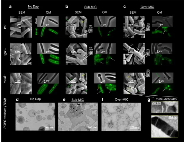
Fig. 1 Dap action on B. subtilis strains (WT,ΔmreB and ΔugtP strains) and on POPG vesicles by EM and OM. a In the absence of Dap, the ΔmreB and ΔugtP mutant strains display abnormal surfaces visible by SEM, whereas the OM does not display such disparities. b Exposed for 15 min to sub-MIC Dap, SEM displays in the WT andΔmreB strains, but not in the ΔugtP strain, the creasing of the bacterial cell wall surface (yellow arrows). In all the strains, we observe pronounced deformations at the cell poles (green arrow) and submicron protuberances emerging, seemly of lipidic nature given its spherical shape (white arrow and insets). In contrast, the OM displays bulbous micron-sized swellings, primarily coming out of the bacterial poles in the two mutants, but not in the WT cells. Thus, the cell wall delimits the Dap-induced morphological alterations.c Exposed for 15 min to over-MIC, the OM also displays bulbous micron-sized swellings in the WT B. subtilis. All the rest of deformations observed at sub-MIC are found also at over-MIC in all the strains.d–f TEM of POPG vesicles exposed to Dap: In the absence of Dap, the POPG vesicles display spherical shape; at sub-MIC, a number of tubulations emerge from, and interconnect, the vesicles (black arrows); at over-MIC, the tubulations are omnipresent interconnecting the vesicles and the surface of the vesicles becomes creased.g Detail of the tubulations that appear in theΔmreB cells at over-MIC visualised by SEM. Their size, shape, and interconnective character, is comparable to those observed in POPG vesicles shown ine and f. These tubules are only noticed withΔmreB mutant; thus, they are not created by the SEM sample preparation.
showing tighter lipid acyl chain packing at the poles and looser packing at the sides. In the WT cells, low concentrations of Dap seem to slightly increase packing at the poles. However, higher Dap concentrations increase the heterogeneity in the distribution: We observe tight lipid packing over the totality of some cells (Fig.2a, WT, over-MIC, orange arrow), while others maintain the loose packing found in the absence of Dap (Fig. 2a, WT, over-MIC, blue arrow); as a result the packing distribution is no longer bimodal but stretched and with a little peak at higher levels of packing. Furthermore, not only there is a heterogeneity between cells but the intra-cellular heterogeneity also increases. The Dap induced deformations of the membranes are heterogeneous and result in zones of high and low lipid packing with higher levels of lipid packing at the protrusions (Fig. 2a, WT, over-MIC, zoom, orange and blue arrows). Similar observations were made by Müller et al. in 2016 using the hydration of the Laurdan membrane probe for membrane fluidity assessment19, more
dependent on the lipid composition than the FliptR lipid packing assessment (moreover, the intrinsic kynurenine fluorescence of Dap could have caused data misinterpretation, as it was not corrected19). In line with the cellular morphological observations,
the lipid packing observations show that on the mutant strains smaller concentrations of Dap induce the same effects as higher concentrations of Dap on the WT (Fig. 2b, c); the finding reinforces the idea that a normal cell wall is probable capable of limiting Dap-induced deformations.
To gain further insight, vesicles with lipid composition similar to that of the pathogens Clostridium difficile72and S. aureus—
both medically treated with Dap—1-palmitoyl-2-oleoyl, (PO, 16:0-18:1 acyl chains) PG (POPG) were characterised with same set of measurements as the bacteria. We avoided mixing the
anionic POPG with non-bacterial zwitterionic PC lipid as many previous authors have18,21,24–27,31,33,35,37,38,44,48,49,60,61. TEM
shows that, unsurprisingly and as in bacteria, Dap deformations are more important at higher than at lower Dap concentrations (Fig. 1d–f). We observed that the predominant type of
deformation was the formation of membrane tubules that resembled, in shape and dimensions, those that emerge from the surface of mreB-mutant cells at high Dap concentrations (Fig.1g). Significantly, the fact that similar tubules were noticed
in one out of the three B. subtilis strains shows that they are not artefacts from the SEM sample preparation procedures. The observation of such tubules could correlate with the lipid extraction model derived from similar observations of (7:3) DOPC:DOPG vesicles31. By tracking the development of the lipid
packing on POPG vesicles following exposure to Dap using FliptR (Fig.2d), we observed a two-step process: after an initial stage of increase in packing, there is a dramatic reduction in lipid packing, correlating to intense deformation of the vesicles. This two-step process, with both reduced and increased lipid packing, is consistent with the heterogeneity of zones identified in the bacteria exposed to Dap; the POPG vesicles exposed to Dap mimic the morphological and the lipid packing effects observed in bacteria, thus supporting the use POPG supported model membrane for the assessment of the activity of Dap by AFM.
HS-AFM on supported POPG bilayers. In order to observe at the molecular-level the effects of Dap on lipid bilayers we imaged supported POPG bilayers by HS-AFM that featured an imaging rate of about ten frames per second on areas of hundreds of nanometres square at sub-nm resolution.
a
b
c
d
2.6 Lifetime (ns) 3.8
Low Lipid packing High
No Dap mreB– Zoom 1 μm No Dap Sub-MIC Over-MIC Zoom Pixel count 1 μm WT No Dap Zoom ugtP– 1 μm 24 μm Dap IN Mean of pixels with value ≠ 0 4.5 4 3.5 3 2.5 0 5 10 15 Lifetime (ns) Time (min) Over-MIC Sub-MIC Sub-MIC Over-MIC
Fig. 2 Dap action on B. subtilis strains (WT,ΔmreB and ΔugtP strains) and POPG vesicles by the FliptR lipid packing probe. FliptR signal: lower lifetimes correlate to lower lipid packings and vice versa.a FliptR image on WT cells: in the absence of Dap, all the cells keep the same distribution, the bacterial poles display higher packing levels than the bacterial sides, a bimodal distribution of lipid packing values appears; at sub-MIC, at the poles, lipid packing level rises; at over-MIC, an inter-cell heterogeneity appears, certain cells show high lipid packing disseminated over the full cell (orange arrow) while others maintain their previous packing distribution (blue arrow), the distribution of packing stretches and a little peak at higher levels of packing becomes apparent. At higher magnification (zoom) also heterogeneity at the intra-cell level becomes evident, zones of high (orange arrow) and low (blue arrow) lipid packing disseminate inside the cell, in agreement with previous observations19. It is observed that the deformations protruding from the cells present
higher levels of lipid packing than the zones that do not protrude.b, c For theΔmreB and ΔugtP mutants, FliptR signals are akin to WT strain. Yet, the inter-cell heterogeneity appears at lower Dap concentration, sub-MIC instead of over-MIC, supporting that‘healthier’ bacterial cell wall limits the deformations induced by Dap.d Tracking of the progression of the lipid packing on POPG vesicles exposed to over-MIC Dap by FliptR. Dap is injected at the‘Dap IN’ label and a two-step process takes place. First the vesicles rigidify, then they collapse and the lipid packing is drastically reduced to values below the initial ones. This two-step process could explain the two zones of low and high lipid packing identified in the bacteria cells exposed to Dap.
The HS-AFM allowed us to observe the development of the dynamics of interaction at the molecular-level over several hours. Over the course of the experiment many different structures were observed forming and then disappearing giving rise to new structures. At thefirst stages, measured at low Dap concentration and 37 °C, it was possible to observe small particles occupying an area fraction of ~4% of the total surface diffusing rapidly on the POPG (Fig. 3a and Supplementary movie 1). These particles diffused, as expected52, at speeds of 300 ± 200 µm/s, or in terms of
diffusion coefficient a few µm2/s, which is consistent with the
particles being Dap oligomers. Understanding the oligomerization states is critical to understand the function-structure relations of AMPs73. The images confirm that the HS-AFM imaging enables
molecular-level visualisation of the transients of AMP oligomers in diffusion on membranes label-free, hence without financial cost and the incertitude related to the labelling of AMPs28,49,58 that
hampers pharmaceutical development. In these initial stages of action, the oligomers created temporary ensembles (Fig.3a, slashed ellipses), but they did not form permanent clusters. Nevertheless, we noticed that the Dap oligomers form transiently stable assemblies: mainly pairs of oligomers (Fig. 3a light blue circles; Fig. 3c). These transient assemblies of oligomers diffused and rotated and re-separated (Fig. 3c, last frames). Their presence
indicates that at sufficiently short distances there is a net attractive force between the Dap oligomers. Further detail was obtained by following two interacting oligomers, we obtained a movie of 36 frames where the oligomers rattle in mutual attraction after correcting for their lateral diffusion (Fig. 3c), we measured the probability distribution (in time) with respect to the centre-to-centre distance (d) and derived the energy profile U(d) considering an equilibrium Boltzmann distribution74(Fig.3c, right), moreover
using the spatial distribution of the ensemble of oligomers (which is equivalent by ergodicity to time probability distribution) we further evidenced the oligomer-oligomer attraction and the shape of U(d). We could quantify that the U(d) is attractive for d < 20 nm with a global minimum of energy of−3.4 kBT at d= 7 nm, and a local minimum of −2.8 kBT at d= 16 nm; such multi-minima interaction energy profile could cause bi-stable behaviours in the membrane deformations induced by Dap. To date, the membrane-mediated lipopeptide oligomer-oligomer interactions were not studied neither theoretically nor numerically, only the formation of the individual oligomers was so far simulated75. Nevertheless, there
have been experimental reports of the aggregation of Dap oligomers; by Förster resonance energy transfer (FRET), where it was found that the signal of the oligomer-oligomer interaction exceeds pure random interactions27; and by OM on living cells,
Particle analy sis 15 nm Oligomers diffusion 0 s 0.073 s 0.146 s 0.219 s 0.292 s 0.365 s 0.438 s 8 4 0 2 4 6 8 10 nm2 Co u n t 15 nm Area oligomer d (nm) 0 10 20 30 –0.5 –1.5 –2.5 –3.5 d (nm) 0 10 20 30 U (kB T) –0.5 –1.5 –2.5 –3.5 0.000 s 0.091 s 0.182 s 0.273 s 0.364 s 0.455 s 0.546 s 0.637 s 0.728 s 0.819 s 0.910 s 1.001 s 1.092 s 1.183 s 1.274 s 1.365 s 1.456 s 1.547 s 1.638 s 1.729 s 1.820 s 1.911 s 2.002 s 2.093 s 2.184 s 2.275 s 2.366 s 2.457 s 2.548 s 2.639 s 2.730 s 2.821 s 2.912 s 3.003 s 3.094 s 3.185 s a b c U (kB T)
Oligomer transient assemblies and interaction energy
Count 25 20 15 10 5 0 28 frames Count ( ) 0 10 20 0 20 Count ~ 28 • 0.04 ~ 1.12 30 40 50 B Oligomer-oligomer distance (d)
Area fraction oligomers: ~ 0.04 10 20 30 40 d
d
(nm)
5 nm
Mica
Fig. 3 Sub-MIC Dap on POPG at 37 °C. First minutes. Initial stages. a Oligomers of Dap in diffusion on a membrane (slashed circles). The upper left image shows the membrane and the underlying mica substrate (colour scale: 4 nm). The rest of the images are close-ups on top of the membrane (colour scale: 2 nm): The oligomers formed temporary ensembles (slashed ellipses), but they did not form permanent clusters. Movie details: frame rate 74 ms; full image size 180 nm × 60 nm; full-frame size 256 × 80 pixels.a, right, The particle analysis (right bottom, one of the frames analysed; right top, histogram) shows a spread in the area of the oligomers from 2 to 10 nm2, a peak value of 4.3 nm2, and a mean and standard deviation of 5.4 ± 3.2 nm2; which
corresponds to a stoichiometric variability maximal of 1 to 6 monomers, and mean and standard deviation of 3.6 ± 2.1 monomers. The oligomers experience mutual attraction at short distances.b, left, Oligomer-oligomer energy profile obtained from the spatial distribution of oligomer-oligomer distances. The area fraction of the oligomers on the membrane is of ~0.04.b, centre, Minima are found at distances between oligomers of ~7 and ~16 nm. The count at far distance (count∞) can be calculated to be ~1.12 (area fraction occupied by the oligomers (0.04; ~4% of the total membrane area) multiplied by the number of frames counted (n= 28), horizontal white line at d > 30 nm) in agreement with the count at d > 30 nm. b, right, Equilibrium Boltzmann distribution energy profile derived from U(d) = −kbTln[count (d)/count∞]; the depthless of the energy wells is estimated to be of −3.4 kBT at d= 7 nm and −2.8 kBT
at d= 16 nm. c These 32 frames (2.821 s) show a pair of oligomers in interaction. c, right, From the count of d, the energy profile features one global (d = 7 nm) and one local (d= 16 nm) minima. Because the oligomer-oligomer interaction is ergodic and, as expected, the interaction profile in space is similar to interaction profile in time. This allows to derive the energy values of the interaction profile in time from the in space results.
where clusters of Bodipy-labelled Dap molecules were identified on B. subtilis58. Curiously, the attractive interaction detected for the Dap oligomer contrasts with the repulsive interaction expected for membrane-bound circular and small curvature-inducers76; besides
the lack of theory, the fatty acyl side chain of Dap is probable a crucial point to explain the observed attraction. Structurally, the oligomers show a diversity of sizes, the analysis indicates (Fig.3a, right) a range of areas from 2 to 10 nm2, with a median of 4.3 nm2, and a mean and standard deviation of 5.4 ± 3.2 nm2. In view of the
diameter of the Dap hydrophilic peptide of 1.4 nm, and its area of 1.5 nm2, the Dap oligomers stoichiometry ranges 1 to 6 monomers, with a mean and standard deviation of 3.6 ± 2.1 monomers. This range compares well, although with higher spread, with a previous FRET estimation (even if that calculation supposes homogeneous stoichiometry) of the Dap oligomer containing 6 to 7 monomers27, and a more recent estimation of the stoichiometry of Dap oligomers using electrophysiology renders a spread closer to our findings77.
In summary, during thefirst minutes of exposure, Dap is able to insert the exposed lipid leaflet and once inserted, the monomers diffuse rapidly. The HS-AFM allowed us to confirm the formation of oligomers without the uncertainty26–28linked to
label-based approaches. Here, we observed a slightly wider distribution than previous measurements27 of the Dap stoichio-metry by FRET that ranged from 1 to 6 monomers, but close to the estimations by electrophysiology77. In addition, the extent of
our size distribution could be partially issued from the uncertainties derived from process of AFM imaging of the fast moving (300 ± 200 µm/s) oligomers. Significantly, our data points to an oligomer-oligomer attraction for centre-to-centre distance below 20 nm in which a partially bi-stable energy well is present: circumstance that could cause the emergence particular supra-molecular organisations on the membrane that could be relevant for Dap activity.
After a few tens of minutes of exposure, the observable structures change. Our imaging observed‘dimples’, circular zones of decreased membrane thickness, as proposed by Zhang et al.25, diffusing on the membrane along with the oligomers (Fig.4a) with an instantaneous speed (0.7 ± 0.5 µm/s, Fig.4b, c) that is roughly six times slower than that of the Dap oligomers; still the AFM speeds from 6 to 60 µs/pixel that the dimples required for their imaging were too fast for the conventional AFM rates of 20044to
100032–38µs/pixel. The dimples had slightly variable diameters of 7 ± 2 nm and depths of 0.9 ± 0.3 nm (88 measurements) suggesting
15nm
Oligomers and pores
0 s 0.097 s 0.194 s 0.511 s 0.388 s 15 nm Pores diffuse 15 nm Mica 0.835 s 0.918 s 1.00 s 1.085 s 1.169 s 1.252 s 1.336 s 1.419 s 1.503 s 1.586 s 0 s 0.083 s 0.167 s 0.250 s 0.334 s 0.417 s 0.501 s 0.584 s 0.668 s 0.751 s 0 s 0.074 s 0.148 s 0.222 s 0.296 s 0.370 s 0.444 s 0 s 0.074 s 0.148 s 0.222 s 0.296 s 0.370 s 0.444 s 0.518 s 0.592 s
Clusters of pores in rearrangement Pores diffuse and clusters of pores
2 nm
0 nm 9
POPG + sub-MIC Dap
d i o r o t e t a e r c d n a e s u f e l p m i d d n a p m u h d e t a g n o l E s p m u h d e t a g n o l E al pore 0 s 0.074 s 0.1 s 0.222 s 0.296 s 0.370 s 15 nm 0 s 1.9 s 15 nm a b c d e f 0 10 20 30 –1.25 –2.50 U (k B T) nm
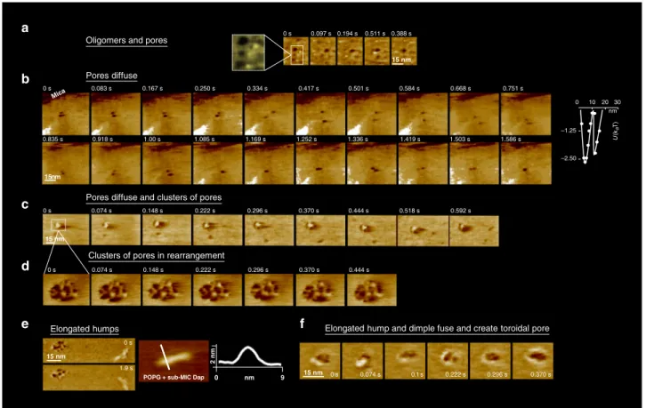
Fig. 4 Sub-MIC Dap on POPG at 37 °C. Tens of minutes. Intermediate stages a A new structure appeared: dimples, zones of thinner membrane thickness, whose diameter was in the range 7 ± 2 nm. Most dimples diffuse, but some remained static (colour scale: 3 nm). Movie details: frame rate 97 ms; zoom of a full image of 150 nm × 90 nm and 256 × 160 pixels.b The dimple diffusion consisted of swinging trajectories, implying membrane-mediated dimple-dimple attraction (colour scale: 3 nm).b, right, Energy profile of the interaction of the dimples obtained derived from 120 centre-to-centre distance measurements that contains as the oligomers two energy minima. Movie details: frame rate 83 ms; full image of 150 nm × 150 nm and 256 × 256 pixels.c In some membrane zones, clusters of dimples, reminiscent of cubic phases, developed (colour scale: 4 nm). Movie details: frame rate 74 ms; full image of 90 nm × 60 nm and 256 × 160 pixels.d The clusters of dimples were moderately dynamical in time, with moderate internal rearrangements (colour scale: 4 nm). Movie details: frame rate 74 ms; full image of 25 nm × 16 nm and 256 × 160 pixels.e The other deformation found was elongated-humps on top of the POPG membrane.e, left, An elongated-hump in the proximity of a cluster of dimples (colour scale: 4 nm). e, right, A close-up and a profile of an elongated-hump. Additional images of elongated-humps on Supplementary Fig. 1. Movie details: frame rate 479 ms; zoom of full image of 250 nm × 200 nm and 300 × 256 pixels.f It was observed that the dimples and the elongated-humps fused and gave yield to pores of toroidal structure where a protruding ring surrounds the pore (colour scale: 4 nm). Movie details: frame rate 74 ms; full image of 40 nm × 40 nm and 256 × 160 pixels.
little variations in the number of molecules forming them. From the swinging trajectories of diffusing dimples, it was clear that the membrane tension they generated resulted in attractive forces (Fig. 4b and Supplementary movie 2). It was also clear that the dimples of the same leaflet can fuse and give yield to dimples of larger size in that same leaflet (Fig.4b, last frame); not be confused with the fusion of dimples from opposite membrane leaflets that is proposed in the existing model of Dap pore formation25. In more detail, their energy profile (Fig. 4b right, from n= 120 dimple-dimple distance measurements) shows a similar pattern that the oligomers, two minima appear at d ~ 5 nm, where the dimples are in a side-by-side contact, and at d ~ 13 nm, where they interact by the stressfield they generate in the bilayer. In agreement with the attraction between dimples, we were able to observe clusters of dimples (Fig.4c), not previously described in the literature, slightly reminiscent of cubic phases, located statically on the membrane, although with internal dynamic rearrangements on the time scale of several seconds (Fig.4d).
In addition to the oligomers and dimples, at slightly longer times after exposure to Dap, another type of deformation, previously unreported in the literature, that we named ‘elongated-humps’ formed on top of the POPG bilayer surface (Fig.4e and Supplementary Fig. 1), and presented rather uniform widths and heights, with 4 nm and 2 nm, respectively, but variable lengths ranging from several to tens of nanometres.
Significantly, due to the fast imaging speed of the HS-AFM, we could determine the process of fusion between the elongated-humps and the dimples that ended in the formation of toroidal-shaped pores (Fig. 4, 2f). In this process, never observed nor proposed for Dap molecular activity, an elongated-hump embraces the dimple forming a ring protruding 2 nm above the membrane, which then develops into a toroidal membrane pore. To later stages of Dap action, after several hours, the objects visible in the membrane had evolved further. Toroidally shaped pores, of similar shape than the pores formed by the fusion of humps and dimples, appeared on the POPG membrane (Fig.5a), the AFM visualisation reinforces the toroidal pore model of Dap action25,30, and the timescale of formation of the pores relates
well to the Dap-induced depolarisation process observed on bacteria that lasts several tens-of-minutes16,17. The toroidal pores were differentiated from other structures by their larger diameter, a protruding external ring, and their lack of mobility that
contrasted strongly with the dynamic dimples. These differences suggest that the toroidal pores could traverse the membrane, as proposed by the toroidal pore model. It would be possible that the toroidal pores may not diffuse because they interact with the underlying mica substrate. Unfortunately, the HS-AFM probe tip is too large to fit into the toroidal pores, and consequently we cannot determine whether the toroidal pores traverse the membrane or not (Fig. 5a and Supplementary Fig. 2). The geometry of the pores often deviated from ideal toroidal shapes, often the pore showed an elliptical or incomplete couronne; in an ensemble of 16 pores, the couronnes of the pores range in their peak-to-peak distance between 7 and 12 nm (Supplementary Fig. 2). Another structure that formed at the long times were tubulations with diameters between ~8 and ~12 nm, these were mostly observed at the membrane edges (Fig.4b), and rarely lying on the POPG bilayer (Fig.4c). These structures may be related to the tubules visualised on POPG vesicles and mreB mutant cells (Fig.1f, g). They are probably also related to the elongated-humps observed. Significantly, the tubulations where not uniform but displayed regions of thicker and thinner cross-section with a periodicity of ~34 nm (Fig.5b): only the most rectilinear sections of the tubules showed periodicity, at the membrane edges the tubules were highly rectilinear, on the tubules lying on the bilayer the curved sections did not show the periodicity that arose in the most rectilinear sections (Fig. 2b, c). This periodic structure, alternating positive and negative curvature, indicates in the present case that the tubules are composed of more than one species, presumably POPG and Dap; since POPG, a cylindrical lipid, forms lamellar phases, while Dap alone forms spherical micelles78. Provided their respective curvatures, we reason that
the thinner and thicker regions of the tubules are enriched in Dap and POPG, respectively. In sum, our results on living bacteria and membranes are consistent with the lipid extraction model31that
was before witnessed on membranes of 70% content of PC a lipid not found in Gram+ bacteria that may alter Dap activity18.
It should be remembered that the geometry of this experiment means that, even though the concentration of Dap is low, the Dap to lipid ratio is rather high, as the lipids are confined to the mica surface. Thus, a considerable amount of Dap can partition into the supported membrane. A further peculiarity of the geometry is that the supported bilayer is attached to the mica surface and thus the leaflet in contact with the support is inaccessible for Dap
Tubulation lying on bilayer
Tubulations at membrane edge Toroidal pores Mica b2 b3 c1 b1 Mica 12 45 6 100 6 100 b1 b2 b3 c1 nm nm nm 6 100 nm a b c
Mica POPG + sub-MIC Dap
3 nm 5 pores and rotational
6 nm
1 nm
average
0 nm 55
15 nm
Fig. 5 Sub-MIC Dap on POPG at 37 °C. Hours. Final stages. a Toroidal pores of similar topography than those formed from the fusion of a hump and a dimple formed.a, left, Two toroidal pores in close to the membrane border, the underlying mica support is indicated (colour scale: 3 nm). a, right, An average, including rotational average, of 5 toroidal pores. Toroidal pores did not diffuse, in sharp contrast to the diffusive dimples. The AFM tip radius was too large for the height profile to assess the depth of the toroidal pores, and thus, if they traversed the membrane (white arrow in height profile). Supplementary Fig. 2 provides further details the AFM topographic profiles of the toroidal pores. b Tubulations formed, mostly at the edge of the POPG membranes. The tubulations were nearly rectilinear and displayed regions of larger and thinner cross sections that periodically distributed with a ~34 nm period, which is symptomatic of a two components composition in the tubules, Dap and POPG, at the thinner and larger cross section regions, respectively. Height profiles across and along the tubules are shown. c More rarely, tubules were found lying on the POPG bilayer, some sections, the more rectilinear ones (k1), present periodicity in their topography, other, the less rectilinear, do not show topographic periodicity.
insertion. Membrane attachment also is likely to hider the deformations of the lower leaflet necessary for the formation of toroidal pores. Nevertheless, we observed a number of structures probably pertinent for Dap action: The fast diffusing (300 ± 200 µm/s) small oligomeric clusters of thefirst minutes developed into slower (0.7 ± 0.5 µm/s) diffusing dimples, elongated-humps on top of the membranes, and static clusters of dimples, that ultimately gave rise to pores, cubic phases and tubules. Our visualisations are consistent with both the toroidal pore and lipid extraction models.
The picture was rather different at higher Dap concentration. Thefirst seconds after exposure, on the edge of the bilayer, tubules formed and branched into and hexagonal honeycomb structures of variable and time-changing unit cell size that evolved into a smaller and more uniform unit cells of ~55 nm (Fig.6a). After the first minutes after exposure, the membrane was fully covered with a heterogeneous ensemble of deformations. However, no dimples or toroidal pores, common at lower concentrations, were observed. The deformations were very dynamic as the system reconfigured. The structures were clearly in a non-equilibrium situation and material flowed across the membrane (Fig.6b and Supplementary movie 3). Theflow of material streamed across a pronounced structural heterogeneity: theflow seemed to originate at a structure similar to an Im3m cubic phase (25 nm of pitch) reminiscent of the aggregations of pores at lower concentration and the hexagonal tubular honeycomb, and end at a tubulation located at the edge of the membrane patch, the flow traversed a rippled membrane (15 nm of pitch) where the ripples, slightly reminiscent of the membrane humps, moved and indicated the
sense of theflow (Fig.6b, white and blue lines); even if only at the rippled membrane could theflow of material be assessed by the HS-AFM, provided the conservation of theflux continuity, even if not seen by HS-AFM, material shouldflow too across the cubic phase domain and the tubulation.
After 1 h, the dynamicflow came to a stop, equilibrium was reached, which enabled us to achieve higher resolution AFM imaging. We imaged tubules that were similar to those observed when the membrane was exposed to sub-MIC Dap concentra-tions (Fig.5b): also they presented periodically distributed regions with smaller and thicker cross sections, their periodicity was of ~32 nm, hence, similar to the ~34 nm period measured at sub-MIC (Fig. 6c), indicating that the ratio Dap:POPG inside the tubules is relatively invariant with respect the concentration of Dap in solution. The equilibrium staticity also facilitated us a better imaging of the ripple phase, as well as the Im3m and pn3m cubic phases (Fig.6d, e). The spacing of the ripple phase regions was evaluated at 13.1 ± 0.9 nm (Supplementary Fig. 3), and of the Pn3m and Im3m cubic phases at 11.2 nm and 16.7 nm, respectively, the sides of the hexagon and a square patterns (Supplementary Fig. 4).
The observations at over-MIC are less easy to relate to the physiological action of Dap. However, they highlight a number of important points such as the relaxation of an out of equilibrium situation through the formation of Dap-POPG tubules. The situation at over-MIC, in which Dap binding to the membrane is much faster, results rapidly in a very rich and congested membrane with little space available for the formation of dimples or toroidal pores.
a b c
d e
Fig. 6 Over-MIC Dap on POPG at 37 °C. a Thefirst seconds after exposure to over-MIC Dap, at the POPG membrane border tubules of diameter of ~15 nm branch in hexagonal directions (arrow) and create in seconds a tubular honeycomb structure. Movie details: frame rate 848 ms; zoom of a full image of 150 nm × 90 nm and 256 × 160 pixels (colour scale: 16 nm).b Minutes after exposure, the POPG membrane showed an heterogenous and dynamic environment where different regions of the membrane showed different Dap-induced deformations and where materialflowed from region to region (low passedfiltered images to highlight the contrast are shown). The flow of material, observable on a zone of ripple phase of 15 nm pitch (please note that the white and blue lines and the arrows indicate the direction of displacement of the ripples), indicative of a non-equilibrium situation, went from the cubic phase type Im3m of 25 nm of pitch to the tubulation that emerged from the border of the POPG patch. Movie details: frame rate 1530 ms; full image of 390 nm × 350 nm and 256 × 256 pixels.c Once the membrane exposed reached equilibrium (1 h after exposure), higher resolution imaging of the effects of over-MIC Dap exposure was possible. The tubulations (of Fig.3b) were observed to be of periodic shape with zones of thicker and thinner diameters distributed with a 32 ± 4 nm period. Compared to the tubulations found at sub-MIC, the zones of thinner diameter were appreciably longer, given the higher content on Dap at over-MIC than at sub-MIC, the zones of thinner diameter were associated with zones enriched in Dap and, inversely, the zones of thicker diameter with zones enriched in POPG.d At equilibrium, also the ripple phase could be imaged in more detail, as well as e Im3m and Pn3m cubic phases.
In sum, the HS-AFM made possible the first molecular-level visualisation of the structures and dynamics of the AMP activity on membranes from its initial to final stages under infection-like conditions. We were able to determine unexplored details of the multifaceted pathways of the AMP action correlated to their dependence on the concentration and the elapsed time. We confirmed or disconfirmed so far hypothetical models and detected unknown molecular mechanisms and quantifications of interaction energies out of the reach of other techniques, including computer simulations48,49. Finally, biological and medical relevance of the
approach was enhanced by the correlation of the molecular AFM information with information from SEM, TEM and OM on B. subtilis WT and mutant cells and non-supported POPG vesicles. Resistance to Dap by an increase of the CL membrane content. As already mentioned, bacterial resistance to Dap is increasing. In some strains, of Enterococcus faecalis, bacterial resistance to Dap
has been directly related to increased content of cardiolipin in the membrane63,64. To investigate the mechanism of bacterial resis-tance, a model membrane composed of 20% molar TOCL (CL) 18:1 (1’,3’-bis[1,2-dioleoyl-sn-glycerol-3-phospho]-sn-gly-cerol) and 80% molar POPG was exposed to Dap. CL and POPG have similar alcyl chains and are mutually soluble79.
We exposed non-supported CL/POPG vesicles to Dap and characterised it by FliptR measurements. We found that when CL was present, the lipid packing remained constant under Dap exposure, and that it was concentration-independent in the range studied (Fig. 7a, histograms). 20% of CL content, then, sharply changes the response of the membrane with respect the observations for POPG membranes and WT B. subtilis where Dap caused a relevant alteration of the lipid packing measured by FliptR (Fig. 2). It is interesting to mention that the B. subtilis 168 strain that we used contains 2.3% of CL at the exponential growth phase80. TEM observations confirmed that the CL/POPG
a
b
c
d
e
Fig. 7 Dap effect on TOCL(CL)/POPG vesicles and model membrane by TEM, FliptR and HS-AFM. The incorporation of 20% molar of TOCL to the POPG model membrane inhibited Dap action creating CL-specific membrane dynamics. a By TEM, the TOCL/POPG vesicles do not deform at sub-MIC, whereas at over-MIC only minor deformations, scarce tubulations, are formed. By FliptR lifetime (standard deviation error shown), the lipid packing is not altered by Dap.b, left, On TOCL/POPG supported membranes exposed (tens of minutes) on the outer leaflet to sub-MIC Dap (colour scale: 4 nm), deformations were not observed, but, at over-MIC (b, right), membrane buckling appeared; periodically distributed lines with a 5 nm pitch reminiscent of a ripple phase (colour scale: 3 nm). The buckling static allowed high-resolution imaging, its substructure was composed of particles of 1.5 nm in diameter; possibly the head groups of Dap and/or TOCL molecules.c TOCL/POPG supported membrane of symmetrized inter-leaflet Dap distribution; for it, the POPG vesicles opened and adsorbed onto the mica substrate in the presence of Dap in the solution, toroidal pores formed of structure similar to the toroidal pores formed on POPG membranes (colour scale: 2 nm). The toroidal pores in TOCL/POPG showed a tendency to cluster (asterisk).d We induced inter-leaflet de-symmetrization by exposing the outer leaflet to supplementary quantities of Dap added to the imaging solution. This induced the activation of a possible membrane tension relief mechanism that, by a cyclic accumulation process seems to eject material out of the membrane; some material appears next to the pore at 1.30 s (Supplementary Movie 3) (Full colour scale: 3 nm).d, right, Histogram of the time-lapses between consecutive accumulations, the time variability spans from hundreds of milliseconds to several seconds, signature of the stochastic nature of the accumulation process. Afitting to an exponential curve is shown. e The pore recovers its initial state without any visible traces remaining from the material accumulation. Moreover, the symmetrization of Dap inter-leaflet distribution and the probable ejection of material from the pore caused the disappearance of the bucking deformation from the TOCL/POPG membranes, indicative of a membrane tension relief (Full colour scale: 3 nm).
vesicles were not deformed by the sub-MIC concentrations of Dap (Fig. 7a, images). However, higher concentrations of Dap created some tubulations, these could be imaged by TEM (Fig.7a, right) but were undetected by the FliptR averaged signal.
On supported CL/POPG membranes, in line with the results on non-supported CL/POPG vesicles, sub-MIC exposure did not cause any deformations visible by AFM, neither dimples, toroidal pores nor tubules were observed (Fig.7b) after tens of minutes of exposure. At higher concentration, instead, a high degree of static buckling appeared (Fig. 7b, right). At higher magnification,
periodically distributed lines with a 5 nm pitch, reminiscent of ripple phase or the elongated humps appeared (Fig.7b, inset), the lines displayed a periodically distribution (1.4 nm spacing) indicating a substructure of particles, probably constituted by head groups of Dap considering their dimensions81,82.
Overall, the AFM data confirm that higher contents of CL in the membrane strongly diminish the capability of Dap to perturb the POPG membrane. It has previously been suggested that the extra volume of the acyl-chain of CL would better accommodate Dap in the hydrophobic core of the membrane, avoiding the formation of energetically unfavourable voids, and thus, reducing curvature induction by Dap30. Our observations are
certainly consistent with the idea that CL results in less deformation of the bilayer. However, it is surprising if Dap inserts stably into the bilayer that it does not change lipid packing as observed by FliptR, although it can agree with the observation of a CL-related stabilisation of membranefluidity as observed on Pseudomonas putida exposed to surfactants83.
Imposed symmetrization and de-symmetrization of the inter-leaflet distribution of Dap in a CL/POPG membrane. The previously described observation of a lack of toroidal pore for-mation on CL/POPG supported membranes exposed to Dap is coherent with previous findings (on CL/PG/PC bilayers) that show that CL inhibits Dap membrane permeabilization25,60. Such
event has been hypothesised could derive from the blockage of the inter-leaflet translocation of Dap by the presence of CL in the membrane25. To investigate this hypothesis, a control experiment
with a symmetric inter-leaflet distribution of Dap was devised. We symmetrized the inter-leaflet distribution of Dap, both in supported POPG and CL/POPG membranes, by incubating the lipid vesicles on the mica substrate in the presence of Dap in the incubation solution. As a result, during the opening of the vesicles on the mica substrate for the formation of the supported lipid bilayer, both leaflets of the membrane became exposed to the Dap, allowing the molecules to reach both leaflets prior to the adsorption of the bilayer on the mica support.
Applying this procedure with low Dap concentrations in solution, toroidal pores formed both in the POPG and the CL/ POPG supported membranes. In the POPG membranes, the toroidal pores that formed (Supplementary Fig. 5) were indistinguishable from the toroidal pores forming in POPG membranes exposed to Dap after attachment (Fig. 5). In CL/ POPG membranes, the pores formed by this procedure were very similar to those observed in POPG membranes, yet, in average, and besides their structural variability and their deviation from an ideal toroidal geometry, their size was slightly smaller than that of the pores in the POPG membranes (Fig. 7c and Supplementary Fig. 2). In the supported CL/POPG membranes, as in the supported POPG membranes, the toroidal pores were immobile. Nevertheless, they showed some tendency for clustering (Fig.7c), although it was significantly smaller than that of the dimples (Fig. 4d), their clustering probably took place before the adsorption of the bilayers on the mica. Thanks to our control experiments we could confirm that, as was hypothesised in the
literature, the CL incorporation blocks the formation of toroidal pores by impeding the translocation of Dap from the outer to the inner leaflet, and, thus, we support the model of Zhang et al.25
given that we proved that the lack of dimples stops the translocation of Dap molecules from the outer to the inner leaflet of the membrane; which leads us to reason that the dimples reduction of the membrane thickness increases the probability of Dap translocation.
As the symmetrization of the Dap distribution caused the appearance of pores, a subsequent de-symmetrization could induce other effects. We therefore induced inter-leaflet de-symmetrization by exposing the outer leaflet of the symmetrized CL/POPG membrane to extra Dap after the bilayers where adsorbed on the mica surface.
When the concentration of Dap in the imaging solution was increased to over-MIC the toroidal pores responded dramatically and dynamically. The HS-AFM imaging permitted us (Supple-mentary movie 4) to image a cyclic process occurring in the pores that consisted of three steps: Step 1: material, of undefined nature, accumulated at the centre of the pores (Fig.7d, time 0.52 s). Step 2: the accumulation grew until it reached a diameter similar to the diameter of the pore, our AFM measurement (it applies tens to hundreds of piconewton forces) records a maximal height of ~3 nm that could be higher in reality (Fig.7d, time 1.04 s). Step 3: the accumulated material in the pore disappeared, (it could be due either to the ejection of the material or to the re-insertion of the material in the membrane, the former seems more plausible provided the flux of Dap molecules from the solution inserted into the outer leaflet, plus the observation of material that appears next to the pore at the end of the accumulation process at the time of 1.30 s). In addition, the pore recovered its initial state without any visible traces remaining from the material ejection process (Fig. 7d, time 1.56 s). The process repeated with a new accumulation starting to grow at the centre of the pore (Fig.7e), the time-lapses between the accumulations were irregular and the duration of the different cycles ranged from hundreds of milliseconds to several seconds (Fig.7d), signature of a stochastic process (Fig.7d right, the histogram of the time-lapses wasfitted to an exponential). Moreover, the symmetrization of Dap inter-leaflet distribution and the ejection of material from the pores caused that at over-MIC the bucking deformation did not appear on the CL/POPG membranes (Fig.7d).
Discussion
A persistent problem in the studies of membrane-active anti-microbial peptides is the lack of experimental6,7 and in-silico48,49 methods for investigating their molecular configurations and processes in the membranes29. The leitmotif for this work was to
develop new tools and protocols capable of scrutinising with molecular detail the multifaceted actions of antimicrobial lipo-peptide molecules. There is a high intrinsic complexity in lipopeptide-membrane interactions that renders the design and development of lipopeptides antibiotics highly costly. At the same time, the complexity of the multifaceted actions of lipopeptides, and their easy access to their predominant target, the bacterial plasma membrane, renders the lipopeptides highly attractive can-didates for the research of new antibiotic molecules. New advanced computer-assisted design strategies45and new synthetic
lipopep-tides11 are improving the cost-effectiveness and the spectrum of potential lipopeptides. We focused on Dap, one of the few anti-biotics that has achieved commercialisation in the last two decades for the treatment of Gram+ infections of which the details of its mode of action remains, as for any other lipopeptide, elusive.
The HS-AFM imaging correlated with EM and OM enabled unprecedented assessment of structures and dynamics of the
activity Dap on the membranes of living bacteria and mimic bilayers. We provided visual proof of previously hypothetical models, quantified interactions, and found unexpected new phenomena. Based on ourfindings, we discuss next our view on the activity of Dap on membranes (schematized in Fig.8).
When Dap is added to POPG bilayers it inserts into the exposed monolayer thanks to its hydrophobic tail. This insertion results in an increase in the lateral pressure, as the area of the monolayer tries to increase but is retained by the geometry of the other monolayer. The inserted Dap molecules can form small dynamic aggregates in the exposed monolayer, as we have observed. Nevertheless, the system would like to reduce the lateral pressure: we found two different pathways of action for this. In a first mechanism the lateral pressure is reduced by flip-flop with Dap molecules passing to the second, unexposed, monolayer. This pathway leads to the formation of toroidal pores and is associated with killing by membrane permeation. In a second mechanism, the exposed monolayer can extrude POPG/Dap tubules with a relatively high Dap composition. These tubules are somewhat reminiscent of cylindrical mixed micelles and this pathway is associated with killing by lipid loss.
In the context of this model for Dap action we would suggest that the flip-flop mechanism requires a relatively low Dap con-centration in the outer-leaflet, as when it is over-occupied the lateral pressure prevents the passage of the large head-group into the membrane. This is consistent with the role proposed for the dimples, that we have observed, in catalysing inter-leaflet transfer, as this structure also increases monolayer surface area or lateral pressure. We suggest that at relatively high surface densities Dap aggregates are able to reduce the lateral pressure by forming elongated-humps in the membrane. Such humps can evolve in several ways. Indeed, they can aggregate to form the ripple like
phase that we observed, they can fuse with a dimple pushing Dap to the other side of the membrane in the process or they can develop into tubules.
The model also allows a coherent vision of the different dynamical processes that we have observed. The membrane leaflet exposed to high Dap concentrations rapidly accumulates large amounts of Dap and the lateral pressure rises. This initial unstable system relaxes, over a period of hours, as the lateral pressure drops and tubules form. Toroidal pores are prevented from formation by the absence of dimples that catalyses inter-leaflet transport of Dap. Equally, the membrane containing pores exposed to extra Dap produces (under the presence of CL) not tubes but objects escaping from toroidal pores. These, we believe to be Dap rich mixed micelles whose production is catalysed by the pores. This process is powered by the relief of the accumu-lated pressure caused by the insertions of Dap and reminiscent of the process of relief of accumulated pressure of geysers.
We expect that the potential of the HS-AFM imaging will stimulate new models and insight on the structure-activity rela-tionship of membrane-interacting molecules. For example, U.S. Food and Drug Administration recently approved antibiotics with similarities with Dap (oritavancin, dalbavancin, and tela-vancin), also medically used for complicated skin infections by Gram+ bacteria. These molecules, such as Dap, anchor the bacterial membrane by a lipophilic side chains, but the molecular mechanisms involved in membrane rupture are undetermined; they may involve either the formation of membrane pores or the lysis of the membrane84, Hence, for the pharmaceutical industry,
HS-AFM label-free assessment that does not require biochemical synthesis and provides novel information, represents an oppor-tunity to considerably increase the throughput of screening of molecular candidates. In addition, it also permits to better point
Low CL content High CL content Dap Dap Time (min) 0 2 10 15 Pressure rises Pressure dr ops Dap flip-flop Toroidal pore Permeation Ejections Ejections
from pores Buckling
Tubulations Elongated
humps
Dimples andhumps fuse
Dap reac hes inner leaflet
(Impr obab le) PG PG CL Oligomers Oligomers Dimples
Fig. 8 Schematics of the action pathways of Dap on Gram+ membranes. The timescale is indicated. The insertion of Dap molecules in the membrane rises the pressure. The pressure relief drives Dap actions: Left, When the membrane does not contain CL, the formation of Dap oligomers is followed by the formation of dimples in the membrane, followed by the formation of elongated-humps. The dimples favour the translocation of Dap to the inner membrane leaflet by the flip-flop mechanism. When Dap is present in the inner and outer leaflets toroidal pores form, the membrane is permeated, and the bacteria die. Alternatively, Dap can also induce ejection of material from the membrane by the formation of tubulations; equally destabilising the membrane and killing the bacteria. Right, Concerning the bacteria containing a high content of CL in the membrane (>20% molar), Dap action is drastically modified. In this case, the Dapflip-flop does not take place and the dimples are not formed. Instead, the membrane buckles but it does not break. Hence, despite Dap insertion, the membrane still separates the cytoplasm from the exterior. In the improbable case that some Dap molecules reach the inner leaflet of the membrane, pores form as in the case CL is present. The particularity of the pores formed under high CL content is that they are able of ejecting material out of the membrane. These pores behave‘like geysers’, and relief the pressure built on the outer leaflet by the insertions of Dap molecules in cycles of activity. Both CL-related processes maintain the membrane integrity and the bacteria alive.