HAL Id: hal-02055769
https://hal-univ-rennes1.archives-ouvertes.fr/hal-02055769
Submitted on 16 Apr 2019HAL is a multi-disciplinary open access archive for the deposit and dissemination of sci-entific research documents, whether they are pub-lished or not. The documents may come from teaching and research institutions in France or
L’archive ouverte pluridisciplinaire HAL, est destinée au dépôt et à la diffusion de documents scientifiques de niveau recherche, publiés ou non, émanant des établissements d’enseignement et de recherche français ou étrangers, des laboratoires
simultaneously acetylcholinesterase and glycogen
synthase kinase-3
Killian Oukoloff, Nicolas Coquelle, Manuela Bartolini, Marina Naldi, Rémy Le
Guével, Stéphane Bach, Béatrice Josselin, Sandrine Ruchaud, Marco Catto,
Leonardo Pisani, et al.
To cite this version:
Killian Oukoloff, Nicolas Coquelle, Manuela Bartolini, Marina Naldi, Rémy Le Guével, et al.. De-sign, biological evaluation and X-ray crystallography of nanomolar multifunctional ligands targeting simultaneously acetylcholinesterase and glycogen synthase kinase-3. European Journal of Medicinal Chemistry, Elsevier, 2019, 168, pp.58-77. �10.1016/j.ejmech.2018.12.063�. �hal-02055769�
M
AN
US
CR
IP
T
AC
CE
M
AN
US
CR
IP
T
AC
CE
PT
ED
Design, Biological Evaluation and X-ray Crystallography of Nanomolar
Multifunctional Ligands Targeting Simultaneously Acetylcholinesterase
and Glycogen Synthase Kinase-3
Killian Oukoloff,a Nicolas Coquelle, b,c Manuela Bartolini,d Marina Naldi,d Rémy Le Guevel,e Stéphane Bach,f Béatrice Josselin f Sandrine Ruchaud,f Marco Catto,g Leonardo Pisani,g Nunzio Denora,g Rosa Maria Iacobazzi,h Israel Silman,i Joel L. Sussman,j Frédéric Buron,k Jacques-Philippe Colletier,b Ludovic Jean,*,a Sylvain Routier,k and Pierre-Yves Renard*,a
a
Normandie Univ, UNIROUEN, INSA Rouen, CNRS, COBRA (UMR 6014), 76000 Rouen, France
b
Institut de Biologie Structurale, Université Grenoble Alpes, Centre National de la Recherche Scientifique (CNRS)-Commissariat à l’Énergie Atomique (CEA) (UMR 5075), F-38054 Grenoble, France
c
Institut Laue Langevin, 71, avenue des Martyrs - CS 20156, 38042 Grenoble cedex 9, France d
Department of Pharmacy and Biotechnology, Alma Mater Studiorum University of Bologna, Via Belmeloro 6, I-40126, Bologna, Italy
e
Plateforme ImPACcell-SFR BIOSIT UMS-CNRS3480 UMS-INSERM018, Université de Rennes1, 35043, Rennes Cedex, France
f
Sorbonne Université, CNRS USR 3151, Protein Phosphorylation & Human Diseases, Station Biologique de Roscoff, CS 90074, Roscoff Cedex F-29688, France
g
Dipartimento di Farmacia-Scienze del Farmaco, Università degli Studi di Bari "Aldo Moro", via Edoardo Orabona 4, 70125 Bari, Italy
h
Istituto Tumori IRCCS Giovanni Paolo II, via le Orazio Flacco 65, 70124 Bari, Italy i
Department of Neurobiology, Weizmann Institute of Science, 6100 Rehovot, Israel j
Department of Structural Biology, Weizmann Institute of Science, 76100 Rehovot, Israel k
Institut de Chimie Organique et Analytique, Université d’Orléans, UMR CNRS 7311, rue de Chartres, BP 6759, 45067 Orléans Cedex 2, France
M
AN
US
CR
IP
T
AC
CE
PT
ED
Corresponding Author* For L. J.: phone: +33 2 35 52 24 51; E-mail: ludovic.jean@univ-rouen.fr
* For P.-Y. R.: phone: +33 2 35 52 24 76; E-mail: pierre-yves.renard@univ-rouen.fr.
Keywords
Alzheimer’s disease, acetylcholinesterase, butyrylcholinesterase, kinases, glycogen synthase kinase-3, cyclin-dependent kinase 5, synthesis, SAR, crystallography.
Abstract
Both cholinesterases (AChE and BChE) and kinases, such as GSK-3α/β, are associated with the pathology of Alzheimer’s disease. Two scaffolds, targeting AChE (tacrine) and GSK-3α/β (valmerin) simultaneously, were assembled, using copper(I)-catalysed azide alkyne cycloaddition (CuAAC), to generate a new series of multifunctional ligands. A series of eight multi-target directed ligands (MTDLs) was synthesized and evaluated in vitro and in cell cultures. Molecular docking studies, together with the crystal structures of three MTDL/TcAChE complexes, with three tacrine-valmerin hybrids allowed designing an appropriate linker containing a 1,2,3-triazole moiety whose incorporation preserved, and even increased, the original inhibitory potencies of the two selected pharmacophores toward the two targets. Most of the new derivatives exhibited nanomolar affinity for both targets, and the most potent compound of the series displayed inhibitory potencies of 9.5 nM for human acetylcholinesterase (hAChE) and 7 nM for GSK-3α/β. These novel dual MTDLs may serve as suitable leads for further development, since, in the micromolar range, they exhibited low cytotoxicity on a panel of representative human cell lines including the human neuroblastoma cell line SH-SY5Y.
M
AN
US
CR
IP
T
AC
CE
PT
ED
Moreover, these tacrine-valmerin hybrids displayed a good ability to penetrate the blood-brain barrier (BBB) without interacting with efflux pumps such as P-gp.
1. Introduction
Alzheimer’s disease (AD), the most common cause of senile dementia, is a major public health concern with devastating social impact. In 2016, Alzheimer’s Disease International, the worldwide federation of Alzheimer associations, estimated that 47 million people were affected by this type of dementia, and predicted that 131 million people will be affected in 2050. Thus, the economic impact of the disease on future long-term care costs will be enormous. Already in 2018 the cost of treatment of dementia is estimated to reach US$ 1 trillion, rising to US$ 2 trillion by 2030.[1]
AD results from a neurodegenerative process occurring in the central nervous system (CNS). It is clinically characterized by loss of memory and cognitive impairment, which parallel the deterioration of cholinergic neurons in the basal-forebrain with concurrent reduced levels of the neurotransmitter acetylcholine (ACh). The disease is histologically characterized by extracellular deposits of β-amyloid peptide (Aβ) and intracellular formation of neurofibrillary tangles (NFTs).[2, 3] Other hallmarks include oxidative stress, dys-homeostasis of biometals and calcium, inflammation and loss of synaptic connections.[4] Current therapeutic options for treatment of AD mostly aim at restoring physiological ACh levels by inhibiting the enzyme acetylcholinesterase (AChE), which is responsible for the hydrolysis of ACh. Therefore, three drugs approved by the Food and Drug administration (FDA) for the management of AD are AChE inhibitors (AChEIs), namely donepezil, rivastigmine and galantamine.[5] However, treatment with AChEIs is mostly symptomatic, and effective only for AD patients with mild-to-moderate symptoms, enabling in some cases the progression of cognitive and functional
M
AN
US
CR
IP
T
AC
CE
PT
ED
impairments to be retarded. Memantine, which is an N-methyl-D-aspartate (NMDA) receptor antagonist, is the latest drug approved for the treatment of the disease.[6] The efficacy of memantine is similar to that of the AChEIs among patients with mild-to-moderate symptoms, but has been reported to be more useful for treating patients at more advanced stages of the disease.[7-9] Yet, all four drugs only provide temporary symptomatic relief, but do not provide a cure. Despite efforts over the last few decades of both academic research teams and pharmaceutical companies, no curative treatment has emerged yet.[10-12]
An attrition factor in the development of curative drugs is the fact that the aetiology of AD is not fully understood. It is recognized, however, that the processes resulting in its development and progression are complex and multifactorial.[13] Thus, it is conceivable to hypothesize that a combination of synergetic strategies targeting the various players involved in the onset and development of the disease should be more beneficial, and should present a higher rate of success. The “multifactorial hypothesis” has fuelled the so-called multi-target-directed ligand (MTDL) strategy, which foresees the development of a single molecule able to affect several key targets/pathways.[14-22] Besides the advantage of acting simultaneously on more than one biological target, MTDLs have the advantages of a higher patient compliance (a parameter of paramount importance for such neurodegenerative disease), lower risk of drug-drug interactions, and lower costs to perform ADMET studies.[23]
Kinases have been shown to be associated with the progression of AD, and have received increased attention during the past decade.[24, 25] Some members of this family are involved in the hyperphosphorylation of the tau protein, which results in the production of intracellular NFTs. GSK-3β [26] and CDK5 [27] are the two main kinases involved in this process, whereas GSK-3α appears to regulate the production of Aβ.[28, 29] Thus, GSK inhibitors may be useful
M
AN
US
CR
IP
T
AC
CE
PT
ED
for treatment of AD, as well as other neurodegenerative diseases.[30, 31] A study identified novel mechanisms linking GSK-3 with the Aβ pathology. The inhibition of GSK-3 reversed AD pathogenesis via lysosomal acidification and reactivation/restoration of the mammalian target of rapamycin (mTOR) in a mouse model of AD.[32] Furthermore, evidence has been presented that GSK-3 inhibitors may reduce Aβ-oligomer-induced neuronal toxicity and may promote neurogenesis in vitro and in vivo.[33, 34] These various studies indicate that GSK-3 inhibitors should be further studied as candidates for treatment of AD.[35]
A renewed interest in AChEIs for AD treatment arose when evidence was presented that, in addition to AChE inhibition, certain AChEIs may block the involvement of AChE in mediating the aggregation and deposition of Aβ peptides.[36-38] Thus, AChE has been reported to accelerate the formation of neurotoxic Aβ aggregates via a mechanism that involves its peripheral anionic site (PAS).[39] This resulted in numerous studies aimed at developing dual binding site AChEIs interacting with both the catalytic site and the PAS.[21, 40, 41] Moreover, recent studies have suggested a possible role for AChE in hyperphosphorylated tau (P-Tau) dysregulation.[42] There is thus also still a great interest in developing MTDLs endowed with an anti-ChE activity.
To combine the beneficial effects of ChE inhibition and GSK-3 inhibition within a single molecule, a tacrine scaffold was selected to provide activity toward ChEs (Figure 1). This selection was also based on the fact that the tacrine pharmacophore can easily be substituted on its primary amino group while retaining a satisfactory anticholinesterase activity. Consequently, many tacrine-based MTDLs exhibiting high affinity for AChE and/or BChE have been reported in the past decade.[41, 43-45] However, only one study describing GSK-3/AChE bifunctional
M
AN
US
CR
IP
T
AC
CE
PT
ED
inhibitors has appeared.[46] These MDTLs presented good inhibitory activities towards both enzymes, and alleviated cognitive impairment in the mouse model treated with scopolamine.
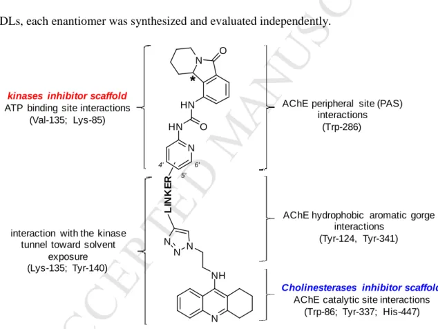
Amongst the GSK-3 kinase inhibitors developed for application to the CNS,[47] we selected the valmerins (Figure 1), which contain a tetrahydropyrido[1,2-a]isoindolone core[48, 49] linked to an heteroaryl moiety by a ureido group. The pharmacophore that interacts with the kinase active site consists of the isoindolone-urea moiety linked to the pyridine ring at its C-2’ position.
N H NH O N O I IC50GSK-3α/β= 170 nM IC50 CDK5/p25 = 80 nM II IC50GSK-3α/β= 22 nM IC50CDK5/p25 = 25 nM N N H NH O N O N Br N NH2 Tacrine
Figure 1. Structures of tacrine, a ChE inhibitor and valmerins I and II, which display potent
CDK5 and GSK-3α/β inhibition.[49]
Docking studies and SAR studies suggested that substitutions at the pyridine C4’, C5’ and C6’ positions should not adversely affect affinity for either GSK-3α/β or CDK5. Indeed, as seen in Fig. 1, the presence of a bromine atom on C4’ actually enhances inhibitory capacity several fold.[49] Thus, effective valmerin-tacrine based MTDLs could be obtained using either of these positions to link the two functional groups. Linkage of the two pharmacophores could be advantageously performed using the copper(I)-catalysed azide alkyne cycloaddition (CuAAC) (Figure 2) as an interesting convergent synthetic strategy for MTDLs. Furthermore, the introduction of a 1,2,3-triazole ring within the linker connecting the two pharmacophores would allow formation of favourable hydrogen bond interactions within the active-site gorge of
M
AN
US
CR
IP
T
AC
CE
PT
ED
AChE.[50-52] The crystal structure of the complex of mouse AChE (mAChE) with one of the most active bifunctional AChEIs, anti-TZ2-PA6 (PDB 1Q84), which contains a triazole ring attached to a tacrine moiety through a two-carbon linker, revealed additional hydrogen bonds between the triazole moiety and well identified residues.[50] Taking these different factors into consideration, we designed a series of MTDLs based on the skeleton depicted in Figure 2. These hybrids present a single chiral centre on the tetrahydroisoindolone moiety. Since this moiety could potentially interact with both of the targeted enzymes, for the most promising candidate MTDLs, each enantiomer was synthesized and evaluated independently.
N NH N N N L IN K E R N HN HN O N O
*
4' 5' 6'kinases inhibitor scaffold
ATP binding site interactions (Val-135; Lys-85)
AChE peripheral site (PAS) interactions
(Trp-286)
AChE hydrophobic aromatic gorge interactions
(Tyr-124, Tyr-341)
Cholinesterases inhibitor scaffold
AChE catalytic site interactions (Trp-86; Tyr-337; His-447) interaction with the kinase
tunnel toward solvent exposure (Lys-135; Tyr-140)
Figure 2. Design strategy of proposed MTDLs targeting both ChEs and GSK-3 kinases.
2. Results and discussion
M
AN
US
CR
IP
T
AC
CE
PT
ED
Before investing synthetic efforts in the preparation of a large number of MTDLs, we performed modelling studies to determine the optimal length and chemical characteristics of the linker connecting the two pharmacophores. These modelling studies took as a starting point the crystal structure of the anti-TZ2-PA6 complex with mAChE that was referred to above (PDB 1Q84) [50]. The active-site gorge of AChE is known to display some conformational flexibility. Binding of bifunctional ligands can induce some side-chain rotations, e.g., of Trp286, and movement of the protein backbone at the PAS and within the region of the gorge between the PAS and the catalytic site.[50, 53-55] Taking into account this flexibility, we modelled eight MTDLs containing different linkers between the triazole and the valmerin pyridine ring by varying the length (0-3 methylene units) and the chemical nature of the linker (a simple alkyl chain or a chain containing an oxygen atom or a tertiary amine). The docking studies were performed with both enantiomers of the tetrahydropyridoisoindolone moiety, and the linker was attached to the pyridine C-4’ atom since it had been earlier reported that this position allows substitution without altering the binding activity for the targeted kinases.[56] The binding energies for AChE of these MTDLs, as calculated by use of Autodock Vina,[57] are listed in Table 1. Low binding energies, below -15.0 kcal/mol, were obtained for both enantiomers of all the tested compounds, except for the S enantiomer of 1, which displayed a higher value, -12.8 kcal/mol. All docking poses showed a productive conformation of the tetrahydroacridine scaffold within the active site of AChE (Figure 3), with an optimal double π-π stacking interaction with Trp86 and Tyr337, and formation of a hydrogen bond with the backbone carbonyl group of His447. Furthermore, it superimposed very well with that of anti-TZ2-PA6 (see additional docking poses in supporting information). Moreover, the lower binding energy predicted for 2,
M
AN
US
CR
IP
T
AC
CE
PT
ED
relative to 3, could be explained by an additional π-π stacking interaction observed between Trp286 and the valmerin pyridine ring.
N H N N N N X * 4' N NH N H O N O 5' n' 6' n
Table 1. Stabilization energies calculated for MTDLs 1-8.
Entry Compound Linker position on pyridine X n n’
Binding energy (kcal/mol)
R enantiomer S enantiomer 1 1 4’ CH2 1 1 -14.9 -12.8 2 2 4’ O 1 1 -15.2 -15.2 3 3 4’ N-Me 1 1 -14.3 -14.7 4 4 4’ CH2 1 0 -14.6 -14.9 5 5 4’ CH2 0 0 -15.8 -15.6 6 6 4’ Ø 0 0 -16.6 -16.8 7 7 5’ Ø 0 0 -16.0 -16.2 8 8 6’ Ø 0 0 -16.3 -16.6 9 anti-TZ2-PA6 - - - -14.6
Figure 3. Molecular docking into hAChE of (R)-2 (left panel), (R)-3 (centre panel) and (R)-6
M
AN
US
CR
IP
T
AC
CE
PT
ED
In addition, as reported for the crystal structure of the anti-TZ2PA6/mAChE complex, molecular docking revealed potential hydrogen bonds between the 1,2,3-triazole moiety and Tyr124 and Tyr337. The lowest stabilization energies were obtained for the two enantiomers of
6. This could be explained by an additional π-π stacking interaction of the triazole with Tyr341.
These low stabilization energies could also be explained by hydrogen bonds formed between the C=O of the urea moiety of the valmerins and Tyr286, and between the backbone carbonyl group of Tyr341 and the NH of the urea. In addition, we modelled MTDLs 7 and 8 substituted respectively at positions C5’ and C6’ on the pyridine ring of the valmerins. This docking study also showed promising binding affinity for AChE.
As a further step, MTLDs 1-8 were docked into the crystal structure of GSK-3β (PDB code 5K5N).[58] The lowest binding energy, -10.6 kcal/mol, was obtained for the S enantiomer of 6 (Figure 4). In the docked structure, the tetrahydropyridoisoindole scaffold points toward the hinge region, forming a hydrogen bond between Val135 and the C=O of the isoindolone.[56] In addition, a hydrogen bond between Lys183 and triazole N-3, and a cation-π interaction between Lys183 and the pyridine ring, were observed. As reported in our earlier SAR studies,[48, 56, 59] better binding was predicted for compounds whose linker is attached to C4’ of the pyridine ring. Indeed, docking poses showed that the tacrine scaffold is oriented towards the solvent, thus minimizing interaction with the kinase. Moreover, the principal docking pose seen for 6 revealed a T-shaped interaction between the tacrine scaffold and Tyr140.
M
AN
US
CR
IP
T
AC
CE
PT
ED
Figure 4. Docking of (S)-6 into GSK-3β.
To validate these molecular modelling studies, we decided to synthesize the MTDLs 2, 3, 4,
5, 6, 7 and 8 as racemic mixtures, as well as both enantiomers of 6, in order to determine
experimentally their inhibitory activities on ChEs and selected kinases. Since docking of compound 1 presenting higher binding energies, especially for the S enantiomer, we focused our synthetic efforts on molecules presenting binding energies lower than -14 kcal/mol for both enantiomers.
2.2Chemistry
First, the tetrahydroacridine scaffold 14, bearing a clickable azide moiety, was prepared from the commercially available 2-aminobenzoic acid 9 and cyclohexanone 10 (Scheme 1). Compound 14 was thus obtained in four steps, with an overall yield of 18%. The detailed procedures for synthesis of 14 are descried in Supporting Information.
M
AN
US
CR
IP
T
AC
CE
PT
ED
Scheme 1. Reagents and conditions. (i) POCl3 (10.0 equiv), 0 °C, then 12 h reflux, 52%; (ii)
ethanolamine (3.0 equiv), 18 h reflux, 76%; (iii) SOCl2 (21.0 equiv), 45 min reflux, 75%; (iv) NaN3 (4.0 equiv), DMF, 80 °C, 24 h, 61%.
The synthesis of the pyridine moieties 19 and 23 (Scheme 2) started with esterification of the commercially available 4-(hydroxymethyl)picolinic acid 15 to give the corresponding methyl ester 16 in 89% yield. Preparation of compound 17 involved treatment of 16 with sodium hydride at 0 °C in DMF followed by the addition of propargyl bromide, giving 17 in 72% yield. Treatment with an excess of hydrazine in methanol gave the acyl hydrazine 18 in near quantitative yield. Finally, the acyl azide 19 was synthesized using sodium nitrite in aqueous 12 N HCl at 0 °C in 88% yield. In conclusion, acyl azide 19 was obtained in four steps from 4-(hydroxymethyl)picolinic acid 15 in a 55% overall yield.
The first step in the preparation of compound 23 involved the conversion of alcohol 16 to the corresponding chloride 20, followed by the nucleophilic substitution with N-methylpropargyl amine in the presence of potassium carbonate in refluxing acetonitrile, to give compound 21 in 74% yield. The following step involved conversion of methyl ester 21 to the corresponding acyl azide 23. In conclusion, derivative 23 was obtained in five steps from 4-(hydroxymethyl)picolinic acid 15, in a 50% overall yield.
M
AN
US
CR
IP
T
AC
CE
PT
ED
Scheme 2. Reagents and conditions. (i) SOCl2 (4.0 equiv), MeOH, reflux, 48 h, 89%; (ii) NaH
(2.0 equiv), propargyl bromide (80% in toluene) (2.0 equiv), DMF, 0 °C to rt, 18 h, 72%; (iii) hydrazine monohydrate (7.0 equiv), MeOH, 45 min rt, 99% for 18 and 22; (iv) NaNO2 (2.0 equiv), 12 N aqueous HCl, 0 °C, 2 h, 88% (for 19) and 83% (for 23); (v) Mesyl chloride (1.5 equiv), Et3N (2.0 equiv), CH2Cl2, 0 °C, then 24 h reflux, 92%; (vi) N-methylpropargylamine (1.5 equiv), K2CO3 (2.0 equiv), CH3CN, 24 h reflux, 74%.
Curtius rearrangement between acyl azides 19 and 23 and tetrahydropyrido[2,1-a]isoindolone (±)-24 [60] in refluxing 1,4-dioxane led to the desired ureas (±)-25 and (±)-26 in 74% and 91% yield, respectively. Finally, CuAAC reaction with 14 using copper sulfate (0.3 equiv) and sodium ascorbate (0.6 equiv) in DMF for 48 h produced (±)-2 and (±)-3 in 65% and 72% yield, respectively (Scheme 3).
M
AN
US
CR
IP
T
AC
CE
PT
ED
N X N3 O N X N H O N H N O N HN N N N X N NH O NH N O 19 (X = O) 23 (X = NMe) (±)-25 (X = O) (±)-26 (X = NMe) (±)-2 (X = O) (±)-3 (X = NMe) i ii H2N N O (±)-24Scheme 3. Reagents and conditions. (i) (±)-24 (1.0 equiv), 1,4-dioxane, reflux, 24 h, 74% (for
(±)-25) and 91% (for (±)-26); (ii) 14 (1 equiv), CuSO4.5H2O (0.3 equiv), sodium ascorbate (0.6 equiv), DMF, 48 h rt, 65% (for (±)-2), 72% (for (±)-3).
We subsequently decided to synthesize several fragments of 2 in order to determine their structural contributions (especially those of the tacrine and triazole scaffolds) to the inhibition of the kinases (Figure 5). Thus, 27, 28A-C and 29 were also prepared as described below.
M
AN
US
CR
IP
T
AC
CE
PT
ED
Figure 5. Structures of isoindolone and tacrine-based fragments of 2 that were synthesized.
The first step in the synthesis of (±)-27 involved the CuAAC reaction between trimethylsilylmethyl azide and compound 17 to furnish 30 in 74% yield (Scheme 4). Treatment with TBAF yielded the N-methyl triazole 31 in 76% yield. The following step was formation of the acyl azide 33 from the methyl ester 31 via a similar route to that previously described for 2, giving 33 in 24% overall yield. Then, Curtius rearrangement between 33 and tetrahydropyrido[2,1-a]isoindolone, (±)-24, resulted in the desired product (±)-27. Thus, (±)-27 was synthesized in five steps from 17 in 3% overall yield.
M
AN
US
CR
IP
T
AC
CE
PT
ED
Scheme 4. Reagents and conditions. (i) trimethylsilylmethyl azide (1.1 equiv), CuSO4.5H2O
(0.3 equiv), sodium ascorbate (0.6 equiv), DMF, 24 h, rt, 74%; (ii) TBAF (1.0 M in THF) (2.0 equiv), THF, 0 °C, then 24 h rt, 76%; (iii) hydrazine monohydrate (7.0 equiv), MeOH, 45 min, rt, 99%; (iv) NaNO2 (2.0 equiv), 12 N aqueous HCl, 2 h, 0 °C, 25%; (v) (±)-24 (1.0 equiv), 1,4-dioxane, 24 h, reflux, 25%.
The syntheses of isoindolone-based fragments 28A-C involved the CuAAC reaction between alkyne (±)-25 and azides 34A,[61] 34B and 34C. The latter were prepared from 3-phenylpropan-1-ol, 4-aminopyridine and 4,7-dichloroquinoline, respectively (see supporting information). Using 0.3 equiv of CuSO4 and 0.6 equiv of sodium ascorbate, 28A-C were obtained in 99, 41 and 47% yields, respectively (Scheme 5).
M
AN
US
CR
IP
T
AC
CE
PT
ED
Scheme 5. Reagents and conditions. (i) 34A-C (1.0 equiv), CuSO4.5H2O (0.3 equiv), sodium
ascorbate (0.6 equiv), DMF, 48 h, rt, 99% (for (±)-28A), 41% (for (±)-28B), 47% (for (±)-28C).
Finally, the tacrine-based fragment 29 was synthesized from 14 as shown in Scheme 6. The CuAAC reaction between 14 and trimethylsilylacetylene, followed by treatment with tetra-n-butylammonium fluoride, gave 29 in two steps in 50% overall yield.
Scheme 6. Reagents and conditions. (i) trimethylsilylacetylene (1.5 equiv), CuSO4.5H2O (0.3
equiv), sodium ascorbate (0.6 equiv), DMF, 3 h, rt, 99%; (ii) TBAF (1.0 M in THF) (2.0 equiv), THF, 3 h, reflux, 50%.
We then prepared the precursor alkynes (±)-47 and (±)-48 for synthesis of the MTDLs (±)-4 and (±)-6, respectively (Scheme 7). Oxidation of the primary alcohols 16 and 37 to the
M
AN
US
CR
IP
T
AC
CE
PT
ED
corresponding aldehydes 38 and 40 was performed using 2-iodoxybenzoic acid (IBX) in refluxed EtOAc in a near quantitative yield. Unfortunately, all attempts to prepare aldehyde 39 by oxidation of alcohol 36 failed. Oxidation using IBX, Dess-Martin periodinane, PCC or Swern conditions led only to degradation, probably due to the low stability of 39. We were thus unable to synthesis the bifunctional ligand 5. Alcohols 36 and 37 were prepared in five steps from commercially available 4-pyridineethanol and 4-pyridinepropanol, respectively (see Supporting Information). Then, Seyferth-Gilbert homologation was carried out using 1.5 equivalent of the Ohira-Bestmann reagent to give the desired alkynes 41 and 42 in 36% and 48% yield, respectively. Conversion of 41 and 42 to the corresponding acyl azides 45 and 46 was performed in two steps in 63% and 82% yields, respectively, as described earlier for 19 and 21. Finally, Curtius rearrangement between acyl azides 45 and 46 and (±)-24 gave (±)-47 and (±)-48 in 92% and 40% yields, respectively.
Scheme 7. Reagents and conditions. (i) IBX (3.0 equiv), EtOAc, 3 h reflux for 38, and 12 h
for 40, 99%; (ii) Ohira-Bestmann reagent (1.5 equiv), K2CO3 (2.0 equiv), MeOH, rt, 36% (for
41), 48% (for 42); (iii) hydrazine monohydrate (7.0 equiv), MeOH, 45 min, rt, 99%; (iv) NaNO2
(2.0 equiv), 12 N aqueous HCl, 2 h, 0 °C, 64% (for 45), 83% (for 46); (v) (±)-24 (1.0 equiv), 1,4-dioxane, 24 h, reflux, 92% (for (±)-47), 40% (for (±)-48).
M
AN
US
CR
IP
T
AC
CE
PT
ED
The final step in accessing the MTDLs (±)-4 and (±)-6 consisted of a CuAAC reaction between azide 14 and alkynes (±)-47 and (±)-48 (Scheme 8). Following this procedure, (±)-4 and (±)-6 were obtained in 19% and 18% yields, respectively.
Scheme 8. Reagents and conditions. (i) 14 (1.0 equiv), CuSO4.5H2O (0.3 equiv), sodium
ascorbate (0.6 equiv), DMF, 48 h, rt, 18% for (±)-6, and 19% for (±)-4.
The syntheses of single enantiomers of 6 started with the enantioseparation of both enantiomers of (±)-24 by supercritical fluid chromatography (SFC) (scheme 9) (see Supporting Information). The absolute stereochemistry of (R)-24 was determined by single crystal X-ray diffraction after crystallization by vapour diffusion (CH2Cl2/pentane) (see Supporting Information). Then, Curtius rearrangement between each enantiomer of 24 and acyl azide 45 gave the corresponding optically pure ureas (R)-47 and (S)-47 in 73% and 76% yield, respectively. The final step consisted of a CuAAC reaction between azide 14 and alkyne (R)-47 and (S)-47 to furnish (R)-6 and (S)-6 in 56% and 45% yield, respectively. The higher yields in the CuAAC reaction achieved in the synthesis of the pure enantiomers with respect to the racemic mixtures suggests that the poor yields obtained for synthesis of (±)-4 and (±)-6 could be improved.
M
AN
US
CR
IP
T
AC
CE
PT
ED
Scheme 9. Reagents and conditions. (i) Separation of enantiomers by SFC; (ii) 45 (1.0 equiv),
1,4-dioxane, 24 h reflux, 73% for (-)-(R)-47, 76% for (+)-(S)-47; (iii) 14 (1.0 equiv), CuSO4.5H2O (0.3 equiv), sodium ascorbate (0.6 equiv), DMF, 48 h, rt, 56% for (-)-(R)-6, 45% for (+)-(S)-6.
Subsequently, we prepared both regioisomers of MTDL 6 substituted at position C-5’ (Schemes 10 and 11). The syntheses started with a Sonogashira cross-coupling reaction between the commercially available bromomethyl picolinates, 49 and 50, and ethynyltrimethylsilane, to furnish alkynes 51 and 52 in 97% and 96% yield, respectively. Deprotection of the alkynes was performed with potassium fluoride to give compounds 53 and 54 in 77% and 82% yield, respectively. The conversion of esters 53 and 54 to the corresponding acyl azides, 57 and 58, was performed as previously described, in two steps, in 95% and 90% overall yields, respectively. Finally, Curtius rearrangement between acyl azides 57 and 58 and (±)-24 gave (±)-59 and (±)-60 in 65% and 50% yield, respectively. To summarize, ureas (±)-59 and (±)-60 were prepared in five steps from methyl picolinates 49 and 50 in 46% and 35% overall yield, respectively. The last step consisted of the CuAAC reaction between azide 14 and alkynes (±)-59 and (±)-60 to give the MTDLs (±)-7 and (±)-8 in 81% and 94% yield, respectively (Scheme 11).
M
AN
US
CR
IP
T
AC
CE
PT
ED
Scheme 10. Reagents and conditions. (i) Ethynyltrimethylsilane (3.0 equiv), CuI (0.1 equiv),
PdCl2(PPh3)2 (0.05 equiv), Et3N/THF (1:1, v/v), 5 h, 60 °C, 97% for 51, 96% for 52; (ii) KF (3.0 equiv), MeOH/CH2Cl2 (1:1, v/v), 12 h, rt, 77% for 53, 82% for 54; (iii) hydrazine monohydrate (7.0 equiv), MeOH, 45 min, rt, 99%; (iv) NaNO2 (2.0 equiv), 12 N aqueous HCl, 2 h 0 °C, 96% for 57, 91% for 58; (v) (±)-24 (1.0 equiv), 1,4-dioxane, reflux for 24 h, 65% for (±)-59, 50% for (±)-60.
Scheme 11. (i) 14 (1.0 equiv), CuSO4.5H2O (0.3 equiv), sodium ascorbate (0.6 equiv), DMF, 48 h rt, 81% for (±)-7, 94% for (±)-8.
2.3In vitro Assays
The newly synthesized MTDLs (±)-2-4, (±)-6-8, (R)-6 and (S)-6, were evaluated for their inhibitory potency on kinases GSK-3α/β and CKD5/p25, and on human AChE (hAChE) and human BChE (hBChE) (Table 2).
M
AN
US
CR
IP
T
AC
CE
PT
ED
Table 2. Inhibition of ChEs and kinases by MTDLs (±)-2-4, (±)-6-8, (R)-6 and (S)-6.
Entry Compound hAChE (nM)a hBChE
(nM)a SI b GSK-3α/β (nM)c CDK5/p25 (nM)c 1 (±)-2 20.8 ± 0.9 169 ± 6 8.1 10 300 2 (±)-3 58.6 ± 3.1 206 ± 7 3.5 10 310 3 (±)-4 23.6 ± 2.3 65.7 ± 3.5 2.8 21 800 4 (±)-6 11.4 ± 1.7 301 ± 14 26 16 800 5 (R)-6 9.5 ± 0.4 395 ± 27 41.5 7 500 6 (S)-6 13.7 ± 1.0 254 ± 18 18.5 19 1100 7 (±)-7 0.8 ± 0.1 185 ± 47 227 >10 000 >10 000 8 (±)-8 133 ± 6 21.1 ± 1.6 0.16 4200 3100 9 Tacrine 424 ± 21 33.5 ± 1.0 0.08 >10 000 >10 000 10 Valmerin I - - - 170 80 11 Valmerin II - - - 22 25 a
Recombinant human AChE and BChE from human serum were used.Values are expressed as the mean ± SEM of two independent experiments each performed in triplicate. b Selectivity Index SI: IC50 (hBChE)/IC50 (hAChE). cAll data points for construction of dose-response curves were recorded in triplicate. Typically, the standard deviation of single data points was below 10%.
In addition, the inhibitory activity, expressed as IC50 values, toward GSK-3α/β and CKD5/p25 of all isoindolone-based fragments, i.e., (±)-25-27, (±)-28A-C, (±)-47, (R)-47, (S)-47, (±)-48, (±)-59 and (±)-60 and of the tacrine-based fragment 29 were determined and are displayed in Table 3.
M
AN
US
CR
IP
T
AC
CE
PT
ED
Table 3. Kinase inhibition by isoindolone-based and tacrine-based fragments.
IC50 (nM)a Entry Compound GSK-3α/β CDK5/p25 1 (±)-25 60 150 2 (±)-26 11 90 3 (±)-27 25 250 4 (±)-28A 8 260 5 (±)-28B 30 200 6 (±)-28C >10 000 240 7 29 >10 000 >10 000 8 (±)-47 41 68 9 (R)-47 33 18 10 (S)-47 90 50 11 (±)-48 15 70 12 (±)-59 >10 000 >10 000 13 (±)-60 300 80 14 Valmerin I 170 80 15 Valmerin II 22 25 a
All data points for construction of dose-response curves were recorded in triplicate. Typically, the standard deviation of single data points was below 10%.
M
AN
US
CR
IP
T
AC
CE
PT
ED
The most active inhibitor (±)-7 showed very high inhibitory potency in the subnanomolar range (IC50 = 815 pM). However, all the other assayed hybrids were active against hAChE in the nanomolar range (Table 2). In detail, the hybrid (±)-2 was 2.8-fold more potent than (±)-3, probably due to better interaction of the pyridine ring with Trp286 at the PAS of AChE. In addition, as predicted by docking studies, the shorter the linker between triazole and pyridine ring is, the better the inhibitory potency. For instance, compound (±)-6 was 2-fold more potent than compound (±)-4, 1.8-fold more potent than compound (±)-2 and 5-fold more potent than compound (±)-3. Regarding the enantiomers of hybrid 6, the enantiomer (R)-6 was only 1.5-fold more potent than the enantiomer (S)-6. All MTDLs, except (±)-8, showed selectivity of one order of magnitude toward hAChE relative to hBChE, except for compound (±)-7, which was 227-fold more potent on hAChE. Conversely. (±)-8 acted as a BChE selective inhibitor. Among all the hybrids, compound (±)-8 showed the highest potency and selectivity toward hBChE with an IC50 value of 21.1 nM and a selectivity index relative to hAChE of 6.3. As has been reported, BChE inhibition could also be of benefit for AD patients.[62]
Interestingly, compounds (±)-2, (±)-3, (±)-4 and (±)-6 showed inhibitory activity in the nanomolar range toward GSK-3 α/β (Table 2). As reported in previous studies, the substitution of the linker on pyridine position C-4’ proved to be the most suitable to preserve a strong inhibitory activity toward GSK-3 α/β. In detail, compound (±)-6 was 625-fold and 262-fold more potent than compounds (±)-7 and (±)-8, whose linkers are attached on pyridine positions C-5’ and 6’, respectively. Conversely to the parent valmerins (Figure 1), all new hybrids showed low inhibitory potency toward CDK5 kinases, and thus a selectivity for GSK-3 α/β (Table 2). As far as the influence of the absolute configuration of the stereocentre of the isoindolone scaffold is
M
AN
US
CR
IP
T
AC
CE
PT
ED
concerned, (R)-6 exhibited a GSK-3 kinase inhibition potency ~3-fold higher than the (S)-enantiomer. This relatively small difference indicates that the shape differences of the isoindolone scaffold related to its stereocentre absolute configuration have a limited effect.
Determination of the inhibitory activity of the isoindolone-based and tacrine-based fragments (Table 3) provided additional information for the SAR study, especially regarding the contribution of the tacrine and triazole moieties to kinase inhibition. Indeed, the racemic hybrid (±)-2 was 6-fold more potent than its precursor (±)-25, indicating that the triazole or/and tacrine scaffolds contributed to the kinase inhibition. This difference of potency was not observed for compound (±)-3, or for its precursor (±)-26, probably due to an additional interaction of the N-Me group within the GSK-3 active site. Addition of the triazole ring allowed a good GSK-3 binding mode. For instance, hybrid (±)-27, containing a triazole ring, was 2.4-fold better inhibitor than (±)-25. In contrast, depletion of the isoindolone scaffold, such as in compound 29, led to a complete loss of activity toward GSK-3 (IC50 > 10 µM). These results suggested an unexpected synergistic effect of the triazole and isoindolone scaffolds in GSK-3 inhibition. The addition of a hydrophobic moiety led to an increase in the inhibitory potency of the isoindolone. In comparison with compound (±)-27, the isoindolone-based fragment (±)-28A was a 3-fold more potent GSK-3 inhibitor. In contrast, the addition of more polar substituents (e.g., 4-aminopyridine and 4-aminoquinoline) decreased the binding affinity toward GSK-3 kinase (up to 1250-fold lower potency). in vitro results confirmed the importance of the triazole in kinase inhibition. Indeed, (S)-6 and (R)-6 were up to 4.7-fold more potent than their precursors, (S)-47 and (R)-47, respectively. Finally, as expected, substitution at position 5 or 6 on the pyridine ring had a deleterious effect for the inhibition of GSK-3 kinase. Only the isoindolone-based fragment (R)-47 showed inhibition potency toward CDK5 in the nanomolar range (IC50 = 18 nM). In
M
AN
US
CR
IP
T
AC
CE
PT
ED
conclusion, the hybrid (R)-6 showed the most promising MTDL profile for the inhibition of hAChE and GSK-3α/β with potencies in the nanomolar range for both enzymes (e.g., IC50 = 9.5 nM for hAChE and 7 nM for GSK-3 α/β). These results also highlighted the synergistic effect of the triazole and isoindolone scaffolds in the inhibition of GSK-3α/β.
The selectivity of (±)-6 (10 µM) was also evaluated on a panel of 468 kinases (Discoverx, KinomescanTM). The results showed that (±)-6 displayed poor selectivity, with several kinases are targeted (see supporting information). At this concentration (10 µM), (±)-6 displayed weak inhibition of GSK-3β (17%), whereas total inhibition of GSK-3α was observed.
2.4Cell-based assays: Cytotoxicity and Brain Penetration
The cytotoxicity of these new MTDL hybrids was evaluated against a panel of representative human cell lines including HuH7 (liver), Caco2 (colon), MDA-MB231 (breast), HCT-116 (colon), PC3 (prostate), NCIH727 (lung), HaCaT (skin). The concentrations that produce 50% inhibition of cell growth are reported in Table 4. Interestingly, the new hybrids were significantly less cytotoxic, whereas the tacrine fragment alone exhibited IC50 values in the micromolar range, and the isoindolone fragments alone in the nanomolar range, thus both being quite toxic. In general, (±)-2-4, (±)-6-8, (R)-6 and (S)-6 assembling the two fragments were, on the contrary weakly cytotoxic, with IC50 values in the micromolar range regardless of the cell line. Hybrids
(±)-7 and (±)-8 presented the lowest cytotoxicity, with a double-digit micromolar IC50 range
(higher than 25 µM for (±)-7), while compound (±)-4 showed slightly higher cytotoxicity, with IC50 values of 1.1 ± 0.4 µ M for colon HCT 116 cell lines.
M
AN
US
CR
IP
T
AC
CE
PT
ED
Table 4: Cytotoxicity of MTDLs on human cell lines
IC50 (µM) on Human cell linesa
Entry MTDL HuH7 Caco2
MDA-MB231 HCT-116 PC3 NCI-H727 HaCaT 1 (±)-2 4.0 ± 0.4 20 ± 3 4.0 ± 0.4 4.0 ± 0.4 7.0 ± 0.1 9 ± 2 4.0 ± 0.4 2 (±)-3 2.0 ± 0.3 9.0 ± 0.3 2.0 ± 0.1 2.0 ± 0.1 3.0 ± 0.3 4.0 ± 0.6 2.0 ± 0.1 3 (±)-4 2.9 ± 0.4 4.8 ± 0.6 2.0 ± 0.1 1.1 ± 0.4 1.6 ± 0.1 6.3 ± 1.5 1.9 ± 0.2 4 (±)-6 3.0 ± 0.9 3.0 ± 0.6 2.0 ± 0.1 3.0 ± 0.5 5 ± 1 2.0 ± 0.3 5.0 ± 0.2 5 (R)-6 3.5 ± 0.3 8 ± 1 2.0 ± 0.2 2.2 ± 0.1 3.0 ± 0.3 5.0 ± 0.9 - b 6 (S)-6 2.6 ± 0.5 10 ± 1 4.0 ± 0.3 2.7 ± 0.1 6.0 ± 0.9 7.0 ± 0.8 - b 7 (±)-7 >25 >25 >25 >25 >25 >25 - b 8 (±)-8 18 ± 7 24 ± 3 17 ± 1 17 ± 2 27 ± 3 >25 - b a
M
AN
US
CR
IP
T
AC
CE
PT
ED
Then, the ability of compounds (±)-2, (±)-3 and (±)-6 to decrease the viability of neuroblastoma cells SHSY5Y after incubation for 24 h and 72 h was determined as a function of drug concentrations of 0.1-100 µM. After 24 h, reduction in cell viability of SHSY5Y was observed for all selected compounds. At the highest tested concentration (100 µ M) cell viability was decreased about 50% relative to untreated cells. After 72 h, SHSY5Y cells showed a higher sensitivity to the compounds (Figure 6) and it was possible to determine the concentrations that induced 50% cell death (IC50, µM). The IC50 values of compounds (±)-2, (±)-3 and (±)-6 were 22±2, 25±2 and 13±2 µ M, respectively.
In order to exclude any cytotoxic effect of the compounds on MDCK-MDR1 cells during the permeability experiments, we performed the MTT assay on MDCK-MDR1 cells after incubation with compounds for 2 h at 75 µM. None of the compounds affected the cell viability of the cells under the conditions described above (data not shown).
Figure 6. Percentage of cell viability of SHSY5Y neuroblastoma cells after 24 h and 72 h of
co-incubation with compounds at a concentration of 100 µM.
24 h 72 h 0 20 40 60 80 100 (±)-3 (±)-2 (±)-6 [Conc. 100 µµµµM of Drug]
%
C
e
ll
V
ia
b
il
it
y
M
AN
US
CR
IP
T
AC
CE
PT
ED
Table 5: Bidirectional Transport across MDCKII-MDR1 Cells of Compounds (±)-2, (±)-3 and (±)-6
Compound PappAP (×10 -5cm/sec) PappBL (×10 -5cm/sec) ERa
(±)-2 1.35 ± 0.21 2.08 ± 0.18 1.54 (±)-3 1.01 ± 0.30 0.84± 0.16 0.83 (±)-6 1.11 ± 0.25 1.68 ± 0.11 1.51 Diazepam 2.02 ± 0.15 1.43 ± 0.21 0.70 FD4 0.69 ± 0.10 0.64 ± 0.16 0.93 a
Efflux ratio (ER) was calculated using the following equation: ER = PappBL/PappAP. An efflux ratio greater than 2 indicates that the test compound is likely to be a substrate for P-gp transport.
The MDCK-MDR1 cell line expressing P-gp represents a well-established model to mimic the BBB.[63] According to a protocol previously described,[64] we determined the Apical (AP) to Basolateral (BL) (PappAP) and the Basolateral to Apical (PappBL) permeabilities of sample
compounds and of the markers of transcellular and paracellular pathways (Diazepam and FD4, respectively). The results reported in Table 5 showed that compounds (±)-2, (±)-3 and (±)-6 have high permeability values, comparable to that of diazepam. Since they have an efflux ratio less than 2, they cannot be considered substrates for P-gp.
2.5Crystal structures of MTDL/TcAChE complexes
To obtain structural insight into the mode of interaction of these novel MTDLs with AChE, crystal structures of the complexes of (R)-2, (R)-3, and (S)-6 with Torpedo californica AChE
(TcAChE) were obtained. The crystalline complexes were obtained by soaking the TcAChE
M
AN
US
CR
IP
T
AC
CE
PT
ED
did not yield crystals that diffracted satisfactorily. Data collection and processing, and structure refinement, are described under Experimental section and in Supporting Information.
In the following text and in the accompanying figures, residue numbering will be that for
TcAChE, but for further clarity and to ensure complementarity with the molecular docking
section, mAChE numbering will be shown in brackets. All three inhibitors for which crystalline
complexes were obtained are tacrine-based, and the tacrine moiety bound as previously observed for tacrine itself. Thus, in all three crystal structures, the tacrine moiety is stacked between Trp84 (Trp86) and Phe330 (Tyr337) (Figure 7). It is also H-bonded to the carbonyl of His440 (His447), and fits into the hydrophobic groove of the active-site, which is composed of Trp432 (Trp439), Phe330 (Tyr337), Ile439 (Pro446), and Tyr442 (Tyr449). In all 3 complexes, the triazole ring is in the narrowest region of the gorge, interacting via perpendicular π-stacking interactions with
Phe330 (Tyr337) and Tyr121 (Tyr124).
Here, two complex crystal structures illustrate the binding mode of compounds with an identical linker length (3 atoms), but with different chemical compositions: a single oxygen atom in 2 (PDB code 6H12) and an N-Me group in 3 (PDB code 6H13). These two compounds present
identical binding modes, with the pyridine ring of the valmerin engaged in a π-stacking interaction with Trp279 (Trp286) of the PAS (Figures 7A and 7B). However, there is a 2-fold difference in the inhibition potency of these compounds, with 2 binding more strongly (Table 2). The Protein-Ligand Interaction Profiler (plip) server,[65] which identifies non-covalent interactions between ligands and proteins, was used to finely compare the binding modes of 2 and 3. Two perpendicular π-stacking interactions were observed between 2 and Trp 279 (Trp 286), but only one is present for 3. The linker of 2, which contains an oxygen atom, seems to afford more flexibility, thus permitting enhanced interaction between the pyridine ring of the
M
AN
US
CR
IP
T
AC
CE
PT
ED
valmerin and Trp279 (Trp286). This is the only structural difference that could be accounted for in the crystal structures of TcAChE in complex with 2 and 3, and thus could be considered
responsible for the higher affinity of 2 for AChE. In neither of these two crystal structures is any interaction observed between the protein and the tetrahydropyridoisoindolone core of the valmerin.
The third crystal structure presented here is the complex of 6 with TcAChE (PDB code
6H14). Unlike in compounds 2 and 3, there is no linker between the two pharmacophores. While the tacrine moiety of 6 binds like tacrine itself, and like the tacrine moiety in both 2 and 3, the absence of a linker induces a slight reorientation of the triazole ring in the active-site gorge; furthermore, the side-chain of Tyr334 moves closer to the triazole ring, so that its phenol ring is perpendicularly π-stacked against it (Figure 7C). The pyridine ring of valmerin does not interact with the PAS, but lies more deeply in the enzyme, interacting in an hydrophobic region of the gorge, specifically with residues Phe290 (Phe297) and Phe331 (Phe338), while its nitrogen atom is H-bonded to the main-chain nitrogen of Phe288 (Phe295). The higher affinity of 6 for AChE, as compared to 2 and 3, may be attibuted to these additional interactions. In this crystal structure, two conformations are observed for the tetrahydropyridoisoindolone core of the valmerin. While
one of these conformations involves a symmetry-related copy of the enzyme, and is thus most probably not biologically relevant, the second conformation reveals a π-stacking interaction between the conjugated ring of the tetrahydropyridoisoindolone and Trp279 (Trp286), as well as
hydrophobic interactions with both Trp279 (Trp286) and Leu282 (Leu289) (Figure 7C). The enhanced inhibitory potency of 6, which is devoided of a linker, when compared to 2 and 3, most probably results from a larger number of interactions between the valmerin moiety and the protein. Finally, the position of the linker on the pyridine ring seems to influence the inhibitory
M
AN
US
CR
IP
T
AC
CE
PT
ED
capacity for AChE of these MTLDs. While for 6 substitution is at position 4, substitution at positions 5 (compound 7) and 6 (compound 8) increases AChE inhibition ~10-fold, and decreases it ~11.6-fold, respectively. Although no crystal structures could be obtained for 7 or 8, these results suggest that the position of the linker substitution on the pyridine ring affects the capacity of the tetrahydropyridoisoindolone moiety to interact with the PAS.
Figure 7. Crystal structures of complexes of TcAChE with compound (R)-2 (left-hand panel)
(PDB code 6H12), compound (R)-3 (centre panel) (PDB code 6H13) and compound (S)-6
(right-hand panel) (PDB code 6H14). The protein main chain is displayed in cartoon mode, with key residues involved in binding the ligand depicted as grey sticks. Parallel and perpendicular π
-stacking interactions are shown as green and black dashed lines, respectively. A hydrogen bond in the right-hand panel is represented as red lines. If represented as sticks, an aromatic residue is in hydrophobic interaction with the compound.
3. Conclusions
Based on rational design, backed by molecular docking studies performed on AChE and GSK-3β, we synthesized a series of eight new hybrid MTDLs containing tacrine and isoindolone scaffolds, making use of the CuAAC reaction to link the two moieties. Amongst these novel
M
AN
US
CR
IP
T
AC
CE
PT
ED
MTDLs, compound (R)-6 showed the most promising in vitro potencies, inhibiting both human
AChE and GSK-3α/β in the nanomolar range (9.5 and 7 nM, respectively). The crystal structures of TcAChE complexed with (R)-2, (R)-3 and (S)-6 revealed how the linker can modify the
affinity of the compound for its target, and how the plasticity of the active-site gorge permits accommodation of such MTDLs. The SAR study also revealed the anticipated importance of the triazole moiety in the inhibition of AChE, as well as its unanticipated involvement in the inhibition of GSK-3α/β. Relative to their isoindolone- and tacrine-based fragments, all these
MTDLs displayed weak cytotoxicity toward a panel of cell lines, including the liver HuH7 cell line, thus predicting low hepatotoxicity for this series of MTDLs that target two enzymes associated with AD, namely human AChE and GSK-3α/β. Moreover, bidirectional transport studies on MDCKII-MDR1 model showed good BBB penetration of these MTDLs without interaction with the P-gp efflux system.
4. Experimental section
4.1 Chemistry
4.1.1 General
Solvents were purified by a dry solvent station MB-SPS-800 (MBraun) immediately prior to use. Triethylamine was distilled from CaH2 and stored over BaO or KOH. All reagents were obtained from commercial suppliers (Sigma Aldrich, Acros, TCI) unless otherwise stated. Column chromatography purifications were performed on silica gel (40–63 µm) from Macherey-Nagel. Thin-layer chromatography (TLC) was carried out on Merck DC Kieselgel 60 F-254 aluminium sheets. Compounds were visualized by UV irradiation and/or spraying with a solution of potassium permanganate, followed by charring at 150 °C. 1H and 13C NMR spectra were
M
AN
US
CR
IP
T
AC
CE
PT
ED
recorded with a Bruker DPX 300 spectrometer (Bruker, Wissembourg, France). Chemical shifts are expressed in parts per million (ppm) from CDCl3 (δH = 7.26 ppm, δC = 77.16 ppm), and CD3OD (δH = 3.31 ppm, δC = 49.00 ppm). J values are expressed in Hz. Mass spectra were obtained with a Finnigan LCQ Advantage MAX (ion trap) apparatus equipped with an electrospray source. High-resolution mass spectra were obtained with a Varian MAT 311 spectrometer using electrospray analysis. Analytical HPLC was performed on a Thermo Electron Surveyor instrument equipped with a PDA detector under the following conditions: Thermo Hypersil GOLD C18 column (5 µm, 4.6 x 100 mm), with 0.1% aq. TFA/ CH3CN (90/10) as eluent (5 min), followed by a linear 10-100% CH3CN gradient (45 min), at a flow rate of 1.0 mL/min and with UV detection Max Plot 220−360 nm. Optical rotations were measured at room
temperature in a 10 cm cell on a Perkin–Elmer 341 LC polarimeter. Specific rotation values are given in units of 10-1 deg.cm2.g-1. Supercritical fluid chromatography was performed using a Waters Investigator SFC system under the following conditions: IA column (4.6 x 250 mm) with isocratic elution (ethanol/ isopropylamine, 70/30) at a flow rate of 4 mL/min, with a pressure of 120 bars, at 35 °C, monitoring at 282 nm. The synthesis of (±)-24 was reported previously [60], and separation of the enantiomers by SFC is reported in the Supporting Information. The syntheses of azides 34A-C are also reported in the Supporting Information.
4.1.2. General procedures A for the synthesis of acyl hydrazine.
To a stirred solution of methyl ester (1.0 equiv) in MeOH (0.3 mol/L) was added hydrazine monohydrate (7.0 equiv). After 45 min, the mixture was concentrated under reduced pressure without further purification.
M
AN
US
CR
IP
T
AC
CE
PT
ED
4.1.3. General procedure B for the synthesis of acyl azide.
To a cooled solution of HCl 12 N (22.0 equiv) was added by portion at 0 °C acyl hydrazine (1.0 equiv). After solubilization, NaNO2 (2.5 equiv) in water (0.6 g/mL) was slowly added. The mixture was stirred at 0 °C for 2 h, and quenched with a saturated aqueous solution of NaHCO3, pH, 8.0. The aqueous layer was extracted three times with Et2O. The organic phase was washed with brine, dried over MgSO4, filtered, and concentrated under reduced pressure without further purification.
4.1.4. General procedure C for the synthesis of urea (Curtius rearrangement)
An acyl azide derivate (1.0 equiv) and tetrahydropyrido[2,1-a]isoindolone (±)-24 (1.0 equiv)
were added to dry 1,4-dioxane at final concentrations of 0.025 M. The mixture was stirred under reflux for 24 h. After cooling, the mixture was concentrated under reduced pressure, and directly purified by chromatography on silica gel.
4.1.5. General procedure D for CuAAC
A mixture of alkyne (1 equiv), azide (1.0 equiv), CuSO4.H2O (0.3 equiv.) and sodium ascorbate (0.6 equiv) in DMF was stirred for 24 h, concentrated under reduced pressure, and purified by chromatography on silica gel.
4.1.6. Methyl 4-(hydroxymethyl)picolinate (16).
To a stirred solution of 4-(hydroxymethyl)picolinic acid 15 (4.00 g, 26.1 mmol) in MeOH (130 mL) at 0 °C was slowly added thionyl chloride (3.81mL, 52.6 mmol), followed by reflux
M
AN
US
CR
IP
T
AC
CE
PT
ED
for 24 h. After cooling to 0 °C, thionyl chloride (3.81 mL, 52.6 mmol) was again added, followed by reflux for 24 h. The mixture was then concentrated in vacuo, and saturated aqueous
NaHCO3 was added until pH 9 was reached. The aqueous layer was then extracted three times with EtOAc. The organic phase was washed with brine, dried over MgSO4, filtered, and concentrated in vacuo without further purification to afford 16 as a white solid in 89% yield
(3.90 g). 1H NMR (300 MHz, CDCl3) δ 8.71 (dd, J = 5.0, 0.6 Hz, 1H), 8.13 (dd, J = 1.7, 0.8 Hz,
1H), 7.51 (m, 1H), 4.84 (s, 2H), 4.01 (s, 3H). 13C NMR (75 MHz, CDCl3) δ 165.46, 152.82, 149.35, 147.32, 124.37, 122.55, 62.56, 52.77. MS (ESI+): m/z (%): 168 (100) [M+H]+.
4.1.7. Methyl 4-((prop-2-ynyloxy)methyl)picolinate (17).
To a stirred solution of methyl 4-(hydroxymethyl)picolinate 16 (1.5 g, 8.9 mmol) in dry DMF (125 mL) at 0 °C was added NaH (60% dispersion in mineral oil) (718 mg, 17.9 mmol). After 1 h, propargyl bromide (80% in toluene) (1.93 mL, 17.9 mmol) was slowly added, followed by stirring for 1 h. The mixture was then stirred at room temperature for 18 h, and quenched with saturated aqueous NH4Cl. The resulting mixture was concentrated in vacuo and water then
added. The aqueous layer was extracted with EtOAc (x3). The organic phase was washed with brine, dried over MgSO4, filtered and concentrated in vacuo. The residue was purified by
chromatography on silica gel (CH2Cl2/EtOAc 90/10, v/v) to afford 17 as a brown oil in 72% yield (1.33g). 1H NMR (300 MHz, CDCl3) δ 8.71 (dd, J = 4.9, 0.6 Hz, 1H), 8.11 (dd, J = 1.6, 0.8
Hz, 1H), 7.48 (dd, J = 4.9, 1.7 Hz, 1H), 4.69 (s, 2H), 4.27 (d, J = 2.4 Hz, 2H), 4.01 (s, 3H), 2.50
(t, J = 2.4 Hz, 1H). 13C NMR (75 MHz, CDCl3) δ 165.8, 150.1, 148.5, 148.2, 125.1, 123.5, 78.9,
M
AN
US
CR
IP
T
AC
CE
PT
ED
4.1.8. 4-((Prop-2-yn-1-yloxy)methyl)picolinohydrazide (18).General procedure A was followed using methyl ester 17 (700 mg, 3.4 mmol) to give 18 as a yellow solid (700 mg) in 99% yield. 1H NMR (300 MHz, CDCl3) δ 8.97 (s, 1H), 8.52 (d, J = 5.0
Hz, 1H), 8.11 (dd, J = 1.6, 0.8 Hz, 1H), 7.46 (dd, J = 5.0, 1.7 Hz, 1H), 4.69 (s, 2H), 4.26 (d, J =
2.4 Hz, 2H), 4.08 (s, 2H), 2.50 (t, J = 2.4 Hz, 1H). 13C NMR (75 MHz, CDCl3) δ 164.8, 149.3,
148.7, 148.7, 124.7, 120.6, 79.0, 75.6, 69.7, 58.2. MS (ESI+): m/z (%): 206 (100) [M+H]+.
4.1.9. 4-((Prop-2-yn-1-yloxy)methyl)picolinoyl azide (19).
General procedure B was followed, using acyl hydrazine 18 (460 mg) to give the desired acyl azide 19 (426 mg) as a white solid in 88% yield. 1H NMR (300 MHz, CDCl3) δ 8.69 (d, J = 4.9
Hz, 1H), 8.12 (d, J = 0.7 Hz, 1H), 7.54 (dd, J = 4.9, 1.5 Hz, 1H), 4.70 (s, 2H), 4.28 (d, J = 2.4
Hz, 2H), 2.51 (t, J = 2.4 Hz, 1H). 13C NMR (75 MHz, CDCl3) δ 172.06, 150.08, 148.86, 148.12,
126.13, 122.94, 78.77, 75.70, 69.33, 58.29. MS (ESI+): m/z (%): 216 (100) [M+H]+.
4.1.10.
(±)-1-(6-Oxo-1,2,3,4,6,10b-hexahydropyrido[2,1-a]isoindol-10-yl)-3-(4-((prop-2-yn-1-yloxy)methyl)pyridin-2-yl)urea ((±)-25).
General procedure C was followed, using the acyl azide 19 (59 mg) and
tetrahydropyrido[2,1-a]isoindolone (±)-24 (50 mg). Purification by flash chromatography using
(CH2Cl2/MeOH/NH4OH, 98:2:1, v/v/v) gave the urea (±)-25 (70 mg) as a beige solid in 74% yield. 1H NMR (300 MHz, DMSO) δ 11.22 (s, 1H), 9.93 (s, 1H), 8.27 – 8.22 (m, 2H), 7.46 (t, J
M
AN
US
CR
IP
T
AC
CE
PT
ED
2H), 4.27 (d, J = 2.4 Hz, 2H), 3.54 (t, J = 2.4 Hz, 1H), 3.05 (t, J = 11.4 Hz, 1H), 2.72 (dd, J = 7.2, 5.4 Hz, 1H), 2.29 – 2.24 (m, 1H), 1.92 (d, J = 13.2 Hz, 1H), 1.76 (t, J = 12.2 Hz, 2H), 1.34 – 1.16 (m, 1H), 0.96 – 0.81 (m, 2H). 13C NMR (75 MHz, DMSO) δ 164.6, 153.1, 152.1, 150.0, 146.1, 134.8, 134.0, 132.9, 128.9, 122.2, 117.3, 115.9, 109.6, 79.8, 77.9, 69.2, 57.5, 57.3, 30.0, 25.1, 23.0. MS (ESI+): m/z (%): 780 (100) [2M+H]+ , 391 (32) [M+H]+. HRMS (ESI+): m/z calc.for C22H23N4O3 391.1692; found 391.1770. HPLC: tR = 22.35 min (purity = 94.5%).
4.1.11.
(±)-1-(6-Oxo-1,2,3,4,6,10b-hexahydropyrido[2,1-a]isoindol-10-yl)-3-(4-(((1-(2-
((1,2,3,4-tetrahydroacridin-9-yl)amino)ethyl)-1H-1,2,3-triazol-4-yl)methoxy)methyl)pyridin-2-yl)urea ((±)-2).
General procedure D was followed, using the alkyne (±)-25 (74 mg) and azide 14 (51 mg). Purification by flash chromatography on silica gel (CH2Cl2/MeOH/NH4OH 97:3:1, v/v/v) gave the desired compound (±)-2 (81 mg) in 65% yield. 1H NMR (300 MHz, DMSO) δ 11.21 (bs, 1H), 9.96 (s, 1H), 8.26 – 8.19 (m, 2H), 8.06 (s, 1H), 8.02 (d, J = 8.1 Hz, 1H), 7.74 (d, J = 7.5 Hz, 1H), 7.61 (t, J = 7.5 Hz, 1H), 7.49 – 7.32 (m, 3H), 7.26 (s, 1H), 6.95 (d, J = 5.3 Hz, 1H), 6.18 (bs, 1H), 4.68 – 4.58 (m, 3H), 4.56 (s, 2H), 4.49 (s, 2H), 4.28 (m, 1H), 4.01 (m, 2H), 3.04 (t, J = 11.5 Hz, 1H), 2.89 (t, J = 6.0 Hz, 2H), 2.71 (d, J = 13.3 Hz, 1H), 2.57 (t, J = 5.6 Hz, 2H), 1.91 (m, 1H), 1.75 (m, 6H), 1.25 (m, 1H), 0.88 (dd, J = 23.1, 10.9 Hz, 1H). 13C NMR (75 MHz, DMSO) δ 164.6, 153.0, 152.2, 150.5, 146.0, 143.4, 134.8, 134.0, 132.9, 128.9, 124.7, 124.1, 123.4, 122.2, 117.3, 115.8, 115.5, 109.5, 69.4, 63.2, 57.3, 49.7, 47.5, 30.0, 25.1, 24.5, 23.0, 22.2, 21.7. (Eight carbons are missing despite an extended acquisition time, due to a DMSO peak that masks some signals). MS (ESI+): m/z (%): 658 (100) [M+H]+. HRMS (ESI+) m/z calc. for
M
AN
US
CR
IP
T
AC
CE
PT
ED
4.1.12. Methyl 4-(chloromethyl)picolinate (20).To a stirred solution of methyl 4-(hydroxymethyl)picolinate 16 (0.70 g, 4.1 mmol) in dry CH2Cl2 (40 mL) was added Et3N (1.16 mL, 8.3 mmol) . The mixture was cooled to 0 °C, and mesyl chloride (0.49 mL, 6.3 mmol) was slowly added. The mixture was then heated for 24 h, and quenched at room temperature with a saturated solution of NaHCO3. The aqueous layer was extracted with EtOAc (x3). The organic phase was washed with brine, dried over MgSO4, filtered, and concentrated in vacuo. The residue was purified by silica gel chromatography (99/1
CH2Cl2/MeOH) to afford 20 as a brown oil in 92% yield (0.72 g). 1H NMR (300 MHz, CDCl3) δ 8.75 (d, J = 4.9 Hz, 1H), 8.16 (d, J = 1.0 Hz, 1H), 7.52 (dd, J = 5.0, 1.7 Hz, 1H), 4.61 (s, 2H),
4.03 (s, 3H). 13C NMR (75 MHz, CDCl3) δ 164.9, 149.9, 148.0, 147.3, 125.9, 124.1, 52.7, 43.3. MS (ESI+): m/z (%): 188 (33), 186 (100) [M+H]+
4.1.13. Methyl 4-((methyl(prop-2-ynyl)amino)methyl)picolinate (21).
To a stirred solution of methyl 4-(chloromethyl)picolinate 20 (602 mg, 3.2 mmol) in dry CH3CN (60 mL) were added K2CO3 (896 mg, 6.4 mmol) and N-methylpropargyl amine (0.41
mL, 4.8 mmol). The reaction mixture was refluxed for 24 h. The mixture was concentrated in
vacuo, and water was added, followed by extraction with EtOAc (x3). The organic phase was
washed with brine, dried over MgSO4, filtered, and concentrated in vacuo. The residue was
purified by chromatography on silica gel (CH2Cl2/MeOH 99/1, v/v) to afford 21 as a beige solid in 74% yield (524 mg). 1H NMR (300 MHz, CDCl3) δ 8.68 (d, J = 4.9 Hz, 1H), 8.12 (d, J = 0.8
M
AN
US
CR
IP
T
AC
CE
PT
ED
= 2.4 Hz, 1H). 13C NMR (75 MHz, CDCl3) δ 165.7, 149.8, 149.4, 148.0, 126.9, 125.2, 77.6, 77.2, 76.7, 73.8, 58.5, 52.8, 45.3, 41.7. MS (ESI+): m/z (%): 219 (100) [M+H]+. 4.1.14. 4-((Methyl(prop-2-yn-1-yl)amino)methyl)picolinohydrazide (22).General procedure A was followed, using methyl ester 21 (195 mg, 0.9 mmol), to give 22 as a yellow solid (195 mg) in 99% yield. 1H NMR (300 MHz, CDCl3) δ 8.97 (s, 1H), 8.48 (dd, J =
4.9, 0.6 Hz, 1H), 8.13 (dd, J = 1.6, 0.7 Hz, 1H), 7.46 (dd, J = 4.9, 1.7 Hz, 1H), 4.07 (s, 2H), 3.65
(s, 2H), 3.33 (d, J = 2.4 Hz, 2H), 2.33 (s, 3H), 2.28 (t, J = 2.4 Hz, 1H). 13C NMR (75 MHz,
CDCl3) δ 164.9, 149.8, 149.3, 148.6, 126.5, 122.5, 78.0, 73.9, 58.7, 45.5, 41.9. MS (ESI+): m/z
(%): 219 (100) [M+H]+.
4.1.15. 4-((Methyl(prop-2-yn-1-yl)amino)methyl)picolinoyl azide (23).
General procedure B was followed, using acyl hydrazine 22 (300 mg), to give the desired acyl azide 23 (260 mg) as a white solid in 83% yield. 1H NMR (300 MHz, CDCl3) δ 8.67 (dd, J = 4.9,
0.6 Hz, 1H), 8.14 (dd, J = 1.5, 0.7 Hz, 1H), 7.54 (dd, J = 4.9, 1.6 Hz, 1H), 3.67 (s, 2H), 3.34 (d, J = 2.4 Hz, 2H), 2.33 (s, 3H), 2.29 (t, J = 2.4 Hz, 1H). 13C NMR (75 MHz, CDCl3) δ 172.22, 150.10, 150.02, 148.21, 128.17, 124.96, 77.86, 74.02, 58.56, 45.55, 41.90. MS (ESI+): m/z (%): 229 (100) [M+H]+. 4.1.16. (±)-1-(4-((Methyl(prop-2-yn-1-yl)amino)methyl)pyridin-2-yl)-3-(6-oxo-1,2,3,4,6,10b-hexahydropyrido[2,1-a]isoindol-10-yl)urea ((±)-26).
General procedure C was followed, using the acyl azide 23 (60 mg) and
![Figure 1. Structures of tacrine, a ChE inhibitor and valmerins I and II, which display potent CDK5 and GSK-3α/β inhibition.[49]](https://thumb-eu.123doks.com/thumbv2/123doknet/13396271.405902/8.892.289.670.399.633/figure-structures-tacrine-inhibitor-valmerins-display-potent-inhibition.webp)