HAL Id: hal-01404030
https://hal.univ-grenoble-alpes.fr/hal-01404030
Submitted on 28 Nov 2016
HAL is a multi-disciplinary open access
archive for the deposit and dissemination of
sci-entific research documents, whether they are
pub-lished or not. The documents may come from
teaching and research institutions in France or
abroad, or from public or private research centers.
L’archive ouverte pluridisciplinaire HAL, est
destinée au dépôt et à la diffusion de documents
scientifiques de niveau recherche, publiés ou non,
émanant des établissements d’enseignement et de
recherche français ou étrangers, des laboratoires
publics ou privés.
Periodontal Ehlers-Danlos Syndrome Is Caused by
Mutations in C1R and C1S, which Encode
Subcomponents C1r and C1s of Complement
Ines Kapferer-Seebacher, Melanie Pepin, Roland Werner, Timothy Aitman,
Ann Nordgren, Heribert Stoiber, Nicole Thielens, Christine Gaboriaud, Albert
Amberger, Anna Schossig, et al.
To cite this version:
Ines Kapferer-Seebacher, Melanie Pepin, Roland Werner, Timothy Aitman, Ann Nordgren, et al..
Periodontal Ehlers-Danlos Syndrome Is Caused by Mutations in C1R and C1S, which Encode
Sub-components C1r and C1s of Complement. American Journal of Human Genetics, Elsevier (Cell Press),
2016, 99 (5), pp.1005-1014. �10.1016/j.ajhg.2016.08.019�. �hal-01404030�
ARTICLE
Periodontal Ehlers-Danlos Syndrome Is Caused
by Mutations in
C1R and C1S, which Encode
Subcomponents C1r and C1s of Complement
Ines Kapferer-Seebacher,1 Melanie Pepin,2 Roland Werner,3 Timothy J. Aitman,4,5 Ann Nordgren,6,7
Heribert Stoiber,8 Nicole Thielens,9 Christine Gaboriaud,9 Albert Amberger,3 Anna Schossig,3
Robert Gruber,3,10 Cecilia Giunta,11 Michael Bamshad,12,13,14,15 Erik Bjo¨rck,6,7 Christina Chen,13
David Chitayat,16,17 Michael Dorschner,2 Marcus Schmitt-Egenolf,18 Christopher J. Hale,2
David Hanna,2 Hans Christian Hennies,3,10,19,20 Irene Heiss-Kisielewsky,1 Anna Lindstrand,6,7
Pernilla Lundberg,21 Anna L. Mitchell,22 Deborah A. Nickerson,13 Eyal Reinstein,23
Marianne Rohrbach,11 Nikolaus Romani,10 Matthias Schmuth,10 Rachel Silver,16,17 Fulya Taylan,6
Anthony Vandersteen,24 Jana Vandrovcova,25 Ruwan Weerakkody,26 Margaret Yang,2
F. Michael Pope,27,28 Molecular Basis of Periodontal EDS Consortium, Peter H. Byers,2,29,*
and Johannes Zschocke3,*
Periodontal Ehlers-Danlos syndrome (pEDS) is an autosomal-dominant disorder characterized by early-onset periodontitis leading to premature loss of teeth, joint hypermobility, and mild skin findings. A locus was mapped to an approximately 5.8 Mb region at 12p13.1 but no candidate gene was identified. In an international consortium we recruited 19 independent families comprising 107 in-dividuals with pEDS to identify the locus, characterize the clinical details in those with defined genetic causes, and try to understand the physiological basis of the condition. In 17 of these families, we identified heterozygous missense or in-frame insertion/deletion mutations inC1R (15 families) or C1S (2 families), contiguous genes in the mapped locus that encode subunits C1r and C1s of the first component of the classical complement pathway. These two proteins form a heterotetramer that then combines with six C1q subunits. Pathogenic variants involve the subunit interfaces or inter-domain hinges of C1r and C1s and are associated with intracellular retention and mild endoplasmic reticulum enlargement. Clinical features of affected individuals in these families include rapidly progressing peri-odontitis with onset in the teens or childhood, a previously unrecognized lack of attached gingiva, pretibial hyperpigmentation, skin and vascular fragility, easy bruising, and variable musculoskeletal symptoms. Our findings open a connection between the inflammatory classical complement pathway and connective tissue homeostasis.
Introduction
Ehlers-Danlos syndrome (EDS) is a clinically and geneti-cally heterogeneous group of connective tissue disorders defined by joint laxity and skin alterations that include hyperextensibility, atrophic scarring, and bruising.1 Peri-odontal EDS (pEDS, previously EDS VIII), a specific subtype of EDS with autosomal-dominant inheritance, was first
identified by Stewart et al. in 19772and has been subse-quently reported in 29 case reports and seven pedigree an-alyses3–7(MIM: 130080). The defining feature is an EDS phenotype combined with severe periodontal inflamma-tion. In childhood, periodontal inflammation in pEDS is characterized by extensive gingivitis in response to mild plaque accumulation. In the teens, early-onset peri-odontitis (EOP) leads to inflammatory destruction of
1Department of Operative and Restorative Dentistry, Medical University of Innsbruck, Innsbruck 6020, Austria;2Department of Pathology, Collagen
Diag-nostic Laboratory, University of Washington, Seattle, WA 98195-7655, USA;3Division of Human Genetics, Medical University of Innsbruck, Innsbruck
6020, Austria;4MRC Clinical Sciences Centre and Department of Medicine, Imperial College London, London W12 0NN, UK;5Institute of Genetics
and Molecular Medicine, University of Edinburgh, Edinburgh EH4 2XU, UK;6Department of Molecular Medicine and Surgery and Centre for Molecular
Medicine, Karolinska Institute, Stockholm 171 76, Sweden;7Department of Clinical Genetics, Karolinska University Hospital, Stockholm 171 76, Sweden;
8Division of Virology, Medical University of Innsbruck, Innsbruck 6020, Austria;9Institut de Biologie Structurale (IBS), University Grenoble-Alpes, CEA,
CNRS, Grenoble 38044, France;10Department of Dermatology, Venereology and Allergology, Medical University of Innsbruck, Innsbruck 6020, Austria; 11Connective Tissue Unit, Division of Metabolism and Children’s Research Centre (CRC), University Children’s Hospital, Zurich 8032, Switzerland; 12Department of Pediatrics, University of Washington, Seattle, WA 98195-6320, USA;13Department of Genome Sciences, University of Washington,
Seat-tle, WA 98195-5065, USA;14Center for Mendelian Genomics, University of Washington, Seattle, WA 98195, USA;15Seattle Children’s Research Institute,
Seattle, WA 98195-7655, USA;16The Prenatal Diagnosis and Medical Genetics Program, Department of Obstetrics and Gynecology, Mount Sinai Hospital,
University of Toronto, Toronto, ON M5G 1X5, Canada;17Division of Clinical and Metabolic Genetics, Department of Pediatrics, The Hospital for Sick
Chil-dren, University of Toronto, Toronto, ON M5G 1X8, Canada;18Department of Public Health and Clinical Medicine, Dermatology, Umea˚ University, Umea˚
901 87, Sweden;19Cologne Center for Genomics, University of Cologne, Cologne 50931, Germany;20Department of Biological Sciences, University of Huddersfield, Huddersfield HD1 3DH, UK;21Department of Molecular Periodontology, Umea˚ University, Umea˚ 901 87, Sweden;22Departments of Genetics
and Genome Sciences and Pediatrics, Case Western Reserve University Medical Center, Cleveland, OH 44106, USA;23Medical Genetics Institute, Meir
Med-ical Center, Kfar Saba 44100, Israel;24Maritime Medical Genetics Service, IWK Health Centre, Halifax, NS B3K 6R8, Canada;25King’s College London,
Department of Medical & Molecular Genetics, Guy’s Hospital, London WC2R 2LS, UK;26Department of Surgery and Cancer, Imperial College London,
London W12 0NN, UK;27West Middlesex University Hospital, Isleworth, Middlesex TW7 6AF, UK;28Hospital of St John & St Elizabeth, London NW8
9NH, UK;29Department of Medicine (Medical Genetics), University of Washington, Seattle, WA 98195, USA
*Correspondence:pbyers@u.washington.edu(P.H.B.),johannes.zschocke@i-med.ac.at(J.Z.)
http://dx.doi.org/10.1016/j.ajhg.2016.08.019.
dental attachment and premature loss of teeth. Other clinical features previously reported include pretibial hy-perpigmentation, acrogeria, skin and gum fragility, scar-ring, generalized and/or distal joint hypermobility, and bruising out of proportion to trauma. There are single case reports of life-threatening complications like arterial or gastrointestinal ruptures.8
In three families, pEDS was previously mapped to a 7 cM (5.8 MB) interval on chromosome 12p134but so far the genetic cause of the condition has not been identified. We have found that in 17 of 19 families we studied, pEDS is associated with heterozygous mutations in either of two adjacent genes in the linked region: C1R (MIM: 613785) (in 15 families) orC1S (MIM: 120580) (in 2 fam-ilies). This identifies a unique link between connective tis-sue pathology and the classical complement pathway in a monogenic condition.
Subjects and Methods
Ethical Considerations
The study was conducted in accordance with the Helsinki Declara-tion of 1975, as revised in 2000, and was approved as part of the Biobank for Rare Diseases by the Ethics Committee of the Medical University Innsbruck, Austria (study no. UN4501). UK patients were recruited according to Ethics Protocol Reference 11/LO/ 0883 (West London Research Ethics Committee). US study partic-ipants were consented through the University of Washington Research Repository of Heritable Disorders of Bone, Blood Vessels and Skin (IRB protocol 27083) or Cedars-Sinai Medical Center IRB protocols 0359 and 0463. The study was part of the Institution Review Board-approved Repository of Heritable Connective Tissue Disorders at the University of Washington. Each individual or the parents of under-age individuals signed informed written consent before investigation. Consent of individuals was obtained to publish their intraoral photographs.
Genomic Analysis
Exome-sequence analysis was performed in ten families (families 1, 2, 4, 5, 11, 15–19) by four different groups (Innsbruck, Edinburgh, Seattle Center for Mendelian Genomics, and Seattle Center for Precision Diagnostics), using standard methods. In Innsbruck, the analysis was preceded by linkage studies to define the regions within the previously linked locus that co-segregated with the phenotype. In the others, whole-exome analysis was completed and the analysis performed genome-wide with attention to the region previously identified on chromosome 12. Presence of the same mutation was confirmed in all available affected family members and excluded in the non-affected individuals by Sanger sequencing.
Once we identified two candidate genes, C1R and C1S, we searched our available laboratory databases for additional families with the possible diagnosis of pEDS and analyzedC1R and C1S by Sanger sequencing in samples from families 3 and 6–13. Addition-ally,C1R and C1S were analyzed by Sanger sequencing in samples from 11 individuals who had been referred for diagnostic testing to exclude vascular EDS (MIM: 130050) and in samples from 71 in-dividuals diagnosed with aggressive periodontitis. Aggressive peri-odontitis (MIM: 170650) is a main differential diagnosis of pEDS. It is a complex genetic disease and is characterized by a high rate of
disease progression, an early age of onset, and the absence of systemic diseases.9
Clinical Investigations
Clinical data were obtained from all mutation-positive fam-ilies (famfam-ilies 1–17) through detailed questionnaires (available from the authors on request), which were completed with the attending physicians or—if not otherwise possible—by the family members.
In families 1 and 14, the clinical diagnosis of early-onset peri-odontitis was based on four or more interproximal sites with clinical attachment lossR 6 mm (not on the same tooth) and four or more interproximal sites with probing pocket depth R 5 mm, or history of complete tooth loss due to tooth mobility at an age of%35 years. In other individuals the case finding de-pended on severe periodontal bone loss or tooth loss due to tooth mobility at young ages (<35 years), validated radiographically or by history and recollection. Additional investigations in family 1 included electron microscopic analysis of cultured fibroblasts of skin biopsy samples and collagen biochemical analyses, as well as activity analyses of the classical complement pathway (CH50-assay), using standard methods.10–12
Statistical Methods
Standard descriptive methods were used to summarize the clinical parameters studied.
Variant Modeling
To map the position of identified variants, 3D models of C1r and C1s were constructed using previously determined X-ray struc-tures. The C1s model is a composite structure obtained after super-imposing the PDB structures 1ELV and 4LMFA onto 4LOT.13The C1r model combines the X-ray structure of its CCP1-CCP2-SP structure14 and a model of the CUB1-EGF-CUB2 interaction
domain based on its homology with C1s.15Pymol was used to
draw the structural illustrations.16 Expression Studies
C1R mutations c.149_150TC>AT (p.Val50Asp), c.927C>G (p.Cys309Trp), and c.1113C>G (p.Cys371Trp), as well as a 26 bp frameshift insertion at position c.899_900 as non-functional con-trol, were generated by site-directed mutagenesis (QuikChange Lightning kit, Agilent Technologies) in a mammalian C1R expression vector (GenScript). Vectors were transfected into C1R-negative HEK293 cells (Sigma Aldrich). Test for mycoplasma contamination (Minerva Biolabs) in cells was negative. Stably transfected cells were selected in the presence of G418 (600 ng/mL; Sigma Aldrich). Cells were rinsed two times in PBS to remove serum components and incubated in serum-free me-dium (LONZA Inc.) for 3 days. Thereafter cells and supernatants were harvested separately; supernatants were concentrated to 1/20 volume using centrifugal concentrators (Sartorius). For pro-tein isolation, cells were disrupted using RIPA buffer containing protease inhibitor (SIGMA), and the protein concentration was photometrically determined using Bradford reagent (BIORAD).
Western blot analysis of cell lysates and supernatants was per-formed with C1r-specific primary antibody diluted 1:1,000 (Ab-cam cat# ab66751, RRID: AB_1860204; which recognizes the first 100 residues of the A chain) as described.17Normal human serum
(diluted 1:10) and non-transfected HEK293 cells were used as controls.
Transfected and non-transfected HEK293 cells were fixed in sus-pension with Karnovsky’s formaldehyde-glutaraldehyde fixative for 1 hr, followed by rinsing in 0.1 M Cacodylate buffer. All spec-imens were postfixed in 3% aqueous osmium tetroxide, contrasted with 0.5% veronal-buffered uranyl acetate, embedded in Epon 812 resin. Sections were examined by transmission electron micro-scopy (Phillips EM 400, FEI Company Electron Optics; operating voltage 80 kV) as described.18
Results
Genetic Results
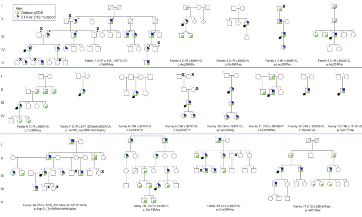
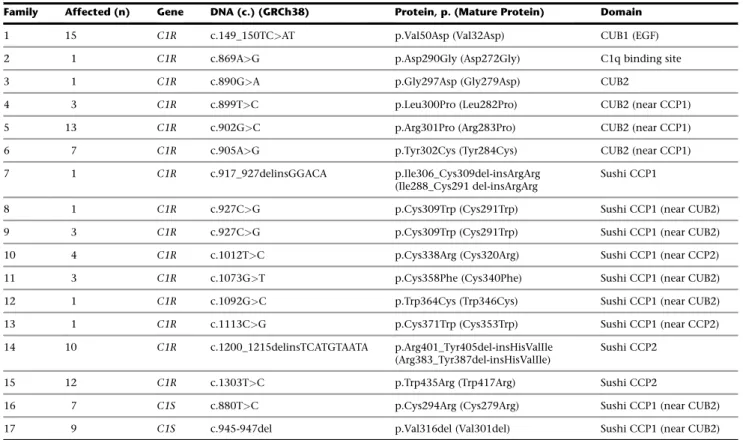
A total of 19 families from USA and Europe comprising 107 individuals with the clinical diagnosis of pEDS were avail-able for molecular investigations. Genome-wide linkage analysis in family 1 confirmed the previously reported locus for pEDS on chromosome 12p13.1. Exome sequencing identified sequence variants in C1R (GenBank: NM_ 001733.4) in six families and in C1S (GenBank: NM_ 201442.2) in two families. Subsequent targeted sequencing revealedC1R sequence variants in nine additional families (Figure 1,Table 1). None of the identified variants was listed in the ExAC database of more than 60,000 exomes of normal individuals, the 1000 Genomes database, ClinVar, or the SNP data base (last accessed 03/2016).
No potentially pathogenic mutations inC1R or C1S were identified in families 18 and 19, previously reported to be
affected by pEDS but not available for clinical re-assess-ment,5,19 in 11 individuals clinically diagnosed with vascular EDS, or in 71 individuals diagnosed with aggres-sive periodontitis but without EDS-like features.C1Q was sequenced in families 18 and 19, but no potentially path-ogenic variants were identified by exome sequencing.
Protein Variant Modeling
C1r and C1s are multidomain proteins that share similar structures (Figure 3A). C1r and C1s are assembled into a Ca2þ-dependent C1s-C1r-C1r-C1s tetramer that associates with the recognition protein C1q (Figures 3A–3C).13,20 Most of the alterations in C1r and C1s structure involved the domains CUB2 and CCP1 in C1r and the domain CCP1 in C1s (Table 1,Figures 3D and 3E). The C-terminal catalytic serine-protease domains were unaffected. The C1r variants in families 7–11 and 13 affected paired cysteines involved in disulfide bonds that stabilize the C1r CCP1 module. The variant in family 16 substitutes a cysteine in the CCP1 module of C1s. The introduction of an addi-tional cysteine in C1r CUB2 or CCP1 (families 6 and 12) could affect the native disulfide bond formation. The dele-tion of five residues and inserdele-tion of three amino acids in C1r CCP2 in family 14 changes the structure adjacent to a cysteine (position 406) involved in the disulfide bond (406–447) that stabilizes the CCP2 module.
Figure 1. The Pedigrees for 17 Families withC1S or C1R Mutations
The colored symbols are defined in the key. X denotes individuals with normal result of molecular testing; asterisk (*) indicates samples included in linkage studies in family 1. Hatch sign (#) indicates individual in family 4 described as ‘‘affected’’ in a previous publication3
but not confirmed by molecular testing. For families 5 (Rahman et al.4), 8 (Hartsfield and Kouseff22), and 11 (Stewart et al.2), a more
Expression Studies
To assess the effects of identified variants, we overexpressed mutant C1r (p.Val50Asp, p.Cys309Trp, p.Cys371Trp), wild-type C1r, and a C1r non-functional control as cDNAs in HEK293 cells. Western blot analyses were performed with a monoclonal antibody directed against the N-terminal part of C1r (which includes the binding domains); the anti-body recognizes the full-length protein, the A chain gener-ated by C1r activation, and thea-fragment generated by autoproteolysis (Figure 4).21Analysis of cell culture super-natant showed C1r protein only in medium of cells trans-fected with wild-type C1R (the 35 kDa autoproteolytic a-fragment; Figure 4A). Analysis of lysed cells identified an additional band at approximately 55 kDa (A chain) in cell lines transfected with plasmids harboringC1R missense mutations; this band was not present in the other cell lines (Figure 4B). Electron microscopy showed an increased proportion of dilated rough endoplasmic reticulum (RER) cisternae inC1R mutation-transfected HEK cells compared to wild-type and non-transfected control cells (Figures 4D–4F). Semiquantitative analysis of randomly selected sec-tion profiles showed RER dilatasec-tion in 36/63 profiles in cells transfected with p.Cys371Trp compared to 18/60 profiles in cells transfected with the wild-type sequence and 13/38 profiles in non-transfected cells.
Clinical Characteristics of Periodontal EDS
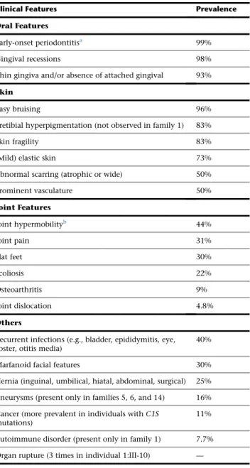
The 17 families with mutations inC1R or C1S comprised 93 individuals with pEDS (Figure 1). Clinical characteris-tics in individuals withC1R or C1S mutations are summa-rized in Table 2. Family descriptions as well as clinical data of each individual are provided in theSupplemental Data.
Defining oral features of pEDS are (1) extensive gingival inflammation in response to mild dental plaque accumula-tion and (2) early-onset periodontitis (EOP) characterized by a rapid destruction of the periodontal attachment appa-ratus in the teens. EOP was present in 99% of clinically or genetically ascertained adults (which partly reflects EOP as a selection criterion). The median age of the periodontal diagnosis—in some individuals the age of first periodontal tooth loss—was 14 years (range 2–35 years). One affected adult (1:IV-3) did not have periodontitis at the age of 24 years, but had extreme gingival recession. Gingival reces-sion (i.e., receding gums) was diagnosed in 98% of individ-uals. Affected individuals, when specifically examined (families 1, 4, 8, 11, 14), had a striking lack of attached gingiva causing oral tissue fragility (Figure 2), which was a unique structural gingival anomaly.
Another defining feature of pEDS was pretibial hyperpig-mentation (83%). No pretibial changes were found in
Table 1. Identified Pathogenic Variants in the Present Cohort with pEDS
Family Affected (n) Gene DNA (c.) (GRCh38) Protein, p. (Mature Protein) Domain
1 15 C1R c.149_150TC>AT p.Val50Asp (Val32Asp) CUB1 (EGF) 2 1 C1R c.869A>G p.Asp290Gly (Asp272Gly) C1q binding site 3 1 C1R c.890G>A p.Gly297Asp (Gly279Asp) CUB2
4 3 C1R c.899T>C p.Leu300Pro (Leu282Pro) CUB2 (near CCP1) 5 13 C1R c.902G>C p.Arg301Pro (Arg283Pro) CUB2 (near CCP1) 6 7 C1R c.905A>G p.Tyr302Cys (Tyr284Cys) CUB2 (near CCP1) 7 1 C1R c.917_927delinsGGACA p.Ile306_Cys309del-insArgArg
(Ile288_Cys291 del-insArgArg
Sushi CCP1
8 1 C1R c.927C>G p.Cys309Trp (Cys291Trp) Sushi CCP1 (near CUB2) 9 3 C1R c.927C>G p.Cys309Trp (Cys291Trp) Sushi CCP1 (near CUB2) 10 4 C1R c.1012T>C p.Cys338Arg (Cys320Arg) Sushi CCP1 (near CCP2) 11 3 C1R c.1073G>T p.Cys358Phe (Cys340Phe) Sushi CCP1 (near CUB2) 12 1 C1R c.1092G>C p.Trp364Cys (Trp346Cys) Sushi CCP1 (near CUB2) 13 1 C1R c.1113C>G p.Cys371Trp (Cys353Trp) Sushi CCP1 (near CCP2) 14 10 C1R c.1200_1215delinsTCATGTAATA p.Arg401_Tyr405del-insHisValIle
(Arg383_Tyr387del-insHisValIle)
Sushi CCP2
15 12 C1R c.1303T>C p.Trp435Arg (Trp417Arg) Sushi CCP2
16 7 C1S c.880T>C p.Cys294Arg (Cys279Arg) Sushi CCP1 (near CUB2) 17 9 C1S c.945-947del p.Val316del (Val301del) Sushi CCP1 (near CUB2) Abbreviations are as follows: n, number; C1R, complement 1 subcomponent r; C1S, complement 1 subcomponent s. For bothC1R and C1S, c.1 is the first nucle-otide of the initiator codon and p.1 is the initiator methionyl residue. The GenBank reference sequences used are NM_001733 and NM_001734 forC1R and C1S, respectively. The signal sequences for C1r and C1s are 18 and 15 amino acids in length, respectively. Most of the literature about these proteins uses p.Ser19 (C1r) and p.Glu16 (C1s) for the start residues of these two proteins. We have included the reference in the mature protein alignment for the sites of the pathogenic variant in parentheses.
family 1 where the skin had normal elasticity but appeared rather soft and dry. Almost all affected individuals had easy bruising (96%), skin fragility (83%), and mild skin hy-perextensibility (73%). Abnormal scars (atrophic or wide) were present in 50% of individuals. Some individuals had additional dermatological findings such as marked facial flushing, thin nails, or thin hair. One individual (9:II-1) re-ported difficulties in wound healing, with open wounds that took months or even years to heal. Joint hypermo-bility was not a consistent finding (44%), and if present was mild and often limited to small joints. Joint pain, scoli-osis, and pes planus were rare. Affected individuals in family 4 reported no musculoskeletal symptoms.
Of the affected individuals, 40% were prone to recurrent infections such as otitis media, herpes zoster, bladder infec-tions, empyema, kidney infecinfec-tions, or pneumonia. There was a history of aneurysms in 16% of affected individuals (families 6, 7, and 15). In total, four individuals had cere-bral aneurysms leading to hemorrhages at ages 23–62, and two individuals died in their mid 40s after aortic dissection. There were two instances of autoimmune disor-ders (Crohn disease and Sjo¨gren syndrome in family 1). In-dividual 2:II-1 had chronic hoarseness that resulted from an abnormality of the cricoarytenoid joint (A.V., unpub-lished data).
Clinical Laboratory Studies
Electron microscopy examination of skin reported in fam-ilies 4 and 5 showed decreased collagen content, abnormal variation in collagen fibril diameter, and some abnormally shaped fibrils.3,4 Similar abnormalities were observed in skin from three individuals from family 1 (Figures S1and
S2) as well as individuals 2:II-1 and 5:V-6.4 Biochemical analysis of collagen in cultured skin fibroblasts in family 1 did not show abnormalities in the production and secre-tion of type I, III, and V collagens. Results of collagen ana-lyses in individuals 5:III-3 and 8:II-2 were reported as normal,4,22 as were those in cells from the probands in families 2, 3, 12, 13, 14, and 16. Complement studies in family 1 (CH50 and circulating levels of C1s and C1r) showed no consistent alterations in classical pathway acti-vation (data not shown).
Discussion
Periodontal EDS is a distinct clinical entity that we have now shown to be caused by mono-allelic missense or in-frame insertion/deletion alterations in C1R or C1S, the genes that encode complement 1 subunits C1r and C1s. The cardinal clinical feature is severe early-onset periodon-titis with marked gingival recessions that in some individ-uals affects primary teeth. In contrast to individindivid-uals with non-syndromic chronic or aggressive periodontitis, those with pEDS have strikingly thin and fragile oral soft tissue with absence of attached gingiva (Figure 2). This feature of pEDS facilitates the clinical diagnosis prior to evident periodontitis: usually, the free gingival margin (the termi-nal edge of the gingiva surrounding the teeth) is contin-uous with the attached gingiva, which is tightly bound to the underlying periostum by collagenous anchoring fi-brils that provide protection during chewing or tooth brushing (Figure 2). In pEDS-affected individuals, attached gingiva is lacking, and the thin and mobile alveolar mu-cosa directly proceeds to the free gingival margin, causing oral tissue fragility. Connective tissue pathology in pEDS also includes atrophic pretibial skin with areas of hyperpig-mentation (83%), easy bruising (96%), and increased risk of arterial aneurysms (16%). Joint symptoms are generally mild, with hypermobility mostly of small joints.
Table 2. Summary of Clinical Features in Periodontal EDS Clinical Features Prevalence
Oral Features
Early-onset periodontitisa 99%
Gingival recessions 98%
Thin gingiva and/or absence of attached gingival 93%
Skin
Easy bruising 96%
Pretibial hyperpigmentation (not observed in family 1) 83%
Skin fragility 83%
(Mild) elastic skin 73%
Abnormal scarring (atrophic or wide) 50%
Prominent vasculature 50% Joint Features Joint hypermobilityb 44% Joint pain 31% Flat feet 30% Scoliosis 22% Osteoarthritis 9% Joint dislocation 4.8% Others
Recurrent infections (e.g., bladder, epididymitis, eye, zoster, otitis media)
40%
Marfanoid facial features 30%
Hernia (inguinal, umbilical, hiatal, abdominal, surgical) 25%
Aneurysms (present only in families 5, 6, and 14) 16%
Cancer (more prevalent in individuals withC1S mutations)
11%
Autoimmune disorder (present only in family 1) 7.7%
Organ rupture (3 times in individual 1:III-10) —
Prevalence rates are based on 93 individuals with mutations inC1R or C1S, and with respective clinical data from the present cohort (Table S1).
aAge of first tooth loss, 2–30 years; age of complete tooth loss, 14–48 years;
prepubertal periodontitis (age<10 years), 16%.
C1r and C1s are structurally similar proteins encoded by C1R and C1S, adjacent genes within the pEDS locus. Both proteins have an identical domain structure characterized by CUB1-EGF-CUB2-CCP1(Sushi)-CCP2(Sushi)-SP(serine protease) (Figure 3). Both proteins have amino-terminal signal sequences that direct them to the lumen of the rough endoplasmic reticulum. C1r and C1s associate as a proenzyme calcium-dependent tetramer that binds to a bouquet-like structure made of six C1q subunits to form the C1 complex. Each C1q subunit is a heterotrimer of A, B, and C chains that form a collagen-like stem (Figure 3). Upon binding of C1q to appropriate targets such as anti-gen-antibody complexes,20C1r is auto-activated by cleav-age at Arg463-Ile464 and can then cleave C1s at the parallel site (Arg447-Ile448) to form the active C1esterase. This enzyme can now cleave C4 and C2 to form the clas-sical pathway C3 convertase (C4b2a).23–25
Heterozygous C1R or C1S mutations we identified in pEDS-affected individuals appear to have gain-of-function effects on as yet unidentified targets either within the cells or in the matrix. In contrast, complete deficiency of C1r or C1s caused by homozygous C1R- or C1S-null muta-tions causes a lupus-erythematosus-like syndrome with increased susceptibility to infections and increased risk of developing autoimmune diseases. Individuals heterozy-gous for C1R- or C1S-null mutations are reported to be asymptomatic, and in particular have not been reported to have periodontal disease.13,23,26Loss of C1 esterase in-hibitor results in intermittent and sometimes life-threat-ening angioedema due to excessive bradykinin production linked to an off-target effect of activated C1s.27This is not a feature of pEDS.
Most mutations in our study alter residues that cluster at the hinges between the CUB2 and CCP1 modules, i.e., the interaction and catalytic domains of C1r and C1s (Figure 3). These hinges are the sites of a conformational change that allows the extended tetramer to fit into the C1q ‘‘cone.’’ Several mutations affect cysteines at positions 309/358 and 338/371 of C1r that form two stabilizing intra-chain disulfide bonds close to the C1r/C1r interface, which are essential for tetramer assembly (Figures 3A and 3C) and stabilization of Sushi modules (complement con-trol protein [CCP] domains in complement and adhesion proteins). Disulfide bond formation could be indirectly affected by other identified mutations such as the C1R deletion-insertion mutation that involves residues 401– 405 adjacent to the 406–447 disulfide bond or mutations that introduce additional cysteines. The C1R mutation c.869A>G (p.Asp290Gly, family 2) involves a C1q binding site and may interfere with the assembly of the C1 complex, as previously shown for p.Asp290Ala.15 The C1r p.Val50Asp substitution may affect the calcium-dependent interaction of C1r with C1s and consequently the interaction of the C1s-C1r-C1r-C1s tetramer with C1q. In order to study the effects of mutations observed in patients with pEDS, we overexpressed C1r variants p.Val50Asp, p.Cys309Trp, and p.Cys371Trp in HEK293 cells. Western blots of cells and supernatants indicated that the abnormal C1r proteins are retained in the cells but can undergo autoactivation that may lead to interac-tion with off-target substrates. Mutainterac-tion-transfected cells showed an increased proportion of dilated RER cisternae (Figure 4), similar to that seen in skin in situ (provided in the Supplemental Data). The C1r-C1s tetramer normally
Figure 2. Oral Features of Periodontal EDS (A) Gingival tissues of a non-affected con-trol child (1:V-1). The gingiva is subdi-vided into the non-attached free gingival margin (FG), the attached gingiva (AG), and the interdental papilla (IP). The gingival epithelium is keratinized and per-forms a protective function during mastica-tion. The attached gingiva is tightly bound to the periostum via collagen structures. The border between attached gingiva and alveolar mucosa (AM) is the mucogingival junction (MGJ). The oral mucosal epithe-lium is non-keratinized and only loosely connected to the periostum; therefore, it is more fragile.
(B and C) Gingival tissues of an affected child (1:V-2) (B) and of an affected adult (1:IV-1) (C). The attached gingiva is missing; the oral mucosa extends to the free gingival margin and the interdental papillae.
(D) Dental radiograph of a non-affected individual (1:IV-4). The alveolar crest is the most cervical rim of the alveolar bone (arrow); in health, it is located approximately 1 mm apical to the cemento-enamel junction (border between dental crown and root).
(E) Dental radiograph of an affected individual 1:IV-2, aged 24 years. Notice periodontal bone loss (BL) in the lower jaw. The alveolar crest is now located more apically.
binds to the N-terminal collagenous domain of C1q that contains a phylogenetically conserved hexapeptide motif Hyp-Gly-Lys-(Val/Asn)-Gly-(Pro//Lys/Met).28,29 Hyp-Gly-Lys-Asn-Gly sequences are present in the triple helical domains of the proa1(I) and proa2(I) chains of type I collagen, as well as the proa1(III) chains of type III collagen, and may represent alternative C1r/C1s binding sites. C1q binding of C1r and C1s is mediated by the CUB domains that are evolutionarily conserved and are present in a number of proteins including procollagen C-proteinase enhancers (PCPE1) and bone-morphogenetic protein (BMP1).30Both PCPE1 and BMP1, as well as C1s, can bind through their CUB domains to the triple helix of collagen and/or propeptides that can be degraded.31–35
This suggests that abnormal binding of (mutated) C1r/ C1s to connective tissue precursors could be a pathoge-netic factor in pEDS.
There is substantial evidence that altered complement function plays an important role in the pathogenesis of non-syndromic periodontitis.36Induction of experimental gingivitis in human volunteers causes progressive comp-lement activation (as determined by C3 conversion in gingival crevicular fluid) that is correlated with increased clinical inflammation.37 Conversely, traditional peri-odontal treatment can lead to decreased complement ac-tivity38and C3 downregulation.39Local inhibition of C3 reduced experimental periodontitis in non-human pri-mates, and this strategy has been suggested as a treatment
Figure 3. C1r and C1s Structure
(A) Modular structure of C1r and C1s and main binding sites to assemble C1. The CUB domain (for complement C1r/C1s, uEGF, BMP1) is a structural motif of approximately 110 residues found almost exclusively in extracellular and plasma membrane-associated proteins. The EGF-like domain is an evolutionary conserved protein domain, which derives its name from the epidermal growth factor where it was first described. It comprises about 40 amino acid residues with six cysteines that form characteristic intra-domain disulfide bonds (1-3, 2-4, and 5-6). CCP (Complement Control Proteins) domains are also termed Sushi domains or Short Consensus Repeats and contain about 60 amino acid residues, each with 4 conserved cysteines that form intradomain disulfide bonds (1-3 and 2-4). These domains are involved in interaction between subunits of proteins and between proteins. The Serine Protease (SP) domains are mostly catalytic domains evolutionary related to the trypsin-chymotrypsin enzymes. The same color code is used for the domains throughout the figure.
(B) C1q (yellow) is a hexamer of heterotrimers that contains in its cone the main protease interfacial domains that are crucial for C1r/C1s tetramer assembly. Each heterotrimer (A, B, C chains) contains a protease binding site in its collagen stem and a C-terminal globular recognition domain. This incomplete C1 model includes two copies each of C1r and C1s interaction domains (violet, red) and two copies of C1r catalytic domains (blue).
(C) Schematic view of the main protease conformational changes during C1 assembly, with strong bending between the interaction and catalytic domains. The central C1r/C1r interface (C/C, blue) involves C1r CCP1 and SP head to tail interactions.
(D and E) Mapping the C1r and C1s variants on 3D structure models. The wild-type residues affected by variants that cause pEDS are shown in colored spheres. The homologous modules are about the same size in the two proteases, which are shown at a different scale.
in humans.40No mutations inC1R or C1S were detected in 71 individuals with aggressive periodontitis, which is a main differential diagnosis to pEDS. Aggressive periodonti-tis is a rare (prevalence 0.1% to 0.5%) complex genetic disease with familial aggregation, characterized by rapid progressing periodontal destruction in otherwise healthy individuals, typically occurring before the age of 35 years. Also, no mutations inC1R or C1S were detected in families 18 and 19. Individuals with suggested pEDS in these fam-ilies presented with periodontitis and EDS-type connective tissue features but had no pretibial plaques.5,19The propo-sita in family 19 had only moderate periodontal
destruc-tion at age 37 (community periodontal index of treatment needs [CPITN] grade III) and a history of severe caries, sug-gesting chronic periodontitis.19Neither family was avail-able for clinical re-assessment.
In conclusion, pEDS in at least the great majority of cases results from specific classes of heterozygous mutations inC1R and C1S. The mechanism of pathogenesis of these mutations differs from homozygous loss of function of these genes and from loss of the C1 esterase inhibitor. Clinical diagnosis of pEDS should be based on severe periodontitis with early onset in combination with absence of attached gingiva, as well as pretibial hyperpigmentation and easy
Figure 4. Analyses of Cells, Lysates, and Serum-free Supernatants of Transfected and Control HEK Cells
(A) Western blot analysis of serum-free supernatants under reducing conditions (n¼ 3). The strong signal at 35 kDa in supernatant of wild-typeC1R transfected HEK239 (WT) corresponds to the a-fragment of autocatalytically cleaved C1r A chain. Cell lines with missense mutations c.149_150TC>AT (p.Val50Asp), c.927C>G (p.Cys309Trp), and c.1113C>G (p.Cys371Trp), non-transfected controls (ctr), and a 26 bp frameshift insertion at position c.899_900 causing a nonsense mutation that is not expected to lead to a functional protein (nonfunctional ctr.) showed no extracellular signal for C1r. Coomassie staining was used as loading control.
(B) Western blot analysis of cell lysates under reducing conditions (n¼ 3). The analysis of cell lysates in cell lines that express missense variants p.Val50Asp, p.Cys309Trp, and p.Cys371Trp showed additional bands at approx. 55 kDa corresponding to the uncleaved A chain of C1r. These bands are absent in non-transfected controls (ctr), transfected nonsense mutations (nonfunctional ctr), and wild-type (WT) C1r samples. This suggests a possible retention of mutated C1r within the cells. Loading control usinga-tubulin antibody shows similar amounts of protein in all samples.
(C) Schematic representation of C1r peptide subunits. C1r is autoactivated by cleavage into the 55 kDa A chain containing the domains CUB1-EGF-CUB2-CCP1-CCP2 and includes the binding domain, and the 28 kDa B chain which represents the serine protease domain; after activation the A chain is autoproteolytically cleaved into several fragments including a 35 kDaa-fragment (CUB1-EGF). The anti-body used (Abcam cat# ab66751; RRID: AB_1860204) recognizes an N-terminal fragment encompassing residues 1–100 of human C1r (A chain).
(D–F) Transmission electron microscopy. Ultrastructure of rough endoplasmic reticulum from mutation-transfected (D), wild-type-trans-fected (E), and untreated (F) HEK293 cells. Semiquantitative analysis of randomly selected section profiles showed RER dilatation (asterisk) in 36/63 profiles in cells transfected with c.1113C>G (p.Cys371Trp) compared to 18/60 profiles in cells transfected with the wild-type sequence, and 13/38 profiles in non-transfected cells. Scale bar represents 200 nm in all panels.
bruising and confirmation by genetic tests. Individuals should receive specific surveillance for aneurysms.
Accession Numbers
All variants have been submitted to ClinVar (submission numbers SUB1882099, SUB1882115, SUB1882116, and SUB1882151).
Supplemental Data
Supplemental Data include detailed descriptions of the individual families, two figures, and one table and can be found with this article online athttp://dx.doi.org/10.1016/j.ajhg.2016.08.019.
Consortia
Additional members of the Molecular Basis of Periodontal EDS Consortium are Kirk Aleck, Zoltan Banki, Joszef Dudas, Herbert Dumfahrt, Hady Haririan, James K. Hartsfield, Charles N. Kagen, Uschi Lindert, Thomas Meitinger, Wilfried Posch, Christian Pritz, David Ross, Richard J. Schroer, Georg Wick, Robert Wildin, and Doris Wilflingseder.
Acknowledgments
We wish to thank the families and individuals with periodontal EDS for their participation in this study. We are grateful to Vincent Offermanns, Alexander Rinner, and Robert Stigler for providing oral tissue samples; Dieter Kotzot for providing additional DNA samples; Britta Berglund, S.M.C. George, and Aparna Sinha for DNA sample collection; and Hella Sto¨ssel for expert help with EM. The study was supported by funds of the Oesterreichische Nationalbank (Anniversary Fund, project number 15408), the Propter Homines Foundation (Liechtenstein), and the FWF (Horos doctoral Program, W1253-B24), by SNF grant number 310030_138288 to C.G. and M.R., by intramural funds from the UK MRC Clinical Sciences Centre, by a Wellcome Clinical Training Fellowship to R.W. (grant no. 100565/Z/12/Z), and by the Freud-mann Fund for Translational Research in Ehlers Danlos syndrome, the Ehlers Danlos Research Fund, and the Center for Precision Diagnostics at the University of Washington. Sequencing was provided by the University of Washington Center for Mendelian Genomics (UW-CMG) and was funded by the National Human Genome Research Institute and the National Heart, Lung, and Blood Institute grant U54HG006493 to D.N., M.B., and S.L. Received: July 8, 2016 Accepted: August 26, 2016 Published: October 13, 2016 Web Resources 1000 Genomes,http://www.1000genomes.org ClinVar,https://www.ncbi.nlm.nih.gov/clinvar/ dbSNP,http://www.ncbi.nlm.nih.gov/projects/SNP/
ExAC Browser,http://exac.broadinstitute.org/
GenBank,http://www.ncbi.nlm.nih.gov/genbank/
OMIM,http://www.omim.org/
RCSB Protein Data Bank, http://www.rcsb.org/pdb/home/ home.do
RRID,https://scicrunch.org/resources
References
1. Vanakker, O., Callewaert, B., Malfait, F., and Coucke, P. (2015). The genetics of soft connective tissue disorders. Annu. Rev. Genomics Hum. Genet.16, 229–255.
2. Stewart, R.E., Hollister, D.W., and Rimoin, D.L. (1977). A new variant of Ehlers-Danlos syndrome: an autosomal dominant disorder of fragile skin, abnormal scarring, and generalized periodontitis. Birth Defects Orig. Artic. Ser.13 (3B), 85–93. 3. Reinstein, E., DeLozier, C.D., Simon, Z., Bannykh, S., Rimoin,
D.L., and Curry, C.J. (2013). Ehlers-Danlos syndrome type VIII is clinically heterogeneous disorder associated primarily with periodontal disease, and variable connective tissue features. Eur. J. Hum. Genet.21, 233–236.
4. Rahman, N., Dunstan, M., Teare, M.D., Hanks, S., Douglas, J., Coleman, K., Bottomly, W.E., Campbell, M.E., Berglund, B., Nordenskjo¨ld, M., et al. (2003). Ehlers-Danlos syndrome with severe early-onset periodontal disease (EDS-VIII) is a distinct, heterogeneous disorder with one predisposition
gene at chromosome 12p13. Am. J. Hum. Genet. 73,
198–204.
5. Reinstein, E., Wang, R.Y., Zhan, L., Rimoin, D.L., and Wilcox, W.R. (2011). Ehlers-Danlos type VIII, periodontitis-type: further delineation of the syndrome in a four-generation pedi-gree. Am. J. Med. Genet. A.155A, 742–747.
6. Biesecker, L.G., Erickson, R.P., Glover, T.W., and Bonadio, J. (1991). Molecular and cytologic studies of Ehlers-Danlos syn-drome type VIII. Am. J. Med. Genet.41, 284–288.
7. Nelson, D.L., and King, R.A. (1981). Ehlers-Danlos syndrome type VIII. J. Am. Acad. Dermatol.5, 297–303.
8. Cıkla, U., Sadighi, A., Bauer, A., and Basxkaya, M.K. (2014). Fatal
ruptured blood blister-like aneurysm of middle cerebral artery associated with Ehlers-Danlos syndrome type VIII (periodonti-tis type). J. Neurol. Surg. Rep.75, e210–e213.
9. Albandar, J.M. (2014). Aggressive periodontitis: case definition and diagnostic criteria. Periodontol. 200065, 13–26. 10. Bonadio, J., and Byers, P.H. (1985). Subtle structural
alter-ations in the chains of type I procollagen produce osteogene-sis imperfecta type II. Nature316, 363–366.
11. Baumann, M., Giunta, C., Krabichler, B., Ru¨schendorf, F., Zoppi, N., Colombi, M., Bittner, R.E., Quijano-Roy, S., Mun-toni, F., Cirak, S., et al. (2012). Mutations in FKBP14 cause a variant of Ehlers-Danlos syndrome with progressive kypho-scoliosis, myopathy, and hearing loss. Am. J. Hum. Genet. 90, 201–216.
12. Costabile, M. (2010). Measuring the 50% haemolytic comple-ment (CH50) activity of serum. J. Vis. Exp.29, 1923. 13. Gaboriaud, C., Ling, W.L., Thielens, N.M., Bally, I., and Rossi,
V. (2014). Deciphering the fine details of c1 assembly and acti-vation mechanisms: ‘‘mission impossible’’? Front. Immunol. 5, 565.
14. Budayova-Spano, M., Lacroix, M., Thielens, N.M., Arlaud, G.J., Fontecilla-Camps, J.C., and Gaboriaud, C. (2002). The crystal structure of the zymogen catalytic domain of complement protease C1r reveals that a disruptive mechanical stress is required to trigger activation of the C1 complex. EMBO J. 21, 231–239.
15. Bally, I., Rossi, V., Lunardi, T., Thielens, N.M., Gaboriaud, C., and Arlaud, G.J. (2009). Identification of the C1q-binding sites of human C1r and C1s: a refined three-dimensional model of the C1 complex of complement. J. Biol. Chem. 284, 19340–19348.
16. DeLano (2002). The PyMOL Molecular Graphics System (Palo Alto, CA: DeLano Scientific LLC).
17. Deutschmann, A.J., Amberger, A., Zavadil, C., Steinbeisser, H., Mayr, J.A., Feichtinger, R.G., Oerum, S., Yue, W.W., and Zschocke, J. (2014). Mutation or knock-down of 17b-hydrox-ysteroid dehydrogenase type 10 cause loss of MRPP1 and impaired processing of mitochondrial heavy strand tran-scripts. Hum. Mol. Genet.23, 3618–3628.
18. Frank, I., Piatak, M., Jr., Stoessel, H., Romani, N., Bonnyay, D., Lifson, J.D., and Pope, M. (2002). Infectious and whole inacti-vated simian immunodeficiency viruses interact similarly with primate dendritic cells (DCs): differential intracellular fate of virions in mature and immature DCs. J. Virol.76, 2936–2951.
19. Reinstein, E., Pariani, M., Lachman, R.S., Nemec, S., and Ri-moin, D.L. (2012). Early-onset osteoarthritis in Ehlers-Danlos syndrome type VIII. Am. J. Med. Genet. A.158A, 938–941. 20. Rossi, V., Bally, I., Lacroix, M., Arlaud, G.J., and Thielens, N.M.
(2014). Classical complement pathway components C1r and C1s: purification from human serum and in recombinant form and functional characterization. Methods Mol. Biol. 1100, 43–60.
21. Arlaud, G.J., Villiers, C.L., Chesne, S., and Colomb, M.G. (1980). Purified proenzyme C1r. Some characteristics of its activation and subsequent proteolytic cleavage. Biochim. Bio-phys. Acta616, 116–129.
22. Hartsfield, J.K., Jr., and Kousseff, B.G. (1990). Phenotypic over-lap of Ehlers-Danlos syndrome types IV and VIII. Am. J. Med. Genet.37, 465–470.
23. Amano, M.T., Ferriani, V.P., Florido, M.P., Reis, E.S., Delcolli, M.I., Azzolini, A.E., Assis-Pandochi, A.I., Sjo¨holm, A.G., Farah, C.S., Jensenius, J.C., and Isaac, L. (2008). Genetic analysis of complement C1s deficiency associated with systemic lupus er-ythematosus highlights alternative splicing of normal C1s gene. Mol. Immunol.45, 1693–1702.
24. Patrick, R.A., Taubman, S.B., and Lepow, I.H. (1970). Cleavage of the fourth component of human complement (C4) by acti-vated Cls. Immunochemistry7, 217–225.
25. Thielens, N.M., Villiers, M.B., Reboul, A., Villiers, C.L., and Colomb, M.G. (1982). Human complement subcomponent C2: purification and proteolytic cleavage in fluid phase by C1s, C1r2-C1s2 and C1. FEBS Lett.141, 19–24.
26. Wu, Y.L., Brookshire, B.P., Verani, R.R., Arnett, F.C., and Yu, C.Y. (2011). Clinical presentations and molecular basis of complement C1r deficiency in a male African-American pa-tient with systemic lupus erythematosus. Lupus20, 1126– 1134.
27. Kaplan, A.P., and Joseph, K. (2010). The bradykinin-forming cascade and its role in hereditary angioedema. Ann. Allergy Asthma Immunol.104, 193–204.
28. Venkatraman Girija, U., Gingras, A.R., Marshall, J.E., Panchal, R., Sheikh, M.A., Ga´l, P., Schwaeble, W.J., Mitchell, D.A., Moody, P.C., and Wallis, R. (2013). Structural basis of the C1q/C1s interaction and its central role in assembly of the
C1 complex of complement activation. Proc. Natl. Acad. Sci. USA110, 13916–13920.
29. Bally, I., Ancelet, S., Moriscot, C., Gonnet, F., Mantovani, A., Daniel, R., Schoehn, G., Arlaud, G.J., and Thielens, N.M. (2013). Expression of recombinant human complement C1q allows identification of the C1r/C1s-binding sites. Proc. Natl. Acad. Sci. USA110, 8650–8655.
30. Bork, P., and Beckmann, G. (1993). The CUB domain. A wide-spread module in developmentally regulated proteins. J. Mol. Biol.231, 539–545.
31. Vadon-Le Goff, S., Kronenberg, D., Bourhis, J.M., Bijakowski, C., Raynal, N., Ruggiero, F., Farndale, R.W., Sto¨cker, W., Hulmes, D.J., and Moali, C. (2011). Procollagen C-proteinase enhancer stimulates procollagen processing by binding to the C-propeptide region only. J. Biol. Chem. 286, 38932– 38938.
32. Steiglitz, B.M., Keene, D.R., and Greenspan, D.S. (2002). PCOLCE2 encodes a functional procollagen C-proteinase enhancer (PCPE2) that is a collagen-binding protein differing in distribution of expression and post-translational modifica-tion from the previously described PCPE1. J. Biol. Chem.277, 49820–49830.
33. Wautier, J.L., Reid, K.B., Legrand, Y., and Caen, J.P. (1980). Re-gion of the Clq molecule involved in the interaction between platelets and subcomponent Clq of the first component of complement. Mol. Immunol.17, 1399–1405.
34. Yamaguchi, K., Sakiyama, H., Matsumoto, M., Moriya, H., and Sakiyama, S. (1990). Degradation of type I and II collagen by human activated C1-s. FEBS Lett.268, 206–208.
35. Bourhis, J.M., Vadon-Le Goff, S., Afrache, H., Mariano, N., Kronenberg, D., Thielens, N., Moali, C., and Hulmes, D.J. (2013). Procollagen C-proteinase enhancer grasps the stalk of the C-propeptide trimer to boost collagen precursor matu-ration. Proc. Natl. Acad. Sci. USA110, 6394–6399.
36. Hajishengallis, G., Maekawa, T., Abe, T., Hajishengallis, E., and Lambris, J.D. (2015). Complement involvement in periodon-titis: molecular mechanisms and rational therapeutic ap-proaches. Adv. Exp. Med. Biol.865, 57–74.
37. Patters, M.R., Niekrash, C.E., and Lang, N.P. (1989). Assess-ment of compleAssess-ment cleavage in gingival fluid during exper-imental gingivitis in man. J. Clin. Periodontol.16, 33–37. 38. Niekrash, C.E., and Patters, M.R. (1985). Simultaneous
assess-ment of compleassess-ment components C3, C4, and B and their cleavage products in human gingival fluid. II. Longitudinal changes during periodontal therapy. J. Periodontal Res. 20, 268–275.
39. Beikler, T., Peters, U., Prior, K., Eisenacher, M., and Flemmig, T.F. (2008). Gene expression in periodontal tissues following treatment. BMC Med. Genomics1, 30.
40. Maekawa, T., Abe, T., Hajishengallis, E., Hosur, K.B., DeAnge-lis, R.A., Ricklin, D., Lambris, J.D., and HajishengalDeAnge-lis, G. (2014). Genetic and intervention studies implicating comple-ment C3 as a major target for the treatcomple-ment of periodontitis. J. Immunol.192, 6020–6027.