POSITION PAPER
Subtrochanteric fractures after long-term treatment
with bisphosphonates: a European Society on Clinical
and Economic Aspects of Osteoporosis and Osteoarthritis,
and International Osteoporosis Foundation Working Group
Report
R. Rizzoli&K. Åkesson&M. Bouxsein&J. A. Kanis&
N. Napoli&S. Papapoulos&J.-Y. Reginster&C. Cooper
Received: 10 August 2010 / Accepted: 30 September 2010 / Published online: 18 November 2010 # The Author(s) 2010. This article is published with open access at Springerlink.com
Abstract
Summary This paper reviews the evidence for an associa-tion between atypical subtrochanteric fractures and long-term bisphosphonate use. Clinical case reports/reviews and case–control studies report this association, but retrospec-tive phase III trial analyses show no increased risk. Bisphosphonate use may be associated with atypical subtrochanteric fractures, but the case is yet unproven.
Introduction A Working Group of the European Society on Clinical and Economic Aspects of Osteoporosis and Osteoarthritis and the International Osteoporosis Founda-tion has reviewed the evidence for a causal associaFounda-tion between subtrochanteric fractures and long-term treat-ment with bisphosphonates, with the aim of identifying areas for further research and providing recommendations for physicians.
R. Rizzoli (*)
Division of Bone Diseases,
Department of Rehabilitation and Geriatrics,
University Hospitals and Faculty of Medicine of Geneva, 1211 Geneva 14, Switzerland
e-mail: Rene.Rizzoli@unige.ch K. Åkesson
Department of Orthopedics Malmö, Skåne University Hospital, Lund University,
Lund, Sweden M. Bouxsein
Department of Orthopaedic Surgery,
Harvard Medical School Center for Advanced Orthopaedic Studies, Beth Israel Deaconess Medical Center,
Boston, MA, USA J. A. Kanis
WHO Collaborating Centre for Metabolic Bone Diseases, University of Sheffield Medical School,
Sheffield, UK N. Napoli
Division of Endocrinology,
Universita’ Campus Bio-Medico di Roma, Rome, Italy
S. Papapoulos
Department of Endocrinology and Metabolic Diseases, Leiden University Medical Center,
Leiden, The Netherlands
J.-Y. Reginster
Department of Public Health Sciences and Department of Physical Medicine and Rehabilitation, University of Liège, CHU Sart-Tilman and BRULL, Liège, Belgium
C. Cooper
MRC Epidemiology Resource Centre, University of Southampton,
Southampton, UK
C. Cooper
NIHR Musculoskeletal Biomedical Research Unit, University of Oxford,
Oxford, UK DOI 10.1007/s00198-010-1453-5
Methods A PubMed search of literature from 1994 to May 2010 was performed using key search terms, and articles pertinent to subtrochanteric fractures following bisphosph-onate use were analysed.
Results Several clinical case reports and case reviews report a possible association between atypical fractures at the subtrochanteric region of the femur in
bisphosphonate-treated patients. Common features of these ‘atypical’
fractures include prodromal pain, occurrence with mini-mal/no trauma, a thickened diaphyseal cortex and
trans-verse fracture pattern. Some small case–control studies
report the same association, but a large register-based study and retrospective analyses of phase III trials of bisphosph-onates do not show an increased risk of subtrochanteric fractures with bisphosphonate use. The number of atypical subtrochanteric fractures in association with bisphospho-nates is an estimated one per 1,000 per year. It is recommended that physicians remain vigilant in assessing their patients treated with bisphosphonates for the treatment or prevention of osteoporosis and advise patients of the potential risks.
Conclusions Bisphosphonate use may be associated with atypical subtrochanteric fractures, but the case is unproven and requires further research. Were the case to be proven, the risk–benefit ratio still remains favourable for use of bisphosphonates to prevent fractures.
Keywords Atypical . Bisphosphonate . Femur . Low trauma . Osteoporosis . Subtrochanteric
Introduction
Treatment with bisphosphonates significantly reduces the risk of fractures in men and women with osteoporosis. The evidence is based on high-quality phase III randomized
controlled trials (RCTs) with fracture as an endpoint [1–10].
The benefits of bisphosphonates also extend to other disorders of bone metabolism such as
glucocorticoid-induced osteoporosis [11], Paget’s disease [12] and bone
metastases [13,14].
Treatment with bisphosphonates is not without adverse effects, but they are generally minor and occur in a minority of patients. The most common adverse effect is gastroin-testinal upset with the oral formulations, the frequency of which decreases with intermittent treatment such as once-weekly or monthly regimens. Intravenous (IV) administra-tion of nitrogen-containing bisphosphonates may induce an acute phase reaction which manifests as fever, myalgia and arthralgia, although these side effects usually resolve within
a few days of onset [3,7, 15]. High doses of
bisphosph-onates given intravenously may impair renal function, and the kidney is a major route of elimination of the
bisphosphonates. For this reason, bisphosphonates are not recommended for use in patients with severe renal
impairment [16–18]. The use of bisphosphonates has been
associated with osteonecrosis of the jaw, but most cases have occurred in patients receiving high-dose IV bisphosphonates for neoplastic bone disease, and osteonec-rosis of the jaw has rarely been reported in patients with
benign bone diseases [19, 20]. An increased risk of atrial
fibrillation has been reported for zoledronic acid [3], but the
association may be coincidental [7]. Other uncommon or
rare side effects of bisphosphonates include anaemia [21],
urticaria [22,23] and symptomatic hypocalcaemia [22].
In recent years, several clinical case reports and case reviews have reported an association between atypical fractures in patients receiving treatment with bisphospho-nates. The majority of these cases have described fractures
at the subtrochanteric region of the femur [24–31].
Against this background, the aim of this report was to critically review the evidence for an increased incidence of subtrochanteric fractures after long-term treatment with bisphosphonates, to identify gaps in our knowledge that warrant further research and to provide guidance for healthcare professionals. A PubMed search of literature from 1994 to May 2010 was performed using the search
terms ‘bisphosphonate(s)’ AND/OR ‘alendronate’ AND/
OR ‘risedronate’ AND/OR ‘ibandronate/ibandronic acid’
AND/OR ‘zoledronate/zoledronic acid’ AND/OR
‘subtro-chanter(ic)’ AND ‘fracture’ AND/OR ‘femur/femoral’
AND/OR ‘atypical’ AND/OR ‘low-trauma’ AND/OR
‘low-energy’. Scientific papers pertinent to subtrochanteric fractures following bisphosphonate use were analysed and included in the evidence base.
Characteristics of subtrochanteric fractures
Subtrochanteric fractures have been defined as occurring in a zone extending from the lesser trochanter to 5 cm distal to
the lesser trochanter [32]. However, this anatomical
classification of subtrochanteric fracture has several
varia-tions [33,34], resulting in variable definitions in published
studies [26,30,35].
Regardless of the definition used, many case reports and case reviews have suggested that there are several common features of subtrochanteric fractures associated with bisphosphonate use. Major features were that the fractures arose with minimal or no trauma and, on radiography, the fracture line was transverse. Minor features were that fractures were commonly preceded by prodromal pain and, on radiographs, there appeared beaking of the cortex
on one side and bilateral thickened diaphyseal cortices [26,
28,36–39]. This fracture pattern has often been referred to
as reviewed below, the distinction between typical and atypical subtrochanteric fractures has not yet been firmly established.
It is worth noting that, on radiography, the appearance of atypical subtrochanteric fractures is similar to that of stress fractures, including a periosteal reaction, linear areas of bone sclerosis and a transverse fracture line. Prodromal
pain prior to diagnosis is also common [43]. However,
stress fractures are more commonly associated with repeated episodes of increased activity (e.g. participation in sports). Nevertheless, stress fractures due to non-traumatic loading in patients with low bone density do
occur (insufficiency fractures) [43]; thus, the terms
‘atyp-ical’ and ‘stress’ are often used interchangeably or in conjunction to describe subtrochanteric fractures in this context. However, this terminology also requires clarifica-tion, as not all stress fractures are atypical.
Epidemiology of subtrochanteric fractures
Subtrochanteric fractures are a relatively rare type of hip
fracture [44–46], usually resulting from high-energy
trau-ma, pathologic fracture or, in the elderly, low-energy injury involving osteoporotic bone. Several series report the
incidence of this fracture [25–28,30,36,37,47], although
the definition of the subtrochanteric site has varied. Nieves et al. reported a large, 11-year epidemiological study of fractures of the hip, subtrochanter, femoral shaft and distal
femur in the US population aged≥50 years using National
Hospital Discharge Survey data from the National Center for Health Statistics and MarketScan® (medical claims
experience) data [46]. Of all femoral fractures, 3% were at
the subtrochanteric region, 5% at the femoral shaft, 5% at the distal femur and 87% were at the proximal femur (i.e. hip). Importantly, this study classified fractures solely according to their location in the femur and did not evaluate the fracture patterns radiographically. Thus, they were not
able to determine the incidence of ‘typical’ vs ‘atypical’
subtrochanteric fractures. In men and in women, the incidence rate of each type of fracture remained stable over
5 years but increased exponentially with age (Fig.1). Each
fracture type was more prevalent in women than in men. Seventy-five percent of all femur fracture cases were in women. The mean age at fracture was 80 years old, and those with a subtrochanteric fracture were of a similar age to those with a hip fracture.
Leung et al. published a retrospective analysis that aimed to document the incidence of low-trauma subtrochanteric or femoral diaphyseal fractures in a Hong Kong hospital over
a 5-year period [42]. In all, 88 cases of subtrochanteric
fractures and 66 of diaphyseal fractures were identified, accounting for 3.9% and 2.9% of all recorded osteoporotic fractures, respectively.
Thus, although the incidence of subtrochanteric fractures is much lower than other femoral fractures, they are not rare and account for about 3% of all femoral fractures in the elderly. If these estimates were applied to the UK, then more than 2,000 subtrochanteric fractures are expected to
occur each year [48], and approximately 48,000 are
expected annually worldwide [49].
Subtrochanteric fractures and bisphosphonate exposure
Case reports and case reviews
Twenty-six published case reports and case reviews were identified that were considered relevant, a similar number
to that identified in a recent review by Giusti et al. [50].
Concern about bisphosphonate use in relation to atypical subtrochanteric fractures arose from case reports that described patients with subtrochanteric fractures who had been exposed to bisphosphonates, particularly long-term treatment with alendronate (Fosamax®/Fosavance®, alendr-onate sodium, Merck Sharp & Dohme Limited). The
Fig. 1 Age-specific incidence of femoral fractures according to fracture site in men (X) and women (O) aged ≥50 years
association between long-term bisphosphonate use and unusual diaphyseal fractures was first described by Odvina
et al. in 2005 [31] who reported nine patients with
osteoporosis or osteopenia who had been treated with alendronate for 3–8 years and sustained atraumatic fractures in the course of their normal daily activities. Three patients had fractures of the femoral shaft and two had fractures of the proximal femur. Of these five patients, fracture healing was radiographically assessed in four. All four patients had delayed or absent fracture healing ranging from 4 months to 2 years while on alendronate treatment.
This and subsequent case reports are summarized in Table1.
The mean and median age of patients was 65 years (range 35–85). All cases involved treatment with alendronate, except for five patients who took risedronate (Actonel®, risedronate sodium, Procter and Gamble Pharmaceuticals) and three who took pamidronate (Aredia®, pamidronate disodium, Novartis Pharmaceuticals Limited). One patient had been taking ibandronate (Bonviva®/Boniva®, ibandronic acid, Roche) for 1 year following long-term alendronate use, and one had been taking risedronate for 5 years following 7 years of pamidronate use. There were no published case reports of subtrochanteric fractures following the use of once-yearly zoledronic acid 5 mg (Aclasta®/Reclast®, zoledronic acid anhydrous, Novartis Pharmaceuticals Limited), although cases following treatment with the monthly 4-mg dose have
been reported [36, 38]. The mean and median duration of
bisphosphonate use was 7.3 and 7.5 years, respectively (range 1–16), and the majority of patients had unilateral fractures (29 out of 43; 67.4%).
In addition to case reports, several case reviews have
been published, which are summarized in Table 2. For
example, the characteristics of low-trauma subtrochanteric and diaphyseal fractures were studied retrospectively by Neviaser et al. in all patients admitted to a US trauma centre
over a 5-year interval (Table 2) [30]. Radiographs were
examined by independent experts to identify fractures with a simple, transverse or short oblique pattern in areas of cortical hypertrophy with a cortical beak. The observers were blinded to patient characteristics, including alendro-nate use. Seventy patients were identified, of whom 25 were treated with alendronate. Nineteen out of 25 (76%) alendronate-treated patients had the radiographic pattern compared with one out of 45 (2%) non-alendronate-treated
patients. Thus, the risk of having an‘atypical’
subtrochan-teric fracture pattern was significantly associated with alendronate use (odds ratio=139; 95% confidence interval (CI) 19–939; p<0.0001). The mean duration of treatment with alendronate was 6.2 years (6.9 years in those who had
the fracture pattern vs 2.5 years in those who did not) [30].
The authors concluded that there are unique features to bisphosphonate-associated fractures.
Controlled studies
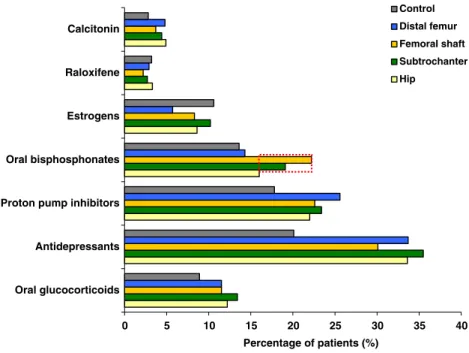
Six studies that utilized control groups were identified that have investigated the association of subtrochanteric frac-tures with the use of bisphosphonates. In the study of Nieves et al. described above, the rate of subtrochanteric and femoral shaft fractures appeared to be higher than that of other fractures in women taking oral bisphosphonates
(Fig. 2) [46], although there is no statistical information
provided. It is not known whether excess fractures were due to trauma or not. The study concluded, however, that there was no evidence of an increase in the incidence of subtrochanteric or femoral shaft fracture between 1996 (around the time that bisphosphonates were first intro-duced) and 2006. Limitations of these data include the lack of radiological and clinical verification and no information on the type of bisphosphonate used or the duration of treatment.
In a study by Leung et al., ten patients with subtrochan-teric fractures who had received alendronate were identified over a 5-year period. This included one patient who had taken alendronate for 1 year followed by ibandronate for
2 years [42]. The crude incidence of subtrochanteric/
femoral diaphyseal fractures associated with prior bisphosphonate use increased over 5 years from 0% in 2003/2004 to 6% in 2004/2005, 8.6% in 2006/2007 and 25% in 2007/2008. This trend was despite a steady annual incidence of subtrochanteric/femoral diaphyseal fractures. It is difficult to draw meaningful conclusions from these data because of the very small sample size (ten subtrochanteric fractures in patients exposed to a bisphosphonate) and the lack of information on bisphosphonate use at other fracture sites. At best, the study documents the increasing use of bisphosphonates over the time of study.
In a small retrospective case–control study, Lenart et al. aimed to identify an association between low-energy subtrochanteric/femoral shaft fractures (according to the Müller AO classification) and long-term bisphosphonate
use [29]. Forty-one low-energy subtrochanteric or femoral
shaft fracture cases were identified and matched by age, body mass index and race to one low-energy intertrochan-teric and femoral neck fracture each.
Fifteen out of the 41 (37%) cases of subtrochanteric or femoral shaft fracture cases were taking bisphosphonates, compared with nine out of 82 (11%) controls (OR=4.4; 95% CI 1.8–11.4; p=0.002). Alendronate was the bisphosphonate taken in all cases. Eight out of nine cases in the control group were taking alendronate (one had previously taken etidronate). A radiographic pattern of a simple transverse or oblique fracture, beaking of the cortex on one side and cortical thickening at the fracture site, was observed in ten of the 15 (67%) subtrochanteric/femoral shaft fracture cases taking bisphosphonate and three of the
T able 1 Case reports of incidents of subtrochanteric fracture following bisphosphonate use (all cases in women unless otherwise indicated) Reference T otal
patients (patients ST/FS/PF fracture) Age (years)
Location
Radiographic features
Bilateral?
Prodromal symptoms (duration) Osteoporosis diagnosis? Prior bisphosphonate Duration of use (years) Concomitant therapy Healing (months of follow-up) Odvina et al. [ 31 ] 9 (5) 52 Femoral shaft No N o (o st eo p en ia ) ALN 8 Ca, D N o (9) 68 a Femoral shaft Y es Y es ALN 8 Ca, D N o (8) 67 Femoral shaft Y es N o (o st eo p en ia ) ALN 5 Oestrogen, Ca, D Y es (5) 49 Proximal femur No Y es (GIO) ALN 3 Pred, Ca, D N o (8) 64 Proximal femur No Y es (GIO) ALN 4 Pred, Ca, D Y es (3) Husada et al. [ 51 ] 1 72 Femoral shaft C o rt ic al th ic k en in g in lateral m id-shaft of contralatera l femu r Y es Severe pain in back and hip (1 month) Y es ALN Not specified Ca, amlodipine, metaprolol, aspirin Schneider [ 52 ] 1 59 Upper femur Cortical thickening No Moderate pain in thigh (3 months) No (family history of osteoporosis) ALN 7 Ca, hormone replacement therapy Y es (>9) Armamento- V illareal et al. [ 53 ] 13 5 a Subtrochanteric femur No ALN 6 C a N o (36) c Cheung et al. [ 54 ] 1 82 Femoral shaft No Y es ALN 10 Ca, glucosamine, chondroitin Demiralp et al. [ 55 ] 1 6 5 Femoral shaft Fracture line, callus, cortical thickening, bowing deformity Y es Incapacitating bilateral femoral shaft pain (1.5 months) Y es ALN 7 Ca, D, steroid,
thyroxine replacement therapy
Lee et al. [ 56 ] 1 73 Femoral diaphysis No Bilateral groin pain, difficulty walking (10 months) Y es ALN 1.5 Y es Sayed-Noor and Sjoden [ 57 ] 1 7 2 Subtrochanteric femur Cortical thickening of lateral femoral cortex, medial beaking at fracture site No Dif fuse pain in hips and thighs (18 months) Y es ALN 7 C a N o (3)/yes (6) V isekruna et al. [ 39 ] 3 5 1 Femoral metadiaphysis Y es Bilateral, lateral hip pain ALN 5 Pred No (3 while on ALN; 12 after stopping ALN) 62 Femoral metadiaphysis Y es Bilateral thigh pain ALN 10 Raloxifene, pred Y es (12) d 75 Femoral metadiaphysis No ALN 10 Pred No (22) Odvina et al. [ 58 ] 1 3 (1 1 ) 5 7 Subtrochanteric, contralateral femur shaft (3 years later) Cortical thickening Y es Pain at fracture site (1 –6 months) N o (o st eo p en ia ) ALN 6 Ca, D Y es (36) 74 Femoral shaft Cortical thickening No Y es ALN 10 Ca, D N o
T able 1 (continued) Reference T otal
patients (patients ST/FS/PF fracture) Age (years)
Location
Radiographic features
Bilateral?
Prodromal symptoms (duration) Osteoporosis diagnosis? Prior bisphosphonate Duration of use (years) Concomitant therapy Healing (months of follow-up) 67 Femoral shaft Cortical thickening No Pain at fracture site (1 –6 months) Y es RIS >5 Ca, D Y es (6) 58 Femoral shaft (fractured twice in 3 years) Cortical thickening No Pain at fracture site (1 –6 months) No ALN 7 Ca, D, tamoxifen Y es (6) 62 Femoral shaft Cortical thickening No N o (o st eo p en ia ) RIS 2 Ca, D, tamoxifen 63 Femoral shaft Cortical thickening No Y es ALN 10 Ca, D, oestrogen Y es (6) 72 Femoral shaft Cortical thickening No Pain at fracture site (1 –6 months) Y es ALN 9 Ca, D, oestrogen Y es 76 Femoral shaft Cortical thickening No Y es (GIO) ALN 1 1 Ca, D, pred Y es (12) 72 Left and right femoral shaft Cortical thickening Y es Pain at fracture site (1 –6 months) Y es (GIO) ALN 10 Ca, D, pred Y es 77 Femoral shaft Cortical thickening No Y es (GIO) ALN 9 Ca, D, pred Y es 38 Left and right femoral shaft Cortical thickening Y es Y es (GIO) ALN 3 Ca, D, pred Y es Ali and Jay [ 59 ] 1 82 Femoral shaft Cortical thickening No ALN 8 Y es (3) Goddard et al. [ 60 ] 1 6 7 Femoral diaphysis Cortical thickening, unicortical beaking No ALN 16 Y es (12) Ibandronate 1 Sayed-Noor and Sjoden [ 61 ] 2 7 8 T ip of femoral stem Cortical thickening No Y es ALN 9 N o (6) 55 Subtrochanteric femur Cortical thickening, medial beaking, cortical thickening on contralateral femur No Dif fuse pain in thighs, walking difficulties (several months) Y es ALN 9 D Y es (9) Cermak et al. [ 62 ] 4 64 Subtrochanteric femur Cortical thickening No Pain in left thigh (3 months) No ALN 5.5 Y es (6) 70 Femur Medial cortical beaking Y es Pain in thighs Y es ALN 6 Y es (4) Other femur (3 months later) Y es (7) 77 Femoral shaft Cortical thickening No Pain in right thigh Y es ALN 12 Y es (12) 59 Subtrochanteric femur Cortical thickening, medial cortical beaking No None Y es ALN 10 Y es (5) Bush and Chew [ 63 ] 1 8 5 Subtrochanteric femur Focal beak of cortical thickening of lateral cortex No Limp, persistent pain in anterior thigh (2–3 months) Y es (GIO) RIS >6 Ca, D, pred Y es (2) Lee [ 64 ] 1 82 Left femoral diaphysis Horizontal fracture lines at thickest part of femoral cortex extending lateral –medial, Y es Y es ALN 8 Ca, D Y es (5) Right femoral diaphysis (4 years later)
Reference
T
otal
patients (patients ST/FS/PF fracture) Age (years)
Location
Radiographic features
Bilateral?
Prodromal symptoms (duration) Osteoporosis diagnosis? Prior bisphosphonate Duration of use (years) Concomitant therapy Healing (months of follow-up) followed by short oblique fracture (identical at both sites) Edwards et al. [ 65 ] 1 6 0 Right femoral diaphysis (T aken after initial, right fracture) Minor lateral cortical thickening on left femur Y es Mild pain in right thigh before right fracture, none before left fracture Y es (GIO) ALN 8 Pred Left femoral diaphysis (2 years later) Giusti et al. [ 50 ] 8 60 Right subtrochanteric femur Y es Pain in right hip No ALN 4 Ca, D, pred, inhaled GCs,
esomeprazole, repaglinide, metformine, azathioprine, rosuvastatin
No (6) Left subtrochanteric femur (9 months later) 36 Femoral shaft No Y es ALN 8 D ,
pred, simvastatine, cyclosporine, amlopidine, atenolol,
lisinopril Ye s 64 Left and right subtrochanteric femur (1 complete, 1 insuff iciency fracture) Y es Pain in right thigh No ALN 2.5 Ca, D, pred,
omeprazole, azathioprine, losartan,
triamteren, HCT No (18) 62 Right and left femoral shaft b Y es Pain in right thigh and hip Y es Oral pamidronate 4 Ca, D, Y es 58 Femoral shaft No Pain in left thigh Y es Intravenous pamidronate 3 Ca, D N o (12) 58 Subtrochanteric femur No Pain in left hip No RIS 5.5 Ca, D, pred, inhaled GCs, omeprazole, pravastatine, ibuprofen No (12) 72 Left subtrochanteric femur Y es Pain in left thigh and hip Y es (GIO) Oral pamidronate followed by ALN 7 + 5 Ca, D, inhaled GCs,
esomeprazole, simvastatine, captopril,
irbesartan, clopidogrel Y es (12) Right subtrochanteric femur (insufficiency fracture 1 year later) 75 Femoral shaft (insuf ficiency fracture) Severe pain in left thigh and hip Y es RIS 6 Ca, D, esomeprazole, etoricoxib Femoral shaft (insufficiency fracture 1 year later) Pain in hip ALN alendronate, BP bisphosphonate, Ca calcium, D vitamin D, FS femoral shaft, GCs glucocorticoids, GIO glucocorticoid-induced osteoporosis, HCT hydrochlorothiazide, PF proximal femur , RIS risedronate, ST subtrochanteric, Pr ed prednisone aMale patient b First fracture prio r to B P treatme nt; con tralateral fra cture follo wing 4 years ’ BP treatme nt; refracture of contralateral fem oral shaft 4 years after second fracture c Patient w as prescri bed alendronate in 1996 and took it for 6 years. Fr acture occurr ed 1 yea r after disc ontin uation and had not completely healed w hen reported in 2006 d Patient beg an teriparatide immediately after fracture
26 (11%) subtrochanteric/femoral shaft fracture cases not taking bisphosphonate (OR = 15.3; 95% CI = 3.1–76.9; p<0.001). The duration of bisphosphonate exposure was
significantly longer in patients with this X-ray pattern [29].
Koh et al. carried out a retrospective clinical and radiological review of geriatric hip fracture patients at a Singapore tertiary centre over 4 years to assess features that
predispose to complete stress fractures [38]. Thirty-two
patients with spontaneous or low-energy fractures with metaphyseal–diaphyseal involvement and on bisphospho-nate therapy were identified. All were on alendrobisphospho-nate therapy except for one who was on monthly zoledronic acid 4 mg and one who had been on risedronate for 6 years following 4 years of alendronate. Of these, 16 patients (median duration of therapy 4.5 years) had radiographic evidence of lateral cortical thickening. Four had cortical stress lesions on the prefracture radiograph (group F) and 12 had cortical stress lesions on the contralateral femur (group C). The type of bisphosphonate taken by patients according to group was not detailed. All patients in group F experienced prodromal thigh discomfort, compared with 25% of patients in group C (p=0.019), and radiographic evidence of a stress line across the cortical thickening occurred in 100% and 8% of patients, respectively (p=0.003). At a median follow-up of 23 months, none of the patients in group C had developed a complete fracture. All of these patients except for one had discontinued bisphosphonate therapy; five had not taken any alternative therapy since discontinuation. Nevertheless, eight out of the 11 were asymptomatic, and no new cortical thickening was detected in any of the patients. The authors concluded that, in people taking long-term bisphosphonate therapy, symp-tomatic cortical stress reactions accompanied by evidence of a stress line across the cortical thickening suggest an
increased risk of a complete stress fracture [38].
In the only population-based study that included radio-logical review of all cases, Schilcher and Aspenberg studied the incidence of stress fractures at the femoral shaft in bisphosphonate-treated patients in four hospitals in Sweden. Women aged over 55 years with fractures of the femoral diaphysis or subtrochanteric region were identified from the operation registry. Preoperative radiographs were examined to identify stress fractures, defined as a transverse fracture of the femoral shaft with cortical thickening. Of 91,956 women identified, 3,087 bisphosphonate users were identified, of whom five had femoral stress fractures. All of these five patients were aged >75 years, and their mean
duration of treatment was 5.8 years [66]. Three patients that
were not treated with bisphosphonates had stress fractures. All were aged <75 years. The annual incidence of femoral shaft stress fractures in bisphosphonate users was 1/1,000 per year (95% CI 0.3–2) vs 0.02/1,000 (0.004–0.1) per year in control patients. Thus, the risk of such fractures was
estimated to be 46 times greater with bisphosphonate use
(95% CI 11–200) [65]. An obvious weakness of the study is
that, although the confidence intervals were corrected for sample size, the findings were based on just eight femoral shaft stress fractures. The results thus raise a hypothesis to be tested on larger samples.
A larger study is provided by Abrahamsen et al. who studied the epidemiology of subtrochanteric and diaphyseal femur fractures in patients in Denmark treated with
alendronate [67]. However, in contrast to the Schilcher
and Aspenberg report, in this study, radiographic fracture patterns were not reviewed, and thus, fractures were identified purely based on their location. In patients aged ≥60 years that had subtrochanteric, diaphyseal femur and hip fractures in 2005, the incidence of subtrochanteric (n= 898) and diaphyseal fractures (n=720) were similar, and the ratio of high-to-low-energy trauma fractures was the same for each of these fracture types (approximately 2.5:1 for each). Exposure to alendronate was also similar between fracture types (approximately 7% each). Patients with subtrochanteric fractures and diaphyseal fractures were more likely to have taken glucocorticoids in the year before fracture than patients with hip fracture (10.9%, 8.4% and 6.5% of patients, respectively).
In a register-based matched cohort analysis, Abrahamsen
et al. investigated whether the increase in risk of‘atypical’
femur fracture in alendronate-treated patients was greater
than the increase in risk of ‘typical’ osteoporotic femur
fractures (‘typical’ and ‘atypical’ were not defined). In total,
15,187 patients who took alendronate for ≥6 months after
the fracture event (the treatment cohort) were compared with two randomly assigned sex-, age- and fracture-matched controls (n=10,374). The use of alendronate was associated with an increase in the hazard ratio (HR; adjusted for baseline comorbidities) for both subtrochan-teric/diaphyseal fractures (HR=1.46; 95% CI 0.91–2.35; p=0.12) and hip fracture (HR=1.45; 95% CI 1.21–1.74; p<0.001). Subtrochanteric/diaphyseal fractures were equal-ly common in the alendronate-treated (14% of hip fractures) and control patients (13%; p=0.70). Both hip fractures and subtrochanteric/diaphyseal fractures were significantly low-er in patients with highlow-er adhlow-erence (HR =0.47 [0.34–0.65; p<0.001] and 0.28 [0.12–0.63; p<0.01], respectively). In a sub-analysis of 178 compliant (medication possession ratio >80%) patients who took alendronate for >6 years, long-term alendronate use was associated with no change in
both hip (HR=1.24 [0.66–2.34]; p=0.52) and
subtrochan-teric/diaphyseal fractures (HR=1.37 [0.22–8.62]; p=0.74). The incidence of subtrochanteric/diaphyseal fractures was similar in the long-term alendronate (10%) and control
(12.5%) groups (10% vs 12.5%, respectively) [67].
This study, in a large number of patients, does not support the hypothesis that exposure to alendronate is
T able 2 Case reviews of incidents of subtrochanteric fracture following bisphosphonate use (all cases in women unless otherwise indicated) Reference Re view loc ation/ pe riod Inclusio n criteria Patients eligib le (n ) Mean age (years [range]) Fracture location Radiogr aphic features (n ) Bilateral? (n ) P
rodromal symptoms (duration) OP diagnosi s? (n ) Prior BP (du ration of use, years) Concomitant therapy (n ) Goh et al. [ 26 ] 2 Singapore hospita ls/May 2005 –Februa ry 2006 ST fracture adue to low-ene rgy trauma 13 ALN (9) 66 .9 (55 –82) NA Corti cal thick ening (6 = lateral, 3 = contralateral) NR 5 pts (2–6 months) Y es (3) ALN (4.2 [2.5 –5]) Ca (all); lo ng-term oral steroids (1) No (4) Unknown (2) No AL N (4) 80 .3 (64 –92) NA NR N one Y es (all) NA Ca (2) Kwek et al. [ 28 ] S ingapore hospita l/May 2005 –Januar y 2007 ST fracture bdue to low-ene rgy trauma in patients tak ing ALN 17 66 (53 –82) NA Lateral co rtical thickening, medi al cortical beaking (all) ST stress fracture (2) Y es, 13 pts (1 week – 24 yea rs) Y es (10 ) ALN (4.4 [2 –8]) [1 patien t takin g RIS after 4 years on ALN] Ca (all); lo ng-term pred nisolone (1) Femor al shaft stress fracture (1) No (6) Femor al shaft fracture (1) Unknown (1) Nevia ser et al. [ 30 ] U S trauma centre/January 2002 –March 2007 Low-ene rgy ST an d mid-sha ft femu r fracture s c 70 (1 1 m ale) 74.7 ST femu r (50) Lateral co rtical
thickening, unicortical beaking
(20 ) d NR N R Y es (31 ) e ALN (6.2 [1 –10]) [25 pts] f NR Femoral shaft (20) Glennon [ 47 ] A
ustralian tertiary hospita
l, 12 mont hs ST stress fracture w ith characteris tic radio logical/ clinical features 66 0– 87 NA T ransverse fracture, unicortical beaking, cortical thickening (all) 1 pat ient P ain in 5 pts (1 week to 6 mont hs) NR ALN (1.5 –16) [5 pts] NR RIS (>3) [1 pt] Ing-L orenzini et al. [ 27 ] S wiss univer sity hospita l/ 2 yea rs Low-ene rgy ST fracture , h istory of BP use 8 (7 females) 67.5 ST femu r (7) Corti cal thick ening, also in contralateral femur in 4 patients Y es, in 4 patients 0.5 –5 y ears after first fracture P ain in 2 pts, 1 lateral side, 1 b o th sides Y es (all; 1 p t GIO) ALN alone (1.5 –8) [3 pt s] Ca (all), gluc ocorticoids (4), pro ton-pump inhi bitors (7) Femoral shaft (1) ALN (3 –10) switched to ibandr onate (1 NK) g[3 pt s] RIS (NK) switched to ALN (2) [1 pt] Pami dronate (5) h [1 pt] Arm amento-V illareal et al. [ 25 ] U S medica l school/ Novembe r 2004 –March 2007 Low-ene rgy fracture , m ainly at cortical sites, 2 years ’ B P therapy , bo ne biopsy 15 (12 females, 3males) 43 –75 Femoral shaft (7) [1 ma le] Y es (2) N R NR ALN (4 –10) [6 pts] Ca (6); vitam in D (6); infliximab (1); triamcinolone (1); tamo xifen (1); lev othyroxine (1); fluticasone (1); HCT (1); mom etazone (1) Other (9) RIS (2) [1 pt] Capec i and T ejwani [ 37 ] U S universi ty hospita l/4 years Bilateral low -energy femo ral di aphyseal or ST fracture, lo ng-term ALN 7 6 1 (53 –75) Simultaneous femo ral diaphysi s (1) Corti cal thick ening, medial beaking (all) Y es (all) T high pain (4 pts with impendi ng ST stress fractures) NR ALN (8.6 [5 –13]) None af fecting bone me tabolism Sequential ST femu r (2) ST and impending contr alateral ST femu r (3) Femoral diaphysis and impending
T able 2 (continued) Reference Re view loc ation/ pe riod Inclusio n criteria Patients eligib le (n ) Mean age (years [range]) Fracture location Radiogr aphic features (n ) Bilateral? (n ) P
rodromal symptoms (duration) OP diagnosi s? (n ) Prior BP (du ration of use, years) Concomitant therapy (n ) contr alateral ST femu r (1) Bunning et al. [ 36 ] U S rehabilitation hospita l/7 years Atypica l low-or no -impact femoral fracture 4 (1 male) 49 –59 Diaphyseal femo ral (3); left ST/righ t diaphyse al femo ral (1) Media l cortical thickening (1) 1 p t P ain in hip (1 –3 months) [all], pain in knee [1 pt] Y es (all) None [1 patien t] NR Pami dronate (0.5)/ zoledronic acid 4 m g (>4.5) [1 pt] ALN (5) [1 pt] ALN (6) [1 pt] ALN alendronate, BP bisphosphonate, Ca calcium, GIO glucocorticoid-induced osteoporosis, HCT hydrochlorothiazide, NA not applicable (described in inclusion criteria), NK not known, NR not reported, OP osteoporosis, Pt patient, RIS risedronate, ST subtrochanteric a In the reg ion of the fem ur which exte nded from the lesser trochanter to the junc tion of the proxima l and middle third of the fem oral shaft b W ithin the reg ion of th e femur 5 cm dista l to the lesser trochanter c Muller AO classification type 32 and type 31 A3 fractures invol ving or extending dista lly to the lesse r trochanter dNineteen had been treated with alendronate eT wenty -one had bee n treated with ale ndrona te f All fem ales. Eighte en cases con firmed through phys icia n/patient con tact. Duration of use esta blished in 16 cases g One patient had been on iban dronate for 1 year . One switched to ibandro nate 4 mont hs bef ore first fracture in Febru ary 2006 ; one sw itched 1 year bef ore second fra cture in Jan 2008 h Stoppe d 1 year bef ore fracture
associated with an increased frequency of subtrochan-teric fractures compared with controls. However, the same study reported that treatment with alendronate was associated with an increased risk of hip fracture. This
should not be interpreted as ‘alendronate causes hip
fracture’, but only that high-risk patients are exposed to alendronate. The finding also illustrates the difficulties in the interpretation of retrospective observational studies, particularly accounting for selection bias that likely confounds the other much smaller observational studies.
Randomized controlled trials
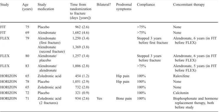
Black et al. recently reported an analysis of subtrochanteric and diaphyseal fractures in the Fracture Intervention Trial
(FIT) of alendronate and its extension [1,2,5,68] and the
HORIZON Pivotal Fracture Trial (PFT) of zoledronic acid
5 mg [3]. Twelve fractures in ten patients were documented
in the subtrochanteric or diaphyseal region (Table 3) a
combined rate of 2.3 per 10,000 patient-years [69].
However, radiographs were not available to confirm typical vs atypical radiographic features. There was no significant increase over placebo in the risk of subtrochanteric/ diaphyseal fractures during the FIT, FIT Long-Term Extension (FLEX) or HORIZON-PFT trials. Compared with placebo, the relative hazard was 1.03 (95% CI 0.1– 16.5) for alendronate use in the FIT trial, 1.5 (95% CI 0.3– 9.0) for zoledronic acid in the HORIZON-PFT and 1.3 (95% CI 0.1–14.7) for continued alendronate use in the FLEX trial. The interpretation of this analysis is limited by the small number of events and the large confidence intervals.
Bilezikian et al. reported the incidence of subtrochanteric fractures in the randomized, placebo-controlled phase III studies of risedronate in post-menopausal osteoporosis, which enrolled more than 15,000 patients. In trials of up to 3 years duration, the mean incidence of subtrochanteric fractures was 0.14% in risedronate 2.5-mg treated patients (n=4,998), 0.13% in risedronate 5-mg treated patients (n= 5,395) and 0.17% in placebo-treated patients (n=5,363)
[70]. In active control studies of risedronate involving
various doses (35 mg once weekly, 75 mg on two consecutive days per month, 150 mg once monthly), no subtrochanteric fractures were reported, and the incidence of hip/femoral fractures was similar to that in the
placebo-controlled studies [70].
The manufacturers of ibandronate have assessed their clinical trials database to determine the incidence of subtrochanteric and diaphyseal femoral fractures in women taking ibandronate for post-menopausal osteoporosis.
Atyp-ical fractures were defined as ‘mostly non-spine fractures
including hip or femur fractures in the subtrochanteric region or shaft and occurring without trauma or in association with low-energy trauma’. For femur fractures, subtrochanteric fracture location was considered as atypical for osteoporosis-related fractures, defined as a region below the lesser trochanter and a junction between the proximal and middle third of the femoral shaft. In the pivotal trials
(MF 4380, BONE, MOBILE and DIVA) [4, 71–73], there
were nine fracture cases corresponding to these defined locations and characteristics (subtrochanteric, femoral shaft, stress or multiple fractures): six occurred in placebo-treated patients (n=1,924) and three in ibandronate-treated patients (n=6,830). In addition, there was one identified case of a
0 5 10 15 20 25 30 35 40
Oral glucocorticoids Antidepressants Proton pump inhibitors Oral bisphosphonates Estrogens Raloxifene Calcitonin Percentage of patients (%) Control Distal femur Femoral shaft Subtrochanter Hip
Fig. 2 Medical and prescription drug history in US female
frac-ture patients (2002–2006)
dur-ing the 1 year before index date
femoral shaft fracture in an ibandronate-treated patient in the extension and major phase IIIb trials (MOBILE LTE,
DIVA LTE, MOTION and PREVENTION; n=2,451) [74–
77]. Some fractures were reported without identifying the
precise location. However, all of these fractures were associated with trauma and thus did not meet the definition for atypical fractures. An additional 5-year analysis of the marketed regimens of ibandronate (150 mg once monthly and 3 mg IV quarterly) was also carried out from the active comparator-controlled trials and their extensions (MOBILE,
DIVA, MOTION, MOBILE LTE and DIVA LTE) [71,72,
74,75,77]. No atypical subtrochanteric/diaphyseal femoral
fractures were found for either of the marketed regimens (150 mg, n=1,279; 3 mg, n=469).
Pharmacovigilance data
Since fractures are the clinical outcome of osteoporosis and no treatments are fully effective, fractures are expected in treated patients. It is likely, however, that the number of reports through pharmacovigilance will be small. The number of postmarketing reports of atypical stress fractures in association with alendronate to circa July 2008 was 115 (of which 84 were femur fractures) and included a large number of the cases reported in the
literature [78].
Bilezikian et al. have reported that in more than 10 years of risedronate post-approval surveillance to September
2008 (18 million patient-years of exposure), the reporting rate for subtrochanteric fractures was <0.1 per 100,000
patient treatment years of exposure [70].
Postmarketing data from the manufacturers of zoledronic acid have revealed a similarly low rate of subtrochanteric fractures with zoledronic acid 5 mg. Using the last cutoff date for worldwide Health Authority Reporting prior to January 2010 (Periodic Safety Update Report v6) and assessing all adverse event reports for zoledronic acid 5 mg (579,501 patient-years of exposure), the rate of femoral subtrochanteric fracture reporting was three per 1,000,000 patient treatment years of exposure.
Postmarketing data from the manufacturers of ibandro-nate have also revealed a low rate of possible atypical fractures occurring in patients receiving ibandronate for the management of postmenopausal osteoporosis. According to their global safety database as of June 2009, cumulative postmarketing exposure of ibandronate yielded a crude reporting rate of possible atypical fractures of approxi-mately one per 1,000,000 patients. Three of the cases involved alendronate treatment followed by ibandronate treatment and were reported in the case series of
Ing-Lorenzini et al. [27].
Regulatory perspective
In July 2008, the Pharmacovigilance Working Party (PhVWP) of the Committee for Medicinal Products for
Table 3 Characteristics of ten patients with 12 low-trauma subtrochanteric or femoral diaphyseal fractures in the FIT, FLEX and HORIZON-PFT
trials (adapted from Black et al. [69])
Study Age (years) Study medication Time from randomization to fracture (days [years]) Bilateral? Prodromal symptoms
Compliance Concomitant therapy
FIT 75 Placebo 962 (2.6) >75% None
FIT 69 Alendronate 1,682 (4.6) >75% None
FLEX 79 Alendronate
(first fracture)
1,250 (3.4) Stopped 3 years
before first fracture
Alendronate, 6 years (in FIT before FLEX) Alendronate (second fracture) 1,369 (3.8) FLEX 80 Alendronate/ placebo 1,257 (3.4) Stopped 3 years before fracture
Alendronate, 6 years (in FIT before FLEX)
FLEX 83 Alendronate/
alendronate
1,006 (2.8) >75% Alendronate, 5 years (in FIT
before FLEX)
HORIZON 65 Zoledronic acid 454 (1.2) Hip pain 100% Raloxifene
HORIZON 78 Placebo 1,051 (2.9) Hip pain 100% None
HORIZON 65 Zoledronic acid 732 (2.0) 100% None
HORIZON 72 Placebo 321 (0.9) 100% Calcitonin
HORIZON 71 Zoledronic acid
(2 fractures)
934 (2.6) Yes Bone pain 100% Bisphosphonate and hormone
replacement therapy, both before study
Human Use (CHMP) initiated a class review on bisphosph-onates and atypical stress fractures. Marketing Authoriza-tion Holders supplied informaAuthoriza-tion about all preclinical, clinical and future studies, published case reports, post-marketing data, possible mechanisms and proposed risk-minimization activities. Following a PhVWP review of these data in December 2008, the CHMP concluded that there was an association between atypical stress fractures and long-term use of alendronate, due to the distinct fracture pattern, prodromal pain and poor fracture healing. However, the benefit–risk balance of alendronate use was considered favourable. The CHMP highlighted that there was uncertainty concerning a class effect for other bisphosphonates and that switching of bisphosphonates should be avoided at this time. Ultimately, the CHMP recommended that information about atypical stress fractures should be added to the product information
for medicinal products containing alendronate [78].
Consequently, the labelling for alendronate (Fosamax®/ Fosavance®, Merck Sharp & Dohme Limited) now includes a special warning/precaution for alendronate use, advising discontinuation of bisphosphonate therapy in patients with stress fracture pending evaluation, based on
an individual benefit–risk assessment [22,79]. Alendronate
is the only bisphosphonate for osteoporosis treatment that currently carries this warning.
In addition to the 2008 class review, the EMEA released a statement in August 2009 highlighting their 2010 priorities for drug safety research with regards to the long-term adverse skeletal effects of bisphospho-nates: (1) generate methodologies to study the link between bisphosphonate use and long-term adverse skeletal events in human populations and (2) measure the incidence of stress/insufficiency fractures in associ-ation with high-dose/long-term use of bisphosphonates by class, compound, mode of administration, dose etc. Methods could include meta-analysis or nested case–
control studies [80].
In June 2008, the US Food and Drug Administration (FDA) initiated a review of bisphosphonates for a possible association with increased risk of atypical subtrochanteric femur fractures. All available case reports and clinical trial data were requested from all bisphosph-onate drug manufacturers and were reviewed alongside the registry data from the large observational study of
Abrahamsen et al. [67]. In March 2010, the FDA
announced that the data reviewed had not shown a clear connection between bisphosphonate use and the risk of atypical subtrochanteric fractures. Physicians were urged to continue to follow the labelling when prescribing bisphosphonates and patients were instructed not to discontinue their medication unless instructed to do so
by their physician [81].
Pathophysiology of subtrochanteric fractures associated with bisphosphonate use
The pathophysiology of atypical low-trauma subtrochan-teric fractures following bisphosphonate use is not known. However, preclinical and clinical studies of the effects of bisphosphonates on bone suggest that there are several possible mechanisms that work either alone or in tandem. The organic matrix of the bone determines its toughness, and this matrix is partly made up of bone collagen, which impacts on the bone’s mechanical properties. Bisphospho-nate use may negatively affect collagen by preventing or
reducing its maturation [82], although this finding has not
been consistently replicated [83]. Bisphosphonates may
also affect bone mineralization density distribution (BMDD). The more heterogeneous the BMDD, the slower that cracks in the bone will develop and the lower the risk
of new cracks and fractures forming [84]. As
bisphospho-nate treatment reduces bone turnover, the increase in overall mineralization leads to more homogeneous bone—as
evidenced by a narrow BMDD [85, 86]—and thus an
increased risk of cracks and fractures. Reduced bone turnover also increases the accumulation of microdamage,
as cracks are not repaired [87], and reduces bone toughness,
which contributes to the increased susceptibility of bone to
new cracks [88–90]. Finally, bisphosphonates have
differ-ing impacts on different types of fracture. Acute fractures of long bone are not affected by bisphosphonates in the initial
healing stages [91–93], as they heal via endochondral
ossification. However, stress fractures heal by normal bone remodelling, and thus, bisphosphonates may prevent or delay healing, increasing the likelihood of a complete fracture with little or no trauma. Several reports have reported on bone quality in people with low-trauma fractures taking bisphosphonate therapy.
For example, Odvina et al. reported that cancellous bone histomorphometry in alendronate-treated patients (3– 8 years) who sustained spontaneous non-vertebral fractures showed markedly suppressed bone formation, with reduced or absent osteoblastic surface in most patients. Osteoclastic surface was also low in most patients, and eroded surface
decreased in half [31]. Odvina et al. reported similar
findings in a later report in a comparable patient population
[58]. In a case report by Armamento-Villareal et al. of a
man who had a low-trauma subtrochanteric fracture after discontinuing 6 years of alendronate treatment, a bone biopsy showed severely decreased trabecular connectivity, a lack of osteoid on trabecular surfaces and an absence of
tetracycline labelling [53]. Armamento-Villareal et al. later
reported that of 15 bisphosphonate-treated patients (2–
10 years; Table2) who underwent bone biopsies following
a low-energy cortical (femoral shaft, pelvis, rib, metatarsal, ankle) fracture, ten had an absence of double-tetracycline
label, reduced osteoid surface and thickness suggestive of suppressed trabecular bone remodelling. However, there was no difference in cortical thickness between patients
with suppressed (n=10) and normal (n=5) turnover [25].
Recent findings by Somford et al., however, suggest an alternative pathophysiology for subtrochanteric fractures associated with bisphosphonate treatment. In a patient who was treated with alendronate for 8 years and subsequently developed spontaneous bilateral subtrochanteric/diaphyseal fractures, biopsies showed a marked decrease in bone formation as expected; however, this was not coupled with the expected decrease in bone resorption. In fact, bone resorption parameters such as osteoclast number were markedly increased in the femur sample. In addition, there was no evidence of hypermineralized bone. This suggests that an imbalance between bone resorption and bone formation at the affected femur—the cause of which is currently unknown—rather than excessive suppression of bone turnover may be the underlying mechanism for subtrochanteric/diaphyseal femoral
frac-tures in bisphosphonate-treated patients [94].
Summary of evidence
The view that bisphosphonates increase the risk of subtrochanteric femoral fractures arises from the case reports and retrospective case reviews that have reported ‘atypical’ subtrochanteric and diaphyseal fractures in patients exposed to bisphosphonates. In all, these data highlight the scope of the problem, i.e. a trend that warrants further investigation. However, the data in their entirety are insufficient proof that long-term bisphospho-nate use is the only cause of atypical low-trauma sub-trochanteric fractures.
There are several limitations to the existing evidence base: lack of a consensus definition of an atypical subtrochanteric fracture, small numbers of patients in-volved, lack of radiographs which precludes characteriza-tion of the radiographic features of the fractures and incomplete reporting of subject characteristics. In addition, subtrochanteric fractures in general are not atypical frac-tures; rather, they are part of the natural history of fragility fractures in osteoporosis. They increase in frequency with age in much the same way as does the incidence of other
osteoporotic fractures [95]. Although their incidence is
much lower than other femoral fractures, they are not rare and account for approximately 3% of all femoral fractures
[46]. Thus, the term ‘atypical’ is not synonymous with
‘unexpected’ which is the common interpretation. Rather, the term should be reserved for subtrochanteric fractures that have atypical features, of which some are similar to with those associated with stress.
Therein lies an additional problem in that it has been difficult to provide characteristics of the fracture that are associated with the use of bisphosphonates. Candidate features, which include the prodromal manifestation of incomplete (fissure) fractures, a thickened cortex and a transverse fracture pattern with cortical beaking may be associated with the use of bisphosphonates but, in the absence of blinded evaluation in all cases, may be subject to large observer biases. In addition, in many instances, cases have been complicated, for example, by concomitant exposure to
glucocorticoids [25–28, 31,39, 50, 55, 58, 63,65], which
appears to be a risk factor for subtrochanteric fractures [46].
In terms of evidence-based medicine, the ultimate arbiter for a causal relationship between subtrochanteric fractures and exposure to bisphosphonates might be expected to derive from information from RCTs. All the information available fail to show an association of this fracture with exposure to bisphosphonates, although all RCTs were completed before attention was drawn to the problem, so the documentation of the sites of fracture and any associated features is inevitably incomplete. Furthermore, the frequency of the event is sufficiently low that even large RCTs are insufficiently powered to identify meaningful associations with drug exposure. Finally, the duration of exposure to bisphosphonates may be too short in the setting of RCTs if, as has been suggested, the complication were to increase in frequency with exposure time. Against this background, data from observational studies might be expected to contribute to our understanding, but such studies are fraught with biases and limitations for which it may be difficult to adjust.
Research agenda
The ultimate question for physicians is what type of patient is at the highest risk of an atypical low-trauma subtrochan-teric fracture. Thus far, apart from long-term alendronate therapy, suggested risk factors include glucocorticoid, proton-pump inhibitor or calcitonin use and female gender
[26,46,67]. Thus, a number of urgent issues and areas for
research have been identified as follows:
1. Standardized definition of ‘subtrochanteric fracture’,
including a definition of ‘atypical’ and ‘typical’
fractures
2. Provision of descriptive epidemiology based on large-scale studies with characterization of radiographic features
3. Definition of fracture incidence by femoral location, mechanism of injury and underlying pathology 4. Identification of risk factors, with greater clarity as to the
5. Pathophysiological studies in relation to risk factors 6. Pathophysiological studies at the tissue level, i.e. is the
mechanism of atraumatic (insufficiency) fractures dif-ferent to that of low-trauma fractures?
7. Long-term, large, prospective, observational studies to assess incidence of subtrochanteric fractures in bisphosphonate-treated vs bisphosphonate-naïve patients. Methods should include (1) futility analysis and (2) radiographic measurements. Outcomes should include (1) adherence, (2) number needed to harm and (3) assessment of temporal relationship between bisphosph-onate treatment and fracture type
8. Long-term, large, prospective, observational studies allowing for systematic follow-up of patients with subtrochanteric fractures treated long-term with bisphosphonates, in order to assess fracture healing characteristics (e.g. time to healing, choice of fracture treatment device, adjuvant bone anabolic intervention etc.)
9. Large, prospective, randomized, controlled clinical trials of the efficacy and safety of pharmacological treatment (e.g. strontium ranelate, teriparatide) for patients with subtrochanteric fractures
Conclusions and recommendations
A sense of proportion may be helpful in alleviating the concerns of the medical community. A plausible scenario is that long-term exposure to bisphosphonates (more than 5 years) increases the risk of subtrochanteric femoral fractures twofold. In the UK, using the guidance of the National Osteoporosis Guideline Group, the relative risk of hip fracture is expected to be approximately threefold increased in postmenopausal women identified for
treat-ment [96]. Assuming that the average population risk of
hip fracture is 1% per year in postmenopausal women, then 300 hip fractures are expected for every 10,000 patients identified to be at high risk. If these patients were treated and assuming an effectiveness of
bisphosph-onates of 36% (RR = 0.64) [97], then 108 hip fractures are
averted by treatment (and approximately 750 fractures at other sites). On the debit side, three subtrochanteric fractures (both typical and atypical) are to be expected, which might increase to six if bisphosphonates doubled the risk of all subtrochanteric fractures. Under the assump-tions of this scenario, the risk–benefit ratio remains very favourable.
Evidence, including that from an EMEA class review, suggests that alendronate use may potentially increase the risk for atypical, low-trauma subtrochanteric fractures, although it is unclear whether this applies to other
bisphosphonates. Irrespective of exposure to bisphospho-nates, the occurrence of subtrochanteric fractures is an expected finding in patients with osteoporosis. If atypical fractures do occur, then their characteristics are poorly defined, their causality with bisphosphonate exposure insecure and their frequency rare. Bisphosphonates as the cause of atypical fractures at the subtrochanteric site is therefore still merely a hypothesis, though no less important for that. Clearly more research is required from well-designed prospective observational studies, meta-analyses and nested case–control studies.
Thus, the available evidence does not suggest that the well-known benefits of bisphosphonate treatment are out-weighed by the risk of these rare, atypical, low-trauma subtrochanteric fractures. Nevertheless, it is recommended that physicians remain vigilant in assessing their patients treated with bisphosphonates for osteoporosis or associated conditions. They should continue to follow the recommen-dations on the drug label when prescribing bisphosphonates and advise patients of the potential risks. Patients with pain in the hips, thighs or femur should be radiologically assessed and, where a stress fracture is evident, the physician should decide whether bisphosphonate therapy should be discontinued pending a full evaluation, based on
an individual benefit–risk assessment. The radiographic
changes should be evaluated for orthopaedic intervention— since surgery prior to fracture completion might be
advantageous—or be closely monitored.
Acknowledgements The Working Group meeting was supported by
an unrestricted educational grant from the European Society for Clinical and Economic Aspects of Osteoporosis and Osteoarthritis. Editorial assistance for the manuscript was provided by Sola Neunie of BioScience Communications, supported by a financial grant from Novartis Pharmaceuticals.
Conflicts of interest Rene Rizzoli has attended paid advisory boards
and received consultancy and lecturing fees from Servier, Novartis, Eli Lilly, Amgen, Roche, Nycomed, Merck Sharp and Dohme and Danone. Kristina Åkesson has received lecturing fees from Medtronics, Novartis, Amgen, Merck and Nycomed. Mary Bouxsein has undertaken consultancy and lecturing commitments for Amgen and Merck & Co. John A. Kanis consults or has received research support from a large number of pharmaceutical companies involved in marketing products for treatment of osteoporosis. He is president of the International Osteoporosis Foundation and serves on its Committee of Scientific Advisors. Nicola Napoli has received grant support from Merck Sharpe and Dohme. Socrates Papapoulos has received consultancy and lecturing fees from Alliance for Better Bone Health, Amgen, Eli Lilly, GSK, Merck & Co, Novartis, Pfizer and Roche. Jean-Yves Reginster has received consulting fees and attended paid advisory boards for Servier, Novartis, Negma, Lilly, Wyeth, Amgen, GlaxoSmithKline, Roche, Merckle, Nycomed, NPS, Theramex and UCB. He has received invited lecture fees from Merck Sharp and Dohme, Lilly, Rottapharm, IBSA, Genevrier, Novartis, Servier, Roche, GlaxoSmithKline, Teijin, Teva, Ebewee Pharma, Zodiac, Analis, Theramex, Nycomed and Novo Nordisk. He has received grant support from Bristol Myers Squibb, Merck Sharp & Dohme, Rottapharm, Teva, Lilly, Novartis, Roche, GlaxoSmithKline,
Amgen and Servier. Cyrus Cooper has undertaken consultancy and lecturing commitments for Alliance for Better Bone Health, Eli Lilly, Novartis, GSK Roche, Servier, MSD and Amgen.
Open Access This article is distributed under the terms of the Creative
Commons Attribution Noncommercial License which permits any noncommercial use, distribution, and reproduction in any medium, provided the original author(s) and source are credited.
References
1. Black DM, Cummings SR, Karpf DB, Cauley JA, Thompson DE, Nevitt MC, Bauer DC, Genant HK, Haskell WL, Marcus R, Ott SM, Torner JC, Quandt SA, Reiss TF, Ensrud KE (1996) Randomised trial of effect of alendronate on risk of fracture in women with existing vertebral fractures. Fracture Intervention
Trial Research Group. Lancet 348:1535–1541
2. Black DM, Schwartz AV, Ensrud KE, Cauley JA, Levis S, Quandt SA, Satterfield S, Wallace RB, Bauer DC, Palermo L, Wehren LE, Lombardi A, Santora AC, Cummings SR (2006) Effects of continuing or stopping alendronate after 5 years of treatment: the Fracture Intervention Trial Long-term Extension (FLEX): a
randomized trial. JAMA 296:2927–2938
3. Black DM, Delmas PD, Eastell R, Reid IR, Boonen S, Cauley JA, Cosman F, Lakatos P, Leung PC, Man Z, Mautalen C, Mesenbrink P, Hu H, Caminis J, Tong K, Rosario-Jansen T, Krasnow J, Hue TF, Sellmeyer D, Eriksen EF, Cummings SR (2007) Once-yearly zoledronic acid for treatment of postmenopausal osteoporosis. N
Engl J Med 356:1809–1822
4. Chesnut CH III, Skag A, Christiansen C, Recker R, Stakkestad JA, Hoiseth A, Felsenberg D, Huss H, Gilbride J, Schimmer RC, Delmas PD (2004) Effects of oral ibandronate administered daily or intermittently on fracture risk in postmenopausal osteoporosis.
J Bone Miner Res 19:1241–1249
5. Cummings SR, Black DM, Thompson DE, Applegate WB, Barrett-Connor E, Musliner TA, Palermo L, Prineas R, Rubin SM, Scott JC, Vogt T, Wallace R, Yates AJ, LaCroix AZ (1998) Effect of alendronate on risk of fracture in women with low bone density but without vertebral fractures: results from the Fracture Intervention Trial. JAMA 280:2077–2082
6. Harris ST, Watts NB, Genant HK, McKeever CD, Hangartner T, Keller M, Chesnut CH III, Brown J, Eriksen EF, Hoseyni MS, Axelrod DW, Miller PD (1999) Effects of risedronate treatment on vertebral and nonvertebral fractures in women with postmenopausal osteoporosis: a randomized controlled trial. Vertebral Efficacy With
Risedronate Therapy (VERT) Study Group. JAMA 282:1344–1352
7. Lyles KW, Colon-Emeric CS, Magaziner JS, Adachi JD, Pieper CF, Mautalen C, Hyldstrup L, Recknor C, Nordsletten L, Moore KA, Lavecchia C, Zhang J, Mesenbrink P, Hodgson PK, Abrams K, Orloff JJ, Horowitz Z, Eriksen EF, Boonen S (2007) Zoledronic acid and clinical fractures and mortality after hip
fracture. N Engl J Med 357:1799–1809
8. Reginster J, Minne HW, Sorensen OH, Hooper M, Roux C, Brandi ML, Lund B, Ethgen D, Pack S, Roumagnac I, Eastell R (2000) Randomized trial of the effects of risedronate on vertebral fractures in women with established postmenopausal osteoporosis. Vertebral Efficacy with Risedronate Therapy (VERT) Study
Group. Osteoporos Int 11:83–91
9. Sorensen OH, Crawford GM, Mulder H, Hosking DJ, Gennari C, Mellstrom D, Pack S, Wenderoth D, Cooper C, Reginster JY (2003) Long-term efficacy of risedronate: a 5-year
placebo-controlled clinical experience. Bone 32:120–126
10. McClung MR, Geusens P, Miller PD, Zippel H, Bensen WG, Roux C, Adami S, Fogelman I, Diamond T, Eastell R, Meunier PJ, Reginster JY (2001) Effect of risedronate on the risk of hip fracture in elderly women. Hip Intervention Program Study Group. N Engl J Med 344:333–340
11. Reid DM, Devogelaer JP, Saag K, Roux C, Lau CS, Reginster JY, Papanastasiou P, Ferreira A, Hartl F, Fashola T, Mesenbrink P, Sambrook PN (2009) Zoledronic acid and risedronate in the prevention and treatment of glucocorticoid-induced osteoporosis (HORIZON): a multicentre, double-blind, double-dummy,
rando-mised controlled trial. Lancet 373:1253–1263
12. Devogelaer JP (2002) Modern therapy for Paget’s disease of bone:
focus on bisphosphonates. Treat Endocrinol 1:241–257
13. Lipton A (2007) Treatment of bone metastases and bone pain with
bisphosphonates. Support Cancer Ther 4:92–100
14. Polascik TJ (2009) Bisphosphonates in oncology: evidence for the prevention of skeletal events in patients with bone metastases.
Drug Des Devel Ther 3:27–40
15. Roche Registration Limited (2009) Bonviva summary of product characteristics. Roche Registration, Hertfordshire
16. Merck SaDL (2007) Fosamax summary of product characteristics. Merck SaDL, Hertfordshire
17. Procter & Gamble Pharmaceuticals (2007) Actonel summary of product characteristics. Procter & Gamble Pharmaceuticals, Weybridge
18. US Food and Drug Administration (FDA) (2009) Drug safety
newsletter. Volume 2, Number 2. http://www.fda.gov/Drugs/
DrugSafety/DrugSafetyNewsletter/default.htm. Accessed 23 Sep
2010
19. Bilezikian JP (2006) Osteonecrosis of the jaw—do
bisphospho-nates pose a risk? N Engl J Med 355:2278–2281
20. Rizzoli R, Burlet N, Cahall D, Delmas PD, Eriksen EF, Felsenberg D, Grbic J, Jontell M, Landesberg R, Laslop A, Wollenhaupt M, Papapoulos S, Sezer O, Sprafka M, Reginster JY (2008) Osteonecrosis of the jaw and bisphosphonate treatment for
osteoporosis. Bone 42:841–847
21. Novartis Europharm Limited (2009) Aclasta summary of product characteristics. Novartis Europharm, Horsham
22. Merck Sharp & Dohme Limited (2009) Fosavance summary of product characteristics. Merck Sharp & Dohme, Hertfordshire 23. Roche Pharmaceuticals (2009) Boniva (ibandronate sodium)
injection prescribing information. Roche Pharmaceuticals, Nutley 24. Guanabens N, Peris P, Monegal A, Pons F, Collado A, Munoz-Gomez J (1994) Lower extremity stress fractures during intermit-tent cyclical etidronate treatment for osteoporosis. Calcif Tissue
Int 54:431–434
25. Armamento-Villareal R, Napoli N, Diemer K, Watkins M, Civitelli R, Teitelbaum S, Novack D (2009) Bone turnover in bone biopsies of patients with low-energy cortical fractures receiving bisphosphonates: a case series. Calcif Tissue Int
85:37–44
26. Goh SK, Yang KY, Koh JS, Wong MK, Chua SY, Chua DT, Howe TS (2007) Subtrochanteric insufficiency fractures in patients on
alendronate therapy: a caution. J Bone Joint Surg Br 89:349–353
27. Ing-Lorenzini K, Desmeules J, Plachta O, Suva D, Dayer P, Peter R (2009) Low-energy femoral fractures associated with the long-term use of bisphosphonates: a case series from a Swiss university hospital. Drug Saf 32:775–785
28. Kwek EB, Goh SK, Koh JS, Png MA, Howe TS (2008) An emerging pattern of subtrochanteric stress fractures: a long-term complication of alendronate therapy? Injury 39:224–231 29. Lenart BA, Neviaser AS, Lyman S, Chang CC, Edobor-Osula F,
Steele B, van der Meulen MC, Lorich DG, Lane JM (2009) Association of low-energy femoral fractures with prolonged bisphosphonate use: a case control study. Osteoporos Int