DOI 10.1007/s00018-005-5253-6 © Birkhäuser Verlag, Basel, 2005
Cellular and Molecular Life Sciences
Review
“Jnking” atherosclerosis
G. Sumara, M. Belwal and R. Ricci*
ETH Zürich (Hönggerberg), Institute of Cell Biology, Schafmattstrasse 18, 8093 Zürich (Switzerland), Fax: +41 44 633 10 69, e-mail: [email protected]
Received 9 June 2005; received after revision 18 July 2005; accepted 18 July 2005 Online First 18 October 2005
Abstract. Numerous studies in animal models estab-lished a key role of the C-jun N-terminal kinase (JNK) family (JNK1, JNK2 and JNK3) in numerous pathologi-cal conditions, including cancer, cardiac hypertrophy and failure, neurodegenerative disorders, diabetes, arthritis and asthma. A possible function of JNK in atheroscle-rosis remained uncertain since conclusions have mainly been based on in vitro studies investigating endothelial cell activation, T-effector cell differentiation and prolif-eration of vascular smooth muscle cells, all of which rep-resent crucial cellular processes involved in atheroscle-rosis. However, recent experiments demonstrated that
macrophage-restricted deletion of JNK2 was suffi cient to effi ciently reduce atherosclerosis in mice. Furthermore, it has been shown that JNK2 specifi cally promotes scaven-ger receptor A-mediated foam cell formation, an essen-tial step during early atherogenesis, which occurs when vascular macrophages internalize modifi ed lipoproteins. Thus, specifi c inhibition of JNK2 activity may emerge as a novel and promising therapeutic approach to attenuate atheroma formation in the future. In this review, we dis-cuss JNK-dependent cellular and molecular mechanisms underlying atherosclerosis.
* Corresponding author.
Key words. JNK1; JNK2; SR-A; atherogenesis; macrophage; foam cell formation; phosphorylation; LDL.
Introduction
So far, three distinct MAPK pathways have been de-scribed in mammalian cells: the extracellular signal-regulated kinase (ERK) pathway, the c-Jun-N-terminal kinase (JNK) pathway and the p38 MAPK pathway. In general, the ERKs are activated by mitogenic stimuli, whereas the JNKs and p38 MAPKs respond to environ-mental stress, including ultraviolet light, heat, osmotic shock and infl ammatory cytokines [1, 2].
The JNK protein kinases are encoded by three genes: JNK1, JNK2 and JNK3. JNK1 and JNK2 are expressed ubiquitously. In contrast, expression of JNK3 is largely
restricted to brain, heart and testis. Transcripts derived from these genes are alternatively spliced to create four JNK1 isoforms, four JNK2 isoforms and two JNK3 isoforms [3]. Extracellular stimuli, including stress and cytokines, lead to the activation of mitogen-activated protein kinase kinase kinases (MAPKKKs) that in turn activate MAP kinase kinase 4 or 7 (MKK4 and MKK7), both of which phosphorylate the tyrosine and threonine residues within the dual phosphorylation motif Thr-Pro-Tyr located in the activation loop of JNK [2, 4]. JNK activation results in phosphorylation of a number of tran-scription factors, including the c-Jun component of the activator protein-1 (AP-1) transcription family, but also other cellular proteins. JNK inactivation is regulated by serine and tyrosine phosphatases, and by a family of dual specifi city MAPK phosphatases [4]. Loss-of-function
studies in mice provided major insights into the function of JNK genes in development and disease. JNK1, JNK2 as well as JNK3 null mice appeared morphologically normal, but simultaneous disruption of JNK1 and JNK2 resulted in embryonic lethality [5, 6]. More detailed analysis revealed that JNK1 and JNK2 knockout mice are immunocompromised due to defects in effector T cell differentiation and cytokine production [7]. Further stud-ies using animal disease models defi ned specifi c roles of JNK genes in a remarkable variety of pathologic condi-tions, such as cancer development [8], cardiac hypertro-phy and heart failure [9], neurodegenerative disorders such as Parkinson’s disease [10, 11] and Alzheimer’s dis-ease [12], obesity-associated type II diabetes [13], type I diabetes [14], arthritis [15, 16] and asthma [17–19]. In this review, we summarize recent fi ndings about JNK and its function in atherogenesis [20].
JNK2 but not JNK1 represents a key player in atherogenesis
In broad terms, atherosclerosis represents a systemic infl ammatory and metabolic disease triggered by a com-plex interplay of numerous cell types, including T cells, macrophages, hepatocytes, enterocytes and vascular cells [21]. Recent studies indicated that JNK becomes highly activated in atherosclerotic lesions in humans [22] and rabbits [23]. Activated JNK was shown to localize mainly in vascular smooth muscle cells and macrophages. In the study conducted by Ricci et al., a very prominent phos-phorylation and thus activation of JNK in total protein extracts from murine atherosclerotic lesions compared with normal vessels was observed [20]. The 10 known splicing variants of the three JNK genes give rise to proteins that have either a molecular weight of 46 kDa (smaller isoforms) or 55 kDa (bigger isoforms). Interest-ingly, only the bigger isoforms were phosphorylated, while similar levels of total JNK was detected at both molecular weights in diseased and control vessels. Ricci et al. then investigated the in vivo function of JNK1 and JNK2 in atherogenesis using the ApoE–/–
mice as a model for atherosclerosis and could demonstrate geneti-cally that JNK2 but not JNK1 represents a key player in atherosclerosis in mice. Furthermore, pharmacological inhibition of JNK activity for a relatively short time period (4 weeks) using the small molecule inhibitor SP600125 effi ciently inhibited atherosclerosis.
A more detailed pharmacological study will be neces-sary to consider JNK inhibition as a possible treatment modality in patients. An important question is whether treatment with a JNK inhibitor leads to inhibition or even regression of atherosclerotic lesions. For that purpose, different time points and treatment periods need to be included in the study. Given the broad activity of JNK
in many cellular processes, treatment with global JNK inhibitors might lead to severe side effects in patients. Therefore, studies with JNK2-selective inhibitors should be considered. At present, most scientists focus on de-velopment and testing of JNK1-specifi c inhibitors. How-ever, the study by Ricci et al. [20] and other recent studies [14, 16] clearly provide evidence for the requirement of inhibitors against JNK2 in specifi c disease settings. How-ever, one should keep in mind that existing mouse models for atherosclerosis do not fully refl ect the situation of hu-man atherosclerosis and that inhibition of JNK2 alone, although probably associated with fewer side effects when compared with broader spectrum inhibitors, will not be suffi cient to effi ciently attenuate atherosclerosis in humans.
In the remaining part of this review we analyze the pos-sible cellular and molecular mechanisms by which JNK exerts its pro-atherogenic functions.
The role of JNK in endothelial cell activation Vascular endothelial cell activation upon pro-infl am-matory stimuli such as tumor necrosis factor a (TNFa) and interlekin (IL)-1 is manifested through expression of adhesion molecules such as vascular cell adhesion 1 (VCAM-1), intercellular adhesion molecule-1 (ICAM-molecule-1) and selectins (P-, E- and L-selectins) [24]. Genetic experiments in vivo revealed importance for these molecules in the early pathogenesis of atheroscle-rosis [25–28].
Previous work mainly established an important role for JNK in TNFa-triggered apoptosis [29–31]. However, several laboratories also reported a possible role for JNK in TNFa-mediated endothelial cell activation. In two studies, it has been demonstrated that the JNK signal-ing pathway is specifi cally required for the induction of E-selectin expression induced by TNFa [32, 33]. Moreover, AP-1 transcription factors, in particular the JNK target c-jun, mediate the effect of TNFa-induced VCAM-1 expression in endothelial cells through inter-action with NF-kappaB [34]. Another research group reported that activation of the JNK pathway leads to ICAM-1 expression [35]. To our knowledge, none of the experiments addressing endothelial cell activation have been conducted in primary JNK1 or JNK2 knock-out endothelial cells. Nevertheless, several experiments suggest that TNFa-mediated cellular processes seem to be controlled mainly by JNK1 and not JNK2 [29, 31, 33]. Consistently, deletion of JNK2 did not result in altered E-selectin, VCAM-1 and ICAM-1 expression in atherosclerotic vessels. As mentioned above, inactivation of JNK1 did not signifi cantly inhibit atherosclerosis in ApoE-defi cient mice. Hence, although a possible role for JNK1 in endothelial cell activation has not been excluded
in the study by Ricci et al. [20], it is reasonable to assume that this process will not be of major relevance in the context of atherosclerosis.
Impact of JNK on vascular smooth muscle cell proliferation and migration
Transition of early plaques to more complex lesions is characterized by migration and proliferation of vascular smooth muscle cells (VSMCs) into the intima [21]. Importantly, the AP-1 member c-jun seems to be required for both platelet-derived growth factor (PDGF)-induced VSMC migration and proliferation [36, 37]. Further-more, inhibition of JNK activity using recombinant adenovirus containing a dominant-negative mutant of JNK attenuates PDGF-induced VSMC migration and proliferation in vitro [38]. Although studies in mouse embryonic fi broblasts showed specifi c roles for JNK1 and JNK2 in proliferation [39], the study by Ricci et al. demonstrated that inactivation of JNK1 and JNK2 did not lead to a signifi cant change in proliferation of VSMCs in vitro in response to PDGF. It has also been demonstrated that migration was not affected in the absence of JNK2. However, it should be noted here that other potential mitogenic factors, such as thrombin, have been shown to activate JNK in VSMCs [40]. But immunohistochemistry using a VSMC-specifi c marker did not reveal increased amounts of VSMCs in plaques of ApoE–/–
compared with ApoE–/–
JNK2–/–
mice [20]. Thus, inactivation of JNK2 seems not to be suffi cient to attenuate VSMC prolifera-tion and migraprolifera-tion, at least not in the context of athero-sclerosis.
JNK is a key player in T effector cell differentiation The role of adaptive immunity in development of athero-sclerosis has been broadly investigated [41]. Within the atheroma, CD4-positive T-helper cells can polarize into those secreting generally pro-infl ammatory cytokines (known as Th1 cells) and those secreting predominantly anti-infl ammatory cytokines (denoted as Th2 cells). In general, Th1 cells predominate in the atheroma. They secrete interferon-g (IFNg), IL-2, and TNF-a and -b, which cause macrophage activation, vascular activation and infl ammation. Th2 cytokines such as IL-4, IL-5 and IL-10 are less abundant than cytokines of the Th1 type in end-stage human atherosclerotic lesion [42].
The JNK signaling pathway has been implicated in the immune response that is mediated through differentiation of CD4+
helper T (Th) cells into Th1 and Th2 effector cells. JNK1-defi cient T cells hyperproliferated, exhib-ited decreased activation-induced cell death, and pref-erentially differentiated to Th2 cells, which in turn led
to enhanced production of Th2 cytokines such as IL-4, IL5 and IL-10. Likewise, the differentiation of precursor CD4+
T cells into Th1 but not Th2 effector cells is im-paired in JNK2-defi cient mice. The inability of IL-12 to differentiate JNK2-defi cient CD4+
T cells fully into Th1 effector cells is caused by a defect in IFNg production during the early stages of differentiation [7, 43–46]. Although clear evidence for a role of T cells has been obtained in mouse models of atherogenesis [47-49], complete lymphocyte defi ciency did not alter develop-ment of atherosclerosis in ApoE-defi cient mice in which acceleration of atherogenesis was induced by feeding mice with a high-cholesterol diet [50, 51]. Therefore, the atherosclerosis model which was used by Ricci et al. very likely was T cell-independent. This is also supported by the fact that there was no effect on atherosclerosis in the absence of JNK1, although very similar T cell defects have been reported for both JNK1 and JNK2, and identi-cal genetic backgrounds have been used. However, these fi ndings do not exclude a possible JNK-dependent effect on lymphocyte differentiation in more chronic and im-munogenic murine and human atherosclerotic lesions.
Impaired lipid processing in JNK2–/–
macrophages In normal circumstances, the endothelial monolayer resists fi rm adhesion of monocytes upon contact with fl owing blood. However, upon exposure with pro-infl am-matory factors, endothelial cells increase the expression of various leukocyte adhesion molecules, which enable monocytes to adhere to the endothelial cell membrane [52]. Adhesion is followed by transmigration of mono-cytes through the endothelial layer into the intima. This is governed by chemotactic factors produced in the sub-endothelial layer. Migrated monocytes become tissue-resident macrophages which in turn develop into lipid-loaded foam cells upon exposure to modifi ed lipoproteins [53]. In foam cells, the membranes become enriched in unesterifi ed cholesterol and cytoplasmic cholesteryl ester droplets as well as lysosomal cholesterol crystals form. On the other hand, the macrophages are able to remove at least partially excessive cellular cholesterol. The cholesterol effl ux process is mediated by high-density lipoproteins (HDLs) and especially particles containing only apolipoprotein A-I (apoA-I). Reverse cholesterol transport from macrophages to the liver is regarded as a central anti-atherogenic process. Recently it has become clear that cholesterol effl ux is a highly regulated process that is mediated by specifi c molecules, including ATP-binding cassette (ABC) transporters [54]. At the same time, macrophages are important regulators of the innate immune system, and infl ammatory pathways induced in activated macrophages are pivotal for the pathogenesis of atherosclerois [55].
As described above, the study by Ricci et al. could largely exclude a function of JNK2 in endothelial cells and vascular smooth muscle cells in the context of atherosclerosis. Importantly, inactivation of JNK2 also did not alter the plasma lipid profi le in ApoE-defi cient mice. However, this study presented clear evidence that JNK2 but not JNK1 is required for effi cient foam cell formation in vitro. Defective foam cell formation in JNK2-defi cient macrophages was caused by defec-tive uptake and degradation of modifi ed lipoproteins (acetylated LDL). Interestingly, JNK2-defi cient mac-rophages also showed a two-fold increase in binding of acetylated LDL (acLDL) (see below). Intriguingly, cellular apoprotein AI (ApoAI)-mediated cholesterol effl ux was markedly increased in the absence of JNK2, which might also contribute to defective foam cell forma-tion in JNK2-defi cient macrophages. Yet the molecular mechanism by which JNK2 attenuates cholesterol effl ux in macrophages is currently unknown. Most important, it has also been shown that macrophage-restricted deletion
of JNK2 was suffi cient to reduce atherosclerosis in mice using bone marrow transplantation experiments [20]. In fact, the bone marrow transplantation experiments further exclude any effect of the absence of JNK2 on endothe-lial cells and VSMCs. However, it is not clear whether JNK2 is also involved in activation of macrophages and controls secretion of infl ammatory cytokines in this cell type, which might also contribute to the development of atherosclerosis. A recent intriguing study suggests that MAPKs, including JNKs, convert cholesterol loading of cells into expression of pro-infl ammatory cytokines at the RNA and protein level [56]. Therefore, more work addressing the role of JNK2 in macrophage-dependent infl ammation is required.
Overall, these data provide evidence that JNK2-depend-ent foam cell formation could represJNK2-depend-ent a crucial evJNK2-depend-ent in atherosclerosis.
JNK2 controls the abundance and phosphorylation of scavenger receptor A
Transition of macrophages into foam cells is triggered by extensive uptake of modifi ed lipoproteins [57]. Uptake of modifi ed lipoproteins has been shown to be mediated mainly by scavenger receptor A (SR-A) and CD36 since uptake of modifi ed low-density lipoproteins (LDLs) is reduced by almost 90% in macrophages lacking SR-A and CD36 simultaneously [58]. Importantly, SR-A alone mediates approximately 30–50% of uptake of modifi ed lipoproteins [58]. However, the role of SR-A in athero-genesis remains controversial. The most striking evidence that demonstrates a pro-atherogenic role for this receptor resulted from the initial study by Suzuki et al. [59] in which atherosclerosis-prone ApoE–/–
mice were crossed with SR-A–/–
mice. These double mutant mice showed re-duced atherosclerosis compared to ApoE–/–
SR-A+/+ mice. Since this initial report, a number of confl icting data on the role of SR-A in atherogenesis have been published. In a study by de Winther et al. [60], mice expressing Apolipoprotein E3Leiden, a dysfunctional ApoE vari-ant associated with familial dysbetalipoproteinemia in humans, were crossed with SR-A–/–
mice. Surprisingly, these mice showed slightly increased lesion formation in the absence of SR-A. Consistent with the study of Suzuki et al. [59], another study showed that mice defi cient for total or macrophage-specifi c expression of SR-A exhib-ited decreased atherosclerosis [61]. Gain-of-function experiments in mice also led to inconsistent results. One study demonstrated no effect on atherosclerosis when human SR-A was ectopically expressed in macrophages in LDL receptor-defi cient mice [62]. Another study even demonstrated decreased atherosclerosis upon macro-phage-specifi c overexpression of bovine SR-A in LDL receptor-defi cient mice [63]. Overall, the experiments
Figure 1. The role of JNK2 in lipid homeostasis and infl ammation in macrophages. JNK2 promotes uptake of modifi ed lipoproteins (mLDLs) and suppresses cholesterol (Chol.) effl ux in macrophages. Extensive uptake of modifi ed lipoproteins triggers foam cell forma-tion, while reverse cholesterol transport from macrophages attenu-ates this process. Foam cell formation is a crucial step in promoting early atherogenesis. JNK2 might also promote infl ammation and plaque formation by regulating secreation of pro-infl amatory cy-tokines in macrophages loaded with cholesterol.
in mice have been conducted in different genetic back-grounds and different mouse models of atherosclerosis. Moreover, the expression of human or bovine SR-A in mice might lead to completely misleading results since even small amino acid sequence differences might give rise to completely aberrant functions in foreign species. Therefore, these experiments should be interpreted very carefully.
The study by Ricci et al. has demonstrated that protein abundance of SR-A is increased in macrophages lacking JNK2 compared with wild-type cells, which has been confi rmed by immunofl uorescence, Western blotting and immunohistochemistry, while RNA levels were unchanged. In addition, JNK2-defi cient macrophages displayed fi lopodia-like projections, which has been associated with increased SR-A expression [64]. Since SR-A is responsible for binding and uptake mainly of the acetylated form of LDL (acLDL), these results would imply that binding and uptake of acLDL would be increased in JNK2-defi cient cells. Indeed, increased binding of acLDL could be observed. However, it was clearly demonstrated by two independent series of experiments that uptake and degradation of acLDL was severely decreased in the absence of JNK2. Thus, although the receptor seems to be more abundantly ex-pressed and can bind more acLDL, it cannot be internal-ized effi ciently. Three studies indicated that phospho-rylation of specifi c serines located in the cytoplasmic tail of SR-A were required for effi cient SR-A-mediated uptake of modifi ed LDLs [65–67]. Indeed, experiments
revealed that SR-A phosphorylation was markedly reduced in macrophages lacking JNK2. Thus, JNK2 promotes phosphorylation of SR-A and is required for effi cient SR-A-mediated lipid internalization in macro-phages. Moreover, decreased phosphorylation of SR-A could lead to increased stability of this receptor and thus could explain the increased abundance of SR-A. However, the exact mechanisms affecting stability of SR-A in the absence of JNK2 need to be further inves-tigated. It also needs to be determined whether JNK2 is phosphorylating SR-A directly, or whether it induces downstream pathways which in turn lead to phosphor-ylation of SR-A.
Involvement of JNK in the advanced stage of atherosclerosis
In the advanced stage of plaque formation, atheroscle-rotic lesions can rupture at least partially due to degrada-tion of the extracellular matrix by various proteases such as collagenases and matrix metaloproteinases (MMPs), exposing tissue factors to blood components, which leads to coagulation and thrombosis [21, 68].
Various studies reported that MMP9, which is highly expressed in atherosclerotic plaques, is regulated by JNK [69–72]. The expression of MMP1 was found to be at least partly mediated by JNK [73], and likewise MMP13 was shown to be controlled by JNK in a murine arthritis model [15]. These studies indicate that JNK might be involved in the process of plaque rupture.
Platelets play a key role in hemostasis and thrombosis. The formation of a platelet plug is accompanied by the generation of thrombin, which results in the generation of fi brin required for stabilization of the platelet plug. Thrombin is a potent platelet activator, which proceeds through proteolysis of the protease activated receptors (PARs) [74]. Indeed, thrombin as well as von Willebrand factor, another potential activator of platelets, have been shown to activate JNK in human platelets [75, 76]. How-ever, the lack of a well-established mouse model for plaque rupture and thrombosis complicates experimental design, and therefore a possible role for JNK in late-stage atherosclerosis remains highly speculative [77].
Conclusions
JNK has been shown to be implicated in most of the cellular processes involved in the development of athero-sclerosis. The recent in vivo study by Ricci et al. revealed that JNK2 controls lipid homeostasis in macrophages and thus represents a key player in atherosclerosis. These fi ndings rather imply that JNK-dependent effects ob-served in endothelial cells, VSMCs and T cells probably
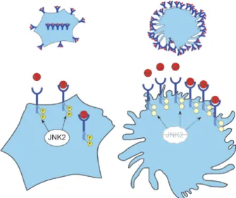
Figure 2. JNK2 promotes SR-A-mediated lipid internalization in macrophages. Left panel: JNK2 indirectly or directly phospho-rylates SR-A (receptor in blue), which allows internalization of receptor-bound lipoproteins (in red). Right panel: In the absence of JNK2, the receptor is less phosphorylated and is more abundant on the surface, allowing more binding of lipoproteins and increased adherence of macrophages. However, internalization is no longer effi cient.
play a minor role in the context of atherosclerosis even though compensatory functions of JNK1 and JNK2 could not be excluded in these cells. Overall, specifi c inhibition of JNK2 could be considered to prevent lesion develop-ment in humans.
Acknowledgement. We would like to thank our colleagues from our
previous and present laboratory for scientifi c contribution and dis-cussion. In particular we would like to thank Thomas F. Lüscher and Christian M. Matter for supporting our work in their laboratory, our collaborators for providing us with interesting results and technical support, especially to Izabela Sumara, Kathryn J. Moore, Mason W. Freeman, Myron I. Cybulsky, Jan Boren, Burkhard Becher and Erwin F. Wagner.
1 Kumar S., Boehm J. and Lee J. C. (2003) p38 MAP kinases: key signalling molecules as therapeutic targets for infl ammatory diseases. Nat. Rev. Drug Discov. 2: 717–726
2 Manning A. M. and Davis R. J. (2003) Targeting JNK for thera-peutic benefi t: from junk to gold? Nat. Rev. Drug Discov. 2: 554–565
3 Gupta S., Barrett T., Whitmarsh A. J., Cavanagh J., Sluss H. K., Derijard B. et al. (1996) Selective interaction of JNK pro-tein kinase isoforms with transcription factors. Embo. J. 15: 2760–2770
4 Davis R. J. (2000) Signal transduction by the JNK group of MAP kinases. Cell 103: 239–252
5 Kuan C. Y., Yang D. D., Samanta Roy D. R., Davis R. J., Rakic P. and Flavell R. A. (1999) The Jnk1 and Jnk2 protein kinases are required for regional specifi c apoptosis during early brain development. Neuron 22: 667–676
6 Sabapathy K., Jochum W., Hochedlinger K., Chang L., Karin M. and Wagner E. F. (1999) Defective neural tube morpho-genesis and altered apoptosis in the absence of both JNK1 and JNK2. Mech. Dev. 89: 115–124
7 Dong C., Davis R. J. and Flavell R. A. (2002) MAP kinases in the immune response. Annu. Rev. Immunol. 20: 55–72 8 Kennedy N. J. and Davis R. J. (2003) Role of JNK in tumor
development. Cell Cycle 2: 199–201
9 Liang Q. and Molkentin J. D. (2003) Redefi ning the roles of p38 and JNK signaling in cardiac hypertrophy: dichotomy between cultured myocytes and animal models. J. Mol. Cell. Cardiol. 35: 1385–1394
10 Peng J. and Andersen J. K. (2003) The role of c-Jun N-ter-minal kinase (JNK) in Parkinson’s disease. IUBMB Life 55: 267–271
11 Hunot S., Vila M., Teismann P., Davis R. J., Hirsch E. C., Przed-borski S. et al. (2004) JNK-mediated induction of cyclooxyge-nase 2 is required for neurodegeneration in a mouse model of Parkinson’s disease. Proc. Natl. Acad. Sci. USA 101: 665–670 12 Okazawa H. and Estus S. (2002) The JNK/c-Jun cascade and
Alzheimer’s disease. Am. J. Alzheimers Dis. Other Demen. 17: 79–88
13 Hirosumi J., Tuncman G., Chang L., Gorgun C. Z., Uysal K. T., Maeda K. et al. (2002) A central role for JNK in obesity and insulin resistance. Nature 420: 333–336
14 Jaeschke A., Rincon M., Doran B., Reilly J., Neuberg D., Greiner D. L. et al. (2005) Disruption of the Jnk2 (Mapk9) gene reduces destructive insulitis and diabetes in a mouse model of type I diabetes. Proc. Natl. Acad. Sci. USA 102: 6931–6935 15 Han Z., Boyle D. L., Chang L., Bennett B., Karin M., Yang L. et
al. (2001) c-Jun N-terminal kinase is required for metalloprotei-nase expression and joint destruction in infl ammatory arthritis. J. Clin. Invest. 108: 73–81
16 Han Z., Chang L., Yamanishi Y., Karin M. and Firestein G. S. (2002) Joint damage and infl ammation in c-Jun N-terminal kinase 2 knockout mice with passive murine collagen-induced arthritis. Arthritis Rheum. 46: 818–823
17 Nath P., Eynott P., Leung S. Y., Adcock I. M., Bennett B. L. and Chung K. F. (2005) Potential role of c-Jun NH2-terminal kinase in allergic airway infl ammation and remodelling: effects of SP600125. Eur. J. Pharmacol. 506: 273–283
18 Eynott P. R., Xu L., Bennett B. L., Noble A., Leung S. Y., Nath P. et al. (2004) Effect of an inhibitor of Jun N-terminal protein kinase, SP600125, in single allergen challenge in sensitized rats. Immunology 112: 446–453
19 Eynott P. R., Nath P., Leung S. Y., Adcock I. M., Bennett B. L. and Chung K. F. (2003) Allergen-induced infl ammation and airway epithelial and smooth muscle cell proliferation: role of Jun N-terminal kinase. Br. J. Pharmacol. 140: 1373–1380 20 Ricci R., Sumara G., Sumara I., Rozenberg I., Kurrer M.,
Akhmedov A. et al. (2004) Requirement of JNK2 for scavenger receptor A-mediated foam cell formation in atherogenesis. Sci-ence 306: 1558–1561
21 Glass C. K. and Witztum J. L. (2001) Atherosclerosis. the road ahead. Cell 104: 503–516
22 Nishio H., Matsui K., Tsuji H., Tamura A. and Suzuki K. (2001) Immunohistochemical study of the phosphorylated and activated form of c-Jun NH2-terminal kinase in human aorta. Histochem. J. 33: 167–171
23 Metzler B., Hu Y., Dietrich H. and Xu Q. (2000) Increased ex-pression and activation of stress-activated protein kinases/c-Jun NH(2)-terminal protein kinases in atherosclerotic lesions coin-cide with p53. Am. J. Pathol. 156: 1875–1886
24 Blankenberg S., Barbaux S. and Tiret L. (2003) Adhesion mol-ecules and atherosclerosis. Atherosclerosis 170: 191–203 25 Cybulsky M. I. and Gimbrone M. A. Jr (1991) Endothelial
ex-pression of a mononuclear leukocyte adhesion molecule during atherogenesis. Science 251: 788–791
26 Collins R. G., Velji R., Guevara N. V., Hicks M. J., Chan L. and Beaudet A. L. (2000) P-Selectin or intercellular adhesion molecule (ICAM)-1 defi ciency substantially protects against atherosclerosis in apolipoprotein E-defi cient mice. J. Exp. Med. 191: 189–194
27 Johnson R. C., Chapman S. M., Dong Z. M., Ordovas J. M., Mayadas T. N., Herz J. et al. (1997) Absence of P-selectin delays fatty streak formation in mice. J. Clin. Invest. 99: 1037– 1043
28 Dong Z. M., Chapman S. M., Brown A. A., Frenette P. S., Hynes R. O. and Wagner D. D. (1998) The combined role of P- and E-selectins in atherosclerosis. J. Clin. Invest. 102: 145–152 29 Liu J., Minemoto Y. and Lin A. (2004) c-Jun N-terminal protein
kinase 1 (JNK1), but not JNK2, is essential for tumor necrosis factor alpha-induced c-Jun kinase activation and apoptosis. Mol. Cell. Biol. 24: 10844–10856
30 Deng Y., Ren X., Yang L., Lin Y. and Wu X. (2003) A JNK-de-pendent pathway is required for TNFalpha-induced apoptosis. Cell 115: 61–70
31 Kamata H., Honda S., Maeda S., Chang L., Hirata H. and Karin M. (2005) Reactive oxygen species promote TNFalpha-induced death and sustained JNK activation by inhibiting MAP kinase phosphatases. Cell 120: 649–661
32 Min W. and Pober J. S. (1997) TNF initiates E-selectin tran-scription in human endothelial cells through parallel TRAF-NF-kappa B and TRAF-RAC/CDC42-JNK-c-Jun/ATF2 pathways. J. Immunol. 159: 3508–3518
33 Read M. A., Whitley M. Z., Gupta S., Pierce J. W., Best J., Davis R. J. et al. (1997) Tumor necrosis factor alpha-induced E-selectin expression is activated by the nuclear factor-kappaB and c-JUN N-terminal kinase/p38 mitogen-activated protein kinase pathways. J. Biol. Chem. 272: 2753–2761
34 Ahmad M., Theofanidis P. and Medford R. M. (1998) Role of activating protein-1 in the regulation of the vascular cell adhe-sion molecule-1 gene expresadhe-sion by tumor necrosis factor-al-pha. J. Biol. Chem. 273: 4616–4621
35 De Cesaris P., Starace D., Starace G., Filippini A., Stefanini M. and Ziparo E. (1999) Activation of Jun N-terminal kinase/ stress-activated protein kinase pathway by tumor necrosis factor
alpha leads to intercellular adhesion molecule-1 expression. J. Biol. Chem. 274: 28978–28982
36 Ioroi T., Yamamori M., Yagi K., Hirai M., Zhan Y., Kim S. et al. (2003) Dominant negative c-Jun inhibits platelet-derived growth factor-directed migration by vascular smooth muscle cells. J. Pharmacol. Sci. 91: 145–148
37 Zhan Y., Kim S., Yasumoto H., Namba M., Miyazaki H. and Iwao H. (2002) Effects of dominant-negative c-Jun on plate-let-derived growth factor-induced vascular smooth muscle cell proliferation. Arterioscler. Thromb. Vasc. Biol. 22: 82–88 38 Zhan Y., Kim S., Izumi Y., Izumiya Y., Nakao T., Miyazaki H.
et al. (2003) Role of JNK, p38, and ERK in platelet-derived growth factor-induced vascular proliferation, migration, and gene expression. Arterioscler. Thromb. Vasc. Biol. 23: 795– 801
39 Tournier C., Hess P., Yang D. D., Xu J., Turner T. K., Nimnual A. et al. (2000) Requirement of JNK for stress-induced activa-tion of the cytochrome c-mediated death pathway. Science 288: 870–874
40 Rao G. N. and Runge M. S. (1996) Cyclic AMP inhibition of thrombin-induced growth in vascular smooth muscle cells cor-relates with decreased JNK1 activity and c-Jun expression. J. Biol. Chem. 271: 20805–20810
41 Hansson G. K. (2005) Infl ammation, atherosclerosis and coro-nary artery disease. N. Engl. J. Med. 352: 1685–1695
42 Frostegard J., Ulfgren A. K., Nyberg P., Hedin U., Swedenborg J., Andersson U. et al. (1999) Cytokine expression in advanced human atherosclerotic plaques: dominance of pro-infl ammatory (Th1) and macrophage-stimulating cytokines. Atherosclerosis 145: 33–43
43 Constant S. L., Dong C., Yang D. D., Wysk M., Davis R. J. and Flavell R. A. (2000) JNK1 is required for T cell-mediated im-munity against Leishmania major infection. J. Immunol. 165: 2671–2676
44 Dong C., Yang D. D., Tournier C., Whitmarsh A. J., Xu J., Davis R. J. et al. (2000) JNK is required for effector T-cell function but not for T-cell activation. Nature 405: 91–94
45 Yang D. D., Conze D., Whitmarsh A. J., Barrett T., Davis R. J., Rincon M. et al. (1998) Differentiation of CD4+ T cells to Th1 cells requires MAP kinase JNK2. Immunity 9: 575–585 46 Dong C., Yang D. D., Wysk M., Whitmarsh A. J., Davis R. J.
and Flavell R. A. (1998) Defective T cell differentiation in the absence of Jnk1. Science 282: 2092–2095
47 Hansson G. K. (2001) Immune mechanisms in atherosclerosis. Arterioscler. Thromb. Vasc. Biol. 21: 1876–1890
48 Buono C., Binder C. J., Stavrakis G., Witztum J. L., Glimcher L. H. and Lichtman A. H. (2005) T-bet defi ciency reduces athero-sclerosis and alters plaque antigen-specifi c immune responses. Proc. Natl. Acad. Sci. USA 102: 1596–1601
49 Hansson G. K., Libby P., Schonbeck U. and Yan Z. Q. (2002) Innate and adaptive immunity in the pathogenesis of atheroscle-rosis. Circ. Res. 91: 281–291
50 Dansky H. M., Charlton S. A., Harper M. M. and Smith J. D. (1997) T and B lymphocytes play a minor role in atherosclerotic plaque formation in the apolipoprotein E-defi cient mouse. Proc. Natl. Acad. Sci. USA 94: 4642–4646
51 Daugherty A., Pure E., Delfel-Butteiger D., Chen S., Leferovich J., Roselaar S. E. et al. (1997) The effects of total lymphocyte defi ciency on the extent of atherosclerosis in apolipoprotein E–/– mice. J. Clin. Invest. 100: 1575–1580
52 Libby P. (2002) Infl ammation in atherosclerosis. Nature 420: 868–874
53 Osterud B. and Bjorklid E. (2003) Role of monocytes in athero-genesis. Physiol. Rev. 83: 1069–1112
54 Linsel-Nitschke P. and Tall A. R. (2005) HDL as a target in the treatment of atherosclerotic cardiovascular disease. Nat. Rev. Drug Discov. 4: 193–205
55 Castrillo A. and Tontonoz P. (2004) Nuclear receptors in mac-rophage biology: at the crossroads of lipid metabolism and infl ammation. Annu. Rev. Cell Dev. Biol. 20: 455–480
56 Li Y., Schwabe R. F., Devries-Seimon T., Yao P. M., Gerbod-Giannone M. C., Tall A. R. et al. (2005) Free cholesterol-loaded macrophages are an abundant source of TNF-alpha and IL-6. Model of NF-kappa B- and MAP kinase-dependent infl amma-tion in advanced atherosclerosis. J. Biol. Chem. 290: 21763– 21772
57 Li A. C. and Glass C. K. (2002) The macrophage foam cell as a target for therapeutic intervention. Nat. Med. 8: 1235–1242 58 Kunjathoor V. V., Febbraio M., Podrez E. A., Moore K. J.,
Andersson L., Koehn S. et al. (2002) Scavenger receptors class A-I/II and CD36 are the principal receptors responsible for the uptake of modifi ed low density lipoprotein leading to lipid load-ing in macrophages. J. Biol. Chem. 277: 49982–49988 59 Suzuki H., Kurihara Y., Takeya M., Kamada N., Kataoka M.,
Jishage K. et al. (1997) A role for macrophage scavenger recep-tors in atherosclerosis and susceptibility to infection. Nature 386: 292–296
60 de Winther M. P., Gijbels M. J., van Dijk K. W., van Gorp P. J., suzuki H., Kodama T. et al. (1999) Scavenger receptor defi ciency leads to more complex atherosclerotic lesions in APOE3Leiden transgenic mice. Atherosclerosis 144: 315–321 61 Babaev V. R., Gleaves L. A., Carter K. J., Suzuki H., Kodama
T., Fazio S. et al. (2000) Reduced atherosclerotic lesions in mice defi cient for total or macrophage-specifi c expression of scavenger receptor-A. Arterioscler. Thromb. Vasc. Biol. 20: 2593–2599
62 Herijgers N., de Winther M. P., Van Eck M., Havekes L. M., Hofker M. H., Hoogerbrugge P. M. et al. (2000) Effect of hu-man scavenger receptor class A overexpression in bone mar-row-derived cells on lipoprotein metabolism and atherosclerosis in low density lipoprotein receptor knockout mice. J. Lipid Res. 41: 1402–1409
63 Whitman S. C., Rateri D. L., Szilvassy S. J., Cornicelli J. A. and Daugherty A. (2002) Macrophage-specifi c expression of class A scavenger receptors in LDL receptor(–/–) mice decreases atherosclerosis and changes spleen morphology. J. Lipid Res. 43: 1201–1208
64 Post S. R., Gass C., Rice S., Nikolic D., Crump H. and Post G. R. (2002) Class A scavenger receptors mediate cell adhesion via activation of G(i/o) and formation of focal adhesion complexes. J. Lipid Res. 43: 1829–1836
65 Heider H. and Wintergerst E. S. (2001) Mimicking phosphor-ylation at Ser-48 strongly reduces surface expression of human macrophage scavenger receptor class A: implications on cell motility. FEBS Lett. 505: 185–190
66 Fong L. G. and Le D. (1999) The processing of ligands by the class A scavenger receptor is dependent on signal information located in the cytoplasmic domain. J. Biol. Chem. 274: 36808– 36816
67 Kosswig N., Rice S., Daugherty A. and Post S. R. (2003) Class A scavenger receptor-mediated adhesion and internalization require distinct cytoplasmic domains. J. Biol. Chem. 278: 34219–34225
68 Lusis A. J. (2000) Atherosclerosis. Nature 407: 233–241 69 Shin M., Yan C. and Boyd D. (2002) An inhibitor of c-jun
aminoterminal kinase (SP600125) represses c-Jun activation, DNA-binding and PMA-inducible 92-kDa type IV collagenase expression. Biochim. Biophys. Acta 1589: 311–316
70 Gum R., Wang H., Lengyel E., Juarez J. and Boyd D. (1997) Regulation of 92 kDa type IV collagenase expression by the jun aminoterminal kinase- and the extracellular signal-regulated ki-nase-dependent signaling cascades. Oncogene 14: 1481–1493 71 Crowe D. L., Tsang K. J. and Shemirani B. (2001) Jun
N-termi-nal kinase 1 mediates transcriptioN-termi-nal induction of matrix metal-loproteinase 9 expression. Neoplasia 3: 27–32
72 Crowe D. L. and Brown T. N. (1999) Transcriptional inhibition of matrix metalloproteinase 9 (MMP-9) activity by a c-fos/ estrogen receptor fusion protein is mediated by the proximal AP-1 site of the MMP-9 promoter and correlates with reduced tumor cell invasion. Neoplasia 1: 368–372
73 Westermarck J., Li S., Jaakkola P., Kallunki T., Grenman R. and Kahari V. M. (2000) Activation of fi broblast collagenase-1 expression by tumor cells of squamous cell carcinomas is medi-ated by p38 mitogen-activmedi-ated protein kinase and c-Jun NH2-terminal kinase-2. Cancer Res. 60: 7156–7162
74 Ruggeri Z. M. (2002) Platelets in atherothrombosis. Nat. Med. 8: 1227–1234
75 Song S., Freedman J., Mody M. and Lazarus A. H. (2000) Porcine von Willebrand factor and thrombin induce the acti-vation of c-Jun amino-terminal kinase (JNK/SAPK) whereas only thrombin induces activation of extracellular signal-related
kinase 2 (ERK2) in human platelets. Br. J. Haematol. 109: 851–856
76 Bugaud F., Nadal-Wollbold F., Levy-Toledano S., Rosa J. P. and Bryckaert M. (1999) Regulation of c-jun-NH2 terminal kinase and extracellular-signal regulated kinase in human platelets. Blood 94: 3800–3805
77 Cullen P., Baetta R., Bellosta S., Bernini F., Chinetti G., Cignarella A. et al. (2003) Rupture of the atherosclerotic plaque: does a good animal model exist? Arterioscler. Thromb. Vasc. Biol. 23: 535–542