REVIEW
The role of the inflammasome in cellular responses
to toxins and bacterial effectors
Barbara Freche&Núria Reig&F. Gisou van der Goot
Received: 25 May 2007 / Accepted: 6 July 2007 / Published online: 6 September 2007 # Springer-Verlag 2007
Abstract Invading pathogens are recognized by mammalian cells through dedicated receptors found either at the cell surface or in the cytoplasm. These receptors, like the trans-membrane Toll-like Receptors (TLR) or the cytosolic Nod-like Receptors (NLR), initiate innate immunity after recognition of molecular patterns found in bacteria or viruses, such as LPS, flagellin, or double-stranded RNA. Recognition of molecules produced only by a specific pathogen, such as a viral envelop protein or a bacterial adhesin does not appear to occur. Bacterial protein toxins, however, might compose an interme-diate class. Considering the diversity of toxins in terms of structure, it is unlikely that cells respond to them via specific molecular recognition. It rather appears that different classes of toxins trigger cellular changes that are sensed by the cells as danger signals, such as changes in cellular ion composition after membrane perforation by pore-forming toxins or type III secretion systems. The signaling pathways triggered through toxin-induced cell alterations will likely play a role in modulating host responses to virulent bacteria. We will here describe the few studied cases in which detection of the toxin by the host cell was addressed. The review will include not only toxins but also bacteria effectors secreted by the bacterium in to the host cell cytoplasm.
Keywords Toxins . Inflammasome . Pore-forming . Caspase-1 . Effector . Type III secretion
During infection, pathogenic microbes actively modify the behavior of host cells to allow them to withstand the rigors of the host environment and establish a suitable replication niche. Changes in host cell properties and behavior include modification of plasma membrane permeability, induction, or inhibition of phagocytosis or apoptosis, depending on the pathogen and the host cell type. These changes result from the action of bacterial proteins that have either membrane interacting activity (pore-forming toxins or PFTs, lipases) or an enzymatic activity (protease, phosphatase, ubiquitin ligase, glucosidase, ADP ribosyltransferase, ...)
[4, 16, 50]. These activity-bearing bacterial proteins are
either secreted into the extracellular medium and called exotoxins or toxins, or they can be injected into the host cell cytoplasm through specialized bacterial secretion machin-eries and are then called effectors. In this review we will use the term toxin in its largest sense including both exotoxins and effectors.
Toxins are produced by many bacterial pathogens, whether gram positive or negative, and contribute to, and are sometimes essential for, virulence. Efforts over the last decades have been focused on determining the activity and targets of toxins and how these toxins affect host cell function and behavior. More recently, a novel concern has appeared in the cellular microbiology field, which is to understand whether cells sense the presence or effects of toxins and whether they mount a response, even in the case of nonimmune cells. After a brief introduction on toxins and the mode of delivery of bacterial proteins into the mammalian cytoplasm, we will discuss recent findings that indicate that the effects of toxins can be sensed and that a response is mounted that depends on the nature and amplitude of the toxic insult as well as the type of affected cell.
B. Freche
:
N. Reig:
F. G. van der Goot (*) Global Health Institute,Ecole Polytechnique Fédérale de Lausanne, Station 15,
1015 Lausanne, Switzerland e-mail: [email protected]
The bacterial toolkit
Bacterial toxins and effectors are generally either enzymes or pore-forming proteins, notable exceptions are the so-called super-antigens, which induce massive unspecific
T-cell activation [4]. Note that many pathogenic bacteria
will produce more than one toxin and for those harboring a type 3 secretion system (T3SS), always multiple effectors,
sometimes near to 20 different ones [72]. While novel
enzymatic activities are continuously reported, the list of
characterized activities is already extremely long (Table1),
including activities as diverse as ADP-ribosyltransferase
such as cholera toxin [39], phosphatidyl inositol-4,
5-phosphatase as Salmonella SopB [30], MAP kinase
kinases acetylase as Yersinia YopJ [52], and MAP kinase
phosphatase as Shigella OspF [6].
Pore-forming toxins
In the context of this review, an important class of toxins is that of pore-forming toxins, which compose approximately a third of all characterized bacterial toxins and are produced by both
Gram-positive and Gram-negative bacteria [7,55]. Most PFTs
share the following mode of action: they are released by the bacteria in a soluble, generally monomeric, form that freely diffuses towards target cells. Host cell binding occurs by specific interactions with cell surface molecules that can be either lipids, such as for Listeria monocytogenes listeriolysin O (LLO) or Bacillus thuringiensis crystal toxin; proteins, such as for Streptoccus intermedius Intermedilysin or carbohydrates bound to proteins, such as for Aeromonas
hydrophila aerolysin [7,23]. Once bound to its receptor, the
toxin undergoes circular polymerization into ring-like struc-tures, the stoichiometry of which varies amongst toxins. The amphipathic nature of these complexes leads to spontaneous insertion into the membrane. Depending on the toxin, the pore
diameter varies from 1 up to 50 nm [7,55]. PFTs are generally
produced by bacteria that remain extracellular, and thus pore-formation occurs at the plasma membrane. Some bacteria that replicate intracellularly, such as L. monocytogenes and Group A Streptococci, however, also produce PFTs, listeriolysin O (LLO), and streptolysin O (SLO), respectively. Although LLO will form pores in the plasma membrane upon secretion by extracellular Listeria, it is absolutely required for the bacterium to breach the phagosomal membrane allowing
escape into the cytoplasm [16].
Delivery of bacterial proteins into the host cell cytoplasm
Except for lipases, the targets of all the bacterial toxins with enzymatic activity mentioned above reside in the host
cytoplasm. Thus the first challenge of these molecules is to get across the plasma membrane, an event that is impossible for a soluble protein if not assisted. Evolution has led to two strategies that will be detailed below: (1) the development of a bacterial injection system that brings the enzyme directly from the bacterial cytoplasm to that of the host, (2) the production of a pore-forming protein that escorts the enzyme to the cytoplasm, mediating both host cell recognition and transloca-tion of the enzyme across membranes.
Type III and IV secretion machineries
Some Gram-negative bacteria inject their toxins into the cytosol of host cells through bacterial transport machines
that function as macromolecular syringes [50]. The best
characterized is the type III secretion system (T3SS). T3SSs consist of three parts: a cytoplasmic bulb, a region spanning both the inner and the outer bacterial membranes, and a
hollow ‘needle’ protruding from the bacterial surface. The
needle is involved in sensing the presence of host cells and triggers the so-called contact mediated secretion, via mechanisms that remain unclear. Transport of effectors is thought to proceed through the central 25-Å channel of the needle. Access to the host cytoplasm, however, also requires perforation of the eukaryotic plasma membrane. This occurs through the secretion of early effectors such as IpaB for Shigella flexneri or SipB for Salmonella that have a pore-forming capacity and puncture the membrane at the tip of the secretion needle, thus creating a continuous conduit from the bacterial cytoplasm to that of the host. This secretion machinery is thought to have evolved from the flagellar export system.
Whereas T3SSs are the most widely spread secretion systems amongst pathogenic bacteria, some Gram-negative bacteria express a somewhat different secretion apparatus
termed the Type IV secretion system (T4SSs) [18]. T4SSs
were first identified in plant pathogens but have since been identified in Legionella or Brucella, for which they are crucial to establish infection. The interesting additional property of T4SSs is that they have been shown to transport DNA, in addition to proteins, during infection of plants. Injection of bacterial DNA into mammalian cells has not been documented. Please note that type III and IV secretion systems are solely devoted to introducing bacterial proteins in the host cell cytoplasm. Bacterial secretion of exotoxins such as pore-forming toxins relies on other secretion apparatuses such as type I and type II secretion systems.
Needle type injection machineries have not been found in Gram-positive bacteria. It has, however, been shown that Streptococcus pyogenes utilizes a Cytolysin-mediated translocation (CMT) to introduce an effector protein of
streptococcal origin into the cytoplasm of a host cell [40].
T able 1 T oxins and type III secretion ef fectors mentioned in this review T oxin Or ganism Receptor Cell delivery Activity Cellular outcome Cellular sensor Anthrax toxin (Baldari et al. [ 8 ]) Bacillus anthracis TEM8, CMG2 T oxin induced endocytosis, cytosol delivery from late endosomes 2 catalytic subunits: EF (calmodulin dependent adenylate cyclase) and LF (metalloprotease that cleaves MAPKK) Increase cAMP , impairment MAPK signaling Nalp1b mediated Caspase-1 activation Cholera toxin (Lencer and T sai [ 39 ]) V ibrio cholerae ganglyoside GM1 T oxin induced endocytosis, cytosol delivery from ER ADP-ribosyltransferase of stimulatory Gs! subunit of heterotrimeric G-proteins Increase cAMP , activation of Cl-channels, water ef flux Not known Diphtheria toxin (Collier [ 14 ]) Corynebacterium diphtheriae HB-EGF T oxin induced endocytosis, cytosol delivery from early endosomes ADP-ribosyltransferase of EF-2 Inhibition of protein synthesis Not known T etanus toxin (T urton et al. [ 74 ]) Clostridium tetani gangliosides from presinaptic membranes T oxin induced endocytosis, retrograde transport along neuron axon metaloprotease of synaptic SNARE proteins Inhibition of neurotransmitor release Not known Botulinum toxin (T urton et al. [ 74 ]) Clostridium botulinum gangliosides from presinaptic membranes T oxin induced endocytosis, traf fic to small presinaptic vesicles metaloprotease of synaptic SNARE proteins Inhibition of neurotransmitor release Not known Ef fectors SopB/SigD (Hernandez et al. [ 30 ]) Salmonella sp via T3SS Phosphatidil inositol phosphatase Sustained activation of AKT (antiapoptotic activity), alteration vesicle traf ficking Not known Y opJ (Mukherjee et al. [ 52 ]) Y ersinia pestis via T3SS Acetyltransferase that modifies MAPKK and IKK Impairment MAPK and NFKb signaling pathways Not known OspF (Arbibe et al. [ 6 ]) Shigella flexneri via T3SS MAPK phosphatase Prevents histone H3 phosphorilation, impairs NFKb signaling Not known IpaB (Chen et al. [ 12 , 13 ]) Shigella flexneri via T3SS structural protein of T3SS Cellular delivery of bacterial ef fector through T3SS pore Caspase-1 activation by direct binding? SipB (Hersh et al. [ 31 ]) Salmonella thypimurium via T3SS structural protein of T3SS Cellular delivery of bacterial ef fector through T3SS pore Caspase-1 activation by direct binding? Y opB, Y opD, LcrV (Shin et al. [ 69 ]) Y ersinia enter ocolitica via T3SS structural proteins of T3SS Potassium ef flux Caspase-1 activation PFT s Listeriolysin O (LLO) (Mariathasan et al. [ 44 ]) Listeria monocytogenes lipid rafts Membrane pore formation Phagosomal escape of endocytosed bacteria and ion ef flux Nalp3 inflammasome
T able 1 (continued) T oxin Or ganism Receptor Cell delivery Activity Cellular outcome Cellular sensor Crystal toxins (Cry1 1Aa, Cry1A, Cry5B) (Grif fitts et al. [ 25 ]) Bacillus thuringiensis GPI-APs, lipid rafts, glycolipids Membrane pore formation Ion ef flux Not known Intermedilysin (Giddings et al. [ 24 ]) Str eptococcus intermedius CD59 Membrane pore formation Ion ef flux Not known Aerolysin (Gurcel et al. [ 27 ]) Aer omonas hydr ophila
GPI-Aps (carbohidrate moiety)
Membrane pore formation Potassium ef flux Nalp3 and Ipaf inflammasomes Streptolysin O (SLO) (Kannenganti et al. [ 37 ]) Str eptococcus pyogenes Cholesterol (lipid rafts) Membrane pore formation Ion ef flux and delivery of P AMPs into the cytosol Nalp3 inflammasome α -toxin (W alev et al. [ 79 ]) Staphilococcus aur eus lipid clusters Membrane pore formation Ion ef flux
Nalp3 inflammasome? Caspase-1 activation
Non protein toxins Nigericin (Mariathasan et al. [ 44 ]) Str eptomyces hygr oscopicus Cation selective channel Potassium ef flux Nalp3 inflammasome V alinomycin (Gurcel et al. [ 27 ]) Str eptomyces sp Cation selective channel Potassium ef flux Nalp3 inflammasome Maitotoxin (Mariathasan et al. [ 44 ]) Gambier discus toxicus Cation selective channel Potassium ef flux Nalp3 inflammasome
makes large pores which allow the passage of proteins. It was found that the S. pyogenes NAD-glycohydrolase involved in virulence is translocated into the host cell via the SLO pore. The model proposes that translocation, which might involve contact between the bacterium and the host, is coupled to polarized secretion, as is supported by the fascinating observation that S. pyogenes has a special-ized microdomain in its plasma membrane, positioned at a hemispherical position distal to either cell pole, termed the
ExPortal, which contains the Sec translocons [63].
AB type toxins
Toxins of the AB type are composed either of multiple domains or multiple independent polypeptides. The A subunit bares the enzymatic activity, while the B subunit mediates binding to the target cell membrane, trafficking within the eukaryotic target cell and for many AB toxins, translocation of the A subunit across cellular membranes. Translocation never occurs across the plasma membrane. Therefore the toxin is internalized by the cell, via pathways that are different from one toxin to another, and transported to a final destination, which can be early endosomes, late endosomes, or the endoplasmic reticulum, depending again on the toxin, from which translocation occurs. Whereas translocation at the ER level is thought to be mediated by the cellular translocon, transport at the level of endosomes is mediated by the B subunit, which has pore-forming
activity [2, 61, 64]. Toxins of the AB family include
Cholera toxin, diphtheria toxin, and anthrax toxin (Table1),
for which we will detail the mode of action because it is most relevant to the present review.
Anthrax toxin is composed of three independent poly-peptide chains: PA (protective antigen), EF (edema factor),
and LF (lethal factor) [2,68]. EF is a calmodulin-dependent
adenylate cyclase thus leading to the increase in c-AMP in intoxicated cells. LF is a metalloprotease that specifically cleaves MAP kinase kinases such as MEK1 or MKK3. Finally, PA corresponds to the B subunit. PA is not only responsible for binding to the cell surface receptors, but it also mediates binding of the enzymatic subunit, EF and LF, transport into the endocytic pathway and translocation into the cytoplasm. To mediate translocation, PA forms a heptameric channel, very similar to that of pore-forming toxins. Channel formation, however, only occurs at acidic pH, thus requiring transport to an endosomal compartment. Due to the ubiquitous nature of the anthrax toxin receptors,
Capillary morphogenesis gene 2 (CMG-2) [67] and Tumor
endothelial marker 8 (TEM-8) [10], most if not all cells are
sensitive to MAP kinase kinase cleavage by anthrax toxin. However, in terms of pathogenesis, the effect on cells of the
immune system is most relevant [8]. It was in particular
shown that lethal toxin, i.e., the combination of PA and LF,
leads to impaired antigen presentation and synthesis of stimulatory molecules by macrophages and dendritic cells, and for certain genetic backgrounds, death, impaired
activa-tion of T cells, and inhibiactiva-tion of B-cell proliferaactiva-tion [8].
Sensing bacterial toxins
For decades, scientists have been studying the effect of bacterial toxins on mammalian cells. The observed effects where either directly linked to the enzymatic activity of the toxin, such as the inhibition of neurotransmitter release in tetanus toxin treated cells due to the toxin-mediated proteolytic cleavage of proteins involved in synaptic vesicle
fusion (Table 1), or related to a somewhat unexplained
cellular effect, such as activation of the NF-κB pathway. Recently, a different way of addressing the effects of toxins has emerged, which is to investigate whether cells are able to sense toxins, or their effects, and whether they respond to
the toxic insult [27,29,35,36,60].
Some recent studies indicate that cells can sense the effect of certain toxins, as will be detailed below. The field is, however, still in an embryonic state and, therefore, very few examples will be presented. The notion of sensing toxins is of course reminiscent of that of sensing bacterial pathogens, a field of intense research since the discovery a decade ago that Toll-like receptors (TLR) play an important role in innate
immunity [38]. The concept that emerged from this work is
that through out evolution from flies to mammals, organisms produce microbial sensing proteins that recognize conserved and often structural components of the microorganisms,
called pathogen-associated molecular patterns (PAMPs) [3].
Bacterial cell wall components such as peptidoglycan, bacterial flagellin, and nucleic acid structures, unique to bacteria and viruses, are examples of PAMPs. The first class of these pattern-recognition molecules that were identified were the TLRs, which are transmembrane proteins involved in detection of microbes in the extracellular compartment
and in endosomes (for review, see [3]). The field of innate
immunity has become even more intense since the discovery of a family of cytoplasmic PAMP detectors, which in analogy to the TLRs have been termed NLRs, or Nod-like
receptors [46].
NLRs are multidomain proteins that, similar to TLRs, contain a leucine-rich repeat (LRR) domain thought to be involved in the sensing of PAMPs, a nucleotide-binding and self-oligomerization domain termed NACHT domain, and one or more effector binding domains that can be either caspase recruitment domains (CARD) or pyrin effector
domains PYD (for review, see [45, 46, 73]). NLRs
comprise two large subclasses, the CARD-containing nucleotide oligomerization domain or NODs (NOD1-5) and the PYD-containing domain or Nalps
(NACHT-LRR-PYD-containing protein) group (Nalp1 to Nalp14). For detailed review on NLRs, their structure, regulation, and ligands, the reader is referred to other reviews in this issue. As for TLRs, NLRs are thought to recognize molecular patterns frequently encountered in microorganisms, such as flagellin or fragments of peptidoglycan. As for TLRs, formal evidence that binding is direct is still missing with the exception of Nalp1, for which direct activation by muramil dipeptide, a fragment of proteoglycan has been
reconstituted in vitro with purified molecules [19]. The
principle of NLR function is that the molecule is in an autorepressed state, possibly through intramolecular do-main interaction, in the absence of stimulus. Upon ligand binding, the molecule opens up rendering the NACHT domain accessible for homo or hetero oligomerization with other NACHT-containing proteins and the CARD and/or PYR domains accessible for the interaction with down stream effectors and/or adaptor proteins. The structural opening of the NLRs upon ligand binding thus induces the assembly of large multiprotein complexes. The two major documented down stream effects of NLR activation are NF-κB activation and caspase-1 activation, events that involve different NLR members. In this paper, we will only discuss NLRs involved in caspase-1 activation.
The NLR containing multiprotein complexes involved in caspase-1 activation have been termed, by Tschopp and
co-workers, the inflammasome [45]. Caspase-1 is indeed a key
player in the inflammatory response as this cystein protease is responsible for the processing of the pro-forms of interleukins 1β 18 and 33, crucial pro-inflammatory cytokines. To date, three different inflammasomes have been described
contain-ing Nalp1, Nalp3, and Ipaf (also a NLR family member),
respectively (see Fig. 1). Interactions of these NLRs with
caspase-1 may be either direct, as proposed for Ipaf (Fig.1),
or require the adaptor protein ASC (Apoptosis-associated
Speck-like protein containing a CARD) [46].
As apparent above, both TLRs and NLRs are involved in the detection of common features amongst pathogenic organisms (peptidoglycan, flagellin, ...). As toxins, even within a given family, differ greatly in terms of sequence and even structure, it seems rather unlikely that either TLRs or NLRs would detect the presence of a toxin per se. More likely, if involved, these sensing devices should detect the consequen-ces of the toxins action, which would be similar for toxins with similar activities, enzymatic, or membrane impairing. We will focus on three topics: pore-forming toxins (PFTs)—the largest class of bacterial toxins—anthrax toxin, and finally the type III and IV secretion systems.
Inflammasome activation by pore-forming toxins
Pore formation leads to permeabilization of the plasma membrane to ions, and in particular to calcium and potassium, along their electrochemical gradients, leading to changes in cytoplasmic ion composition. The large pores formed by certain PFTs as the cholesterol dependent toxins LLO, SLO, and pneumolysin allow also the efflux of
cytoplasmic proteins [32, 75]. As a consequence of pore
formation by Staphylococcal α toxin, aerolysin, and LLO,
cells of the immune system such as lymphocytes or dendritic cells were found to produce numerous
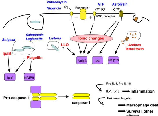
inflamma-Fig. 1 Schematic diagram of the various inflammasome that have been documented to as-semble in response to bacterial toxins or effectors of type III secretion machineries
tory molecules and also to undergo apoptosis [7]. The first evidence that PFTs might activate the inflammasome was the observation that alpha-toxin from Staphylococcus aureus induces the release of IL-1β in LPS pretreated
monocytes [79]. In these early days, it was already
suggested that alpha-toxin triggers the activation of Inter-leukin-converting enzyme or ICE, the former name of caspase-1. Interestingly, these authors found that high extracellular potassium would prevent toxin induced IL-1β secretion, indicating the requirement for a decrease in intracellular potassium.
More recently, it was found that aerolysin from A. hydrophila, a toxin that shares the same mode of action as Staphylococcal alpha toxin and makes pores of similar size
[1,28], also triggers the activation of caspase-1 [27]. With
the increased general knowledge about caspase-1 activa-tion, it was shown that aerolysin induced activation of caspase-1 involves both Nalp3 and Ipaf, in a potassium efflux dependent manner. Caspase-1 activation via the Nalp3 pathway also required the adaptor protein ASC
(Fig. 1). Infection of primary human fibroblasts with A.
hydrophila also triggered caspase-1 activation, in contrast
to the isogenic aerolysin deficient strain [27]. In contrast,
Nalp3-dependent caspase-1 activation was still observed in S. aureus deficient in either alpha, beta, or gamma toxin suggesting that absence of one of the multiple PFTs produced by this bacterium is insufficient to abrogate
caspase-1 activation [44]
Thus one of the sensing events that could be common to all PFTs would be the decrease in cytoplasmic potassium (changes in other ions could also be sensed, possibly by other NLRs), which would trigger caspase-1 activation. Potassium ionophores such as nigericin from Streptomyces hygroscopicus
[44] and valinomycin [27] have also been found to trigger
activation of caspase-1 via Nalp3 inflammasome. In vitro studies have recently shown that NALP inflammasome assembly and caspase-1 recruitment can actually occur
spontaneously at K+concentrations below 90 mM [59]
Potassium efflux can also be triggered by the activation of
the purinergic P2X7 receptor by ATP, which has long been
used as a means to activate caspase-1 [58]. Activation of the
receptor-linked cation selective channel triggers a series of events that are not fully understood. First opening of potassium selective channels occurs, followed by the opening of a larger pore, corresponding to the hemichannel
pannexin-1 [56]. Surprisingly, pannexin-1 was recently
shown to also be crucial for nigericin induced caspase-1
activation as well as that induced by maitotoxin [57].
Maitotoxin is a potent marine nonprotein toxin isolated from
the dinoflagellate Gambierdiscus toxicus [80] for which the
exact mode of action has not been characterized. This highly
cyclic molecule [53] (actually the largest nonprotein
mole-cule known!) first triggers an increase in cytoplasmic
calcium, followed by the opening of medium size pores that allow the passage of <800 Da molecules, finally followed by
the formation of large “death channels.” Combined, these
studies suggest that the initial change in cytoplasmic ion composition triggered by nigericine or maitotoxin is neces-sary but not sufficient to trigger caspase-1 activation, which requires pannexin-1, although apparently not through its
channel-forming activity [57]. Additional studies will be
required to clarify the role of pannexin. It will also be of interest to test whether caspase-1 activation induced by Staphylococcal alpha toxin and aerolysin also involves pannexin-1.
Pannexin-1 channels are reminiscent of the PFTs that form large pores such as SLO and listeriolysin O (LLO), in that they allow the passage of proteins. The similarity was recently extended by the observation that both ATP-induced opening of pannexin-1 channels and SLO allowed the cytosolic access of various PAMPs, such as LPS, synthetic lipid A, lipoteichoic acid, or peptidoglycan, leading to the
activation of caspase-1 [37]. LLO was also recently found
to be implicated in the caspase-1 activation, via the Nalp3
and ASC inflammasome by L. monocytogenes [44,54]. L.
monocytogenes is a Gram-positive facultative intracellular pathogen that can cause life-threatening infections in neonates and immunocompromised hosts. The bacterium
enters into cells via a clathrin-mediated pathway [78] and
then escapes into the cytoplasm through LLO mediated
rupture of the phagosomal membrane [16]. Macrophages
cultured with L. monocytogenes deficient in LLO failed to
secrete IL-1β [44,54]. It is however not clear at what step
LLO pore formation was involved in caspase-1 activation: pore formation in the plasma membrane, as recently
suggested [71] or in the phagosome? Was cytoplasmic
release of the bacterium the required event? In other words, was inflammasome assembly triggered by changes in ion composition or by the presence of PAMPs or both?
Involvement of NLRs in sensing and responding to anthrax toxin
The second toxin to which cells seem to respond in an inflammasome dependent manner is anthrax toxin. Macro-phages treated with anthrax lethal toxin were found to
secrete IL-1β and IL-18 [9, 15]. Direct evidence for the
lethal toxin induced activation of caspase-1 was recently obtained by Boyden and Dietrich and confirmed by others
([51], Reig et al., submitted). Searching for an anthrax
susceptibility gene in mouse macrophages, Boyden and Dietrich found that Nalp1b was involved in lethal toxin induced cell death in mouse macrophages, in a genetic background dependent manner. These authors found that the gene encoding for Nalp1b is highly polymorphic, with 5
alleles identified among 18 mice strains. Allele 1, found for example in Balb/cJ, C3H/HeJ, and 129S1/svImJ mice, was found to encode a Nalp1b protein (termed type 1 Nalp1b) that was able to mediate caspase-1 activation in response to lethal toxin. In contrast, allele 2, found in C57B6 mice, and allele 4, found in DBA/2J mice, encodes a Nalp1b protein that is unable to activate caspase-1 in response to LT (termed type 2 Nalp1b). Whether ASC is required for the type 1 Nalp1b mediated activation of caspase-1 requires further investigation.
The mechanisms that leads to activation of Nalp1b in response to lethal toxin are unclear. It is unlikely, at least for two reasons, that the lethal factor is itself recognized by Nalp1b as a PAMP. As mentioned previously, considering the diversity of bacterial proteins that reach the cytoplasm, it is unlikely that there would be proteins to sense them, the number of sensing proteins would have to be too great. Secondly, the catalytic activity of lethal factor is required to trigger caspase-1 activation, as a translocation competent but catalytically inactive LF was unable to trigger caspase-1 activation, despite its delivery to the cytoplasm (Reig et al., submitted). Sensing thus appears to occur down stream of MAPK kinase cleavage by LF. Determining the sensed events or component will be of great interest.
Bacterial secretion machineries and the inflammasome
A variety of Gram-negative bacteria, such as Salmonella and Shigella, that enter and multiply within cells absolutely require their T3SSs for invasion. Upon infection of macro-phages, both these bacteria induce robust caspase-1 activa-tion, which leads to the release of IL-1β and IL-18 followed
by caspase-1 mediated death [12, 31, 34, 65, 77]. The
proteins Salmonella SipB and Shigella IpaB, which mediate puncturing of the host cell membrane upon Salmonella and Shigella invasion, respectively, seem to have a direct role in caspase-1 induced cell death as microinjection of recombinant SipB or IpaB protein is sufficient to induce apoptosis in
macrophages [13,31]. Furthermore, Salmonella and Shigella
strains defective in IpaB or SipB do not induce macrophage
apoptosis [31,34]. Although direct binding of IpaB and SipB
was thought to activate caspase-1 [31,34], more recent data
suggest that SipB has a more indirect role in caspase-1
activation during infection with Salmonella sp. [22, 47].
Since in the absence of these proteins, none of the other effectors can reach the cytosol, it is unclear whether inflammasome activation is triggered by the secretion system, or some of the effectors, or PAMPs that gain access through IpaB or SipB channels. It was recently shown that the hydrophobic translocators of Yersinia, YopB and YopD, induce caspase-1 activation and IL-1β secretion upon
infection with strains that are deficient in effectors [69].
These observations highlight the importance of membrane impairment in triggering inflammation. Interestingly upon infection with a Yersinia wild type strain, caspase-1 activation did not occur, suggesting that the effectors neutralize the pro-inflammatory phenomenon of the translocator either by inhibiting caspase-1 activation through the GTPase activity of the effector protein YopE (Schotte, Denecker et al. 2004)
or possibly by clogging the translocon [69].
Infection by S. typhymurium and S. flexneri, however, appears to trigger the activation of more than one type of inflammasome as both ASC and Ipaf were found to be
involved in activating caspase-1 [43]. Only the Ipaf
inflammasome was, however, essential for caspase-1
dependent cell death [42]. Although still requiring SipB,
S. typhimurium flagellin was found to trigger Ipaf mediated caspase-1 activation as flagellin deficient Salmonella did
not activate caspase-1 [22, 47, 62]. Furthermore, Ipaf
activation could be recapitulated by the introduction of purified flagellin directly in the cytoplasm. Altogether, these studies indicate that bacteria may trigger caspase-1 activation via more than one pathway.
Activation of caspase-1 has also been observed for other Gram-negative such as the facultative intracellular pathogen Burkholderia pseudomallei, which harbors a T3SS highly homologous to that of S. typhymurium and S. flexneri. B. pseudomallei is the causative agent for meliodosis, an infection that results in a wide spectrum of clinical outcomes including asymptomatic, acute or chronic
pneu-monia, or septicemia [11]. B. pseudomallei infected
macro-phages where found to undergo caspase-1 dependent cell death. The inflammasome components mediating cell death were however not determined, nor was the role of BipB, the Burkholderia homologue of IpaB.
Finally, Legionella pneumophila, a T4SS harboring Gram-negative intracellular pathogen was also found to
trigger caspase-1 activation [82]. Activation was strictly
dependent of Dot-Icm type IV secretion system and led to the assembly of two NLR containing complexes: a Birc1e (Naip5) and an Ipaf inflammasome that were sensitive to
bacterial flagellin [5,62].
Interplay between TLRs and inflammasomes
Activated caspase-1 is essential for the processing and release of biologically active IL-1β and IL-18, two proinflammatory cytokines with critical roles in innate and adaptive immunity
[17]. The secretion of IL-1β is also dependent on the levels
of IL-1β induced at the transcriptional level by pro-inflammatory stimuli including several TLR ligands such as lipopolysaccharide (LPS), lipoteichoic acid (LTA), CpG oligonucleotides, the antiviral compound resiquimod R848,
transduced by TLRs for the induction of pro-IL-1β expres-sion is likely mediated by the transcription factor NF-κB. The cytokine IL-18 is also synthesized as an inactive precursor, but, unlike pro-IL-1β, it is expressed constitutively. Despite the importance of TLR activation in pro-IL-1β synthesis, the relationship between TRL and inflammasome signaling remains poorly understood.
Several lines of evidence indicate that caspase-1 activation does not depend on TLR signaling. It has indeed been shown that inhibition of TLR signaling, for example, upon knock out of MyD88 or Toll/IL-1 receptor (TIR)-domain-containing adaptor-inducing IFN-β (TRIF), two adaptor molecules that are critical for TLR mediated NF-κB activation, up-regulation of pro-IL-1β does not occur but processing of caspase-1 is
unaltered [37,54,81]. In contrast, recent data now indicate
that TLR signaling can be regulated by caspase-1. Upon ligand activation of TLRs, cytosolic TIR domain-containing
adapter proteins are recruited to initiate signaling events [48].
Among them, the MyD88 adaptor-like (MAL) protein is required for signaling by the LPS receptor TLR4 and the bacterial lipoprotein receptor TLR2 and acts as a bridging
adapter for MyD88 recruitment [48]. Recent results reveal
that MAL requires cleavage by caspase-1 to elicit its
downstream signal NF-κB [49]. Consequently, signaling by
TLR2 and TLR4 is impaired in caspase-1 deficient cells. NF-κB activation via Receptor Interacting Protein-2 (RIP2) was
also found to involve caspase-1 [66]. In this case also, NF-κB
activation by LPS was attenuated in caspase-1 deficient macrophages and was inhibited by a catalytically inactive form of caspase-1. Furthermore, the authors point out a
possible regulatory role of ASC in the communication between the inflammasome and the NF-κB pathway.
Over-expression of ASC led to a decrease in NF-κB activity,
whereas ASC knock down led to an increase in NF-κB activity suggesting an inhibitory role of ASC in NF-κB signaling. These findings shed light on an important mechanism for the coordinated activation of host defense responses by TLRs and the inflammasome.
At present, there is no evidence that toxins trigger TLRs. Il-1β secretion upon exposure to pore-forming toxins or anthrax toxin might be due to the synergistic effect of bacterial PAMPs and the toxins. Cellular damage due to bacterial toxins, in particular, PFTs, could also lead to the release of endogenous TLR ligands such as heat shock
proteins [76] or other danger signals.
Consequences of toxins induced caspase-1 activation
The consequences of toxin-induced caspase-1 activation appear to be strongly cell type dependent. In cells of the immune system such as macrophages, the most striking consequence of caspase-1 activation by either the pore-forming effectors of T3SS harboring bacteria or the totally unrelated anthrax toxin is cell death, in addition to processing
and secretion of IL-1β and IL-18 [9, 13, 31, 33]. The
underlying mechanisms, however, remain completely obscure and probably involve yet to be identified targets of caspase-1
(Fig.2). Caspases-2, 3, and 11 do not seem to be involved in
this process, and Bcl-2 overexpression does not block Valinomycin Nigericin P2X7receptor ATP Aerolysin K+ K+ Pannexin-1 pore K+ Ipaf Nalp3 Ionic changes
+
+
Anthrax lethal toxin Nalp1b Flagellin NAIP5 Ipaf Salmonella Legionella IpaB Shigella LLO ? Listeria Pro-caspase-1 caspase-1 Pro-IL-1, Pro-IL-18IL-1, IL-18 Inflammation Unknown targets
Macrophage death Survival, other
effects...
Fig. 2 Schematic representation of bacterial toxins and effectors that activate NLRs, trigger the assembly of inflammasomes and the activation of caspase-1
Shigella-induced cell death in macrophages [34]. Several lines of evidence suggest that caspase-1 induced cell death is mechanistically distinct from the forms of classical apoptosis
and was therefore termed pyroptosis [20]. An interesting
event described in pyroptosis is the caspase-1 dependant formation of plasma membrane pores, between 1.1 and 2.4 nm in diameter, that appear concurrently to the release
of processed IL-1β and IL-18 and precede cell death [21].
These pores also dissipate ionic gradients leading to water uptake and cell swelling, ultimately culminating in osmotic lysis with release of inflammatory cellular contents. Despite of their difference in size, these membrane pores are
reminiscent of the pannexin-1 pore mentioned above [56].
An interesting aspect of caspase-1 mediated cell death in response to anthrax lethal toxin, and presumably others to come, is that cell death is not a direct consequence of the toxins action, as illustrated by the fact that nonimmune cells survive toxin action for days (unpublished observations). Rather, death of immune cells is due to the innate response of the cells to the action of the toxin. This theme is reminiscent of what has been observed in infection where tissue damage is often the results of a massive immune response rather that the action of the bacterium itself.
Many toxins however target nonimmune cells such as epithelial cells. This is in particular the case for the pore-forming toxin aerolysin produced by the enteropathogen A. hydrophila. Aerolysin was shown to trigger caspase-1
activation both in fibroblast like cells [27] as well as human
intestinal cells (Freche and van der Goot, unpublished). These cells express low to undetectable level of Il-1β and thus caspase-1 does not appear to mediate cytokine secretion. These cells are however sensitive to caspase-1 activation. We have recently shown that caspase-1 triggers the activation of the central regulators of lipid metabolism, the sterol respon-sive element binding proteins (SREBPs) after treatment of
epithelial cells with aerolysin and Staphylococcal α toxin
[27]. SREBPs are membrane-bound transcription factors that
control the synthesis of enzymes involved in lipid metabolism
[26]. Caspase-1 mediated SREBP activation was found to
promote cell survival after toxin attack. Although SREBP activation occurs via a proteolytic mechanism, cleavage required the traditional site-1 and site 2-protease indicating that caspase-1 was processing a yet to be identified substrate involved up stream of SREBP.
Concluding remarks
The exquisite action of bacterial toxins on cellular functions of the target cells is a consequence of a long tem coevolution between bacteria and host. But evolution has not only shaped the pathogen but clearly also the host and recent reemergence of the innate immunity field has provided beautiful examples of how hosts detect the
presence of pathogens through the present of particular patterns. Toxins however appear to be detected through their effects rather than by their structure, which are extremely diverse and show not conserved patterns. The coming years are likely to be exciting as sensors for the effect of toxins will be identified and understood.
References
1. Abrami L, Fivaz M et al (2000) Adventures of a pore-forming toxin at the target cell surface. Trends Microbiol 8(4):168–172 2. Abrami L, Reig N et al (2005) Anthrax toxin: the long and
winding road that leads to the kill. Trends Microbiol 13(2):72–78 3. Akira S, Uematsu S et al (2006) Pathogen recognition and innate
immunity. Cell 124(4):783–801
4. Alouf JE, Freer JH (eds) (2005) The comprehensive sourcebook of bacterial protein toxins. Academic Press, London
5. Amer A, Franchi L et al (2006) Regulation of Legionella phagosome maturation and infection through flagellin and host Ipaf. J Biol Chem 281(46):35217–35223
6. Arbibe L, Kim DW et al (2007) An injected bacterial effector targets chromatin access for transcription factor NF-kappaB to alter transcription of host genes involved in immune responses. Nat Immunol 8(1):47–56
7. Aroian R, van der Goot FG (2007) Pore-forming toxins and cellular non-immune defenses (CNIDs). Curr Opin Microbiol 10(1):57–61
8. Baldari CT, Tonello F et al (2006) Anthrax toxins: a paradigm of bacterial immune suppression. Trends Immunol 27(9):434–440 9. Boyden ED, WF Dietrich (2006) Nalp1b controls mouse
macro-phage susceptibility to anthrax lethal toxin. Nat Genet 38(2):240–244 10. Bradley KA, Mogridge J et al (2001) Identification of the cellular
receptor for anthrax toxin. Nature 414(6860):225–229
11. Chaowagul W, White NJ et al (1989) Melioidosis: a major cause of community-acquired septicemia in northeastern Thailand. J Infect Dis 159(5):890–899
12. Chen LM, Kaniga K et al (1996) Salmonella spp. are cytotoxic for cultured macrophages. Mol Microbiol 21(5):1101–1115 13. Chen Y, Smith MR et al (1996) A bacterial invasin induces
macrophage apoptosis by binding directly to ICE. Embo J 15 (15):3853–3860
14. Collier RJ (1990) Diphtheria toxin: Structure and function of a cytocidal protein. ADP-ribosylating toxins and G proteins: Insights into signal transduction. J. M. a. M. Vaughan. Washington DC, American Society of Microbiology: 3–19
15. Cordoba-Rodriguez R, Fang H et al (2004) Anthrax lethal toxin rapidly activates caspase-1/ICE and induces extracellular release of interleukin (IL)-1beta and IL-18. J Biol Chem 279(20):20563–20566 16. Cossart P, Sansonetti PJ (2004) Bacterial invasion: the paradigms
of enteroinvasive pathogens. Science 304(5668):242–248 17. Dinarello CA (1998) Interleukin-1 beta, interleukin-18, and the
interleukin-1 beta converting enzyme. Ann N Y Acad Sci 856:1–11 18. Ding Z, Atmakuri K et al (2003) The outs and ins of bacterial type
IV secretion substrates. Trends Microbiol 11(11):527–535 19. Faustin B, Lartigue L et al (2007) Reconstituted Nalp1
inflam-masome reveals two-step mechanism of caspase-1 activation. Mol Cell 25:713–724
20. Fink SL, Cookson BT (2005) Apoptosis, pyroptosis, and necrosis: mechanistic description of dead and dying eukaryotic cells. Infect Immun 73(4):1907–1916
21. Fink SL, Cookson BT (2006) Caspase-1-dependent pore forma-tion during pyroptosis leads to osmotic lysis of infected host macrophages. Cell Microbiol 8(11):1812–1825
22. Franchi L, Amer A et al (2006) Cytosolic flagellin requires Ipaf for activation of caspase-1 and interleukin 1beta in salmonella-infected macrophages. Nat Immunol 7(6):576–582
23. Geny B, Popoff MR (2006) Bacterial protein toxins and lipids: Pore formation or toxin entry into cells. Biol Cell 98(11):667– 678
24. Giddings KS, Zhao J et al (2004) Human CD59 is a receptor for the cholesterol dependent cytolysin intermedilysin. Nat Struct Mol Biol 11:1173–1178
25. Griffitts JS, Haslam SM et al (2005) Glycolipids as receptors for Bacillus thuringiensis crystal toxin. Science 307:922–925 26. Goldstein JL, DeBose-Boyd RA et al (2006) Protein sensors for
membrane sterols. Cell 124(1):35–46
27. Gurcel L, Abrami L et al (2006) Caspase-1 dependent activation of SREBPS promotes cell survival in response to bacterial pore-forming toxins. Cell 126:1135–1145
28. Gurcel L, Iacovache I et al (2005) Aerolysin and related Aeromonas toxins. The comprehensive sourcebook of bacterial protein toxins. JE Alouf, JH Freer, Academic, London
29. Haugwitz U, Bobkiewicz W et al (2006) Pore-forming Staphylo-coccus aureusalpha-toxin triggers epidermal growth factor receptor-dependent proliferation. Cell Microbiol 8(10):1591–1600
30. Hernandez LD, Hueffer K et al (2004) Salmonella modulates vesicular traffic by altering phosphoinositide metabolism. Science 304(5678):1805–1807
31. Hersh D, Monack DM et al (1999) The Salmonella invasin SipB induces macrophage apoptosis by binding to caspase-1. Proc Natl Acad Sci USA 96(5):2396–2401
32. Heuck AP, Tweten RK et al (2001) Beta-barrel pore-forming toxins: intriguing dimorphic proteins. Biochemistry 40(31):9065– 9073
33. Hilbi H, Chen Y et al (1997) The interleukin 1beta-converting enzyme, caspase 1, is activated during Shigella flexneri-induced apoptosis in human monocyte-derived macrophages. Infect Immun 65(12):5165–5170
34. Hilbi H, Moss JE et al (1998) Shigella-induced apoptosis is dependent on caspase-1 which binds to IpaB. J Biol Chem 273 (49):32895–32900
35. Huffman DL, Abrami L et al (2004) Mitogen-activated protein kinase pathways defend against bacterial pore-forming toxins. Proc Natl Acad Sci USA 101(30):10995–11000
36. Husmann M, Dersch K et al (2006) Differential role of p38 mitogen activated protein kinase for cellular recovery from attack by pore-forming S. aureus alpha-toxin or streptolysin O. Biochem Biophys Res Commun 344(4):1128–1134
37. Kanneganti TD, Lamkanfi M et al (2007) Pannexin-1-mediated recognition of bacterial molecules activates the Cryopyrin inflamma-some independent of Toll-like receptor signaling. Immunity 26 (4):433–443
38. Lemaitre B, Nicolas E et al (1996) The dorsoventral regulatory gene cassette spatzle/Toll/cactus controls the potent antifungal response in Drosophila adults. Cell 86(6):973–983
39. Lencer WI, Tsai B (2003) The intracellular voyage of cholera toxin: going retro. Trends Biochem Sci 28(12):639–645 40. Madden JC, Ruiz N et al (2001) Cytolysin-mediated translocation
(CMT): A functional equivalent of type III secretion in gram-positive bacteria. Cell 104(1):143–152
41. Mariathasan S (2007) ASC, Ipaf and Cryopyrin/Nalp3: Bona fide intracellular adapters of the caspase-1 inflammasome. Microbes Infect 9(5):664–671
42. Mariathasan S, Monack DM (2007) Inflammasome adaptors and sensors: intracellular regulators of infection and inflammation. Nat Rev Immunol 7(1):31–40
43. Mariathasan S, Newton K et al (2004) Differential activation of the inflammasome by caspase-1 adaptors ASC and Ipaf. Nature 430(6996):213–218
44. Mariathasan S, Weiss DS et al (2006) Cryopyrin activates the inflammasome in response to toxins and ATP. Nature 440 (7081):228–232
45. Martinon F, Burns K et al (2002) The inflammasome: a molecular platform triggering activation of inflammatory caspases and processing of proIL-beta. Mol Cell 10(2):417–426
46. Martinon F, Tschopp J (2005) NLRs join TLRs as innate sensors of pathogens. Trends Immunol 26(8):447–454
47. Miao EA, Alpuche-Aranda CM et al (2006) Cytoplasmic flagellin activates caspase-1 and secretion of interleukin 1beta via Ipaf. Nat Immunol 7(6):569–575
48. Miggin SM, O’Neill LA (2006) New insights into the regulation of TLR signaling. J Leukoc Biol 80(2):220–226
49. Miggin SM, Palsson-McDermott E et al (2007) NF-kappaB activation by the Toll-IL-1 receptor domain protein MyD88 adapter-like is regulated by caspase-1. Proc Natl Acad Sci USA 104(9):3372–3377
50. Mota LJ, Sorg I et al (2005) Type III secretion: The bacteria-eukaryotic cell express. FEMS Microbiol Lett 252(1):1–10 51. Muehlbauer SM, Evering TH et al (2007) Anthrax lethal toxin
kills macrophages in a strain-specific manner by apoptosis or caspase-1-mediated necrosis. Cell Cycle 6(6):758–766
52. Mukherjee S, Keitany G et al (2006) Yersinia YopJ acetylates and inhibits kinase activation by blocking phosphorylation. Science 312(5777):1211–1214
53. Nicolaou KC, Frederick MO (2007) On the Structure of Maitotoxin. Angew Chem Int Ed Engl
54. Ozoren N, Masumoto J et al (2006) Distinct roles of TLR2 and the adaptor ASC in IL-1beta/IL-18 secretion in response to Listeria monocytogenes. J Immunol 176(7):4337–4342
55. Parker MW, Feil SC (2005) Pore-forming protein toxins: From structure to function. Prog Biophys Mol Biol 88(1):91–142 56. Pelegrin P, Surprenant A (2006) Pannexin-1 mediates large pore
formation and interleukin-1beta release by the ATP-gated P2X7 receptor. Embo J 25(21):5071–5082
57. Pelegrin P, Surprenant A (2007) Pannexin-1 couples to maitotoxin- and nigericin-induced interleukin-1beta release through a dye uptake-independent pathway. J Biol Chem 282 (4):2386–2394
58. Perregaux D, Gabel CA (1994) Interleukin-1 beta maturation and release in response to ATP and nigericin. Evidence that potassium depletion mediated by these agents is a necessary and common feature of their activity. J Biol Chem 269 (21):15195–15203
59. Petrilli V, Papin S, Dostert C, Mayor A, Martinon F, Tschopp J (2007) Activation of the NALP3 inflammasome is triggered by low intracellular potassium concentration. Cell Death Differ (in press)
60. Ratner AJ, Hippe KR et al (2006) Epithelial cells are sensitive detectors of bacterial pore-forming toxins. J Biol Chem 61. Reig N, van der Goot FG (2006) About lipids and toxins. FEBS
Lett 580(23):5572–5579
62. Ren T, Zamboni DS et al (2006) Flagellin-deficient Legionella mutants evade caspase-1- and Naip5-mediated macrophage immunity. PLoS Pathog 2(3):e18
63. Rosch J, Caparon M (2004) A microdomain for protein secretion in Gram-positive bacteria. Science 304(5676):1513–1515 64. Sandvig K, van Deurs B (2002) Transport of protein toxins into
cells: Pathways used by ricin, cholera toxin and Shiga toxin. FEBS Lett 529(1):49–53
65. Sansonetti PJ, Phalipon A et al (2000) Caspase-1 activation of IL-1beta and IL-18 are essential for Shigella flexneri-induced inflammation. Immunity 12(5):581–590
66. Sarkar A, Duncan M et al (2006) ASC directs NF-kappaB activation by regulating receptor interacting protein-2 (RIP2) caspase-1 interactions. J Immunol 176(8):4979–4986
67. Scobie HM, Rainey GJ et al (2003) Human capillary morphogenesis protein 2 functions as an anthrax toxin receptor. Proc Natl Acad Sci USA 100(9):5170–5174
68. Scobie HM, Young JA (2005) Interactions between anthrax toxin receptors and protective antigen. Curr Opin Microbiol 8(1):106–112 69. Shin H, Cornelis GR (2007) Type III secretion translocation pores of Yersinia enterocolitica trigger maturation and release of Pro-inflammatory IL-1beta. Cell Micro (in press)
70. Sun GW, Lu J et al (2005) Caspase-1 dependent macrophage death induced by Burkholderia pseudomallei. Cell Microbiol 7 (10):1447–1458
71. Sutterwala FS, Ogura Y, Flavell RA (2007) The inflammasome in pathogen recognition and inflammation. J Leukoc Biol (in press) 72. Troisfontaines P, Cornelis GR (2005) Type III secretion: more
systems than you think. Physiology (Bethesda) 20:326–339 73. Tschopp J, Martinon F et al (2003) NALPs: a novel protein family
involved in inflammation. Nat Rev Mol Cell Biol 4(2):95–104 74. Turton K, Chaddock JA et al (2002) Botulinum and tetanus
neurotoxins: Structure, function and therapeutic utility. Trends Biochem Sci 27(11):552–558
75. Tweten RK, Parker MW et al (2001) The cholesterol-dependent cytolysins. Curr Top Microbiol Immunol 257:15–33
76. Vabulas RM, Wagner H, Schild H (2002) Heat shock proteins as ligands of toll-like receptors. Curr Top Microbiol Immunol 270:169–184
77. van der Velden AW, Velasquez M et al (2003) Salmonella rapidly kill dendritic cells via a caspase-1-dependent mechanism. J Immunol 171(12):6742–6749
78. Veiga E, Cossart P (2005) Listeria hijacks the clathrin-dependent endocytic machinery to invade mammalian cells. Nat Cell Biol 7 (9):894–900
79. Walev I, Reske K et al (1995) Potassium-inhibited processing of IL-1 beta in human monocytes. Embo J 14(8):1607–1614 80. Wisnoskey BJ, Estacion M et al (2004) Maitotoxin-induced cell death
cascade in bovine aortic endothelial cells: divalent cation specificity and selectivity. Am J Physiol Cell Physiol 287(2):C345–C356 81. Yamamoto M, Yaginuma K et al (2004) ASC is essential for
LPS-induced activation of procaspase-1 independently of TLR-associated signal adaptor molecules. Genes Cells 9(11):1055–1067
82. Zamboni DS, Kobayashi KS et al (2006) The Birc1e cytosolic pattern-recognition receptor contributes to the detection and control of Legionella pneumophila infection. Nat Immunol 7 (3):318–325