ORIGINAL ARTICLE
Development and validation of a postmortem radiological
alteration index: the RA-Index
C. Egger&P. Vaucher&F. Doenz&C. Palmiere&
P. Mangin&S. Grabherr
Received: 11 October 2011 / Accepted: 21 February 2012 / Published online: 9 March 2012 # Springer-Verlag 2012
Abstract This study aimed to derive an index quantifying the state of alteration of cadavers by quantifying the pres-ence of gas in the body using postmortem multidetector computed tomography (MDCT) imaging, and to validate the index by defining its sensitivity and specificity. The RA (radiological alteration)-index was derived from post-mortem MDCT data from 118 nontraumatically deceased people. To validate the index, 100 additional scanned bodies (50 % traumatically deceased) were retrospectively exam-ined by two independent observers. Presence of gas at 82 sites was assessed by a radiologist, whereas a forensic pathologist only investigated the seven sites used for the RA-index. The RA-index was highly correlated to the over-all presence of gas in over-all 82 sites (R200.98 in the derivation set and 0.85 in the validation set). Semiquantitative evalua-tion of gas presence in each site showed moderate reliability (Cohen's kappa range, 0.41–0.78); nevertheless, the overall RA-index was very reliable (ICC2,100.95; 95 % CI 0.92–
0.96). Examiner using the RA-index detected heart cavities full of gas with a sensitivity of 100 % (95 % CI 51.7–100) and a specificity of 98.8 % (92.6–99.9). We conclude that determining the presence of gas at seven sites is a valid means to measure the distribution of gas due to cadaveric alteration in the entire body. The RA-index is rapid, easy-to-use, and reliable for nonexperienced users, and it is a valid method to suspect the normal presence of gas from cadav-eric alteration. MDCT can be used to screen for gas embo-lism and to give indications for gas composition analysis (gas chromatography).
Keywords Thanatology . Postmortem MDCT . Postmortem changes . Putrefaction gas . Index
Abbreviations
ICC Intraclass correlation coefficient
R2 Coefficient of determination (equivalent to the squared Pearson's correlation coefficient) ROC Receiver operator curve
Introduction
Postmortem imaging examinations are being performed be-fore autopsy with increasing frequency in be-forensic medicine; they are also the subject of numerous studies. Multidetector computed tomography (MDCT) is the most often used tech-nique [1], and is more preferred than MRI for a number of reasons, including the rapid examination, relatively easy handling, and lower cost. Furthermore, the spatial resolution and high sensitivity of MDCT allow the detection of small collections of gas in bodies [1–11], including quantities smaller than those that can be detected using standard au-topsy techniques.
C. Egger (*)
:
P. Vaucher:
P. ManginUniversity Center of Legal Medicine, Lausanne–Geneva,
University of Geneva, Rue Michel-Servet 1, 1211 Geneva 4, Switzerland e-mail: [email protected] F. Doenz
Department of Diagnostic and Interventional Radiology, University Hospital Lausanne,
Rue du Bugnon 46, 1011 Lausanne, Switzerland
C. Palmiere
:
P. Mangin:
S. GrabherrUniversity Center of Legal Medicine, Lausanne–Geneva,
University of Lausanne, Rue du Bugnon 21, 1011 Lausanne, Switzerland
When investigating cause of death, it is fundamental to be able to distinguish gas formed during postmortem cadav-eric alteration from gas formed due to a vital air embolism. As forensic pathologists often have little or no experience in imaging analysis, radiologists need to be trained in forensic imaging in order to identify usual radiologic signs related to postmortem changes [12]. We have previously demonstrated the quantification of cadaveric alteration and gas formation [13]; however, this procedure is time-consuming and requires qualified personnel. There is therefore a need to simplify gas detection procedure and make it accessible to nontrained physicians. Furthermore, there is a need to create an easy-to-use index to distinguish gas due to cadaveric alteration from gas due to other causes that would indicate the requirement for gas composition analysis (gas chromatography).
Although every forensic pathologist knows that the state of cadaveric alteration plays an important role in postmortem investigations, there is no objective way to quantify the alter-ation of a body. Such alteralter-ations are described with subjective descriptions of thanatological changes, such as subcutaneous emphysema, discoloration of teguments, and extractability of hair. Alteration can influence forensic diagnosis, and with increasing alteration, fewer findings can be obtained, and they must be interpreted more carefully. In postmortem radiology, the same problem arises. The more the body is altered, the less the findings can be interpreted. Errors in radiological interpre-tation of images are mostly due to misinterpreinterpre-tation of than-atological changes, especially of the increasing presence of gas in the body.
Here, we have tried to solve this problem by creating an objective alteration score that quantifies the cadaveric alter-ation state of the body, as well as indicates to the interpreting physician whether a diagnosis can be performed when a higher score is reached. The objectives of the present study were to first develop a radiological alteration index (the RA-index) adapted for cases seen in forensic medicine and then to verify whether this index can reliably be used by nonexper-ienced forensic pathologists.
Materials and methods
Our RA-index accuracy study was performed in two steps: the first consisted of deriving an index and the second of validating this index and applying it to cases of traumatic death. We used data collected during a previous study [13] to derive the index, and a new sample of 100 cases was used to validate the index. For recruitment, all cases selected had been scanned prior to external examination, as it is a standard practice in our institute. Examinations were requested by the prosecutor's office. Data collection was therefore planned before the index test, and reference standard were performed.
Derivation set
Subjects
Included in this study were a consecutive series of 118 medi-colegal cases submitted to our institute between April 2008 and August 2009 for external examination. Cases were selected only if they did not present trauma or any invasive medical intervention. Cases were excluded when they had bodily lesions that would allow contamination with external gas (gun-shot injuries, knife wounds, open trauma, or injection marks). Cases comprised 84 men and 35 women, ranging in age from 20 to 101 years (mean age 64.3, SD 16.8). Manner of death included natural circumstances, suicide by hanging, and sui-cide by absorption of a lethal dose of sodium pentobarbital.
MDCT
The MDCT scans were performed in supine position with a LightSpeed Ultra 8-row MDCT from General Electric. Frontal and profile scouts of the entire body were first performed with a tube voltage of 120 kV and 10 mA. The scan was performed in two sets: the first set included the head and neck, and the second set included the arms (down to the proximal half of the arm), the thorax, the abdomen, and the legs (down to the proximal half of the thigh). Technical parameters of the first set were axial scan type, slice thickness of 1.25 mm, interval of reconstruction of 1.25 mm, detector configuration of 8× 1.25 mm, beam collimation of 10 mm, tube voltage of 120 kV and 200 mA, rotation time of 2 s with full length and 8 images per rotation, and head scan field of view (25 cm maximum). Reconstructions were made using soft and bone filters. Tech-nical parameters of the second set were helical scan type, slice thickness of 1.25 mm, interval of reconstruction of 1 mm, detector configuration of 8×1.25 mm, beam collimation of 10 mm, tube voltage of 120 kV and 300 mA, rotation time of 0.8 s with full length and 13.5 mm per rotation, large scan field of view (50 cm maximum), and pitch of 1.35:1. Reconstruc-tions were made using standard and bone filters.
Quantification of gas
Two trained, board-certified radiologists semiquantitatively assessed the amount of gas. Their observations were recorded in a table of selected body sites, including 26 arteries, 28 veins, 4 organs (the four cavities of the heart were analyzed separately), 7 bones, 5 areas of subcutaneous tissues, 5 areas of muscles, the spinal subdural space, and the 3 body cavities.
Four different gas grading systems were used, depending on the element of interest. The major vessels, selected bones, and spinal subdural space were graded as either I (one to a few gas bubbles), II (partly filled with gas), or III
(completely filled with gas). Vessels that could not be iden-tified (N) or that had collapsed (C) were also noted. The cranial cavity was graded as either I (<1 cm gas), II (1–3 cm gas), or III (>3 cm gas). The thoracic and abdominal cavities were graded as either I (1–3 cm gas), II (3–5 cm gas), or III (>5 cm gas). The selected organs (parenchyma), subcutane-ous tissues, and muscles were graded as either I (one to a few gas bubbles), II (moderate emphysema), or III (exten-sive emphysema). For all sites, a grade of 0 indicated no gas. Examples of grading are shown in Fig.1.
Data analysis
We were looking for a simple and rapid method for quantify-ing the state of alteration of the scanned bodies. Statistical analysis of our data enabled us to select seven sites among the 82 initially used. We then observed that gas tended to localize differently at different times, making it possible to define a best-fit model, using only seven sites to describe the overall presence of gas in all 82 sites. The following criteria were used to identify these seven selected sites: (a) isolate new appearing sites for each quartiles of total number of affected sites and select at most two signs per quartile, (b) model with at most eight sites, (c) select sites easily identifiable and investigated by untrained forensic pathologists, and (d) model all possibil-ities using linear regression and select solution with the best likelihood ratio. From the final model, we defined coefficients for each factor using regression coefficients and then weighted them to add up to 100. The RA-index therefore ranges from 0 to 100, indicating no gas to presence of gas in all tissues, respectively. Correlation between the derived index and total number of sites was evaluated in the validation set using linear regression. Coefficients of determination (R2) and their sig-nificant level for them, being different from 0, were reported using likelihood ratio tests.
Validation set
Subjects
The validation set included 100 consecutive bodies that had been scanned prior to external examination between August 2009 and February 2010. In this collective, we deliberately included 50 % deceased without trauma or any invasive medical intervention, and 50 % deceased with either: (a) only one traumatic lesion (such as one gunshot to the head) or (b) cases of polytraumatism (such as a motorcycle acci-dent). Cases were excluded when they suffered major trau-ma, such as decapitation or dismemberment. The subjects
Fig. 1 Examples of grades of gas for the seven sites. The star indicates
the left innominate vein, the capital letter“S” indicates the sternum,
and the capital letter“A” indicates the aorta
comprised 70 men and 30 women, ranging in age from 17 to 98 years (mean age 61.0; SD 19.6). The manner of death included suicide by hanging, suicide by absorption of a lethal dose of sodium pentobarbital, carbon monoxide in-toxication, asphyxia, falls (height varying from 2 to 62 m), gunshot, electrocution, traffic accidents, abdominal internal exsanguination, crushing (by a farm machine or a tree), and combustion.
MDCT
The scanning protocol was identical to the one used for the “derivation set.”
Quantification of gas
The semiquantitative gas grading system was the same as that used for the derivation set and was conducted by two different investigators, each blinded to the other interpreta-tion. On one hand, a trained and board-certified radiologist analyzed the 100 cases according to the initially selected list of 82 sites. On the other hand, a forensic pathologist with no experience in postmortem imaging analyzed only the seven derived sites. During examination of the test cases, the radiologist and the forensic pathologist were separately ob-served by the study principal investigator in order to ensure their understanding of the instructions and to promote their consistency.
Reference standard
The reference standard in both the derivation and the vali-dation set was the detection of gas at all 82 sites by an experienced radiologist. Rationales for using these 82 sites to define the state of cadaveric alteration are reported in a previous publication [13]. There was no time interval be-tween the acquisition of data for measuring RA-index and the reference standard, as both measurements were based on the same images.
Statistical methods
Correlation between reference standard and RA-index was measured using linear regression in the derivation and valida-tion set, and is reported using the coefficient of determinavalida-tion adjusted for the number of factors. This corresponds to the proportion of common variance and is equivalent to the squared Pearson's correlation coefficient (r). Cut-off points for the RA-index were chosen from the derivation set to have 100 % sensitivity in detecting any form of gas in the cranial cavity; gas grade III in the pleural, heart, or peritoneal cavities; or in periumbilical tissues. For each cut-off point, sensitivity, specificity, and area under the receiver operator's curve (ROC)
were each calculated with a 95 % confidence interval (95 % CI). Interpretation of the presence of gas in the 82 sites by the radiologist in the validation set was used as reference stan-dard, and the RA-index evaluated by the masked forensic expert in the same set was used as the index test. Interrater reliability was also calculated using data from the validation set with ICC2,1and 95 % CI, as described by Shrout and Fleiss
[14].
Results
Data collection was completed for all cases with no missing data and no outlier values. In the derivation set, three inde-terminate values for anatomical structures that could not be identified were considered as negative (absence of gas).
Derivation set
To be retained, factors had to be easy to investigate, have little chance of contamination from other sources of gas, and de-termine the overall presence of gas. The derived radiological alteration index based on seven sites is given in Table1. Using only seven sites, we were able to perfectly determine the presence of gas in all 82 sites (R200.98). From the 118 cases analysed in the derivation set, we defined two different cut-off points for the RA-index: >50 for the presence of gas grade III in heart cavities and >60 for presence of gas grade II or III in the cranial cavity. Fifty cases had a null RA-index (42.4 %), 43 (36.4 %) had a RA-index of 15 or less, 14 (11.9 %) had a RA-index of 50 or more, and 13 (11.0 %) had a RA-index of 60 or more. We also noted that gas grade III usually appeared first in the cardiac cavity, then simultaneously and slightly later in both the pleural and the peritoneal cavities. Our obser-vations suggest that there is no association between pneumo-mediastinum and hanging (Fischer's exact test; p00.204), as suspected by Aghayev et al. [6]. We therefore chose not to exclude those cases. Gas was present in 9.5 % of cases (2/21) 6 h or less after death. This proportion rose to 64.4 % (29/45) for cases scanned 7–24 h after death, to 72.7 % (16/22) 25– 48 h after death, 82.3 % (14/17) 49–72 h after death, and 92.8 % (13/14) over3 days after death.
Validation set
Among the seven cases that were scanned 6 h or less after death, five already had gas in their heart cavities (71.4 %). The median observed RA-index was of 1 point and ranged from 0 to 22 points. For the 37 cases scanned 7–24 h after death, 70.3 % had an RA-index above 0, and the RA-index median value was of 6 points (range 0–61). This was similar to what was observed for the 24 cases who were scanned 25–48 h after death (70.8 %>0 points, median 6, range 0–49), but lower
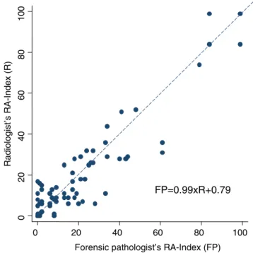
than RA-index observed for the 16 cases that were scanned 49–72 h after death (81.2 %>0 points, median 8, range 0–85), and for those scanned over 3 days after death (n016, 87.5 %> 0 points, median 33, range 0–100). Using seven sites, the inexperienced reader was able to determine the amount of gas present in all 82 sites (R200.85; p<0.0001). The RA-index also was determinant in quantifying the average amount of gas (grades) at each site (R200.58; p<0.0001). Even when only moderate reliability was observed for assessment and semiquantitative evaluation of gas presence in each site (Table1), the overall RA-index was very reliable between experienced and nonexperienced assessors (Fig.2; ICC2,10
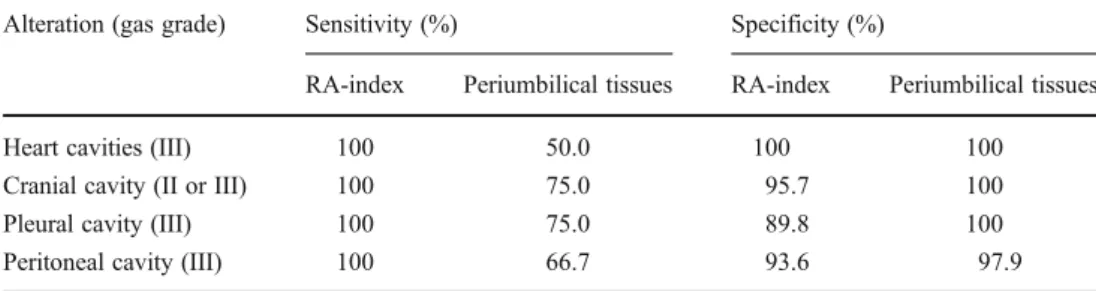
0.95; 95 % CI 0.92–0.96). To measure the sensitivity and specificity of the RA-index in detecting gas in cavities due to cadaveric alteration, we excluded cases with direct gunshot wounds to the head (seven cases) or the thorax (three cases), as those are very likely to have cavities contaminated with external gas. We cannot exclude the possibility that other open wounds contaminated cavities with external gas, especially in the pleural cavity; however, this did not seem to affect the presence of gas in the heart cavities, as determination values remained similar when excluding all cases with trauma (Table2). No cases with an RA-index below 50 presented with gas grade III in the heart (Table3). For other cavities, the
RA-index was more determinant for detecting alteration gas when excluding all traumatic cases (ROC>0.98). Finally, compared to the evaluation of gas in periumbilical tissues, the RA-index was much more determinant for assuming the presence of gas due to cadaveric alteration in different compartments (Table2).
Practical application
Of the nine cases with head gunshot wounds, five had gas grade III in the heart, and three of which were not expected to be due to cadaveric alteration, as they had RA-index values of 25, 28, and 34, which are below the cut-off point of 50. Gas chromatography could therefore have been recommended for these three cases to confirm the presence of vital air embolism.
Discussion
In this study, we derived an index for quantifying the state of cadaveric alteration of bodies, which is exclusively based on postmortem MDCT imaging. We also validated this index by ascertaining the reliability of the results as determined by a forensic pathologist with no experience in postmortem imag-ing, compared to those of a radiologist.
The RA-index is a simple and rapid tool for assessing the alteration state of a body, based on analysis of seven selected sites that are easy to identify even for an untrained forensic pathologist. This study shows that the RA-index remains valid for estimating the degree of cadaveric alteration in cases of traumatical deaths. Indeed, the seven selected sites of the
RA-Table 1 Scores corresponding to gas grades for the seven sites, and reliability
Sites Gas grades Scores Reliability
Cohen's kappa Heart cavities I 1 0.66 II 8 III 17 Liver parenchyma and vessels I 1 0.78 II 5 III 20
Left innominate vena I 5 0.41
II 15 III 15 Abdominal aorta I 8 0.49 II 8 III 8 Kidney parenchyma I 0 0.56 II 7 III 7 Vertebra L3 I 5 0.43 II 10 III 25 Subcutaneous pectoral tissues I 8 0.46 II 8 III 8 RA-index 0–100 ICC00.95 0 20 40 60 80 100 Radiologist’s RA-Index (R) 0 20 40 60 80 100
Forensic pathologist’s RA-Index (FP)
FP=0.99xR+0.79
Fig. 2 Reliability of the RA-index between the radiologist and the forensic pathologist. Regression line is shown
index are sufficiently distributed all over the body to over-come the bias from open wound gas expansion and, therefore, the RA-index is applicable in case of gunshot injury or knife wound. In cases with open wounds, we observed the presence of gas in cavities at earlier stages of alteration; we considered this to be due to local contamination with external gas, and it could have been due to simple gas diffusion rather than bacterial contamination [13]. However, our results suggest that hepatic gas develops in the same way independently of the presence of open wounds or not. Therefore, early gas might not only be due to contamination from an open wound through passive ongoing circulation [15].
The application of the RA-index is easy. The score is calculated following examination of the seven sites using the radiological data. It is very important to note that the image analysis is performed dynamically on successive slices of the MDCT. For each site, the grade of gas present (0, I, II, or III) has to be defined, which is possible even for a forensic pathologist without experience in the interpretation of radio-logical data. For vessels, the quantification of gas includes examination of the entire length of the vessel. Once a grade is assigned for each defined site, the corresponding scores can be found by using Table1; a site with no gas corresponds to a score of zero. Finally, the scores of each site are added, giving the RA-index. An example of RA-index calculation is shown in Fig.3.
The RA-index is a useful tool for postmortem diagnosis of the presence of vital gas on MDCT, which could have multiple applications. Due to its high determination value, the RA-index is able to detect gas grade III due to cadaveric alteration in the heart cavities and in the cranial cavities, based on cut-off points of >50 (specificity 90.9 %) and >60 (specificity 64.9 %), respectively,. This means that the presence of gas grade III
in the heart with an RA-index below 50 is a clear indication to proceed to further investigations (i.e., gas chromatography). Although, it should also be noted that an RA-index over 50 does not mean that a vital air embolism was not present before the development of postmortem gas. The RA-index could be useful in postmortem imaging studies for excluding cases in states of advanced cadaveric alteration. The inclusion or ex-clusion criteria could be based on their radiological alteration state, rather than on the less precise clinical observations made during the external examination of the body. Additionally, the changes occurring during the late postmortem period have not yet been well investigated and, apart from estimations of the postmortem interval based on forensic entomology, their in-terpretation often relies on the subjective personal experience of the forensic pathologist [16].
Secondly, regarding the comparison of bodies in different studies, it could be very interesting to have an objective mean of comparison of the selected collectives, in order to standard-ize the protocols. For example, in the testing of new techni-ques such as postmortem angiography, the study collective could be limited to cases with an RA-index of <50 to possibly avoid artifacts due to postmortem changes that could falsify the results of the tested method. We believe that it would be much more informative to use the RA-index as inclusion criteria, rather than the postmortem interval. As every experi-enced forensic pathologist knows, it is not only the time factor, but also the circumstances in which a body is stored, that influence the alteration. In contrast to the postmortem delay, the RA-index gives the real alteration state and is therefore taking these circumstances into consideration.
The third and, in our opinion, most important application for the RA-index will be its influence on the radiological interpretation of images. Although imaging is revolutionizing
Table 2 Sensitivities and specificities of detecting alteration gas in cavities for nontrauma cases, comparing periumbilical tissues to the RA-index
Alteration (gas grade) Sensitivity (%) Specificity (%)
RA-index Periumbilical tissues RA-index Periumbilical tissues
Heart cavities (III) 100 50.0 100 100
Cranial cavity (II or III) 100 75.0 95.7 100
Pleural cavity (III) 100 75.0 89.8 100
Peritoneal cavity (III) 100 66.7 93.6 97.9
Table 3 Validity of index for detecting gas in the cavities. In n090, cases with gun wounds to the head (n07) or the thorax (n03) were excluded
due to contamination by external gas
Alteration (gas grade) RA-index True positive True negative Sensitivity Specificity ROC
Heart cavities (III) ≥50 6/7 (85.7%) 83/83 (100%) 100% (51.7–100) 98.8% (92.6–99.9) 1.000 (1.0–1.0)
Cranial cavity (II or III) ≥60 4/7 (57.1%) 82/83 (98.8%) 80.0% (29.9–98.9) 96.5% (89.3–99.1) 0.821 (0.488–1.0)
Pleural cavity (III) ≥60 1/7 (14.3%) 79/83 (95.2%) 20.0% (1.1–70.1) 92.9% (84.7–97.1) 0.616 (0.290–0.943)
Fig. 3 Example showing gas grade I in the heart cavities (a), gas grade I in the liver parenchyma and vessels (b), gas grade 0 in the left innominate vein (c), gas grade II in the abdominal aorta (d), gas grade 0 in the kidney parenchyma (e), gas grade 0 in vertebra L3 (f), and gas grade 0 in pectoral subcutaneous tissues (g). Calculation of the RA-index (h)
classical forensic medicine, it is very important to be aware of its limitations. Postmortem radiological diagnosis requires the immense knowledge of both a forensic pathologist and a radiologist, and even with this knowledge, there is relatively little experience in the field of radiological interpretation of postmortem images. Thus, there is a need to establish guide-lines for this interpretation, on which newcomers to the field can lean on. We believe that there are a number of radiological diagnoses that can only be performed on cases with a low RA-index. However, most radiologists are not aware of this fact. Similarly to classical autopsy, the interpretation of findings should be performed more carefully if the body is presenting an advanced alteration. Additionally, the interpretation of the presence of gas in the late postmortem interval can be difficult because it cannot be established with certainty whether vital air embolism was present before the development of postmor-tem gas. Aside from the diagnosis of intravital air embolism, there are other procedures that should be limited to cases with a low RA-index, such as the diagnosis of pneumothorax, ascite, etc., that can be explained by thanalogical changes as well as by pathological processes. These alteration-based guidelines will become even more important if, in addition to performing a native MDCT, minimally invasive techniques, such as postmortem CT-angiography, are also executed. Here, the radiological interpretation becomes even more elaborate, requiring more knowledge and depending even more on the thanathological changes of the body. The need for guidelines in this field has already been highlighted [17].
The principal limitation of this study is that the RA-index cannot be validated for all types of traumas. Another limitation of the study is that only seven cases of the validation set were in an advanced decomposition state with an RA-index of >50.
The validity of the RA-index in estimating the degree of alteration is therefore not necessarily valid for unstudied trau-matically deceased cases, such as decapitation or dismember-ment. Furthermore, concerning the forensic pathologist, an improvement in the scoring method due to a learning effect cannot be excluded. Future plans for the RA-index include the adaptation of the image interpretations according to the alter-ation state of the bodies and the determinalter-ation of the useful-ness of additional postmortem exams in cases with a high RA-index.
Conclusion
This study shows that analysis of the presence of gas from seven sites alone is a valid means to evaluate the distribution of gas due to physiological cadaveric alteration phenome-non in the entire body. The RA-index is an easy-to-use instrument to radiologically determine the alteration state
of the body. This method remains reliable for nonexper-ienced users and is valid for nontraumatically and traumat-ically deceased.
References
1. Grabherr S, Lesta MDM, Rizzo E, Mangin P, Bollmann M (2008) Forensic imaging. Rev Med Suisse 4:1609–1614
2. O'Donnell C, Rotman A, Collett S, Woodford N (2007) Current status of routine post-mortem CT in Melbourne, Australia. Forensic
Sci Med Pathol 3:226–232
3. Bolliger SA, Thali MJ, Ross S, Buck U, Naether S, Vock P (2008) Virtual autopsy using imaging: bridging radiologic and forensic sciences. A review of the virtopsy and similar projects. Eur Radiol
18:273–282
4. Paperno S, Riepert T, Krug B, Rothschild MA, Schultes A, Staak M, Lackner L (2005) Value of postmortem computed tomography
in comparison to autopsy. Rofo 177(1):130–136
5. Payne-James J (2003) Forensic medicine: clinical and pathological aspects. Greenwich Medical Media, San Francisco
6. Aghayev E, Yen K, Sonnenschein M, Jackowski C, Thali M, Vock P, Dirnhofer R (2005) Pneumomediastinum and soft tissue emphy-sema of the neck in postmortem CT and MRI; a new vital sign in hanging? Forensic Sci Int 153(2–3):181–188
7. Dirnhofer R, Jackowski C, Vock P, Potter K, Thali MJ (2006) Virtopsy: minimally invasive, imaging-guided virtual autopsy.
Radiographics 26(5):1305–1333
8. Thali MJ, Jackowski C, Oesterhelweg L, Ross SG, Dirnhofer R
(2007) Virtopsy—the Swiss virtual autopsy approach. Leg Med
(Tokyo) 9(2):100–104
9. Jackowski C, Thali M, Sonnenschein M, Aghayev E, Yen K, Dirnhofer R, Vock P (2004) Visualization and quantification of air embolism structure by processing postmortem MSCT data. J
Forensic Sci 49(6):1339–1342
10. Shiotani S, Kohno M, Ohashi N, Atake S, Yamazaki K, Nakayama H (2005) Cardiovascular gas on non-traumatic postmortem com-puted tomography (PMCT): the influence of cardiopulmonary
resuscitation. Radiat Med 23(4):225–229
11. Levy AD, Harcke HT, Mallak CT (2010) Postmortem imaging: MDCT features of postmortem change and decomposition. Am J Forensic Med Pathol 1:12–17
12. O'Donnell C, Woodford N (2008) Post-mortem radiology—a new sub-speciality? Clin Radiol 63(11):1189–1194
13. Egger C, Bize P, Vaucher P, Mosimann P, Schneider B, Dominguez A, Meuli R, Mangin P, Grabherr S (2011) Distribution of artifac-tual gas on post-mortem multidetector computed tomography
(MDCT). Int J Legal Med 126(1):3–12
14. Shrout PE, Fleiss JL (1979) Intraclass correlations: uses in assessing
rater reliability. Psychol Bull 86(2):420–428
15. Jackowski C, Sonnenschein M, Thali MJ, Aghayev E, Yen K, Dirnhofer R, Vock P (2007) Intrahepatic gas at postmortem com-puted tomography: forensic experience as a potential guide for in
vivo trauma imaging. J Trauma 62(4):979–88
16. Colombo P (2011) Estimation of the time since death in the later postmortem interval: proposition of a compound method based on a Europe-wide survey. Thesis, Department of Clinical Research, University of Bern, Inselspital, Switzerland
17. Grabherr S, Doenz F, Steger B et al (2010) Multi-phase post-mortem CT angiography: development of a standardized protocol. Int J Legal