ARTICLE
A positive interaction between inhibitors of protein synthesis
and cefepime in the fight against methicillin-resistant
Staphylococcus aureus
B. Guignard&J. Vouillamoz&M. Giddey&P. Moreillon
Received: 8 December 2012 / Accepted: 9 January 2013 / Published online: 31 January 2013 # Springer-Verlag Berlin Heidelberg 2013
Abstract Quinupristin–dalfopristin (Q-D) synergizes with cefepime for the treatment of methicillin-resistant Staphylococcus aureus (MRSA). Here, we studied whether the synergism was restricted to MRSA and if it extended to non-beta-lactam cell wall inhibitors or to other inhibitors of protein synthesis. Three MRSA and two methicillin-susceptible S. aureus (MSSA) strains were tested, including an isogenic pair of mecA−/mecA+ S. aureus Newman. The drug interactions were determined by fractional inhibitory concentration (FIC) indices and population analysis profiles. The antibacterial drugs that we used included beta-lactam (cefepime) and non-beta-lactam cell wall inhibitors (D-cyclo-serine, fosfomycin, vancomycin, teicoplanin), inhibitors of protein synthesis (Q-D, erythromycin, chloramphenicol, tetra-cycline, linezolid, fusidic acid), and polynucleotide inhibitors (cotrimoxazole, ciprofloxacin). The addition of each protein inhibitor to cefepime was synergistic (FIC≤ 0.5) or additive (FIC>0.5 but < 1) against MRSA, but mostly indifferent against MSSA (FIC ≥ 1 but ≤4). This segregation was not observed after adding cotrimoxazole or ciprofloxacin to cefe-pime. Population analysis profiles were performed on plates in the presence of increasing concentrations of the cell wall
inhibitors plus 0.25 × minimum inhibitory concentration (MIC) of Q-D. Cefepime combined with Q-D was synergistic against MRSA, but D-cycloserine and glycopeptides were not. Thus, the synergism was specific to beta-lactam antibiotics. Moreover, the synergism was not lost against fem mutants, indicating that it acted at another level. The restriction of the beneficial effect to MRSA suggests that the functionality of penicillin-binding protein 2A (PBP2A) was affected, either directly or indirectly. Further studies are necessary in order to provide a mechanism for this positive interaction.
Introduction
Staphylococcus aureus is a major pathogen that causes both hospital-acquired and community-acquired infections. It is also mastermind in developing resistance to antibacterial agents [1]. It colonizes up to 20 % of the uninfected popu-lation [2] and, thus, is frequently exposed to the antibiotics used to treat infections caused by unrelated pathogens. Therefore, it has a great chance of acquiring resistance to any new antibacterial, even if the drug was originally tar-geted against other bacteria.
Methicillin-resistant S. aureus (MRSA) is a paradigm of this scenario. The loss of activity of beta-lactams against penicillin-binding protein 2A (PBP2A)-positive strains pushed the medical community to use numerous alternative antibacterials; however, MRSA became resistant to all of them [1]. Therefore, unless a number of new molecules with no cross-resistance are made available for the treatment of MRSA, staphylococci will develop resistance against each consecutive new compound and add the resistance mecha-nism to its existing multiresistance panoply [3]. As vanco-mycin is still the preferred treatment used against MRSA, a Part of this work was presented at the 44th Interscience Conference on
Antimicrobial Agents and Chemotherapy (ICAAC), Washington DC, October/November 2004 (abstract C1-1307).
B. Guignard
:
J. Vouillamoz:
M. Giddey:
P. Moreillon Department of Fundamental Microbiology, Biophore Building, University of Lausanne, 1015 Lausanne, Switzerland B. Guignard (*)Division of Pharmacy, University Hospitals of Geneva, Rue Gabrielle-Perret-Gentil 4,
1211 Geneva 14, Switzerland e-mail: [email protected] DOI 10.1007/s10096-013-1824-x
continuous selective pressure in the clinical environment has led to the emergence of vancomycin-intermediate S. aureus (VISA) strains. These strains have a decreased susceptibility to glycopeptides and are associated with vancomycin thera-peutic failure [4]. New agents such as linezolid, tigecycline, and daptomycin are now available for the treatment of MRSA infections, but linezolid and tigecycline are only bacteriostatic against S. aureus and are not approved for invasive and difficult-to-treat infections such as endocardi-tis. Moreover, resistance to these drugs is emerging [5,6].
Producing entirely new molecules is a profound scientific challenge [7]. Thus, it is also useful to explore the unex-pected features of existing drugs. For instance, understand-ing the mechanism of PBP2A-mediated resistance to beta-lactams [1,8] has set the rationale for the development of beta-lactams with improved anti-PBP2A affinity, such as ceftobiprole medocaril and ceftaroline fosamil [9, 10]. Alternatively, drug combinations can be useful by acting in synergy or because their resistance mechanisms may be mutually exclusive, as is the case for methicillin and lysos-taphin in MRSA [11]. The advantages of combining existing drugs are that their intrinsic toxicity is usually known and the compounds are available for use. On the other hand, a disadvantage is that the effects of drug combinations are accomplished through complex interactions that are often incompletely understood.
Recently, we and others have reported a synergism be-tween the streptogramin quinupristin–dalfopristin (Q-D) and beta-lactams in the treatment of MRSA both in vitro and in rats with experimental endocarditis [12–14]. The combina-tion of a low dose of Q-D with cefepime successfully cured MRSA endocarditis in the animals, despite both being inef-fective on their own [12]. In the present study, we tested in vitro combinations of several classes of antibiotics with cefepime, a beta-lactamase-resistant cephalosporin widely used in the hospital setting. We show that the previously observed positive interaction between Q-D and beta-lactams is restricted to MRSA, does not translate to methicillin-susceptible S. aureus (MSSA), and extends to other inhib-itors of protein synthesis but not to mechanistically unrelat-ed compounds.
Materials and methods
Microorganisms and growth conditions
The test organisms are described in Table1. They included the isogenic pair of MSSA and MRSA S. aureus Newman mecA− and mecA+ strains [15], one multiresistant clinical isolate of MSSA (strain P7142) [16], the homogeneously methicillin-resistant MRSA COL strain [17], and the multi-resistant clinical isolate MRSA P8 [18]. In certain experi-ments, we also used three isogenic strains, including the parent strain (MRSA BB270) [19] and two mutants lacking the femB and femAB loci (MRSA BB815 and AS145, re-spectively) [20,21], which are implicated in the synthesis of the pentaglycine cross-bridge (Table 1). The bacteria were routinely grown at 35 °C in either tryptic soy broth (TSB; Difco Laboratories, Detroit, MI) or tryptic soy agar (TSA; Difco). Cation-supplemented Mueller–Hinton broth (Difco) was used for the antibiotic-susceptibility tests that were performed in liquid medium. All of the media were supplemented with 2 % NaCl to increase the level of expression of beta-lactam resistance genes by MRSA. The stocks were kept at −70 °C in TSB supplemented with 10 % (vol/vol) glycerol.
Antibiotics and chemicals
Cefepime was provided by Bristol-Myers Squibb AG (Baar, Switzerland) and Q-D was provided by Aventis Pharma AG (Zürich, Switzerland). All of the other drugs and chemicals were commercially available products.
Susceptibility testing and antibiotic interactions
The minimum inhibitory concentrations (MICs) were deter-mined by a standard broth macrodilution method [22], with a final inoculum of 105 to 106colony-forming units (CFU)/ml. The antibiotic interactions were assessed by the checkerboard method in 96-well microtiter plates (Dynatech Microtiter, Chantilly, VA), as previously described [23]. The Table 1 Strains used in this
study Strain Origin PBP2A Main resistance profile Reference
P7142 Parent − MSSA, CMLSBr, Cmr, Tcr [16]
COL Parent + MRSA, Tcr [17]
P8 Parent + MRSA, CMLSBr, Cmr, Tcr, Gmr [18]
Newman Parent − MSSA [15]
Newman mecA+ Newman mecA + MRSA [15]
BB270 Parent + MRSA [19]
BB815 BB270ΔfemB + MSSA, Emr [20]
wells were filled with 100 μl of media containing twofold serial dilutions of each of the test antibiotics and inoculated with 105CFU/ml of bacteria (final con-centration) from a logarithmic-phase culture; the plates were then incubated for 24 h at 35 °C before visible bacterial growth was determined. The fractional inhibi-tory concentration (FIC) indices were interpreted as follows: ≤0.5 for drug synergism, >0.5 but <1 for ad-dition, ≥1 but ≤4 for indifference, and >4 for antago-nism. For each antibiotic combination, the experiment was performed in triplicate, and the lower of the FIC index values is presented.
Population analysis profiles
The phenotypic expression of cefepime resistance was determined by spreading a large bacterial inoculum (≥109
CFU), as well as the appropriate dilutions, onto NaCl-supplemented agar plates containing twofold serial dilutions of the drug [24]. In certain experiments, the plates were supplemented with a constant subinhibitory concentration (0.25 × MIC) of a partner drug [12]. The numbers of colonies growing on the plates were enu-merated after 48 h of incubation at 35 °C. The results are presented by plotting the numbers of colonies grow-ing on the plates against the cefepime concentration of the plates. The expression of resistance to non-beta-lactam cell wall inhibitors (fosfomycin, D-cycloserine, vancomycin, teicoplanin) was determined in a similar fashion.
Results
Antibiotic susceptibility and FIC indices
The drug susceptibilities of the test organisms are presented in Table2.
The positive or negative interactions between various antibiotics were first determined by FIC indices. The drugs included the beta-lactam cefepime, inhibitors of protein synthesis, and inhibitors of nucleic acid synthesis and as-sembly. Figure1a depicts the results of combining cefepime with the protein inhibitors. Most of these combinations interacted positively against MRSA, as demonstrated by the presence of synergism (FIC≤ 0.5) in 14/18 (78 %) of the cases and addition (FIC>0.5 but < 1) in 4/18 (22 %) of the cases. In contrast, these combinations were only additive (FIC>0.5 but < 1) or indifferent (FIC≥ 1 but ≤ 4) against the two MSSA isolates. This MRSA–MSSA dichotomy was clearly apparent in the mecA− and mecA+ versions of S. aureus Newman (Fig. 1a), suggesting that the presence of PBP2A was involved.
To test whether this mecA-related difference was also a property of other drug classes, the FIC experiments were repeated with cefepime in combination with the two inhib-itors of nucleic acid synthesis and assembly, cotrimoxazole and ciprofloxacin. Figure1b indicates that these combina-tions were not more active against MRSA than MSSA. Hence, the sensitization of MRSA to beta-lactams (in this case, cefepime) [12] was not a conserved feature between all of the drug classes.
Table 2 Minimum inhibitory concentrations (MICs) of several antibiotics for the five isolates used to test the different antibi-otic combinations
Antibiotics MICs in mg/L
Newman mecA− Newman mecA+ P7142 COL P8
Cell wall inhibitors
Cefepime 2 256 2 1,024 32 D-cycloserine 32 16 64 64 32 Fosfomycin 8 8 8 32 16 Vancomycin 2 2 1 2 1 Teicoplanin 1 2 0.5 1 0.25 Protein inhibitors Q-D 0.5 0.5 0.25 0.25 0.125 Erythromycin 0.25 0.25 1,024 0.5 1,024 Chloramphenicol 8 4 64 8 16 Tetracycline 0.5 0.5 256 128 32 Linezolid 2 1 4 2 2 Fusidic acid 0.12 0.03 0.25 0.125 0.25
Nucleic acids inhibitors
Ciprofloxacin 0.25 0.125 0.25 0.125 0.25
Population analysis profiles I: cefepime plus protein inhibitors
The FIC indices are relative values that provide information on the drug interactions independently of the bacterial re-sistance phenotype (e.g., CMLSB
r
, Cmr, Tcr, and Gmr for MSSA 7142 and MRSA P8, as observed in Table 1). To more quantitatively assess the changes in MICs due to the partner compound, the drug combination experiments were repeated in population analysis profiles. A series of agar plates was prepared with increasing concentrations of the drug to be tested plus a fixed subinhibitory concen-tration (0.25 × MIC) of the partner compound. The
mecA− and mecA+ S. aureus Newman strains were used as the model organisms.
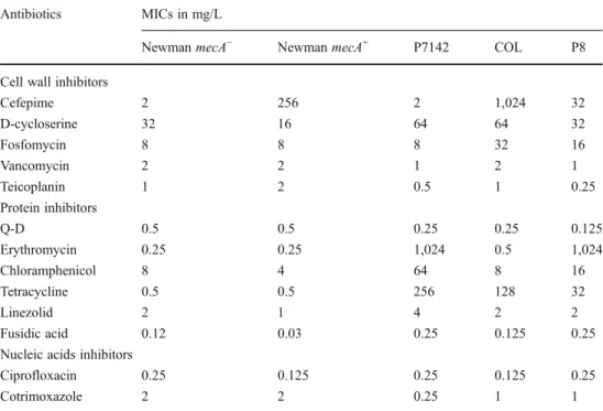
In the absence of an accompanying drug, the mecA+ S. aureus Newman strain grew on plates containing up to 500 mg/L cefepime (Fig. 2a). In the presence of 0.25 × MIC of Q-D or erythromycin, the MIC of cefepime de-creased by 10×and 5×, respectively, in the majority of the cell population (99.9999 %). This positive effect is in ac-cordance with the FIC indices depicted in Fig. 1a and is supported by previous reports indicating that these drugs could affect beta-lactam resistance in MRSA [12, 13]. In comparison, the positive interaction was much less marked in the mecA−S. aureus Newman strain (Fig.2b), a result that is also compatible with the FIC indices presented in Fig.1a. Thus, clinically achievable concentrations of Q-D and eryth-romycin (0.125 and 0.062 mg/L, respectively) could affect methicillin resistance in MRSA, but they barely affected MSSA beta-lactam susceptibility.
When non-MLSB protein inhibitors were used (e.g.,
chloramphenicol, tetracycline, and linezolid), a similar— yet less marked—positive effect was observed against the mecA+ S. aureus Newman strain (Fig. 2c), whereas the effect was, again, virtually non-existent against the mecA− S. aureus Newman strain (Fig. 2d). In contrast, the DNA inhibitor ciprofloxacin did not alter the MIC of cefepime for either of the organisms (Fig.2e, f).
The experiments were repeated to test the opposite set-ting, i.e., whether the subinhibitory concentrations of cefe-pime could affect the susceptibility of the bacteria to inhibitors of protein synthesis. Cefepime was added to the plates at a fixed subinhibitory concentration of 0.25×MIC (125 and 0.5 mg/L for the mecA+ and mecA− S. aureus Newman strains, respectively), whereas the inhibitors of protein synthesis were added at increasing concentrations. No beneficial or detrimental effects were observed with either of the organisms (data not presented). Hence, minor alterations of protein synthesis caused by low concentra-tions of protein inhibitors could enhance the effect of the beta-lactams, whereas marginal alterations of PBP function by subinhibitory concentrations of the beta-lactams could not enhance the effect of the protein synthesis inhibitors. Population analysis profiles II: non-beta-lactam cell wall inhibitors plus protein inhibitors
Because the inhibitors of protein synthesis, especially the MLSBtype, could decrease beta-lactam resistance in MRSA,
the question arose as to whether this effect was restricted to beta-lactams or if non-beta-lactam inhibitors of cell wall syn-thesis were also affected. Population analysis profiles were repeated using the following: (i) D-cycloserine, as a drug acting in the early synthesis of muropeptide precursors and before PBP-mediated transpeptidation (Fig. 3), (ii) vancomycin and
0.25 0.50 0.75 1.00 P8 COL NW + P7142 NW – MRSA MSSA
b
FIC indices 0.25 0.50 0.75 1.00 P8 COL NW + P7142 NW – MRSA MSSAa
FIC indicesFig. 1 Fractional inhibitory concentration (FIC) indices for cefepime combined with other drugs, including a the inhibitors of protein syn-thesis quinupristin–dalfopristin (black squares), erythromycin (white squares), chloramphenicol (black triangles), tetracycline (white trian-gles), linezolid (black circles), and fusidic acid (white circles) and b the inhibitors of nucleic acid synthesis and assembly ciprofloxacin (black diamonds) and cotrimoxazole (white diamonds). The tests were run against the three methicillin-resistant Staphylococcus aureus (MRSA) and two methicillin-susceptible S. aureus (MSSA) strains. The hori-zontal line at 0.5 indicates the limit for synergism (FIC index≤ 0.5). The horizontal lines between 0.5 and 1 delineate the area of addition (FIC index > 0.5 but < 1). The area above 1 indicates indifference (FIC index≥ 1 but ≤ 4). No antagonism between cefepime and any of the other drugs tested was observed
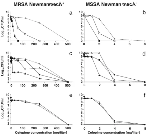
teicoplanin, as drugs acting after precursor maturation and blocking the latest stages of cell wall assembly, and (iii) fosfo-mycin, as a dual inhibitor of early precursor synthesis and PBP2A expression [25] (Fig.3).
Agar plates containing increasing concentrations of these compounds were supplemented, or not, with 0.25×MIC of Q-D and inoculated with the mecA+ and mecA− S. aureus Newman strains as above (Fig.4). Q-D marginally affected the MIC of D-cycloserine for both organisms (Fig.4a, b), suggesting that the beneficial effect of Q-D with the cell wall inhibitors must take place later in cell wall synthesis. At the other extreme of cell wall synthesis, Q-D had no effect in combination with vancomycin and teicoplanin on the two bacteria (Fig.4c–f), suggesting that the beneficial
interac-tion did not involve blockage of the precursor at the latest steps in cell wall assembly. Finally, Q-D interacted positive-ly with the dualpositive-ly active fosfomycin, but this interaction was much more marked against the mecA+(>10× decrease in the MIC of fosfomycin) than against the mecA−(2×decrease in the MIC of fosfomycin) S. aureus Newman strain (Fig.4g, h). Given that the D-cycloserine results indicated that the early steps of precursor synthesis were not involved, and
because fosfomycin affects both early precursor assembly and the expression of PBP2, 2A, and 4 [25] (Fig. 3), the anti-MRSA effect of Q-D plus fosfomycin most likely op-erated at the level of PBP functionality.
Effect of Q-D on the fem mutants
One critical feature of the function of PBP2A is its requirement for pentaglycine-decorated stem peptides in the muropeptide precursors (Fig.3) [26]. The femA and femB genes ensure that the pentaglycine side chains are added in a stepwise manner [21]. To test whether the positive effect of the protein inhib-itors could operate by altering the pentaglycine decorations, the Q-D and cefepime FIC experiments were repeated against the two fem mutants femB BB815 and femAB AS145, which carry triglycine and monoglycine decorations instead of pen-taglycine, respectively (Table 1), and their MRSA parent BB270 [19–21]. The FIC indices were unaltered by the fem mutations (0.6, 0.6, and 0.75 for the two mutants and the parent, respectively), indicating that the positive effect of the drug combination against the mecA+ staphylococci was an addition, and not a synonym, of the fem mutations.
MRSA NewmanmecA+ MSSA Newman mecA
-0 100 200 300 400 500 2 3 4 5 6 7 8 9 10 Log 10 CFU/ml
a
0 2 4 6 8b
0 100 200 300 400 500c
0 2 4 6 8Cefepime concentration [mg/liter]
f
0 100 200 300 400 500
Cefepime concentration [mg/liter]
e
0 2 4 6 8d
2 3 4 5 6 7 8 9 10 Log 10 CFU/ml 2 3 4 5 6 7 8 9 10 Log 10 CFU/ml 2 3 4 5 6 7 8 9 10 2 3 4 5 6 7 8 9 10 2 3 4 5 6 7 8 9 10Fig. 2 Population analysis profiles for the isogenic strains of MRSA Newman mecA+(a, c, e) and MSSA Newman mecA−(b, d, f) plated on increasing concentrations of cefepime in combination, or not, with a subinhibitory concentration (0.25 × MIC) of various partner drugs. Large numbers of bacteria [≥ 109
colony-forming units (CFU)] and the appropriate dilutions were plated, and the colonies were enumerat-ed after 24 h of incubation at 35 °C and plottenumerat-ed against the
concentration of antibiotic. The details are as follows: cefepime alone (+) or cefepime combined with a constant subinhibitory concentration of quinupristin–dalfopristin (0.125 mg/L, black squares), erythromycin (0.062 mg/L, white squares), chloramphenicol (2 mg/L, black trian-gles), tetracycline (0.125 mg/L, white triantrian-gles), linezolid (0.25 mg/L, black circles), or ciprofloxacin (0.062 mg/L, black diamonds)
Discussion
These results highlight some salient features of the previ-ously described synergism between Q-D and beta-lactam against S. aureus [12–14]. First, the positive interaction between the two drug classes was restricted to MRSA and was much less potent in MSSA. This was clearly apparent in the mecA+ and mecA− versions of S. aureus Newman and was also observed across the five test isolates. Second, this positive interaction extended to other inhibitors of protein synthesis, although it was less marked with the non-MLSB
compounds. Third, this positive interaction did not extend to mechanistically unrelated compounds such as DNA inhibitors and non-beta-lactam inhibitors of cell wall biosynthesis, except for fosfomycin. Together, these fea-tures help to delineate, at least partially, the mechanism of the drug interactions.
The restriction of the beneficial effect to MRSA indicates that the inhibitors of protein synthesis affected the functional-ity of PBP2A, either directly or indirectly. However, it does not specify the mechanism by which the positive interaction occurred. Our results obtained by combining the protein in-hibitor Q-D with the non-beta-lactam cell wall inin-hibitors
suggested that the level of the interaction was between the two extremes of the wall assembly line. At the early stage of wall assembly, the positive effect of Q-D most likely occurred after the addition of the D-ala-D-ala terminal to the muropep-tide precursor, as no positive interaction was observed be-tween Q-D and D-cycloserine. At the late stage of wall assembly, Q-D did not synergize with direct blockers of the precursors, such as glycopeptides, suggesting that the function of PBP and/or the earlier steps were involved.
One possibility was that an interaction with the maturation of muropeptide precursors was occurring, possibly at the level of adding pentaglycine decorations to the stem peptides. PBP2A requires pentaglycine-decorated precursors to be ef-fective [26–28]. Indeed, mutations in the fem genes, which block the addition of glycines to the precursors at various levels, block the expression of methicillin resistance, regard-less of the amount of PBP2A [17,28,29]. Protein inhibitors could affect the fem pathway by decreasing the amounts of glycine-adding enzymes, thus, formally replacing the fem effect. If the protein inhibitors do affect the fem pathway, then the Q-D plus beta-lactam synergism should be lost against the fem mutants, while it should persist against the parent MRSA strain. The experiments indicate that the beneficial effect of
Q-GlcNAc UDP MurNAc UDP MurNAc UDP Ala Glx Lys Ala Ala MurNAc UDP Ala Ala Ala Glx Lys MurNAc Ala Ala Ala Glx Lys PP MurNAc Ala Ala Ala Glx Lys PP Ala Ala MurNAc Ala Ala Ala Glx Lys PP MurNAc Ala GlcNAc FO S F O M Y C IN fmhB, fe mA , fe mB PLAS MAMEM BRANE LIPID I LIPID II TEICOPLANIN D-CYCLOSERINE β-LACTAMS LIPID II PEPTIDOGLYCAN GGGGG GGGGG GGGGG Ala Ala Ala Glx Lys VANCOMYCIN MurNAc Ala Ala Ala Glx Lys GlcNAc GGGGGAla Glx Lys MurNAc Ala GlcNAc GGGGG TRANSPEPTIDATION GlcNAc GlcNAc Ala Glx Lys MurNAc Ala GlcNAc GGGGG Ala TRANSGLYCOSYLATION T R A N S L O C A T IO N
Fig. 3 Principal steps of peptidoglycan assembly in S.aureus and locations of inhibition by some cell wall inhibitors. Cell wall precur-sors are synthesized in the cytoplasm from uridine diphosphate N-acetylglucosamine (UDP-GlcNAc) and transformed into uridine di-phosphate N-acetylmuramic acid (UDP-MurNAc). This is followed by the successive addition of one L-alanine (Ala), one D-isoglutamine or isoglutamate (Glx), one L-lysine (Lys), and the dipeptide D-Ala-D-Ala. The MurNAc pentapeptide is then linked to the plasma membrane to form lipid I. A cytoplasmic transglycosylase adds a GlcNAc to the
MurNac moiety and five glycine (G) residues to theε-NH2terminal of L-lysine, by the products of the fmhB, femA, and femB genes, to complete the formation of lipid II. After membrane translocation, the precursors are processed by the membrane penicillin-binding proteins (PBPs) through the transglycosylation and transpeptidation steps. The steps affected by beta-lactams, glycopeptides (vancomycin or teicopla-nin), D-cycloserine, and fosfomycin are indicated within the figure. Fosfomycin was also reported to decrease the expression of PBP2, 2A, and 4 (not depicted in the figure) [25]
D in combination with cefepime was identical in both the parent and the femB and femAB mutant strains, as shown by the FIC indices of 0.75, 0.6, and 0.6, respectively. Therefore, the drug combination operated in addition to the fem muta-tions and not instead of them.
Q-D had a positive interaction with fosfomycin, which was consistent with recent reports of synergism between fosfomycin and other protein synthesis inhibitors, such as linezolid [30]. Fosfomycin disrupts the expression of PBP2, 2A, and 4. It is tempting to hypothesize that the expression of the PBPs could also be altered, albeit in a different way, following protein synthesis inhibition by Q-D or other pro-tein inhibitors. This would explain the observed positive interaction with fosfomycin, by complementary alterations in the expression of the PBPs, as well as the beta-lactam susceptibility restoration. A decrease in the amount of PBP2A would be the simplest explanation for the observed restoration of cefepime susceptibility in the tested MRSA strains and the absence of this effect in the MSSA strains.
However, a decrease in PBP2 activity would also provide a rationale for the MRSA-specific synergism observed. Native S. aureus PBP2 is a class A PBP providing trans-glycosylase activity for the glycan chain elongation step in the cell wall building process. Hence, it is indispensable for the functionality of the other purely transpeptidase PBPs, including PBP2A. When MRSA are exposed to beta-lactams, all of their transpeptidase sites are blocked except for the low-affinity PBP2A site and the indispensable PBP2 transglycosylase site [31]. If this transglycosylase is further inhibited by either decreasing its amount, as hypothesized herein, or by specific drugs such as moenomycin, then PBP2A can no longer function and the beta-lactams regain their antibacterial activity. In MSSA, in contrast, the beta-lactams need only to block the high-affinity native trans-peptidases to block bacterial growth. This occurs at very low drug concentrations, and additional inhibition of the trans-glycosylase does not provide any additional effects. Thus, the dichotomy of synergism between MRSA and MSSA
2 3 4 5 6 7 8 9 10 0 25 50 75 100 125
D-cycloserine concentration [mg/liter]
Log 10 CFU/ml
a
2 3 4 5 6 7 8 9 10 0 25 50 75 100 125D-cycloserine concentration [mg/liter]
b
2 3 4 5 6 7 8 9 10 0 1 2 3 4Vancomycin concentration [mg/liter]
Log 10 CFU/ml
c
2 3 4 5 6 7 8 9 10 0 1 2 3 4Vancomycin concentration [mg/liter]
d
2 3 4 5 6 7 8 9 10 0 1 2 3 4Teicoplanin concentration [mg/liter]
Log 10 CFU/ml
e
2 3 4 5 6 7 8 9 10 0 1 2 3 4Teicoplanin concentration [mg/liter]
f
2 3 4 5 6 7 8 9 10 0 50 100 150 200 250Fosfomycin concentration [mg/liter]
Log 10 CFU/ml
g
2 3 4 5 6 7 8 9 10 0 50 100 150 200 250Fosfomycin concentration [mg/liter]
h
MRSA Newman mecA+ MSSA Newman mecA
-Fig. 4 Population analysis profiles of the isogenic strains of MRSA Newman mecA+(a, c, e, g) and MSSA Newman mecA−(b, d, f, h) plated on increasing concentrations of the non-beta-lactam cell wall inhibitors D-cycloserine (white circles), vancomycin (white triangles), teicoplanin (white diamonds), or fosfomycin (white squares) either alone (open symbols and dashed lines) or in combination with a constant subinhibitory concentration (0.125 mg/L) of quinupristin–dalfopristin (closed symbols and continuous lines). The details are the same as in Fig.2
could be explained by the hypotheses reported above. Further investigations, including the simultaneous titration of PBPs (mainly PBP2 and 2A) in the presence or absence of Q-D or other protein synthesis inhibitors at subinhibitory concentrations, would be required.
Several additional genes affecting methicillin resistance could also be involved. These include glnR (femC), which is responsible for glutamic acid amidation in precursor stem peptides, glmM (femD), which is responsible for UDP-N-ace-tylglucosamine biosynthesis, and murE (femF), which is re-sponsible for lysine addition in precursor stem peptides [28]. Moreover, protein inhibitors such as chloramphenicol and tet-racycline can inhibit the stringent response of Escherichia coli and promote the assembly of an abnormally thickened cell wall [32]. S. aureus can also undergo the stringent response [33], and subinhibitory concentrations of Q-D have been shown to induce cell wall thickening in this very organism [34].
Proteomic and DNA array analysis of S. aureus exposed to cell wall inhibitors revealed an ample alteration in gene expression through a phenomenon known as the cell-wall-stress stimulon [35, 36]. Among the altered genes, the expression of PBP2 was increased after induction of the vraSR genes. A possible interference of Q-D (or other protein inhibitors) with the physiological beta-lactam stress response (e.g., the VraSR system) would be another indirect rationale for the Q-D–beta-lactam effect.
Answering the numerous questions requires the addition-al titration of gene expression by microarrays. While such an approach is being attempted, the present observation offers further characterization of the previously observed anti-MRSA synergism between Q-D and the beta-lactams. Moreover, this precedent with MRSA may provide the rationale for testing this synergism using additional organ-isms that resist beta-lactams via decreased PBP affinity, including pneumococci and enterococci, as well as examin-ing new combinations of bactericidal beta-lactams with the new anti-MRSA protein inhibitors linezolid and tigecycline. Acknowledgments This work was supported by grants 3200-47099.96 and 3200-0458.95/2 from the Swiss National Funds for Scientific Research and an unrestricted grant from the Foundation for Advances in Medical Microbiology and Infectious Diseases.
We thank Prof. Brigitte Berger-Bächi, Institute of Microbiology, University of Zürich, for generously providing the mutant strains. Conflict of interest The authors declare that they have no conflict of interest.
References
1. Guignard B, Entenza JM, Moreillon P (2005) Beta-lactams against methicillin-resistant Staphylococcus aureus. Curr Opin Pharmacol 5(5):479–489
2. Wertheim HF, Melles DC, Vos MC, van Leeuwen W, van Belkum A, Verbrugh HA, Nouwen JL (2005) The role of nasal carriage in Staphylococcus aureus infections. Lancet Infect Dis 5(12):751– 762
3. Kuroda M, Ohta T, Uchiyama I, Baba T, Yuzawa H, Kobayashi I, Cui L, Oguchi A, Aoki K, Nagai Y, Lian J, Ito T, Kanamori M, Matsumaru H, Maruyama A, Murakami H, Hosoyama A, Mizutani-Ui Y, Takahashi NK, Sawano T, Inoue R, Kaito C, Sekimizu K, Hirakawa H, Kuhara S, Goto S, Yabuzaki J, Kanehisa M, Yamashita A, Oshima K, Furuya K, Yoshino C, Shiba T, Hattori M, Ogasawara N, Hayashi H, Hiramatsu K (2001) Whole genome sequencing of meticillin-resistant Staphylococcus aureus. Lancet 357(9264):1225–1240
4. Howden BP, Davies JK, Johnson PD, Stinear TP, Grayson ML (2010) Reduced vancomycin susceptibility in Staphylococcus au-reus, including vancomycin-intermediate and heterogeneous vancomycin-intermediate strains: resistance mechanisms, labora-tory detection, and clinical implications. Clin Microbiol Rev 23 (1):99–139
5. Sorlozano A, Gutierrez J, Martinez T, Yuste ME, Perez-Lopez JA, Vindel A, Guillen J, Boquete T (2010) Detection of new mutations conferring resistance to linezolid in glycopeptide-intermediate sus-ceptibility Staphylococcus hominis subspecies hominis circulating in an intensive care unit. Eur J Clin Microbiol Infect Dis 29(1):73– 80
6. Shakil S, Akram M, Khan AU (2008) Tigecycline: a critical update. J Chemother 20(4):411–419
7. Boucher HW, Talbot GH, Bradley JS, Edwards JE, Gilbert D, Rice LB, Scheld M, Spellberg B, Bartlett J (2009) Bad bugs, no drugs: no ESKAPE! An update from the Infectious Diseases Society of America. Clin Infect Dis 48(1):1–12
8. Lim D, Strynadka NC (2002) Structural basis for the beta lactam resistance of PBP2a from methicillin-resistant Staphylococcus au-reus. Nat Struct Biol 9(11):870–876
9. Moreillon P (2008) New and emerging treatment of Staphylococcus aureus infections in the hospital setting. Clin Microbiol Infect 14 (Suppl 3):32–41
10. Llarrull LI, Fisher JF, Mobashery S (2009) Molecular basis and phenotype of methicillin resistance in Staphylococcus aureus and insights into new beta-lactams that meet the challenge. Antimicrob Agents Chemother 53(10):4051–4063
11. Climo MW, Ehlert K, Archer GL (2001) Mechanism and suppres-sion of lysostaphin resistance in oxacillin-resistant Staphylococcus aureus. Antimicrob Agents Chemother 45(5):1431–1437 12. Vouillamoz J, Entenza JM, Féger C, Glauser MP, Moreillon P (2000)
Quinupristin–dalfopristin combined with beta-lactams for treatment of experimental endocarditis due to Staphylococcus aureus constitu-tively resistant to macrolide–lincosamide–streptogramin B antibiot-ics. Antimicrob Agents Chemother 44(7):1789–1795
13. Sieradzki K, Tomasz A (1997) Suppression of beta-lactam antibi-otic resistance in a methicillin-resistant Staphylococcus aureus through synergic action of early cell wall inhibitors and some other antibiotics. J Antimicrob Chemother 39(Suppl A):47–51 14. Allen GP, Cha R, Rybak MJ (2002) In vitro activities of
quinu-pristin–dalfopristin and cefepime, alone and in combination with various antimicrobials, against multidrug-resistant staphylococci and enterococci in an in vitro pharmacodynamic model. Antimicrob Agents Chemother 46(8):2606–2612
15. Vaudaux PE, Monzillo V, Francois P, Lew DP, Foster TJ, Berger-Bächi B (1998) Introduction of the mec element (methicillin resis-tance) into Staphylococcus aureus alters in vitro functional activ-ities of fibrinogen and fibronectin adhesins. Antimicrob Agents Chemother 42(3):564–570
16. Blanc DS, Petignat C, Moreillon P, Entenza JM, Eisenring M, Kleiber H, Wenger A, Troillet N, Blanc C, Francioli P (1999) Unusual spread of a penicillin-susceptible methicillin-resistant
Staphylococcus aureus clone in a geographic area of low inci-dence. Clin Infect Dis 29(6):1512–1518
17. Murakami K, Tomasz A (1989) Involvement of multiple genetic determinants in high-level methicillin resistance in Staphylococcus aureus. J Bacteriol 171(2):874–879
18. Franciolli M, Bille J, Glauser MP, Moreillon P (1991) Beta-lactam resistance mechanisms of methicillin-resistant Staphylococcus au-reus. J Infect Dis 163(3):514–523
19. Beck WD, Berger-Bächi B, Kayser FH (1986) Additional DNA in methicillin-resistant Staphylococcus aureus and molecular cloning of mec-specific DNA. J Bacteriol 165(2):373–378
20. Henze U, Sidow T, Wecke J, Labischinski H, Berger-Bächi B (1993) Influence of femB on methicillin resistance and peptidogly-can metabolism in Staphylococcus aureus. J Bacteriol 175 (6):1612–1620
21. Strandén AM, Ehlert K, Labischinski H, Berger-Bächi B (1997) Cell wall monoglycine cross-bridges and methicillin hypersuscep-tibility in a femAB null mutant of methicillin-resistant Staphylococcus aureus. J Bacteriol 179(1):9–16
22. Amsterdam D (1996) Susceptibility testing of antimicrobials in liquid media. In: Lorian V (ed) Antibiotics in laboratory medicine. The Williams & Wilkins Co., Baltimore, pp 52–111
23. Eliopoulos GM, Moellering RC Jr (1996) Antimicrobial combina-tions. In: Lorian V (ed) Antibiotics in laboratory medicine. The Williams & Wilkins Co., Baltimore, pp 330–396
24. Que YA, Entenza JM, Francioli P, Moreillon P (1998) The impact of penicillinase on cefamandole treatment and prophylaxis of experi-mental endocarditis due to methicillin-resistant Staphylococcus au-reus. J Infect Dis 177(1):146–154
25. Utsui Y, Ohya S, Magaribuchi T, Tajima M, Yokota T (1986) Antibacterial activity of cefmetazole alone and in combination with fosfomycin against methicillin- and cephem-resistant Staphylococcus aureus. Antimicrob Agents Chemother 30(6):917–922
26. de Jonge BL, Tomasz A (1993) Abnormal peptidoglycan produced in a methicillin-resistant strain of Staphylococcus aureus grown in the presence of methicillin: functional role for penicillin-binding protein 2A in cell wall synthesis. Antimicrob Agents Chemother 37(2):342–346
27. Berger-Bächi B, Rohrer S (2002) Factors influencing methicillin resistance in staphylococci. Arch Microbiol 178(3):165–171 28. de Lencastre H, de Jonge BL, Matthews PR, Tomasz A (1994)
Molecular aspects of methicillin resistance in Staphylococcus au-reus. J Antimicrob Chemother 33(1):7–24
29. Rohrer S, Berger-Bächi B (2003) FemABX peptidyl transferases: a link between branched-chain cell wall peptide formation and beta-lactam resistance in gram-positive cocci. Antimicrob Agents Chemother 47(3):837–846
30. Pachón-Ibáñez ME, Ribes S, Domínguez MA, Fernández R, Tubau F, Ariza J, Gudiol F, Cabellos C (2011) Efficacy of fosfo-mycin and its combination with linezolid, vancofosfo-mycin and imipe-nem in an experimental peritonitis model caused by a Staphylococcus aureus strain with reduced susceptibility to vanco-mycin. Eur J Clin Microbiol Infect Dis 30(1):89–95
31. Pinho MG, Filipe SR, de Lencastre H, Tomasz A (2001) Complementation of the essential peptidoglycan transpeptidase function of penicillin-binding protein 2 (PBP2) by the drug resis-tance protein PBP2A in Staphylococcus aureus. J Bacteriol 183 (22):6525–6531
32. Kusser W, Ishiguro EE (1986) Lysis of nongrowing Escherichia coli by combinations of beta-lactam antibiotics and inhibitors of ribosome function. Antimicrob Agents Chemother 29(3):451–455 33. Cassels R, Oliva B, Knowles D (1995) Occurrence of the regula-tory nucleotides ppGpp and pppGpp following induction of the stringent response in staphylococci. J Bacteriol 177(17):5161– 5165
34. Lorian V, Amaral L, Fernandes F (1995) RP 59500 postantibiotic effect defined by bacterial ultrastructure. Drugs Exp Clin Res 21 (3):125–128
35. Utaida S, Dunman PM, Macapagal D, Murphy E, Projan SJ, Singh VK, Jayaswal RK, Wilkinson BJ (2003) Genome-wide transcrip-tional profiling of the response of Staphylococcus aureus to cell-wall-active antibiotics reveals a cell-wall-stress stimulon. Microbiology 149(Pt 10):2719–2732
36. Jordan S, Hutchings MI, Mascher T (2008) Cell envelope stress response in Gram-positive bacteria. FEMS Microbiol Rev 32 (1):107–146