O R I G I N A L A R T I C L E – B R E A S T O N C O L O G Y
Pathological Processing Techniques and Final Diagnosis of Breast
Cancer Sentinel Lymph Nodes
Florian Rudolf Fritzsche, MD1, Tanja Reineke, MD1, Lars Morawietz, MD2, Glen Kristiansen, MD1,
Manfred Dietel, MD2, Daniel Fink, MD3, Christoph Rageth, MD4, Christoph Honegger, MD5, Rosmarie Caduff, MD1, Holger Moch, MD1, and Zsuzsanna Varga, MD1
1Institute of Surgical Pathology, University Hospital Zurich, Zurich, Switzerland;2Institute of Pathology, Charite´—
Universita¨tsmedizin Berlin, Berlin, Germany;3Department of Gynecology, University Hospital Zurich, Zurich, Switzerland;4Breast Centre Seefeld, Zurich, Switzerland;5Section of Gynecology, Hospital Uster, Zurich, Switzerland
ABSTRACT
Background. Recommendations for intraoperative and postoperative breast sentinel lymph node (SLN) processing differ widely. Micrometastases and isolated tumor cells (ITC) have recently been proposed as prognostically and therapeutically relevant. We compared 3 SLN protocols with regard to intraoperative and postoperative diagnosis. Materials and Methods. SLN in cohort I (270 patients) were intraoperatively assessed by stereomicroscopy. Intraoperative frozen section (IFS) was used only in ste-reomicroscopically suspicious SLN. In cohort II (197 patients), all SLN were examined with only 1 IFS. Final SLN workup in cohorts I and II consisted of complete step sectioning with immunohistochemistry. In cohort III (268 patients) 2 or more IFS were performed followed by 3 step sections and immunohistochemistry.
Results. pN1 stages were significantly higher in cohorts I and II (33.3% and 34.0% respectively) than in cohort III (24.6%). Intraoperative false negativity for the detection of metastases (pN1) ranged from 54.4% (cohort I) and 35.8% (cohort II) to 21.2% (cohort III). In contrast, ITC were detected significantly more frequently in cohort I (9.3%) and cohort II (14.7%) than in cohort III (1.9%).
Conclusions. Higher rates of SLN metastases and ITC in cohort I/II compared to cohort III suggest that IFS may result in tissue loss thus increasing the risk of missing metastases. Sparse IFS but complete postoperative SLN workup with step sectioning and immunohistochemistry provides more
accurate information regarding minimal disease in SLN, but often results in delayed axillary lymph node dissection. This is important for preoperative patient information and rec-ommendations in SLN processing protocols.
The axillary nodal status is one of the most important prognostic factors in breast cancer.1,2Intraoperative senti-nel lymph node (SLN) biopsy is a good predictor of the axillary nodal status and is now a standard method in the assessment of patients with early breast cancer.3–6Despite the general acceptance of the SLN model, protocols for intraoperative and postoperative processing of SLN differ widely.7–11 Intraoperative evaluation ranges from only gross examination, imprint cytology, 1 single intraopera-tive frozen section (IFS) to complete intraoperaintraopera-tive step sectioning with steps as small as 50 lm including intra-operative immunohistochemistry and other molecular-based methods.12–15 Postoperative evaluation includes hematoxylin and eosin (H&E) step sections, serial sec-tioning with or without auxiliary immunohistochemical stainings.12 The accuracy of IFS and the rate of false negative intraoperative results depend on the intraoperative workup of the SLN and the size of the metastasis. Some studies have demonstrated a good accuracy of IFS for macrometastases, but not for small metastases or isolated tumor cells.3,6 Isolated tumor cells (staged as pN0(i ?), with deposits B0.2 mm) and micrometastases (staged as pN1mi, with deposits [0.2 to B2.0 mm) have been assigned separate categories in the TNM staging.16 The cutoff value of 0.2 mm was chosen arbitrarily. Recently, it has been suggested that patients with ITC or micrometas-tases have reduced disease-free survival and may profit from adjuvant therapy.1,2
Ó Society of Surgical Oncology 2010 First Received: 8 January 2010; Published Online: 4 May 2010 F. R. Fritzsche, MD
e-mail: [email protected] DOI 10.1245/s10434-010-1097-x
Because of the relevance of minimal metastatic disease in axillary lymph nodes, the intraoperative and postopera-tive examination of SLNs is of increasing importance. It is not surprising that increasing the precision the postopera-tive SLN workup (e.g., with extensive use of cytokeratin step sections), will result in more intraoperative false negative cases, where the tumor was missed on a single or few H&E sections. The complete intraoperative processing of the SLN with alternating H&E and cytokeratin step sections at 50-lm intervals was proposed by Viale et al.13 The Swiss Section of Gynecological Pathologists has rec-ommended an approach of a single IFS followed by complete postoperative step sectioning using 200-lm intervals. Varga et al. have recently proposed the use of a stereomicroscope for the intraoperative assessment of SLN and preselection for IFS.9Stereomicroscopy consists of the examination of native unstained tissue samples with a magnification factor between 6 and 40, avoiding potential tissue loss associated with IFS of SLN.9
In this study, we compared 3 large cohorts of patients with axillary SLN biopsies. In 1 cohort the primary ste-reomicroscopic approach with or without IFS was used, the second cohort was diagnosed using the conventional approach of a single IFS, and diagnosis for the third cohort allowed 2 or more step sections for the IFS. The postop-erative workup varied from 3 step sections to complete step sectioning.
METHODS
Study Cohorts and Protocols
Cohort I (Stereomicroscopy with 1 Facultative Intra-operative Frozen Section) This cohort included 270 consecutive breast cancer patients (651 SLN), diagnosed at the Institute of Surgical Pathology, University Hospital Zurich from August 2007 until end of July 2008. Sentinel lymph nodes were detected with standard radioactive tracers,
partially in combination with blue dye.6 SLN from these patients were processed as follows (Table1): Axillary SLN were longitudinally bisected (or sectioned in 2-mm slices in SLN larger than 5 mm) and primarily assessed with a stereomicroscope. If the cut surface of the lymph node was clearly involved by the tumor or highly suspicious for malignancy, 1 single frozen section of the lymph node was performed.9In all other cases, lymph nodes were submitted to paraffin embedding and complete histological sectioning: paraffin blocks were completely step sectioned (200-lm steps) with 1 hematoxylin-eosin (H&E) staining and 1 unstained slide at each step. No residual lymph node structures remained in the paraffin block. If no tumor was found on the H&E slides, all unstained slides were immunostained with pan-cytokeratin (Lu5, 1:250, Roche, Basel, Switzerland) using standard immunohistochemical protocols and Ventana Benchmark autostainer platforms (Ventana, Tucson, AZ). Lymph nodes that were found positive (pN1) at IFS were fixated with formalin and embedded in paraffin and stained with 1 H&E. Additional cytokeratin immunostaining was applied if considered necessary.
The mean number of lymph nodes per case was 2.4 (median 2; range 1–8). The mean size of the lymph node was 11.2 mm (median 10 mm). The median size of the invasive carcinomas in this cohort was 16 mm (mean 19.9 mm). The median size of lymph node metastases was 3.6 mm (mean 5.1 mm; range 0.2–25 mm).
Cohort II (Only One Compulsory Intraoperative Frozen Section) This cohort included 197 consecutive breast cancer patients (476 SLN), diagnosed at the Institute of Surgical Pathology, University Hospital Zurich from August 2004 until end of July 2005. SLN from these patients were processed as follows (Table1): All SLN were bisected (or sectioned deeper in 2-mm slices if large enough) and 1 intraoperative frozen section stained with H&E was performed on each SLN irrespective of the gross
TABLE 1 Three different protocols for the intraoperative and postoperative assessment of sentinel lymph nodes (SLN)
Cohort I Cohort II Cohort III
Longitudinal bisection of the SLN Longitudinal bisection of the SLN Longitudinal bisection of the SLN
Stereomicroscopic examination 1 IFS C2 IFS
Suspicious for cancer Not suspicious Positive Negative Positive Negative
1 IFS No IFS
If positive
1 H&E staining Complete workup of SLN in H&E step sections (200 lm) and unstained slides for cytokeratin
1 H&E staining
Complete workup of SLN in H&E step sections (200 lm) and unstained slides for cytokeratin 1 H&E staining Workup of SLN in 3 H&E step sections (150 lm) and 1 cytokeratin staining
If negative see right column
appearance of the SLN. The residual SLN tissue was submitted to paraffin embedding and complete histological sectioning as described for cohort I.
The mean number of lymph nodes per case was 2.4 (median 2; range 1–8). The mean size of the lymph node was 10.2 mm (median 10 mm). The median size of the invasive carcinomas in this cohort was 16 mm (mean 17.7). The median size of these metastases was 3 mm (mean 4.8 mm; range 0.2–20 mm).
Cohort III (At Least Two or More Intraoperative Frozen Sections) This group included 268 consecutive breast cancer patients (472 SLN), diagnosed at the Institute of Pathology, Charite´ University Hospital Berlin from August 2007 until end of July 2008. SLN from these patients were examined as follows (Table1): All SLN were bisected (or sectioned in 2-mm slices in larger SLN). At least 2 (optionally more) IFS with H&E were performed on each SLN regardless of the gross appearance of the SLN. If the IFS was positive, the residual SLN tissue was embedded in paraffin and only 1 H&E stain was made. If the IFS was negative, residual SLN tissue was submitted to paraffin embedding and 3 H&E step sections (150-lm steps) were performed, accompanied by 1 pan-cytokeratin immunostain-ing (MNF116, 1:1000, Dako, Glostrup, Denmark) usimmunostain-ing Benchmark XT autostainers (Ventana).
The mean number of lymph nodes per case was 1.8 (median 1; range 1–7). The mean size of the lymph node was 11.4 mm (median 10 mm). The median size of the invasive carcinomas in this cohort was 18 mm (mean 20.3 mm). The median size of metastases was 5 mm (mean 6.9 mm; range 0.2–35 mm).
Assessment of Potential Tissue Loss
To evaluate the potential effects of tissue loss on the number and size of lymph node metastasis we analyzed 2 subgroups of the cohorts I and II. The first subgroup comprised all cases from cohort I that were inconspicuous in stereomicroscopy and thus left uncut in the IFS. The second group included cases that had 1 single IFS from cohort I and II but were considered negative in the IFS. Statistical Analyses
We analyzed the sensitivity, specificity, positive and negative predictive values, and accuracy of each protocol. Further, we assessed differences between the cohorts and for cases with and without IFS concerning nodal metastases and presence of ITC. Furthermore, all 3 cohorts were compared in terms of clinicopathological data to ensure comparability (Mann–Whitney U). Statistical analyses
TABLE 2 Tumor and lymph node characteristics of cohorts I, II, and III
Parameter Cohort I (%) Cohort II (%) Cohort III (%)
Number of cases 270 (100%) 197 (100%) 268 (100%)
Tumor histology
Invasive ductal carcinoma 216 (80.0%) 162 (82.2%) 209 (78.0%)
Invasive lobular carcinoma 42 (15.6%) 29 (14.7%) 41 (15.3%)
Other cancer types 12 (4.4%) 6 (3.0%) 18 (6.7%)
pT statusa
pT1 170 (63.0%) 138 (70.1%) 163 (60.8%)
pT2 85 (31.5%) 53 (26.9%) 93 (34.7%)
pT3 14 (5.2%) 5 (2.5%) 11 (4.1%)
Multifocal (m)b 29 (10.7%) 19 (9.7%) 35 (13.1%)
Histological tumor gradinga
G1 48 (17.8%) 48 (24.4%) 51 (19.0)
G2 127 (47.0%) 93 (47.2%) 157 (58.6)
G3 88 (32.6%) 53 (26.9%) 58 (21.6)
Patient nodal status
pN0/pN0 i? 155 (57.4%)/25 (9.3%) 101 (51.3%)/29 (14.7%) 197 (73.5%)/5 (1.9%)
pN1 90 (33.3%) 67 (34.0%) 66 (24.6%)
a No pT status was available for 1 case in each cohort, and no grade was given for 7 cases in cohort I, 3 cases in cohort II, and 2 cases in cohort III
b Multifocal tumors were extracted from other pT stages for comparative reasons and were otherwise included with the pT stage that matched with the largest tumor nodule
were performed using Microsoft Excel 2007 and SPSS Version 18.0. P values \ .05 were considered significant.
RESULTS
Comparability of the Cohorts
There was no statistically significant difference con-cerning the basic tumor parameters tumor type, pT status and histological grading between cohorts I and II (Table2). A high pT status was significantly more frequent in cohort III than in cohort II (P = .031). The histological grade was higher in cohort I than in cohort III (P = .019). There was no statistical difference between the combined cohorts I and II and cohort III.
Definitive Diagnosis—More Frequent pN1 Stages and ITC in Cohort I and II
Cohorts I and II comprised 270 and 197 breast cancer patients with 90 and 67 lymph node metastases, respec-tively (Table2). The frequency of pN1 stages did not differ between these 2 cohorts (33.3% vs. 34.0%). Isolated tumor cells (ITC) were slightly more frequent in cohort II (14.7%) than in cohort I (9.3%). Cohorts I and II (0 or 1 IFS) were compared with cohort III (n = 268) with 2 or more IFS. The frequency of patients with positive SLN (pN1) was significantly lower in cohort III (24.6%) than in cohort I (33.3%; P = .016), cohort II (34%; P = .027) or cohort I and II combined (P = .008). ITC were detected in 5 patients (1.9%) of cohort III. This was significantly less than in cohorts I (9.3%) and II (14.7%), either taken soli-tary or in a combined analysis (each P \ .001).
Intraoperative Diagnosis—False Negativity is Related to Sparse IFS
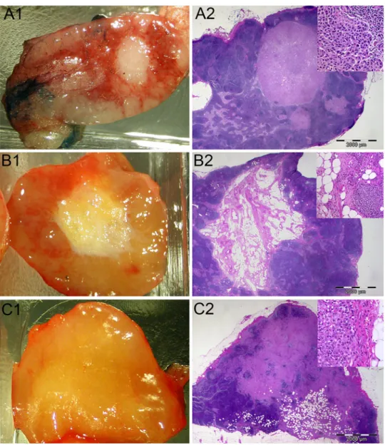
The sensitivity of the IFS for the detection of metastatic deposits was higher in cohort II (64.2%) compared with cohort I (45.6%, Table3). Likewise the negative predictive value (NPV) (84.3% versus 78.6%) and the accuracy (87.3% versus 81.9%) were higher in cohort II. Specificity and positive predictive values (PPV) were between 97.7% and 100% in both cohorts. In cohort II, 1 IFS was false positive because of a misinterpretation of large cells with mitotic figures as micrometastasis. Histological tumor type was not a relevant factor in any of the 3 protocols for intraoperative or postoperative detection of metastasis. Representative cases of corresponding stereomicroscopic and histological results are displayed in Fig.1. Theses cases also demonstrate typical difficulties encountered with stereomicroscopic assessments.
With 78.8%, the sensitivity of IFS in cohort III was clearly above that of cohort I and II. The NPV (93.5%) and accuracy (94.8%) in cohort III were also distinctly above that of either of the other cohorts. Specificity and PPV were 100%. One SLN was intraoperatively false positive because of the misinterpretation of an adenosis nodule.
Within cohort I, lymph node metastases were more frequent in those lymph nodes without an IFS (cohort I) compared with SLN with 1 IFS (cohort I and II; 13.9% vs. 7.6%; P = .002; Table4), whereas ITC were only slightly more often detected in the group with IFS (5.3% vs. 8%). DISCUSSION
The 3 different protocols for axillary SLN processing analyzed in this study revealed significantly higher rates of SLN metastases and ITC in protocols with few IFS com-bined with complete postoperative workup. In contrast, few
TABLE 3 Comparison of intraoperative frozen section diagnosis with final diagnosis (after formalin fixation and paraffin embedding)
Intraoperative result Final result Sum
Positive Negative Cohort I, section 1 Positive 41 (45.6%) 0 (0%) 41 Negative 49 (54.4%) 180 (100%) 229 Sum 90 (100%) 180 (100%) 270 Cohort I, section 2 Positive 48 (37.2%) 0 (0%) 48 Negative 81 (62.8%) 522 (100%) 603 Sum 129 (100%) 522 (100%) 651
Cohort II, section 1
Positive 43 (64.2%) 1 (0.8%) 44
Negative 24 (35.8%) 129 (99.2%) 153
Sum 67 (100%) 130 (100%) 197
Cohort II, section 2
Positive 62 (68.9%) 1 (0.3%) 63
Negative 28 (31.1%) 385 (99.7%) 413
Sum 90 (100%) 386 (100%) 476
Cohort III, section 1
Positive 52 (78.8%) 0 (0%) 52
Negative 14 (21.2%) 202 (100%) 216
Sum 66 (100%) 202 (100%) 268
Cohort III, section 2
Positive 75 (83.3%) 1 (0.3%) 76
Negative 15 (16.7%) 381 (99.7%) 396
Sum 90 (100%) 382 (100%) 472
Section 1 displays the patient result irrespective of the number of lymph nodes submitted to frozen section. Section 2 depicts the results for single lymph nodes, since often more than 1 sentinel lymph node was submitted
IFS increased the rate of intraoperative false negative SLN diagnosis with the potential consequence of delayed axil-lary lymph node dissection. The protocol of SLN processing is therefore relevant in the debate of short- or long-term outcome data of clinical trials.2,17
Very recently, de Boer et al. presented a large study demonstrating that patients with ITC had decreased
disease-free survival and that chemotherapy improved survival in these patients.2This study renewed interest in the implications of the presence of ITC.18–20 Our study demonstrates how significantly the results of the SLN biopsy are influenced by the pathological workup protocol applied. We found a more than 4-fold difference between cohort III and the other 2 cohorts in the detection of ITC. Because this study was observational in design, this cer-tainly implicates potential biases of the results. However, our analyses of the most important tumor parameters (pT status, grading, tumor type) did not reveal relevant differ-ences between the 3 cohorts that could explain such variances. Therefore, the demonstrated differences in the amount of ITC are almost certainly the result of the lesser extent of immunohistological workup in cohort III. This implies that the validity of results of clinical trials studying the association of ITC and outcome of breast cancer should take into account the extent of the SLN workup in
FIG. 1 Stereomicroscopic and matching histological results (magnification 69, inlet 2009). aSLN with macrometastasis visible in stereomicroscopy (A1) and in the matching H&E staining (A2). b Stereomicroscopically suspect SLN (B1) with fibrosis and lipomatosis in the histological section (B2).
cStereomicroscopically unsuspicious SLN (C1) with macrometastasis on the first H&E section (C2)
TABLE 4 Comparison of lymph node status in sentinel lymph nodes without intraoperative frozen section (cohort 1) and with intraopera-tive frozen section (cohort 1 and 2 combined)
Parameter One IFS
performed (%)
No IFS performed (%)
P value
Lymph node status .002
pN0/pN0 i? 420 (84.3%)/ 40 (8.0%)
412 (80.8%)/ 27 (5.3%)
pathology. The biological relevance of ITC has not been clearly determined yet.21–24 Rutgers suggested only using SLN processing protocols that identify lymph node metastases of known clinical relevance.25 Different SLN protocols in various breast centers, sufficient follow-up time, and adequate clinical screening for metastases are potential limitations and prerequisites of clinical studies to evaluate the relationship between ITC or micrometastases with clinical outcome.26
Our results further show that 2 or more intraoperative step sections resulted in a better intraoperative sensitivity to detect lymph node metastases compared with a single IFS-only model, which can be regarded as a clear argument to use 2 or more intraoperative step sections. Higher intraoperative sensitivity and accuracy are helpful to avoid delayed axillary lymph node dissection, thereby lowering iatrogenic morbidity. Importantly, our findings suggest that a higher intraoperative detection rate for metastases with more than 1 IFS was also accompanied by a significantly lower rate of metastases in the final diagnosis. This sup-ports the hypothesis that tissue loss occurs during the procedure of IFS, which might have been the reason for recommendations in the United Kingdom of not perform-ing IFS.9,12To avoid tissue loss was also the basic concept for using stereomicroscopic preselection of SLN for IFS as described by Varga et al.9
Concerning stereomicroscopy, our data suggest that the macroscopic/stereomicroscopic preselection of SLN cannot be recommended, because this approach does not lead to significantly higher rates of postoperatively detected metastases compared with a protocol with a single IFS. Instead stereomicroscopic preselection led to a very high rate of intraoperative false negativity (54.4%). As seen in Fig.1 some metastases are invisible to the naked eye or stereomicroscopy. Theoretically, specimens with intense
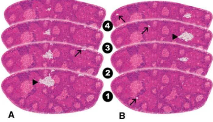
blue dye staining could also make it harder for the pathologist to interpret the stereomicroscopic results. However, the latter situation constituted no major problem in our experience. Because the cohorts without or with 1 IFS showed similar rates of pN1 stages (33.3% vs. 34%) and ITC (14.7% vs. 9.3%), this supports a recommendation to do at least 1 IFS intraoperatively. Nonetheless, it has to be noted that the intraoperative false negativity rate with a single IFS was still high (35.8%) compared with the use of 2 and more IFS (21.2%). This phenomenon can be explained by the location of the metastatic deposits in deeper planes of the SLN. Because a single IFS only detects metastases located on the cutting plane of the lymph node, we analyzed how many of the metastases were found on the cutting plane and how many on deeper step sections only. From 157 patients with SLN metastases in cohorts I and II, 48 (30.6%) were not detectable on the first step section. From the 54 patients where ITC were the only finding, for 28 (51.9%) cases the ITC were only detectable on deeper step sections. Figure2 illustrates the problem of metastases in deeper cutting planes. None of the pro-tocols showed any tumor type specific advantages or disadvantages.
Economical questions have to be also taken into account for the recommendation of SLN processing protocol. Viale et al. propose the complete intraoperative processing of the SLN with alternating H&E and cytokeratin step sections at 50 lm intervals.13 Considering that the number of SLN submitted might easily exceed 1 or 2, the costs and time of such a complete intraoperative workup as well as the obligation to meet also the intraoperative demands of other surgical specialities, such a protocol is difficult to imple-ment in a cost- and time-efficient way in most pathology laboratories. Although we did not specifically compare the exact IFS times of the protocols, in this context it should be noted that none of the 3 evaluated protocols exceeded a 30-minute time limit for IFS.
In conclusion, our presented data support a strong dependence between the use of IFS and extent of step sectioning with intraoperative sensitivity/accuracy and potential tissue loss. Some surgeons believe that the pos-sibility for an axillary lymph node dissection during initial surgery outweighs the risk of missed metastases.6,27In our opinion, this is a point that should be decided under con-sideration of the patients physical condition and preferences. The highest rate of metastasis detection is reached by omission of IFS or a single IFS followed by a thorough and immunohistochemically supported workup of the SLN. Stereomicroscopic-based assessment could not be recommended due to high intraoperative false negativity. If the presence of ITC should become decisive for treatment selection, the current high variability of protocols in the detection of ITC has to be seriously reconsidered.
FIG. 2 Two step-sectioned SLN with different tumor localizations. In contrast to the macrometastasis (arrowhead) in SLN a, maximum in the cutting plane, the 1 in SLN b would probably be missed by stereoscopic or single IFS assessment. Detecting micrometastases or ITC (arrows) is a challenge in the H&E staining regardless of the localization
ACKNOWLEDGMENT We are grateful to Deborah Ina¨bnit, Aytac Altuncevahir (USZ), and Gu¨nter John (Charite´) for their logistical support of this study and Miss Heather Dawson and Dr. Victoria Salter for copy editing the manuscript.
REFERENCES
1. Soerjomataram I, Louwman MW, Ribot JG, Roukema JA, Coe-bergh JW. An overview of prognostic factors for long-term survivors of breast cancer. Breast Cancer Res Treat. 2008;107:309–30.
2. de Boer M, van Deurzen CH, van Dijck JA, Borm GF, van Diest PJ, Adang EM, et al. Micrometastases or isolated tumor cells and the outcome of breast cancer. N Engl J Med. 2009;361:653–63. 3. van de Vrande S, Meijer J, Rijnders A, Klinkenbijl JH. The value
of intraoperative frozen section examination of sentinel lymph nodes in breast cancer. Eur J Surg Oncol. 2009;35:276–80. 4. Goyal A, Mansel RE. Recent advances in sentinel lymph node
biopsy for breast cancer. Curr Opin Oncol. 2008;20:621–6. 5. Pugliese MS, Tickman R, Wang NP, Atwood M, Beatty JD. The
utility of intraoperative evaluation of sentinel lymph nodes in breast cancer. Ann Surg Oncol. 2007;14:1024–30.
6. Langer I, Guller U, Berclaz G, Koechli OR, Moch H, Schaer G, et al. Accuracy of frozen section of sentinel lymph nodes: a prospective analysis of 659 breast cancer patients of the Swiss multicenter study. Breast Cancer Res Treat. 2009;113:129–36. 7. Cserni G. Evaluation of sentinel lymph nodes in breast cancer.
Histopathology. 2005;46:697–702.
8. Grabau DA, Rank F, Friis E. Intraoperative frozen section examination of axillary sentinel lymph nodes in breast cancer. APMIS. 2005;113:7–12.
9. Varga Z, Rageth C, Saurenmann E, Honegger C, von Orelli S, Fehr M, et al. Use of intraoperative stereomicroscopy for pre-venting loss of metastases during frozen sectioning of sentinel lymph nodes in breast cancer. Histopathology. 2008;52:597–604. 10. Viale G, Mastropasqua MG, Maiorano E, Mazzarol G. Pathologic examination of the axillary sentinel lymph nodes in patients with early-stage breast carcinoma: current and resolving controversies on the basis of the European Institute of Oncology experience. Virchows Arch. 2006;448:241–7.
11. Weaver DL. Pathological evaluation of sentinel lymph nodes in breast cancer: a practical academic perspective from America. Histopathology. 2005;46:702–6.
12. Cserni G. Histopathologic examination of the sentinel lymph nodes. Breast J. 2006;12:S152–6.
13. Viale G, Sonzogni A, Pruneri G, Maffini F, Masullo M, Dell’Orto P, et al. Histopathologic examination of axillary sentinel lymph nodes in breast carcinoma patients. J Surg Oncol. 2004;85:123–8. 14. Douglas-Jones AG, Woods V. Molecular assessment of sentinel lymph node in breast cancer management. Histopathology. 2009;55:107–13.
15. Tamaki Y, Akiyama F, Iwase T, Kaneko T, Tsuda H, Sato K, et al. Molecular detection of lymph node metastases in breast cancer patients: results of a multicenter trial using the one-step nucleic acid amplification assay. Clin Cancer Res. 2009;15:2879– 84.
16. Sobin LH, Wittekind C (eds) TNM Classification of Malignant Tumours. 6th ed. New York: Wiley-Liss, 2002.
17. Langer I, Guller U, Viehl CT, Moch H, Wight E, Harder F, et al. Axillary lymph node dissection for sentinel lymph node mi-crometastases may be safely omitted in early-stage breast cancer patients: long-term outcomes of a prospective study. Ann Surg Oncol. 2009;16:3366–74.
18. Sonke GS, Linn SC. Isolated tumor cells in breast cancer. N Engl J Med. 2009;361:1995; author reply 1996.
19. Roukos DH. Isolated tumor cells in breast cancer. N Engl J Med. 2009;361:1994–5; author reply 1995–6.
20. Lyman GH, Peppercorn J. Isolated tumor cells in breast cancer. N Engl J Med. 2009;361:1994; author reply 1995–6.
21. Bulte CS, van der Heiden-van der Loo M, Hennipman A. Axil-lary recurrence rate after tumour negative and micrometastatic positive sentinel node procedures in breast cancer patients, a population based multicenter study. Eur J Surg Oncol. 2009;35: 25–31.
22. Langer I, Marti WR, Guller U, Moch H, Harder F, Oertli D, et al. Axillary recurrence rate in breast cancer patients with negative sentinel lymph node (SLN) or SLN micrometastases: prospective analysis of 150 patients after SLN biopsy. Ann Surg. 2005;241:152–8.
23. Querzoli P, Pedriali M, Rinaldi R, Lombardi AR, Biganzoli E, Boracchi P, et al. Axillary lymph node nanometastases are prognostic factors for disease-free survival and metastatic relapse in breast cancer patients. Clin Cancer Res. 2006;12:6696–701. 24. Kahn HJ, Hanna WM, Chapman JA, Trudeau ME, Lickley HL,
Mobbs BG, et al. Biological significance of occult micrometas-tases in histologically negative axillary lymph nodes in breast cancer patients using the recent American Joint Committee on Cancer breast cancer staging system. Breast J. 2006;12:294–301. 25. Rutgers EJ. Sentinel node biopsy: interpretation and management of patients with immunohistochemistry-positive sentinel nodes and those with micrometastases. J Clin Oncol. 2008;26:698–702. 26. Turner RR, Weaver DL, Cserni G, Lester SC, Hirsch K, Elashoff DA, et al. Nodal stage classification for breast carcinoma: improving interobserver reproducibility through standardized histologic criteria and image-based training. J Clin Oncol. 2008;26:258–63.
27. van Diest PJ, Torrenga H, Meijer S, Meijer CJ. Pathologic analysis of sentinel lymph nodes. Semin Surg Oncol. 2001;20: 238–45.