HAL Id: tel-00812718
https://tel.archives-ouvertes.fr/tel-00812718
Submitted on 12 Apr 2013HAL is a multi-disciplinary open access archive for the deposit and dissemination of sci-entific research documents, whether they are pub-lished or not. The documents may come from teaching and research institutions in France or abroad, or from public or private research centers.
L’archive ouverte pluridisciplinaire HAL, est destinée au dépôt et à la diffusion de documents scientifiques de niveau recherche, publiés ou non, émanant des établissements d’enseignement et de recherche français ou étrangers, des laboratoires publics ou privés.
Prédire l’âge de personnes à partir de photos du visage :
une étude fondée sur la caractérisation et l’analyse de
signes du vieillissement
Alex A. Nkengne
To cite this version:
Alex A. Nkengne. Prédire l’âge de personnes à partir de photos du visage : une étude fondée sur la caractérisation et l’analyse de signes du vieillissement. Ingénierie biomédicale. Université Pierre et Marie Curie - Paris VI, 2008. Français. �NNT : 2008PA066082�. �tel-00812718�
THESE DE DOCTORAT DE L’UNIVERSITE PIERRE ET MARIE CURIE
Informatique Biomédicale ……….
ED 393 – SANTE PUBLIQUE : Epidémiologie et sciences de l’Information Biomédicale
Présentée par
M NKENGNE NGUIMEZONG Alex Albert Pour obtenir le grade de
DOCTEUR de l’UNIVERSITÉ PIERRE ET MARIE CURIE Sujet de la thèse :
Prédire l’âge de personnes à partir de photos du visage :
une étude fondée sur la caractérisation et l’analyse de signes du vieillissement Predicting people’s age from their facial image:
a study based on the characterization and the analysis of the signs of aging Soutenue le 13 Juin 2008
Devant le jury composé de :
Philippe HUMBERT Professeur, Université de Franche-Comté Rapporteur Alice CAPLIER Professeur assistante, ENSERG Rapporteur Bernard FERTIL Directeur de recherche, CNRS Directeur de thèse Nathalie ISSACHAR Directeur de recherche, Johnson & Johnson Co-encadrante NIkiforos KOLLIAS Directeur de recherche, Johnson & Johnson Examinateur Jean Gabriel GANASCIA Professeur, Université de Paris VI Examinateur Maurice MILGRAM Professeur, Université de Paris VI Examinateur
Université Pierre & Marie Curie - Paris 6
Bureau d’accueil, inscription des doctorants et base de données Esc G, 2ème étage
15 rue de l’école de médecine 75270-PARIS CEDEX 06
Tél. Secrétariat : 01 42 34 68 35 Fax : 01 42 34 68 40
Tél. pour les étudiants de A à EM : 01 42 34 69 54 Tél. pour les étudiants de EN à ME : 01 42 34 68 41
Tél. pour les étudiants de MF à Z : 01 42 34 68 51 E-mail : scolarite.doctorat@upmc.fr
Au Père Marie Elie, A mes chers parents
Remerciements 1
Remerciements
Merci { vous qui avez conduit mes pas depuis la tendre enfance, { vous qui m’avez entouré de votre affection, choyé, protégé, encouragé.
Merci à vous ma famille, mes amis, mes nombreux pères et mères. Ce travail de thèse, est le fruit de votre Amour et de votre Sueur, soyez en fiers.
Merci à vous qui avez suscité en moi une vocation pour la science et m’avez montré les chemins de l’excellence.
Merci, { vous qui patiemment m’avez appris { décrypter les mystères du monde et { lier le bois au bois.
Merci { vous qui m’avez transmis votre flamme, votre passion de la science et de la vérité. Merci à vous tous, enseignants et guides. Puissiez-vous retrouver dans cette thèse quelques unes des idées que vous vous êtes donnés tant de mal { m’inculquer.
Merci { vous qui avez accueilli { bras ouvert l’inconnu et m’avez donné une place { votre table.
Merci { vous qui m’avez donné l’opportunité de traverser les mers pour poursuivre un idéal.
Merci { vous, amis et mécènes qui avez cru en moi. Ce travail, je l’espère est { la mesure de la confiance que vous m’avez accordé.
La vie est faite de rencontres qui nous enrichissent et nous portent vers de nouveaux horizons. Maintenant que s’achève cette étape du voyage, je souhaiterais en particulier exprimer ma gratitude à ceux qui ont partagé ces années de thèse.
Mes premiers mots de remerciement vont à ceux qui ont rendu possible cette aventure, Nathalie Issachar et Bernard Fertil. Vous avez cru en moi et en mon projet et m’avez accueilli à bras ouvert dans vos équipes ; je vous en suis très reconnaissant.
Remerciements 2 J’exprime également toute ma reconnaissance aux membres de l’unité UMR 678 et en particulier à son directeur Alain Herment. Les moments passés avec vous ont été source d’émulation et d’inspiration.
J’ai une pensée particulière pour Alain Giron, véritable poumon de l’équipe IV ; ton dévouement m’aura redonné courage dans les moments d’incertitude.
L’équipe IV a été un véritable lieu de vie, ou j’ai eu le plaisir de me lier d’amitié { des étudiants tous plus généreux les uns que les autres. Arthur, Sylvain, Jean-François (mon super binôme), Caroline, Sahra et bien entendu Clara (par adoption), je n’oublierais pas les excellents moments passés en votre compagnie.
Une autre équipe qui aura su m’ouvrir ses bras est celle du Skin Care Research Institute. Grâce à vous, Christiane, Arlette, Ana, Valérie, Anne, Romain, Aline, Emilie, Estelle, Marion, Fanny, Maryline, Stéphanie, Delphine j’ai passé et continue de passer de très bons moments. Je vous exprime ma reconnaissance pour cette bonne humeur que vous savez si bien distiller. J’ai une attention particulière pour Christiane, Stéphanie, Delphine et Arlette qui ont largement contribué { la collecte et { l’analyse des données présentées dans ce travail. Merci de votre précieuse collaboration.
Je voudrais également remercier tous ceux dont les réflexions et l’énergie ont contribué { améliorer mon travail de thèse et ce document final. Christiane Bertin, Georgios Stamatas, l’increvable Alain et bien sur Bernard ; merci { vous pour le temps et l’énergie consacrée. Toute ma reconnaissance va également { Rachid Belaroussi qui s’est dévoué six mois durant pour me fournir une méthode adaptée de segmentation.
Enfin, j’ai l’honneur et le plaisir d’avoir dans mon jury les Professeurs Philippe Humbert, Alice Caplier, Maurice Milgram et Jean Gabriel Ganascia ainsi que les Docteurs Nikiforos Kollias, Nathalie Issachar et Bernard Fertil. Vos travaux à tous suscitent mon admiration. Je vous suis donc infiniment reconnaissant d’avoir accepté d’être membres de mon jury de thèse.
Résumé 3
Résumé
L’âge a de tout temps constitué un attribut identitaire important. Nous avons développé au fil de l’évolution une aptitude innée { classer les individus en fonction de leur âge. Cette classification s’appuie en grande partie sur le visage et sur les transformations anatomiques qu’il subit au cours du temps. De plus en plus de traitements cosmétiques, dermatologiques et d’interventions chirurgicales s’attaquant { un signe ou un groupe de signes spécifiques du vieillissement sont mis en œuvre pour annuler, ou tout au moins masquer partiellement l’effet du temps sur le visage. On peut dès lors s’interroger sur l’influence de chacun des signes sur notre capacité { prédire l’âge d’un individu en observant son visage. Afin de construire un algorithme capable de déterminer l’âge d’individus { partir de leurs photos, nous nous sommes intéressés aux signes du vieillissement et { leur impact sur l’âge apparent. Dans un premier temps, nous avons déterminé et analysé les transformations anatomiques qui altèrent le visage { partir de l’âge adulte (au-delà de 20 ans). Puis nous avons étudié les signes sur lequel on se base pour prédire l’âge d’une personne. Enfin, nous avons construit et validé un modèle prédictif de l’âge en s’appuyant sur les observations précédentes.
Transformations anatomiques du visage avec l’âge : La prévalence d’un certain nombre
de signes de vieillissement (rides, tâches brunes, forme du visage,…) a été mesurée sur un panel représentatif de femmes volontaires âgées de 20 à 74 ans. Ces données ont permis d’établir la cinétique d’apparition de ces signes.
Appréciation subjective de l’âge: Il s’agissait de déterminer les signes sur lesquels un
observateur s’appuie lorsqu’il évalue l’âge d’un sujet. Pour ce faire, nous avons demandé { un panel constitué de 48 observateurs d’attribuer un âge aux volontaires sur lesquelles nous avions précédemment mesuré les signes du vieillissement. Nous avons confirmé avec ce groupe d’observateurs que la perception de l’âge est liée au sexe et { l’âge de l’observateur. De plus, { l’aide d’une régression PLS (Partial Least Square régression), nous avons établi des relations entre les signes du vieillissement et l’âge observé et démontré que selon que l’on soit jeune ou âgé, un homme ou une femme, on n’exploite pas les mêmes signes de vieillissement pour prédire l’âge.
Modèle de prédiction : Enfin, nous avons proposé un modèle s’appuyant sur la régression
Résumé 4 la particularité d’associer, dans une approche unifiée, les signes relatifs { la couleur, { la forme et { la texture du visage { l’âge des sujets. A l’instar des Modèles Actifs D’apparence (AAM), le modèle construit vise { réduire fortement l’information portée par l’ensemble des pixels du visage. Toutefois, ce dernier est supervisé : Il est donc très approprié dans notre contexte puisque que l’on peut mettre en œuvre une procédure d’apprentissage pilotée par le but. Les performances sont de fait comparables à celles des humains.
Summary 5
Summary
Age has always been an important identity attribute. Mankind has developed, throw the evolutionary process, an innate ability to classify individuals according to their ages. This classification is partly driven by the face and the anatomical transformations that occur with time. Many dermatological, cosmetic or surgical procedures are developed to fight again signs of aging. Therefore, we can wonder how these procedures modify the perceived age of people who achieve them.
In order to build an algorithm that will predict someone age from his front face picture, we have study the signs of facial aging and their incidence on perceived age. Firstly we have analyzed the anatomical transformations that alter the adult face. Secondly, we have determined the signs of aging which mostly drive the human perception of age. Finally, we have built and validated a predictive model of age thanks to the results of the first two steps.
Anatomical transformation of the face with age: The importance of 21 signs of aging has
been measured on a representative panel of Caucasian women aged between 20 and 74 years. This data have enabled to build the kinetic of facial aging.
Subjective judgment of age: The objective was to determine which signs drive observers’
perception when evaluating people age. Forty height observers were therefore asked to give an age to the volunteers whose aging signs were previously measured. The perception of age was shown to be bias by the age and the gender of the observers. Moreover, the relation between the signs of aging and the perceived age was evaluated using Partial Least Square (PLS) regression. Particularly, it was shown that depending on the age or the gender of the graders, they do not similarly use the signs aging when predicting peoples’ age.
Summary 6
Age prediction model: Finally a model was proposed to predict peoples’ age from their
front face image using PLS regressions. The proposed model combines and the signs of aging related with the color, the shape and the texture of the face. Like the Active Appearance Model (AAM), the proposed model allows to strongly reduce the dimensionality of the information carried by the pixels values. However, this model is supervised and is thus appropriate in our context of learning by sample. The ability of our model to predict peoples’ age is finally comparable with human.
Table of contents 7
Table of contents
French long summary _________________________________________________________ 15
I. Introduction ________________________________________________________________ 15 II. Vieillissement du visage _______________________________________________________ 16 III. Vieillissement du visage des femmes Caucasiennes ______________________________ 17 IV. Perception du vieillissement _________________________________________________ 19 V. Prédiction de l’âge à partir de photos ___________________________________________ 20 VI. Conclusion et perspectives __________________________________________________ 21
Introduction ________________________________________________________________ 25
Objectives and motivation _________________________________________________________ 26 Overview _______________________________________________________________________ 26 Thesis contributions ______________________________________________________________ 27 Structure of the thesis _____________________________________________________________ 27
Chapter 1 Facial ageing ____________________________________________________ 29
I. Facial anatomy ______________________________________________________________ 30
I.1. Bones __________________________________________________________________________ 30 I.2. Muscles and fat __________________________________________________________________ 31 I.3. Skin ___________________________________________________________________________ 32
II. Changes with age ____________________________________________________________ 34
II.1. Changes on shape ________________________________________________________________ 35 II.2. Changes on texture _______________________________________________________________ 37 II.3. Changes on color _________________________________________________________________ 38
III. Factors influencing facial aging ______________________________________________ 38
III.1. Ethnicity _______________________________________________________________________ 38 III.2. Gender _________________________________________________________________________ 39 III.3. Environment and lifestyle __________________________________________________________ 39
Table of contents 8 Chapter 2 Changes on Caucasian women’s face with age _________________________ 43
I. Population and method _______________________________________________________ 44
I.1. Population ______________________________________________________________________ 44 I.2. Evaluation of skin attributes ________________________________________________________ 44
II. Facial changes _______________________________________________________________ 49
II.1. Changes on shape ________________________________________________________________ 50 II.2. Changes on texture _______________________________________________________________ 55 II.3. Changes in color _________________________________________________________________ 58 II.4. Discussion ______________________________________________________________________ 60
III. Accuracy of clinical grading _________________________________________________ 61 IV. Conclusion _______________________________________________________________ 64
Chapter 3 Age perception ___________________________________________________ 65
I. Previous work _______________________________________________________________ 65
I.1. Biases in age perception ___________________________________________________________ 66 I.2. Influence of facial attributes on perceived age __________________________________________ 68
II. Influence of facial features on perceived age of caucasian women ____________________ 68
II.1. Apparent age ____________________________________________________________________ 68 II.2. Age and gender biases _____________________________________________________________ 74 II.3. Influence of facial attributes on the perceived age _______________________________________ 76
III. Discussion ________________________________________________________________ 82 IV. Conclusion _______________________________________________________________ 84
Chapter 4 Age prediction ____________________________________________________ 85
I. Previous works ______________________________________________________________ 86
I.1. Feature based methods ____________________________________________________________ 86 I.2. Statistical based methods __________________________________________________________ 87 I.3. Aging patterns based method _______________________________________________________ 88 I.4. Comparative summary of the different approaches _______________________________________ 90
II. Age prediction using Supervised Facial Model ____________________________________ 91
II.1. Introduction _____________________________________________________________________ 91 II.2. Statistical model of facial appearance _________________________________________________ 92 II.3. Supervised Face Model ____________________________________________________________ 94 II.4. Age prediction ___________________________________________________________________ 98
III. Results from JNJ database __________________________________________________ 99
III.1. Supervised face model ___________________________________________________________ 100 III.2. Linear regression using PLS _______________________________________________________ 104 III.3. Comparison with AAM ___________________________________________________________ 109 III.4. Non linear regression using neural network ___________________________________________ 110
IV. Discussion _______________________________________________________________ 111 V. Conclusion _________________________________________________________________ 112 Conclusion and perspectives ___________________________________________________ 113
Table of contents 9 Annexes _______________________________________________________________ 117
Annex 1: Colorimeter measurements _______________________________________________ 117
Color perception _______________________________________________________________________ 118
Annex 2: Model validation ________________________________________________________ 122
Quality of the model (Leave-one-out) _______________________________________________________ 122 Confidence interval for coefficient values (Bootstrap) __________________________________________ 122
Annex 3: Comparison between PLS and PCA ________________________________________ 124
List of figures 10
List of figures
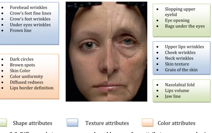
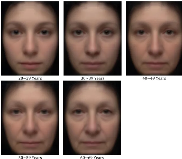
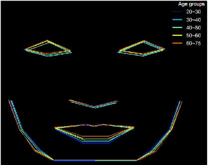
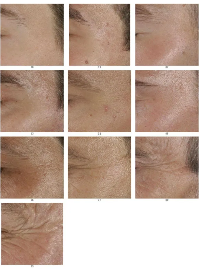
Figure 1-1: Main facial bones __________________________________________________________________ 30 Figure 1-2: Facial muscles _____________________________________________________________________ 32 Figure 1-3: skin anatomy ______________________________________________________________________ 33 Figure 1-4: Resorption of the mandible after the extraction of a tooth (Atwood 1971) _______________________ 35 Figure 1-5: Fat atrophy (green) and accumulation (purple) (Sclafani and Romo 3rd 2000) ___________________ 36 Figure 2-1: Distribution of subjects per age groups __________________________________________________ 44 Figure 2-2: Example of average face _____________________________________________________________ 49 Figure 2-3: Difference between a young and an old woman face; attributes are assessed using VAS ____________ 50 Figure 2-4: Average face per group of age _________________________________________________________ 51 Figure 2-5: Changes on shape attributes with age ___________________________________________________ 52 Figure 2-6: Changes in the eyes area with age______________________________________________________ 54 Figure 2-7: Changes of the mean facial shape with age _______________________________________________ 55 Figure 2-8: Changes on skin textural attributes with age ______________________________________________ 57 Figure 2-9: Changes on skin color attributes with age ________________________________________________ 59 Figure 2-10: Nine grade scale built to assess the severity of crow's feet wrinkles ___________________________ 63 Figure 2-11: Improvement of graders agreement after the use of a proposed image scale, for the crow’s feet area. 64 Figure 3-1: The thirteen faces image. _____________________________________________________________ 66 Figure 3-2: Distribution of graders among age and gender groups ______________________________________ 69 Figure 3-3: Agreement on perceived ages for four different subjects _____________________________________ 72 Figure 3-4: Correlation between accuracy and agreement among the graders. ____________________________ 73 Figure 3-5: Mean age versus perceived age ________________________________________________________ 74 Figure 3-6: Validation of the five PLS models built for age prediction ___________________________________ 79 Figure 3-7: Comparison between PLS model of real age and perceived age _______________________________ 80 Figure 3-8: Comparison between PLS models of perceived ages for different age groups of graders____________ 81 Figure 3-9: Comparison between PLS model of perceived ages from different gender groups of graders ________ 82 Figure 4-1: Vectorization of the aging pattern. (Geng and Smith-Miles 2007) _____________________________ 89 Figure 4-2: First four parameters of appearance and their variations (± 3 sd) (Cootes, Edwards et al. 2001) ____ 93 Figure 4-3: Diagram of the age prediction algorithm ________________________________________________ 94 Figure 4-4: Manual landmarks on faces ___________________________________________________________ 95 Figure 4-5: RMSE in age prediction based on the position of the landmarks as a function of the number of latent variables __________________________________________________________________________________ 100 Figure 4-6: First two modes of shape variation ____________________________________________________ 101 Figure 4-7: RMSE in age prediction based on the pixels’ values in each color channel as a function of the number of latent variables _____________________________________________________________________________ 101 Figure 4-8: The four first weight b1, b2, b3 and b4 of the PLS model of texture ____________________________ 102
Figure 4-9: RMSE in age prediction based on the latent variables from the shape and texture models as a function of the number of latent variables__________________________________________________________________ 103
List of figures 11
Figure 4-10: First two modes of face variation ____________________________________________________ 104 Figure 4-11: Correlation between real and predicted age for the validation set of data _____________________ 105 Figure 4-12: Residual plot of the age regression model ______________________________________________ 106 Figure 4-13: Subjects with an error of more than ten years in age prediction _____________________________ 107 Figure 4-14: Cumulative distribution of the mean absolute error in age estimation ________________________ 108 Figure 4-15: Correlation between perceived and predicted age on the validation set of data (PLS regression) ___ 109 Figure 4-16: Correlation between perceived and predicted age on the validation set of data (AAM used for data image compression) _________________________________________________________________________ 110 Figure 4-17: correlation between real and predicted age on the validation set of data (neural network model with PLS latent variables). ________________________________________________________________________ 110
List of tables 12
List of tables
Table 2-1: Parameters assessed using a Visual Analog Scale ... 47 Table 2-2: Results of linear regressions made to compare the agreement among two graders ... 62 Table 3-1: Means and standard deviations of absolute values of errors in age prediction. ... 75 Table 3-2: Parameters of the linear regression model between the absolute value of error in age estimation and subjects’ and graders’ ages ... 75 Table 4-1: Summary of the different works published on age prediction... 90 Table 4-2: Correlation coefficients between the first components of the shape and texture models. ... 98
Main references 13
Main references
Cootes, T. F., G. J. Edwards, et al. (2001). "Active appearance models." IEEE Transactions on Pattern Analysis and Machine Intelligence 23(6): 681-685.
Cootes, T. F., C. J. Taylor, et al. (1995). "Active shape models-their training and application." Computer Vision and Image Understanding 61(1): 38-59.
CrimeLib (2002). Age Progression. Criminal Minds and Methods. C. T. s. C. Library.
Edwards, G. J., C. J. Taylor, et al. (1998). "Interpreting face images using active appearance models." 3 rdInternational Conference on Automatic Face and Gesture Recognition: 300–305. Fu, Y., Y. Xu, et al. (2007). ESTIMATING HUMAN AGE BY MANIFOLD ANALYSIS OF FACE PICTURES AND REGRESSION ON AGING FEATURES. International Conference on Multimedia & Expo. Beijing, China.
Gandhi, M. R. (2004). A Method for automatic Synthesis of Aged Human Facial Images. Department of Electrical & Computer Engeneering. Montreal, Canada, McGill University. Master
Degree: 107.
Geng, X. and K. Smith-Miles (2007). "Automatic Age Estimation Based on Facial Aging Patterns." IEEE TRANSACTIONS ON PATTERN ANALYSIS AND MACHINE INTELLIGENCE 29(12): 2234-2240. Geng, X., Z. H. Zhou, et al. (2006). "Learning from facial aging patterns for automatic age estimation." Proceedings of the 14th annual ACM international conference on Multimedia: 307-316.
Hayashi, J., M. Yasumoto, et al. (2002). "Age and gender estimation from facial image processing." SICE 2002. Proceedings of the 41st SICE Annual Conference 1.
Horng, W. B., C. P. Lee, et al. (2001). "Classification of Age Groups Based on Facial Features." Tamkang Journal of Science and Engineering 4(3): 183-192.
Main references 14 Hubball, D., M. Chen, et al. (2006). Evolutionary Morphing of Facial Images for Aging Simulation, University of Wales Swansea.
Kwon, Y. H. and N. da Vitoria Lobo (1994). "Age classification from facial images." Computer Vision and Pattern Recognition, 1994. Proceedings CVPR'94., 1994 IEEE Computer Society Conference on: 762-767.
Lanitis, A., C. Draganova, et al. (2004). "Comparing different classifiers for automatic age estimation." Systems, Man and Cybernetics, Part B, IEEE Transactions on 34(1): 621-628.
Liu, J., N. Zheng, et al. (2007). "Estimating Aging Pattern by Aging Increment Distribution for Re-rendering of Facial Age Effects." LECTURE NOTES IN COMPUTER SCIENCE 4681: 782.
Lobo, N. and Y. Kwon (1998). Automatic feature detection and age classification of human faces in digital images, Google Patents.
Ramanathan, N. and R. Chellappa (2006). "Face verification across age progression." IEEE transactions on image processing 15(11): 3349-3361.
Ramanathan, N. and R. Chellappa (2006). "Modeling Age Progression in Young Faces." Computer Vision and Pattern Recognition, 2006 IEEE Computer Society Conference on 1.
Ricanek Jr, K. and T. Tesafaye (2006). "MORPH: a longitudinal image database of normal adult age-progression." Automatic Face and Gesture Recognition, 2006. FGR 2006. 7th International Conference on: 341-345.
Seoul, K. (2004). Extraction and Manipulation of Wrinkles and Spots for Facial Image Synthesis. Proceedings of the Sixth IEEE International Conference on Automatic Face and Gesture Recognition.
Tiddeman, B. P., M. R. Stirrat, et al. (2005). "Towards Realism in Facial Image Transformation: Results of a Wavelet MRF Method." Computer Graphics Forum 24(3): 449-456.
French long summary 15
French long summary
I. Introduction
L’âge a toujours été un facteur important d’interaction sociale. La posture, le vocabulaire et l’intonation sont autant d’éléments qu’on adapte { l’âge de son interlocuteur. De manière plus large, on peut noter que la société propose des activités et des loisirs différents en fonction de l’âge des individus. Ainsi, pouvoir prédire l’âge demeure une aptitude importante qui se développe dès la tendre enfance. Cette aptitude s’appuie sur de nombreux signes qui peuvent aussi bien être liés à la tenue vestimentaire qu’{ la posture ou aux traits du visage.
Dans cette thèse, nous nous intéressons en particulier au visage et aux transformations qui l’affectent avec l’âge, { la capacité d’un individu { s’appuyer sur ces transformations pour prédire l’âge et { la possibilité de construire un algorithme automatique de prédiction. Les objectifs de ce travail sont donc de:
1. Décrire les changements qui affectent le visage au cours du temps 2. Analyser l’influence de ces changements sur la perception de l’âge
3. Proposer un algorithme permettant de prédire l’âge d’individus { partir de leurs photos
Les effets du temps étant variables d’un sexe { un autre et d’une race { une autre, nous nous restreignons dans cette thèse à une population de femmes Caucasiennes.
Ce document est divisé en quatre chapitres. Le premier chapitre s’intéresse au vieillissement anatomique du visage et présente un résumé des connaissances acquises. Il décrit les phénomènes biologiques liés { l’âge et leur influence sur l’apparence du visage. Dans le second chapitre, nous nous intéressons en particulier aux femmes Caucasiennes et présentons une étude menée afin de quantifier les transformations anatomiques du visage liées { l’âge. Nous évaluons l’évolution de 21 paramètres en fonction de l’âge sur une
French long summary 16 population constituée de 173 femmes âgées de 20 à 73 ans et établissons la cinétique du vieillissement.
Dans le troisième chapitre, nous mesurons l’influence relative des signes du vieillissement sur l’âge perçu. Nous demandons { 48 observateurs d’évaluer l’âge des 173 volontaires précédentes. Un modèle PLS permet ensuite d’établir une pondération des signes du vieillissement relativement { la perception d’âge.
Le quatrième chapitre s’appuie sur les résultats précédents pour proposer un modèle de prédiction d’âge à partir de photos. Ce modèle est utilisé de manière concluante sur les photos des 173 précédentes volontaires.
II. Vieillissement du visage
Le temps qui passe altère irrémédiablement le visage. De nombreuses publications décrivent l’ensemble des transformations qui modifient aussi bien la forme, que la texture et la couleur du visage. Le résumé de ces transformations est le suivant :
1. La forme du visage:
La forme du visage est liée à la disposition des os, des tissus mous (muscles et graisse) et à la fermeté de la peau. Au cours du temps, chacun de ces éléments subit des changements importants. Quelques unes de ces transformations sont :
Au niveau du squelette :
o La réduction de la hauteur du visage
o La perte des dents et l’ostéoporose des os maxillaires et mandibulaires o L’augmentation da la largeur du visage
Au niveau des tissus mous
o La myopathie de tous les muscles du corps
o L’hypertrophie des muscles orbiculaires et frontaux entrainant l’apparition d’une ptose sur les sourcils et des poches sous les yeux o L’infiltration de graisse dans le nez provocant la ptose de la pointe de
nez
o La redistribution de graisses sur l’ensemble du visage le rendant plus concave
Au niveau de la peau
o L’apparition de sillons marqués tels que le sillon nasogélien
o La perte de fermeté favorisant le relâchement des paupières et sur la partie base du visage l’apparition de bajoues ou d’un double menton 2. La couleur du visage:
La couleur du visage est principalement liée { l’épaisseur de l’épiderme et { la distribution des différents chromophores (mélanine et hémoglobine). Au fil du temps, la peau devient plus fine et les vaisseaux sous-jacents plus visibles, ce qui
French long summary 17 entraine un ternissement et un jaunissement du teint. Par ailleurs, l’exposition du visage au soleil favorise la non homogénéisation de la distribution de la mélanine provocant l’apparition de tâches pigmentaires.
3. La texture du visage :
La texture du visage est altérée suite { l’apparition des rides et ridules. On peut distinguer les rides d’expressions qui apparaissent sur les parties du visage en mouvement telles que le front et des rides liés { l’âge. Toutes les rides se creusent avec le temps, suite { la dégradation du collagène et de l’élastine contenus dans le derme.
Bien que toutes les transformations décrites ci-dessus soient générales, elles s’opèrent plus ou moins vite en fonction de nombreux facteurs tels que l’origine raciale, le sexe, l’exposition au soleil, la pollution, la consommation de tabac, … La prochaine section s’intéresse spécifiquement au vieillissement de femmes Caucasiennes.
III.
Vieillissement du visage des femmes Caucasiennes
Répondre aux attentes des femmes soucieuses de ralentir les effets du temps est l’une des principales missions des cosméticiens et chirurgiens plastiques. Dans ce contexte, la mesure quantitative des changements liés { l’âge permet de proposer de meilleurs traitements d’une part, et d’autre part de vérifier l’efficacité des procédures disponibles. La métrologie cutanée est la science qui s’est développée afin de mesurer les attributs cutanés et de suivre leur évolution. Johnson & Johnson dispose d’un laboratoire de métrologie au sein duquel une étude a été menée sur 173 femmes Caucasiennes âgées de 20 à 74 ans afin de mesurer les changements affectant leurs visages et liés { l’âge.
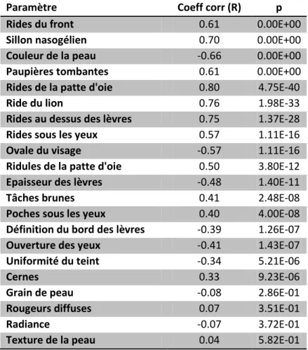
Pendant cette expérience 21 attributs choisis suite à une revue de la littérature ont été évalués par un expert { l’aide d’échelles visuelles analogiques. Le tableau ci-dessous fait le récapitulatif des attributs évalués et de leur corrélation avec l’âge.
French long summary 18
Paramètre Coeff corr (R) p
Rides du front 0.61 0.00E+00
Sillon nasogélien 0.70 0.00E+00
Couleur de la peau -0.66 0.00E+00
Paupières tombantes 0.61 0.00E+00
Rides de la patte d'oie 0.80 4.75E-40
Ride du lion 0.76 1.98E-33
Rides au dessus des lèvres 0.75 1.37E-28
Rides sous les yeux 0.57 1.11E-16
Ovale du visage -0.57 1.11E-16
Ridules de la patte d'oie 0.50 3.80E-12
Epaisseur des lèvres -0.48 1.40E-11
Tâches brunes 0.41 2.48E-08
Poches sous les yeux 0.40 4.00E-08
Définition du bord des lèvres -0.39 1.26E-07
Ouverture des yeux -0.41 1.43E-07
Uniformité du teint -0.34 5.21E-06
Cernes 0.33 9.23E-06
Grain de peau -0.08 2.86E-01
Rougeurs diffuses 0.07 3.51E-01
Radiance -0.07 3.72E-01
Texture de la peau 0.04 5.82E-01
Table 0-1: Corrélation entre les attributs mesurés et l’âge des femmes
Comme le montre ce tableau de nombreux paramètres mesurés sont significativement liés à l’âge. En regardant la forme des courbes, nous mettons par ailleurs en évidence l’incidence de la ménopause sur 13 attributs. Sept attributs semble être négativement affectés par la ménopause (accélération de la dégradation), tandis que six d’entre eux sont positivement affectés (ralentissement).
Nous nous intéressons aussi à la reproductibilité de la méthode utilisée et montrons qu’elle convient pour mesurer la plupart des paramètres cutanés de manière reproductible. Deux experts sont invités à évaluer 31 volontaires et les résultats de leurs évaluations sont comparés en utilisant le coefficient de corrélation de Pearson. Nous obtenons une corrélation significative pour 70% des attributs comparés. Nous proposons néanmoins l’usage d’échelles construites { partir de photos afin de faciliter l’entrainement des évaluateurs et d’améliorer leur reproductibilité.
Les attributs évalués dans cette section sont utilisés par la suite pour comprendre les signes sur lesquels s’appuie un observateur pour prédire l’âge d’un sujet.
French long summary 19
IV.
Perception du vieillissement
Une revue de la littérature nous permets d’établir que :
- La capacité { prédire l’âge est acquise dans les premières années de l’enfance
- Cette capacité est sujette { un biais lié { la race, { l’âge et au sexe des observateurs ; chacun étant plus { même de prédire l’âge de personnes du même groupe que lui. - La taille du visage, la couleur de la peau, la présence de rides, la vitalité du regard
sont autant d’éléments qui permettent de juger de l’âge d’un individu. Néanmoins aucune hiérarchisation de ces paramètres ne semble faire le consensus, probablement à cause des protocoles expérimentaux utilisés qui ne permettent pas de directement comparer l’influence des différents attributs faciaux (approches unidimensionnelles).
Afin de déterminer les attributs influençant le plus la prédiction d’âge nous proposons une nouvelle méthodologie s’appuyant sur la régression PLS et permettant d’avoir une approche multidimensionnelle du problème.
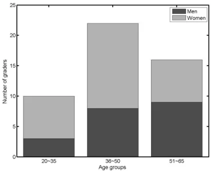
Quarante huit observateurs ; 20 hommes et 28 femmes ; âgés de 20 { 64 ans évaluent l’âge des 173 précédentes volontaires à partir de photos de visage prises dans des conditions standardisées. L’âge apparent est définit comme l’âge moyen donné par les évaluateurs. Dans un premier temps, la comparaison des évaluateurs masculins et féminins confirme que les femmes sont plus précises pour prédire l’âge d’autres femmes comme le laissait prévoir la littérature. Par ailleurs, la comparaison de trois groupes d’évaluateurs : les jeunes (20-35 ans), les personnes d’âge intermédiaire (36-55 ans), et les personnes âgées (56-65 ans) met en évidence des différences liées { l’âge des évaluateurs. Le groupe de jeunes montre une meilleure précision que les autres groupes. Néanmoins, nous ne confirmons pas sur ces données que les individus d’un groupe d’âge sont plus précis lorsqu’ils évaluent des gens de la même tranche d’âge.
Enfin, nous construisons sept modèles de régression PLS pour prédire l’âge { partir des 21 attributs mesurés. Chacun des modèles prédit respectivement l’âge réel, l’âge apparent donné par l’ensemble des évaluateurs, les âges apparents donnés par les groupes d’hommes et de femmes et finalement les âges apparents donnés par les trois groupes d’âge (Jeunes-âge intermédiaire, personnes âgées).
L’importance relative des pondérations du modèle PLS nous permet de connaître l’importance de chacun des attributs pour le modèle de prédiction d’âge. Nous établissons ainsi que :
- Les attributs les plus importants sont dans un ordre décroissant l’épaisseur des lèvres, la forme de l’ovale du visage, les rides autour de la bouche et l’uniformité du teint.
French long summary 20 - Les hommes et les femmes ne présentent pas de différence significative concernant
l’utilisation des différents attributs pour la prédiction d’âge.
- L’importance du sillon nasogélien, des cernes, des poches sous les yeux, de l’uniformité du teint décroit avec l’âge des observateurs. Au contraire, celle des rides de la patte d’oie, de l’ouverture des yeux, de l’épaisseur des lèvres augmente avec l’âge des volontaires.
Ces résultats qui vont au-delà de ce que décrit la littérature sont en cours de publication et gagneraient { être confirmés par d’autres expériences.
V. Prédiction de l’âge { partir de photos
Nous proposons un algorithme de prédiction de l’âge en s’appuyant sur les observations faites dans les parties précédentes. Cet algorithme qui s’inspire des Modèles Actifs d’Apparence (AAM) peut se décomposer en trois grandes étapes :
- D’abord les principaux traits du visage (contour, yeux, bouche, nez) sont localisés { l’aide de points d’encrages. Toutes les images sont alignées { l’aide de ces points de manières à ce que les traits principaux se retrouvent à la même position.
- Deux séries de modèles PLS sont construit pour prédire l’âge { partir des informations provenant des photos. Le modèle de forme s’appuie sur la position des points d’encrage et décrit les changements relatifs { l’âge dans la forme des visages. La deuxième série est constituée de trois modèles PLS décrivant les variations d’intensité des pixels sur chacun des canaux de couleur (RVB) en fonction de l’âge. Chacun de ces modèles constitue donc un descripteur de la texture du visage. Tous ces modèles fournissent un ensemble de variables latentes qui résument l’information relative { l’âge des individus de la base d’apprentissage.
- Les variables latentes des quatre précédents modèles sont normalisées avant de servir de variables d’entrée pour la construction d’un modèle global de prédiction d’âge.
Appliquée à notre base de données, cette approche nous permet de visualiser les attributs les plus importants pour la prédiction d’âge ainsi que leur transformation au cours du temps. Elle met ainsi en exergue l’importance de la forme de la bouche, du menton (ovale du visage) et de la taille des yeux. Elle permet aussi de rendre compte de l’importance de la région autour des yeux, ainsi que du sillon nasogélien et des bajoues.
Sur une base de test, le modèle proposé prédit l’âge avec une Erreur Moyenne Absolue (MAE) de 5.98 ans, qui est comparable { l’erreur humaine pour la même tâche. La capacité de prédiction s’améliore (MAE = 4.87 ans) lorsqu’on apprend { prédire l’âge perçu plutôt que l’âge réel. Ce résultat laisse entendre que le modèle s’appuie sur le même type d’informations que les observateurs humains.
French long summary 21
VI.
Conclusion et perspectives
Dans ce travail de thèse, nous nous sommes intéressés aux changements liés au vieillissement du visage et à leur perception. Dans un premier temps, nous avons quantifié sur une population de femmes caucasiennes l’évolution de signes liés { la couleur et { la texture de la peau, à la forme du visage et des lèvres ainsi qu’au contour des yeux. Les observations effectuées ont montré la corrélation de la plupart des signes choisis avec l’âge, et ceux malgré les variations interindividuelles. Par la suite nous avons analysé l’importance des signes du vieillissement sur la perception d’âge. Pour se faire nous avons proposé une approche originale, reposant sur la régression PLS. A notre surprise, nous avons trouvé que les personnes jeunes et les personnes âgées utilisent différemment les attributs du visage lorsqu’elles prédisent l’âge. Néanmoins, nous avons montré que les principaux attributs guidant la perception sont liés { l’épaisseur des lèvres, la forme de l’ovale du visage, les rides autour de la bouche et l’uniformité du teint. Tous ces enseignements ont alimentés notre approche pour la proposition d’un algorithme de prédiction automatique de l’âge { partir de photos.
Le modèle que nous avons proposé pour prédire l’âge s’appuie sur des informations aussi bien liées { la forme qu’{ la couleur et { la texture du visage. Ce modèle utilise un nouvel algorithme de compression supervisée des visages, le Modèle Supervisé de Visage (SFM) reposant sur la régression PLS. Le SFM permet de réduire l’information portée par l’ensemble des pixels de l’image (plusieurs milliers) en un nombreux restreint de paramètres (quelques dizaines) portant l’information relative { l’âge. Cette approche nous permet de prédire l’âge avec une précision comparable avec celle d’un observateur humain. Néanmoins, quelques améliorations pourraient encore augmenter les performances de cet algorithme. Dans un premier temps, il serait surprenant que la relation entre la valeur des pixels de l’image et l’âge des sujets n’ait pas une composante non-linéaire. Par conséquent, l’utilisation d’un modèle non-linéaire pour la compression des images du visage pourrait améliorer la qualité de la prédiction. Par ailleurs, le modèle de texture utilisé n’exploite que faiblement les informations contenues dans les hautes fréquences (rides et ridules). Des paramètres issus d’une analyse fréquentielle (Fourier ou ondelettes) permettraient de mieux encoder ces informations. Enfin, le modèle pourrait être étendu aux images en 3 dimensions (3D) afin non seulement de prédire l’âge { partir d’image 3D, mais surtout de simuler le vieillissement.
Introduction 25
Introduction
Coming from an African country where people are respected for the graying of their hair and beard, one may be surprised in the western societies by the high number of products and procedures invented everyday to make people look younger. In fact, since Alexander the Great have started searching for the legendary Fountain of Youth, a great part of the humanity has struggled to stop or at least to slow down the effects of time on people’s appearance.
Meanwhile, people’s age remains an important attribute of social interactions. The way we act, the vocabulary we choose, our body attitude in front of someone else depends on his age. Moreover specific works, entertainment, lectures, movies, etc. are assigned to groups of age. Therefore people since childhood develop a capacity to estimate other people’s age based on physical attributes. These attributes can be related to dress code or body gesture, but they are primarily linked to facial appearance. It is obvious that anyone can distinguish a baby face from an adult and from a senior one since some evident characteristics such as the size of the head and the number of wrinkles are affected dramatically by age. Consequently it would be useful to understand all the changes that occur on faces with age and their effect on people’s appearance.
Furthermore, nowadays and even more so in the near future we will need to interact not only with human beings but also with computers and robots. A lot of research focusing on improving human to machine interactions is made in order to enable natural communication. One of the most accessible applications of these research results is the ability for the newest digital cameras to locate the faces in images and to capture pictures when people are smiling. Other applications include people identification at secured access points or faces tracking in video movies. However, to our knowledge, little work has been done on age prediction and there is no public accessible application that is commercialized which uses an age prediction algorithm. However many reasons have already been identified that justify the need for age prediction algorithms.
Introduction 26
Objectives and motivation
This thesis focuses on changes that occur with age on Caucasian women’s faces. Our three objectives are:
1. To describe the facial changes with age
2. To explore the incidence of these changes on the perceived age
3. To propose an automatic algorithm for age prediction based on facial images.
Various benefits could be derived from these three objectives in the fields of cosmetic dermatology, psychology and image processing. These benefits will be presented in details in the remaining of the document. Here are some examples:
- By understanding the contribution of each facial attribute to the overall aging appearance, one can design better procedures for facial rejuvenation.
- Understanding how people code and interpret faces is still an open problem in psychology. This work can contribute to better understanding of how signs of aging are read and interpreted.
Automatic algorithms for age prediction could be used for age-based indexing of facial images from a database. Furthermore, the work done for this algorithm could be extended for age simulation algorithms. Age simulation can be used in image processing of a facial image to show how someone will look after several years. Age simulation is useful to find missing children or to capture wanted fugitives.
Overview
Age or ages?
The concept of age measures the number of years that have passed since birth. When dealing with facial aging, one may be interested in three different kinds of aging: chronological aging, photoaging and apparent aging. Chronological aging or intrinsic aging refers to the natural physiological changes in the facial tissues. Those changes are related to the growth and the degeneration of the body over time and occur throughout the full body. Since the face is more exposed than the rest of the body to environmental factors such as sun, photoaging is also well noticeable on facial skin. The effects of photoaging underscore the ones produced by chronological aging leading to a gap between chronological age and apparent age. Apparent age is the average age given to a person by other peoples. It is a subjective judgment: a robust estimation can only be obtained by averaging multiple estimations. Some people look younger or older than their real age, depending on their genetic makeup, sun exposure, smoking habits and mood. Some events in life like the death of a parent or a long disease can accelerate the process of apparent aging. Make up, anti-aging creams or surgery can reduce the apparent age.
Introduction 27
Does everyone age similarly?
We have already pointed out that apparent age depends on many internal and external factors. Changes with age are also tightly related to ethnicity. Within a same ethnic population, a wide variability is observed in physical appearance of individuals within the same age group. There is also a difference between apparent and chronological age. Thus, studies devoted to the measure of the anatomical transformations related to age should include a large number of subjects to account for individual variations. Since we are interested in facial changes with age, it is necessary to eliminate the characteristics that are related to the subject identity.
Thesis contributions
In this thesis we explore three different areas related to facial aging, confirm some recent findings and provide a unified approach of age prediction. The main contribution of this document includes:
On facial aging:
We quantitatively describe the age-related changes on Caucasian women
On age perception:
We confirm that the perception of age is related to the age and the gender of the observer. We also establish a ranking of the signs of aging and show that depending on the observer’s age, he/she gives a different importance to each facial attribute
On age prediction:
We propose a new method to describe a face using a small set of. We also propose a new algorithm that enables to predict people’s age with accuracy comparable to that of human.
Structure of the thesis
The next chapter describes the anatomical changes related to facial aging. It enables to understand the biological phenomena related to aging and their incidence on facial appearance.
In the chapter 2, we focus on aging of Caucasian women and present an experiment performed to record age-related changes in this population. We quantitatively describe the transformation of 21 facial attributes measured clinically on 173 volunteers from 20 to 74 years of age.
In the chapter 3, we focus on the influence of these 21 facial attributes on age perception. Firstly, we assign a perceived age to each of the 173 volunteers by a consensus among 48 assessors who were asked to give an age to these volunteers. Then we use a Partial Least Square (PLS) regression to build explanatory models of age. These models permit us to measure the relative contribution of each attribute to the perception of age.
Introduction 28 The chapter 4 introduces a new age prediction algorithm. Firstly, we present a model that encodes facial images with a small set of parameters. We subsequently use it to predict the age of the 173 volunteers of our study from their facial images.
Facial aging 29
Chapter 1
Facial aging
The process of aging induces several changes on face. Despite of individual variations linked to genetic and living conditions, some general rules are found. These rules are gender and ethnic dependant. Differences between men’s and women’s aging are attributed to sex-specific characteristics such as skin thickness and hormonal activity, especially in relation with menopause (Broniarczyk-Dyla and Joss-Wichman 2001; Wines and Willsteed 2001; Raine-Fenning, Brincat et al. 2003; Sumino, Ichikawa et al. 2004; Quatresooz, Pierard-Franchimont et al. 2006).
Generally speaking, if faces are considered as 3D objects, the transformations that occur with age belong to three groups: shape, texture and color. Although unusual when looking at facial aging, this classification enables us to describe the aging process, keeping in mind that our objective is to extract signs of aging from facial images. Here is a short description of the three levels that we have considered:
- Changes on shape. They occur in the spatial dimensions with a centimetric range. They may be caused by the growth of bones or their osteoporosis, changes in muscle and fat distributions, skin sagging caused by the lost of elasticity and gravity. These changes can be recorded on low resolution images. They are affected by lighting conditions (orientations and shadows) and head positioning.
- Changes on texture. They happen in the spatial dimensions at a millemetric range. They are linked with skin aging and can involved wrinkles, pores and microrelief. They are not recorded on low resolution images.
- Changes on color. They are also linked with skin aging. They involve tone yellowish and unevenness with the apparition of brow spots.
This chapter depicts the anatomic transformations that influence the appearance of the face with age. Firstly, a brief description of the anatomy of the face is made in order to help better understanding these morphological transformations. Then the anatomic changes
Facial aging 30 with age, reported by the literature are presented. Finally, the effects of some factors that influence the aging process are discussed.
I. Facial anatomy
The study of the facial anatomy is important for understanding the appearance of the face. Since anatomy is not the focal topic of this document, description is short: the objective is to highlight the main elements that contribute to the physical shape, color and texture of the face.
Facial anatomy is composed of three main elements: skin, soft tissue, and the underlying skeleton; each of them being affected by age differently.
I.1. Bones
Bones give the body a framework, maintain its shape, and protect vital organs. They also provide a place for muscles, supporting structures to attach. Bones are also sites for mineral storage and blood cells formation.
The face is composed of five main bones as shown on Figure 1-1 and support entry of the digestive and the respiratory systems. They also provide connection for muscles of facial expression and mastication, and they support organs for the main senses.
Figure 1-1: Main facial bones1
Facial aging 31 The relationship between facial bones and visual appearance of the face is not direct. For example, the shape of the eyes and eyelids, the tip of the nose and the lips cannot be predicted from the skull. However, the forehead, margins of the eyes, cheekbones, bridge of the nose, chin and overall facial shape are closely related with skull contours (Evison 2001).
I.2. Muscles and fat
Facial muscles and fat are known in the literature as soft tissues. Their distribution smoothes the bones shape and gives the face its identity. Simpson and Henneberg (Simpson and Henneberg 2002) have explored the relationship of soft tissues with craniometric dimension on Caucasian cadavers (17 males and 23 females). “Significant correlations between many soft-tissue depths and craniometric dimensions were found, suggesting a relationship between the amount of soft tissue present on the face and the size of the underlying bony skeleton. Soft-tissue depths were highly positively correlated with each other; craniometric dimensions were correlated but to a lesser extent.”
The specific contribution of muscle to facial shape is not extensively discussed in the literature, but a detailed description of their anatomy and their contribution to facial expressions and wrinkling is available (Freilinger, Gruber et al. 1987; Waters 1987; Waters and Terzopoulos 1990). Facial muscles can be divided into two groups: cutaneous and bones muscles.
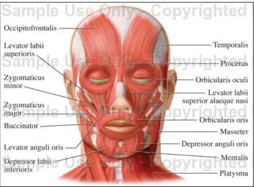
The front face is made of more than sixteen major bones muscles (Figure 1-2) that give life to facial expressions and mouth movements (speech and chewing). The results of muscles movements arise on the skin and can lead to permanent wrinkles known as expression wrinkles.
On the upper face, the Frontalis (Occipitofrontalis) arises from the top of the skull to the orbital area. Contraction of this muscle is responsible for the rise of the brow, and may induce forehead wrinkles. Located in the intercilliary zone, the Procerus draws the skin between the eyebrows and causes horizontal wrinkles in the root of the nose. The Orbicularis oculi are circulars muscles around the orbital cavity. The internal portion of the muscles closes and open the eyelids while the low external portion downwards the eyebrows. The external portion also causes hard occlusion of the eyes, producing crow’s feet wrinkles on the temples.
The middle face is moved by the major and minor Zygomaticus, and the Levator labii. When contracted, the minor Zygomaticus elevates the portion of the lip where it is inserted while the major Zygomaticus lifts upward and laterally the corners of the mouth (smile). The Levator labii raises the later edge of the wing of the nose and a portion of the upper lip. It action leads to the apparition of the nasolabial folds.
Facial aging 32
Figure 1-2: Facial muscles2
The movement of the mouth is also driven by the Buccinator, the Orbicularis oris, the masseter, the Depressor labii and the Levator anguli oris. Their coordination contributes to the mimics of the mouth and the mastication movements.
Some cutaneous muscles are part of the skin. The skin merged with the cutaneous muscles and fat - usually referred in the literature as the superficial musculoaponeurotic system (SMAS) – have an impact on apparent age and are manipulated through dermato-cosmetical procedures with a great benefit on apparent age (Barbara A. Gilchrest 2006).
I.3. Skin
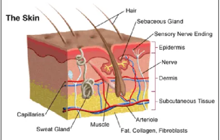
The skin is the most voluminous body organ. Its main function is to be a protective barrier that interfaces against hostile environment including microbes and bacteria Facial skin in particular is exposed to many external aggressive factors such as sun and atmospheric pollution. Consequently, the facial skin aging will results from both chronological and extrinsic aging. To be able to understand changes occurring on the skin with age, one need to firstly describe its three layers organization (Figure 1-3).
Facial aging 33
Figure 1-3: skin anatomy
The epidermis is the outer layer of the skin containing five sub-layers made with keratinocytes at different level of differentiation. Keratinocytes are produced in the basal or generative layer and start migrating to the upper layer while differentiating (Flattening and increasing surface). The top layer of the epidermis, the stratum corneum is made with flat non-nucleus and dead skin cells. The epidermis also contains melanocytes in its basal layer, which produce melanin. Melanin absorbs radiant energy from the sun and protects skin from ultraviolet radiations. The ratio of melanocytes and keratinocytes is approximately 1:4 in sun exposed area.
The primary function of the dermis is to support and sustain the epidermis. Apart from skin appendages and blood vessels the dermis is almost pure collagen. Collagen comprises 70% to 80% of the dry weight of human skin. Collagen fibers provide tensile strength and resistance to shear and other mechanical forces. Elastin fibers also contribute to the mechanical properties of the dermis. Even if they only represent 1% of the weight of the dermis, they resist to deformational forces and give the skin its elasticity. The cutaneous vessels located in the dermis are arranged in a horizontal network parallel to the surface of the skin.
The subcutaneous tissue is a layer made of fat and connective tissues containing larger blood vessels and nerves. This layer is important for the regulation of the skin and the body temperature.
Facial skin is clinically characterized mainly by its wrinkles and its color.
Skin wrinkles
Wrinkles are folds of the skin frequent on the face of aged people. They are caused by the changes in the thickness of the epidermis and structural transformation in the dermis related to aging. According to their aspect, there are three major groups of wrinkles (Kligman, Zheng et al. 1985):
Facial aging 34 - Crinkling-type wrinkles are fine wrinkles that formed from folded skin. They usually appear in old skin (person more than 75 years old) and may be caused by sun-damage on individuals with elastosis.
- The glyphic wrinkles have a crisscross pattern and are frequently seen on the cheek and the neck.
- The deep wrinkles form long and straight major lines or deep grooves. They usually appear on the forehead and the crow’s feet area.
Skin color
Color is an essential component of physical properties of the skin (Andreassi 1995). Skin color arises from the interactions of light with the epidermis and the dermis (Stamatas and Kollias 2004). The perceived color also depends on the detector (eyes, camera, film…). The interaction of light with the skin is related to the concentration of chromophores and the absorption coefficient of the constitutive layers of the skin. With an index of refraction of 1.55, the stratum corneum reflects approximately 5% of an incident perpendicular light (Kollias 1995). Most of the incident light crosses over the 10 µm of the stratum corneum and reaches the viable epidermis. In this area, the major chromophore is melanin. Melanin absorbs light stronger in the blue than in the red, resulting in a color that is rich in red and poor in blue (brown color) (Kollias 1995) .The absorbance spectrum of melanin shows a monotonic decrease towards longer wavelengths. Under the epidermis, the dermis provides the skin with nourishment through blood vessels. The hemoglobin found in the dermal blood is the chromophore responsible for the red appearance of the skin. The type of hemoglobin that carries oxygen molecules is call oxy-hemoglobin (oxy-Hb) while hemoglobin without oxygen is called deoxy-hemoglobin (deoxy-Hb). Each type of hemoglobin has its how absorbance spectrum.
It follows that the visual perception of skin color is the result of the contributions of three chromophores, melanin, oxy-Hb and deoxy-Hb. However, the skin color can hardly be linked with the concentration of one specific chromophore. As an example, Stamatas and Kollias (Stamatas and Kollias 2004)} have shown that skin darkening is not only linked with the concentration of melanin, but also depends on deoxy-Hb.
Another mode of interaction of light with the skin is scattering. Scattering occurs when light changes its trajectory after crossing over two different optical media. The principal component of scattering in the dermis is the collagen. Light scattering is also a function of wavelength. Blue light is more scattered than yellow, which penetrates deeper into the skin.
II. Changes with age
Several authors have described the overall changes on face with age. This section is based on the overall descriptions made by Friedman (Friedman 2005), Vacher (Vacher 2004), Taister & All (Taister, Holliday et al. 2000), Gilchrest & Krutmann (Barbara A. Gilchrest
Facial aging 35 2006) and Kanior & Kerth (Konior and Kerth 1990). The changes described involve those that take place from the twenties up to the eighties.
II.1. Changes on shape
Changes on facial shape with age are related to bones, muscles, fat and skin transformations and are tightly linked to body weight and gravity.
II.1.1. Bone transformation
Bartlett & All have explored age-related changes of the craniofacial skeleton of a Caucasian population (Bartlett, Grossman et al. 1992). They performed anthropometric measurements on 160 skulls selected randomly from 1500 specimens. Observed changes in craniofacial morphology included:
i. Appreciable reduction of facial height,
ii. Most marked in the maxilla and mandible, strongly correlated with loss of teeth, iii. Modest increase in facial width,
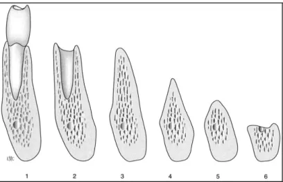
iv. Modest increase in facial depth, except in those regions associated with tooth loss. Vacher (Vacher 2004) considered the osteoporosis and the lost of teeth as the principal skull transformation linked with age. The osteoporosis is caused by the decalcification that occurs in all bones of the body and is even accentuated by the lost of teeth. Carlsson and Persson (Carlsson and Persson 1967) have shown that the front side of the mandible decreases by 2 mm two months after a tooth extraction, 4 mm after one year and approximately 7 mm after 5 years (Figure 1-4). The mandible resorption is more important for females than for males, meaning that it could be related to osteoporosis (Vacher 2004). The lost of teeth also affects the maxilla, but with a resorption of only 0.1 mm per year. Finally, bones changes mostly affect the lower part of the face.
Facial aging 36
II.1.2. Muscles and fat transformation
With increasing age, a degenerative myopathy of all the body muscles takes place due to the reduction of the number and the size of the muscular fibrous and vascular vessels. This leads to a reduction of about 30% of the muscle mass between 30 and 80 years of age (Vacher 2004). As an example, the frontalis loses its straight resulting in a ptosis of the eyebrow. Orbicularis muscles hypertrophy is responsible of the apparition of the orbital rims (Friedman 2005). Facial muscles are also affected by the modifications of the skeleton and facial fat. In most cases, a fat infiltration occurs in the skin muscles causing the nasal tip ptosis.
Fat also redistributes making the face looking more concave, with a hill and valley topography. Donofrio (Donofrio 2000) has pointed out both a fat atrophy in some areas of the face and a fat hypertrophy on other areas. Fat atrophy occurs in the forehead, periorbital, temporal, perioral and buccal areas while fat hypertrophy happens in the jowl, lateral nasolabial fold, lateral labiomental crease and lateral malar areas (Figure 1-5). These changes lead to old faces being more compartmented, with broken, wavy or concave shapes. Arcs, like the cheek arc in profile view or the mandible arc are converted to straight lines, living behind a relative excess of skin. Fat descents along the folds like nasolabial fold. Nasolabial fold can occur at all ages, but like the expression wrinkles, it increases with age (Yousif, Gosain et al. 1994).